Embed Size (px)
DESCRIPTION
Coagulation Disorders and Disseminated Intravascular Coagulation (DIC). Jianzhong Sheng MD, PhD Department of Pathophysiology School of Medicine Zhejiang University. Normal Hemostasis. First step in hemostasis is formation of a platelet aggregate - PowerPoint PPT Presentation
Citation preview

Coagulation Disorders and Disseminated Intravascular Coagulation (DIC)
Jianzhong Sheng MD, PhD
Department of PathophysiologySchool of MedicineZhejiang University

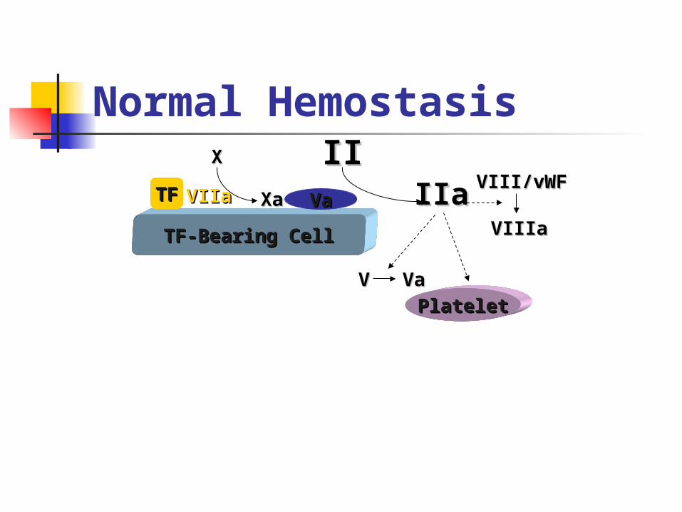
Normal Hemostasis
• First step in hemostasis is formation of a platelet aggregate
• At the molecular level interaction of coagulation factors takes place on the surface of activated platelets
• The Tissue Factor–FVIIa complex is the physiological activator of normal hemostasis
HemostasisSubendothelial matrixSubendothelial matrix
Nitric oxideNitric oxide
Endothelial cellEndothelial cell

Initiation of coagulation

Contact Tissue Factor + VIITissue Factor + VII
XIIIXIIIaa
XIIIXIII
ThrombinThrombin
FibrinFibrin(strong)(strong)
FibrinogenFibrinogen FibrinFibrin(weak)(weak)
IXIX
XIXI
XIXIaa
IXIXaa
XaXaVVaa
XIIXIIaa
ProthrombinProthrombin
TF-VIIaTF-VIIa
(Prothrombinase)(Prothrombinase)
PLPL
PLPL(Tenase)(Tenase)
VIIIVIIIaa
PLPL
XX
Intrinsic Pathway
HKHKaa
Extrinsic Pathway
Common Pathway
TF Pathway
Coagulation Pathways
Protein C, Protein S, Antithrombin III
TF-Bearing CellTF-Bearing Cell
VaVaTFTFVIIaVIIa XaXaXX IIII
IIa (Thrombin)IIa (Thrombin)
Normal Hemostasis

TF-Bearing CellTF-Bearing Cell
VaVaTFTF VIIaVIIa XaXa
XX IIIIIIaIIa
VIII/vWFVIII/vWF
VIIIaVIIIa
Normal Hemostasis
TF-Bearing CellTF-Bearing Cell
VaVaTFTF VIIaVIIa XaXa
XX IIIIIIaIIa VIII/vWFVIII/vWF
VIIIaVIIIa
VV VaVa
PlateletPlatelet
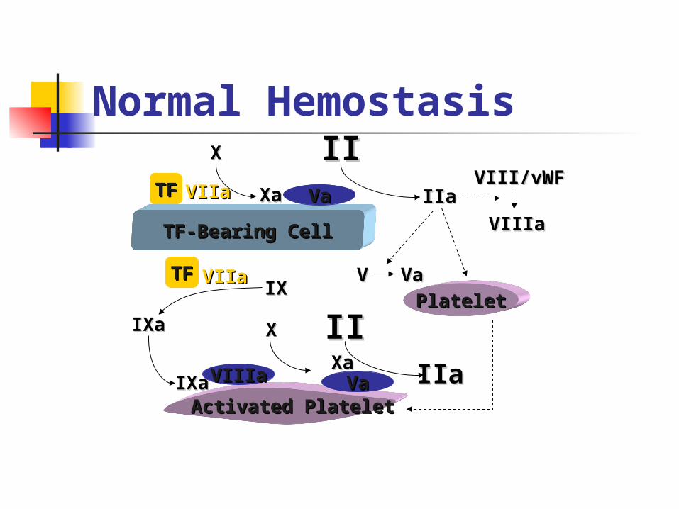
Normal Hemostasis

TF-Bearing CellTF-Bearing Cell
VaVaTFTF VIIaVIIa XaXa
XX IIIIIIaIIa
VIII/vWFVIII/vWF
VIIIaVIIIa
VV VaVa
PlateletPlatelet
Activated PlateletActivated Platelet
Normal Hemostasis

TF-Bearing CellTF-Bearing Cell
VaVaTFTF VIIaVIIa XaXa
XX IIIIIIaIIa
VIII/vWFVIII/vWF
VIIIaVIIIa
VV VaVa
PlateletPlatelet
TFTF VIIaVIIaIXIX
IXaIXa
Activated PlateletActivated Platelet
Normal Hemostasis
TF-Bearing CellTF-Bearing Cell
VaVaTFTF VIIaVIIa XaXa
XX IIIIIIaIIa
VIII/vWFVIII/vWF
VIIIaVIIIa
VV VaVa
PlateletPlatelet
TFTF VIIaVIIaIXIX
IXaIXa
Activated PlateletActivated Platelet
VIIIaVIIIa VaVaIXaIXaXaXa
IIaIIa
IIII
Normal Hemostasis
XX

Hoffman et al. Hoffman et al. Blood Coagul FibrinolysisBlood Coagul Fibrinolysis 1998;9(suppl 1):S61. 1998;9(suppl 1):S61.
Activated PlateletActivated Platelet
PlateletPlatelet
TFTF
VIIIaVIIIa VaVa
VIIaVIIa
XX IIII
TF-Bearing CellTF-Bearing Cell
VaVaTFTF VIIaVIIa XaXa IIaIIa
IXIXVV VaVa
IIII
VIII/vWFVIII/vWF
VIIIaVIIIa
IXaIXa XX
IXaIXa IIaIIaXaXa
Normal Hemostasis

Hoffman et al. Hoffman et al. Blood Coagul FibrinolysisBlood Coagul Fibrinolysis 1998;9(suppl 1):S61. 1998;9(suppl 1):S61.
TF-Bearing CellTF-Bearing Cell
Activated PlateletActivated Platelet
PlateletPlatelet
TFTF
VIIIaVIIIa VaVa
VIIIaVIIIa VaVa
VaVa
VIIaVIIa
TFTF VIIaVIIa XaXa
XX IIIIIIaIIa
IXIXVV VaVa
IIII
VIII/vWFVIII/vWF
VIIIaVIIIa
IIII
IXaIXa
XXIXIX
XX
IXaIXa
IXaIXaVIIaVIIaXaXa
IIaIIa
IIaIIa
XaXa
Normal Hemostasis: Pivotal role of TF/VIIa

Adhesion
GpIIb/IIIa
Platelet Activation Pathways
GpIIb/IIIaGpIIb/IIIa Aggregation
ADP
Adrenaline Platelet GpIb
Exposed Collagen
Endothelium
vWF
COLLAGEN
GpIIb/IIIaGpIIb/IIIa AggregationGpIIb/IIIaGpIIb/IIIa Aggregation
AdhesionAdhesion
ADP
Adrenaline
THROMBINTHROMBINHemophilia
Gp Glycoprotein

TissueTissueFactorFactor Factor Factor
VIIaVIIa
• The first step in all coagulation: The Tissue Factor- The first step in all coagulation: The Tissue Factor- Factor VIIa Factor VIIa complex formationcomplex formation
Bleeding through a Cut in a Vessel WallBleeding through a Cut in a Vessel Wall

TissueFactor- TissueFactor- Factor Factor VIIa VIIa
ComplexComplex
• The first step in all coagulation: The Tissue The first step in all coagulation: The Tissue Factor- Factor- Factor VIIaFactor VIIa complex formation complex formation
• This catalysis the coagulation cascade in This catalysis the coagulation cascade in normalnormal persons persons and in patients with bleeding and in patients with bleeding disordersdisorders
Bleeding through a Cut in a Vessel WallBleeding through a Cut in a Vessel Wall

Platelets
rFactorVIIarFactorVIIa
Recombinant Factor VIIa (rFVIIa)Recombinant Factor VIIa (rFVIIa) in high concentration in high concentration binds to platelets; thisbinds to platelets; this complex catalysis further complex catalysis further coagulation. coagulation. The local coagulation activation is greatly enhancedThe local coagulation activation is greatly enhanced
TissueFactor- TissueFactor- Factor Factor VIIa VIIa
ComplexComplex
Recombinant Factor VIIa Platelet BindingRecombinant Factor VIIa Platelet Binding

rFVIIarFVIIa
Platelets
High peak levels of recombinant Factor VIIaHigh peak levels of recombinant Factor VIIa(rFVIIa) induces formation of a strong fibrin (rFVIIa) induces formation of a strong fibrin
network.network.This network cross-binds and forms a solid This network cross-binds and forms a solid
hemostatic plughemostatic plug
TissueFactor-TissueFactor- rFVIIarFVIIa ComplexComplex
Further Formation of a Hemostatic Further Formation of a Hemostatic PlugPlug

Disseminated Intravascular Coagulation (DIC)

Primarily a thrombotic process Systemic process producing
both thrombosis and hemorrhage
Also called consumption coagulopathy and defibrination syndrome1
Its clinical manifestation may be widespread hemorrhage in acute, fulminant cases2.
-Background -Pathophysiology -Etiology -Clinical Manifestations -Diagnosis -Treatment -Xigris
DIC


Basic pathophysiology Entry into the circulation of procoagulant sub
stances Trigger systemic activation of the coagulation
system and platelets Lead to the disseminated deposition of fibrin-
platelet thrombi. Procoagulant stimulus is tissue factor (most c
ases) Lipoprotein Not normally exposed to blood.
Tissue factor gains access to blood by Tissue injury, Malignant cells, Expression on the surfaces of monocytes and e
ndothelial cells by inflammatory mediators.
-Background -Pathophysiology -Etiology -Clinical Manifestations -Diagnosis -Treatment
DIC


Tissue factor triggers Thrombin
Protease Induces fibrin formation and platele
t activation Other procoagulants
Cysteine protease Mucin (粘液素) Trypsin
-Background -Pathophysiology -Etiology -Clinical Manifestations -Diagnosis -Treatment
DIC
Acute DIC Coagulation factors are consum
ed at a rate in excess of the capacity of the liver to synthesize them,
Platelets are consumed in excess of the capacity of bone marrow megakaryocytes to release them.
-Background -Pathophysiology -Etiology -Clinical Manifestations -Diagnosis -Treatment
DIC

DIC Laboratory manifestations
Prolonged prothrombin time (PT) Prolonged Activated partial thromboplastin ti
me (aPTT) Thrombocytopenia. Increased fibrin formation
Stimulates compensatory process of secondary fibrinolysis,
Plasminogen activators generate plasmin to digest fibrin (and fibrinogen) into fibrin(ogen) degradation products (FDPs).
FDPs are potent circulating anticoagulants that contribute further to the bleeding manifestations of DIC.
Intravascular fibrin deposition can cause fragmentation of red blood cells and lead to the appearance of schistocytes in blood smears
Hemolytic anemia is unusual in DIC. Microvascular thrombosis in DIC can compromis
e the blood supply to some organs and lead to multiorgan failure
-Background -Pathophysiology -Etiology -Clinical Manifestations -Diagnosis -Treatment

-Background -Pathophysiology -Etiology -Clinical Manifestations -Diagnosis -Treatment
DIC

DIC always has an underlying etiology Must be identified and eliminated
to treat the coagulopathy (凝血病) successfully.
The development of DIC in many of these disorders is associated with an unfavorable outcome1.
Occurs in 1% of hospitalized patients Mortality rate approaches 40-80%
-Background -Pathophysiology -Etiology -Clinical Manifestations -Diagnosis -Treatment
DIC

Causes Infection
Most common cause of DIC. The syndrome particularly is associate
d with gram-negative or gram-positive sepsis
Can be triggered by a variety of other Bacterial Fungal Viral Rickettsial, and protozoal microorgani
sms.
-Background -Pathophysiology -Etiology -Clinical Manifestations -Diagnosis -Treatment
DIC

Obstetrics The placenta and uterine c
ontents are rich sources of Tissue factor Other procoagulants th
at normally are excluded from the maternal circulation
-Background -Pathophysiology -Etiology -Clinical Manifestations -Diagnosis -Treatment
DIC

Clinical manifestations of DIC may accompany obstetric complications, especially in the third trimester.
These syndromes range from Acute, fulminant, and often fatal DI
C in amniotic fluid embolism Blood is exposed to large amou
nts of tissue factor in a short period of time creating large amounts of thrombin
Multiorgan failure Chronic or subacute DIC with a retai
ned dead fetus. Exposure to small amounts of ti
ssue factor
-Background -Pathophysiology -Etiology -Clinical Manifestations -Diagnosis -Treatment
DIC

Other obstetric problems associated with DIC include
Abruptio placentae (胎盘剥离)ToxemiaSeptic abortion.
DIC -Background -Pathophysiology -Etiology -Clinical Manifestations -Diagnosis -Treatment

Clinical manifestations Determined by
Nature Intensity Duration of the underlying stimulus.
Chronicity Low-grade DIC is often asymptomatic
Diagnosed only by laboratory abnormalities. Bleeding is most common clinical finding
Generalized or widespread ecchymoses Chronic disease
Thrombotic complications Trousseau's syndrome in cancer Gangrene of the digits or extremities Hemorrhagic necrosis of the skin Purpura fulminans
Enhanced by Coexistence of liver disease
-Background -Pathophysiology -Etiology -Clinical Manifestations -Diagnosis -Treatment
DIC

Diagnosis of severe, acute (easy) Prolongation of PT, aPTT and Thrombin t
ime Due to consumption and inhibition of clot
ting factors Thrombocytopenia Fibrin degradatin products
Increased due to secondary fibrinolysis Measured by latex agglutination or D-dim
er assays. Schistocytes may be seen in the peripher
al blood smear Neither sensitive nor specific for DIC.
DIC -Background -Pathophysiology -Etiology -Clinical Manifestations -Diagnosis -Treatment

Chronic or compensated forms of DIC Highly variable patterns of abnormalities
in "DIC screen" coagulation tests. Increased FDPs and prolonged PT are ge
nerally more sensitive measures than are abnormalities of the aPTT and platelet count.
Overcompensated synthesis of consumed clotting factors and platelets in some chronic forms
Cause shortening of the PT and aPTT and/or thrombocytosis
Though, elevated levels of FDPs indicate secondary fibrinolysis in such cases.
-Background -Pathophysiology -Etiology -Clinical Manifestations -Diagnosis -Treatment
DIC

Treatment Identify underlying cause and
treat All other therapies are
temporizing
DIC -Background -Pathophysiology -Etiology -Clinical Manifestations -Diagnosis -Treatment
Asymptomatic patients with self-limited DIC Have only laboratory manifestati
ons of the coagulopathy No treatment may be necessary.
-Background -Pathophysiology -Etiology -Clinical Manifestations -Diagnosis -Treatment
DIC

Actively bleeding or who are at high risk of bleeding,
Blood component treatments of choice Transfusions of platelets
Improve the thrombocytopenia Fresh-frozen plasma (FFP)
Replace all consumed coagulation factors and correct the prolonged PT and aPTT.
Large volumes of plasma in severe cases The theoretical concern that these blood
products may "fuel the fire" and exacerbate the DIC has not been supported by clinical experience
DIC -Background -Pathophysiology -Etiology -Clinical Manifestations -Diagnosis -Treatment

Special cases Profound hypofibrinogenemia
Additional transfusion of cryoprecipitate,
Plasma concentrate enriched in fibrinogen
Sepsis Infusion of antithrombin III concent
rate may be considered as an adjunctive measure
-Background -Pathophysiology -Etiology -Clinical Manifestations -Diagnosis -Treatment
DIC

Pharmacologic inhibitors of coagulation and fibrinolysis Heparin Theoretical benefit
It blocks thrombin and the secondary fibrinolysis.
Might exacerbate the bleeding tendency Usually reserved for Forms manifested by
Thrombosis Acrocyanosis (发绀) Cancer Vascular malformations Retained dead fetus Acute promyelocytic leukemia.
早幼粒细胞
DIC -Background -Pathophysiology -Etiology -Clinical Manifestations -Diagnosis -Treatment

Antifibrinolytic agents, ε-aminocaproic acid and tranex
amic acid Generally are contraindicated
May precipitate thrombosis May be effective in decreasing lif
e-threatening bleeding
-Background -Pathophysiology -Etiology -Clinical Manifestations -Diagnosis -Treatment
DIC

Thank you