Embed Size (px)
Citation preview

Biogeosciences, 17, 2537–2551, 2020https://doi.org/10.5194/bg-17-2537-2020© Author(s) 2020. This work is distributed underthe Creative Commons Attribution 4.0 License.
Co-occurrence of Fe and P stress in natural populations of themarine diazotroph TrichodesmiumNoelle A. Held1,2, Eric A. Webb3, Matthew M. McIlvin1, David A. Hutchins3, Natalie R. Cohen1, Dawn M. Moran1,Korinna Kunde4, Maeve C. Lohan4, Claire Mahaffey5, E. Malcolm S. Woodward6, and Mak A. Saito1
1Department of Marine Chemistry and Geochemistry, Woods Hole Oceanographic Institution, Woods Hole, MA 02543, USA2Department of Earth, Atmospheric, and Planetary Sciences, Massachusetts Institute of Technology,Cambridge, MA 02139, USA3Marine and Environmental Biology, Department of Biological Sciences, University of Southern California,Los Angeles, CA 90089, USA4Ocean and Earth Science, National Oceanography Centre, University of Southampton, Southampton, UK5Department of Earth, Ocean and Ecological Sciences, University of Liverpool, Liverpool, UK6Plymouth Marine Laboratory, Plymouth, UK
Correspondence: Mak A. Saito ([email protected])
Received: 17 December 2019 – Discussion started: 16 January 2020Revised: 16 March 2020 – Accepted: 30 March 2020 – Published: 12 May 2020
Abstract. Trichodesmium is a globally important marine mi-crobe that provides fixed nitrogen (N) to otherwise N-limitedecosystems. In nature, nitrogen fixation is likely regulated byiron or phosphate availability, but the extent and interactionof these controls are unclear. From metaproteomics analysesusing established protein biomarkers for nutrient stress, wefound that iron–phosphate co-stress is the norm rather thanthe exception for Trichodesmium colonies in the North At-lantic Ocean. Counterintuitively, the nitrogenase enzyme wasmore abundant under co-stress as opposed to single nutrientstress. This is consistent with the idea that Trichodesmiumhas a specific physiological state during nutrient co-stress.Organic nitrogen uptake was observed and occurred simul-taneously with nitrogen fixation. The quantification of thephosphate ABC transporter PstA combined with a cellularmodel of nutrient uptake suggested that Trichodesmium isgenerally confronted by the biophysical limits of membranespace and diffusion rates for iron and phosphate acquisitionin the field. Colony formation may benefit nutrient acquisi-tion from particulate and organic sources, alleviating thesepressures. The results highlight that to predict the behaviorof Trichodesmium, both Fe and P stress must be evaluatedsimultaneously.
1 Introduction
The diazotrophic cyanobacterium Trichodesmium plays animportant ecological and biogeochemical role in the tropi-cal and subtropical oceans globally. By providing bioavail-able nitrogen (N) to otherwise N-limited ecosystems, it sup-ports basin-scale food webs, increasing primary productivityand carbon flux from the surface ocean (Capone et al., 1997;Carpenter and Romans, 1991; Coles et al., 2004; Deutsch etal., 2007; Sohm et al., 2011). Nitrogen fixation is energeti-cally and nutritionally expensive, so it typically occurs whenother sources of N are unavailable, i.e., in N-starved environ-ments (Karl et al., 2002). However, nitrogen availability isnot the sole control on nitrogen fixation, which must be bal-anced against the cell’s overall nutritional status. Because itcan access a theoretically unlimited supply of atmosphericN2, Trichodesmium often becomes phosphorus (P) limited(Frischkorn et al., 2018; Hynes et al., 2009; Orchard, 2010;Sañudo-Wilhelmy et al., 2001; Wu et al., 2000). It also hasa tendency to experience iron (Fe) limitation because the ni-trogenase enzyme is iron-demanding (Bergman et al., 2013;Chappell et al., 2012; Rouco et al., 2018; Sunda, 2012; Wal-worth et al., 2016a).
There is uncertainty about when and where Tri-chodesmium is Fe and P stressed and how this impacts ni-
Published by Copernicus Publications on behalf of the European Geosciences Union.

2538 N. A. Held et al.: Co-occurrence of Fe and P stress in Trichodesmium
trogen fixation in nature. Some reports suggest that Tri-chodesmium is primarily phosphate stressed in the North At-lantic and primarily Fe stressed in the Pacific, owing to rel-ative Fe and P availability in these regions (Bergman et al.,2013; Chappell et al., 2012; Frischkorn et al., 2018; Hyneset al., 2009; Orchard, 2010; Sañudo-Wilhelmy et al., 2001).However, others have suggested that Fe and P can be co-limiting to Trichodesmium; one incubation study found twoexamples of Fe–P co-limitation in the field (Mills et al.,2004). Even less clear is how Fe and/or P stress impacts ni-trogen fixation. For instance, despite the intuitive suggestionthat nitrogen fixation is limited by Fe or P availability, labo-ratory evidence indicated that Trichodesmium is specificallyadapted to co-limited conditions, with higher growth and N2-fixation rates under co-limitation than under single nutrientlimitation (Garcia et al., 2015; Walworth et al., 2016a).
There are several protein biomarkers for Fe and P stress inTrichodesmium, many of which are periplasmic binding pro-teins involved in nutrient acquisition. For Fe, this includesthe IdiA and IsiB proteins, and for phosphorus, specificallyphosphate, it includes the PstS and SphX proteins (see Ta-ble S1 in the Supplement). In Trichodesmium, IdiA, an ABCtransport protein, and IsiB, a flavodoxin, are expressed underFe-limiting conditions, and both are conserved across specieswith high sequence identity (Chappell et al., 2012; Webb etal., 2007). Transcriptomic and proteomic studies have shownthat they are more abundant during Fe stress, though thereis low-level basal level expression (Chappell et al., 2012;Snow et al., 2015; Walworth et al., 2016a). In this dataset,IdiA and IsiB were both highly abundant and correlated toone another (Fig. S1 in the Supplement). IdiA is used as themolecular biomarker of Fe stress in the following discussion,but the same conclusions could be drawn from IsiB distri-butions. Like IdiA and IsiB, SphX and PstS are conservedacross diverse Trichodesmium species (Chappell et al., 2012;Walworth et al., 2016a). SphX is abundant at the transcriptand protein level under phosphate limitation (Orchard et al.,2009; Orchard, 2010). PstS, a homologous protein locateda few genes downstream of SphX, responds less clearly tophosphate stress. In Trichodesmium, the reason may be thatPstS is not preceded by a pho box, a regulatory DNA se-quence which is necessary for P-based regulation (Orchardet al., 2009). Thus, in this study we focused on SphX as amarker of phosphate stress and IdiA as a marker of Fe stress.
Here, we present evidence based on field metaproteomesthat Trichodesmium colonies were simultaneously Fe and Pstressed, particularly in the tropical and subtropical Atlantic.While Fe–P stress has been suggested before, this study pro-vides molecular evidence for co-stress in a broad geograph-ical and temporal survey. This co-stress occurred across sig-nificant gradients in Fe and P concentration, suggesting nu-trient stress was driven not only by biogeochemical gradi-ents but also by Trichodesmium’s response to nutrient deple-tion; we explore possible biophysical and biochemical mech-anisms behind this. Fe and P stress were positively associated
with nitrogen fixation and organic nitrogen uptake, suggest-ing that Trichodesmium’s Fe, P, and N statuses are linked,perhaps via a regulatory network and/or due to the biophys-ical limits of nutrient uptake related to the intersection ofmembrane crowding and nutrient diffusion.
2 Materials and methods
2.1 Sample acquisition
A total of 37 samples were examined in this study. Sam-ples were acquired by the authors on various research ex-peditions, and most exist in biological duplicate or triplicate(Table S2). Trichodesmium colonies were handpicked from200 or 130 µm surface plankton net tows, rinsed thrice in0.2 µm filtered trace-metal-clean surface seawater into trace-metal-clean LDPE bottles, decanted onto 0.2–5 µm filters,and frozen until protein extraction. The samples were ofmixed puff and tuff morphology, depending on the natural di-versity present at the sampling location. The majority of sam-ples considered in this study were taken in the early morningpredawn hours. Details such as filter size, morphology, loca-tion, cruise, date, and time of sampling are provided in Ta-ble S2.
2.2 Protein extraction and digestion
Proteins were extracted by a detergent-based method follow-ing Saito et al. (2014) and Lu and Zhu (2005). To reduceprotein loss and contamination, all tubes were ethanol rinsedand dried prior to use, and all water and organic solventsused were LC–MS grade. Sample filters were placed in a mi-crofuge tube with 1–2 mL 1 % sodium dodecyl sulfate (SDS)extraction buffer (1 % SDS, 0.1 M Tris/HCL pH 7.5, 10 mMEDTA) and incubated for 10 min at 95 ◦C with shaking, thenfor 1 h at room temperature with shaking. The protein ex-tract was decanted and clarified by centrifugation (14 100 g)at room temperature. The crude protein extracts were quanti-fied with the colormetric BCA protein concentration assayusing bovine serum albumin as a standard (Pierce catalognumber 23225). Extracts were concentrated by 5 kD mem-brane centrifugation (Vivaspin spin columns, GE Health-care). The protein extracts were purified by organic precipita-tion (0.5 mM HCl made in 50 % methanol and 50 % acetone)at −20 ◦C for at least 1 week, then collected by centrifuga-tion at 14 100 g for 30 min at 4 ◦C, decanted, and dried byvacuum concentration for 10 min. The protein pellets wereresuspended in a minimum amount of 1 % SDS extractionbuffer and re-quantified by BCA protein concentration assayto assess extraction efficiency.
The proteins were embedded in a 500 µL final volumeacrylamide gel, which was then cut up into 1 mm piecesto maximize surface area and rinsed in 50 : 50 acetoni-trile / 25 mM ammonium bicarbonate overnight at room tem-perature. The next morning, the rinse solution was replaced
Biogeosciences, 17, 2537–2551, 2020 www.biogeosciences.net/17/2537/2020/

N. A. Held et al.: Co-occurrence of Fe and P stress in Trichodesmium 2539
and the rinse repeated for 1 h. Gels were dehydrated thricein acetonitrile, dried by vacuum centrifugation, rehydratedin 10 mM dithiothreitol (DTT) in 25 mM ammonium bi-carbonate, and then incubated for 1 h at 56 ◦C with shak-ing. Unabsorbed DTT solution was removed and the volumerecorded, allowing the total gel volume to be calculated. Gelswere washed in 25 mM ammonium bicarbonate, then incu-bated in 55 mM iodoacetamide for 1 h at room temperaturein the dark. Gels were again dehydrated thrice in acetonitrile.Trypsin (Promega Gold) was added at a ratio of 1 : 20 µg totalprotein in 25 mM ammonium bicarbonate in a volume suffi-cient to barely cover the gel pieces. Proteins were digestedovernight at 37 ◦C with shaking. Any unabsorbed solutionwas then removed to a new tube, and 50 µL of peptide ex-traction buffer (50 % acetonitrile, 5 % formic acid in water)was added and incubated for 20 min at room temperature.The supernatant was then decanted and combined with theunabsorbed solution, and the peptide extraction step was re-peated. The resulting peptide mixture was concentrated byvacuum centrifugation to 1 µg µL−1 concentration based onthe starting protein concentration. Finally, the peptides wereclarified by centrifugation at room temperature, taking thetop 90 % of the volume to reduce the carryover of gel debris.
2.3 Data acquisition
The global proteomes were analyzed by liquidchromatography–active modulation–liquid chromatog-raphy mass spectrometry (LCamLC-MS) using twoorthogonal, in-line reverse-phase columns: a PLRP-S col-umn (200 µm× 150 mm, 3 µm bead size, 300 Å pore size,NanoLCMS Solutions) and a C18 column packed in-house(100 m× 150 mm, 3 µm particle size, 120 Å pore size,C18 Reprosil-God, Dr. Maisch GmbH, packed in a NewObjective PicoFrit column). The first dimension utilizedan 8 h pH= 10 gradient (10 mM ammonium formate and10 mM ammonium formate in 90 % acetonitrile) and wastrapped every 30 min on alternating dual traps, then eluted at500 nL min−1 onto the C18 column with a 30 min gradient(0.1 % formic acid and 0.1 % formic acid in 99.9 % acetoni-trile); 10 µg of protein was injected per run directly onto thefirst column using a Thermo Dionex Ultimate3000 RSLC-nano system (Waltham, MA), and an additional RSLCnanopump was used for the second dimension gradient. Thesamples were then analyzed on a Thermo Orbitrap Fusionmass spectrometer with a Thermo Flex ion source (Waltham,MA). MS1 scans were monitored between m/z 380–1580,with a m/z 1.6 MS2 isolation window (CID mode), 50 msmaximum injection time, and 5 s dynamic exclusion time.
2.4 Relative quantitation of peptides and proteins
Raw spectra were searched with the Sequest algorithm us-ing a custom-built genomic database (Eng et al., 2008).The genomic database consisted of a publicly available Tri-
chodesmium community metagenome available on the JGIIMG platform (IMG ID 2821474806), as well as the en-tire contents of the CyanoGEBA project genomes (Shihet al., 2013). Protein annotations were derived from theoriginal metagenomes. SequestHT mass tolerances were setat ±10 ppm (parent) and ± 0.8 Dalton (fragment). Cys-teine modification of +57.022 and methionine modifica-tion of +16 were included. Protein identifications weremade with Peptide Prophet in Scaffold (Proteome Software)at the 95 % protein and peptide identification levels. Rel-ative abundance was measured by averaging the precur-sor intensity (area under the MS1 peak) of the top threemost abundant peptides in each protein, then by normal-izing this value to total precursor ion intensity. Normal-ization and global false discovery rate (FDR) calculations,which were 0.1 % at the peptide level and 1.2 % at the pro-tein level, were performed in Scaffold (Proteome Software).FDR was calculated by Scaffold using the probabilisticmethod by summing the assigned protein or peptide proba-bilities and dividing by the maximum probability (100 %) foreach. The mass spectrometry proteomics data have been de-posited in the ProteomeXchange Consortium via the PRIDEpartner repository with the dataset identifier PXD016225and can be accessed at https://doi.org/10.6019/PXD016225(Perez-Riverol et al., 2019; Held and Saito, 2019). Sta-tistical tests of relationships between proteins were con-ducted with the SciPy stats package (https://docs.scipy.org/doc/scipy/reference/stats.html, last access: December 2019)using linear Pearson tests when the relationship appeared tobe linear and a Spearman rank order test when this was notthe case.
2.5 Absolute quantitation of peptides
A small number of peptides were selected for absolute quan-titation using a modified heterologous expression system.The peptides were ensured to be specific to Trichodesmiumbased on sequence identity compared to over 300 marine bac-teria genomes, 3 metagenomes, and 956 specialized assem-blies (see http://www.metatryp.whoi.edu, last access: De-cember 2019) (Saito et al., 2015). A custom plasmid was de-signed that contained the Escherichia coli K12 optimized re-verse translation sequences for peptides of interest separatedby tryptic spacers (protein sequence: TPELFR). The peptidesand transition ions included are provided in Table S7. Toavoid repetition of the spacer nucleotide sequence, 12 dif-ferent codons were utilized to encode the spacer. Six equineapomyoglobin and three peptides from the commerciallyavailable Pierce peptide retention time calibration mixture(product number 88320) were also included. The sequencewas inserted into a pet(30a)+ plasmid using the BAMH1 5′
and XhoI 3′ restriction sites.The plasmid was transformed into competent
tuner(DE3)pLys E.coli cells and grown on kanamycin-amended LB agar plates to ensure plasmid incorporation.
www.biogeosciences.net/17/2537/2020/ Biogeosciences, 17, 2537–2551, 2020

2540 N. A. Held et al.: Co-occurrence of Fe and P stress in Trichodesmium
A single colony was used to inoculate a small amount of15N-labeled, kanamycin-containing SOC media (CambridgeIsotope Laboratories) as a starter culture. These cellswere grown overnight and then used to inoculate 10 mLof 15N-labeled, kanamycin-containing SOC media. Cellswere grown to approximately OD600 0.6, then inducedwith 1 mM isopropyl β-D-1-thiogalactopyranoside (IPTG),incubated in the overexpression phase overnight at roomtemperature, and harvested by centrifugation.
Cells were lysed with BugBuster detergent with addedbenzonase nuclease. The extracts were centrifuged and alarge pellet of insoluble cellular material remained. Becausethe plasmid protein was large, this pellet contained a largenumber of inclusion bodies containing nearly pure protein.The inclusion bodies were solubilized in 6 M urea at 4 ◦Covernight. The protein was reduced, alkylated, and trypsindigested in solution to generate a standard peptide mixture.
The standard mixture was calibrated to establish theexact concentration of the peptides. A known amount(10 fmol µL−1) of the commercially available Pierce standardpeptide mixture (Catalog number 88320) and an apomyo-globin digest were spiked into the standard. The ratio ofPierce (isotopically labeled according to JPT standards) orapomyoglobin (light) to heavy standard peptide MS2 peakarea was calculated and used to establish the final concentra-tion of the standard peptide mixture (Fu et al., 2016; Milo,2013). Multiple peptides were used for this calibration, andthe standard deviation among them was approximately 10 %.Finally, the linearity of the peptide standard was tested bygenerating a dilution curve and ensuring that the concen-tration of each standard peptide versus MS2 peak area waslinear between 0.001 and 20 fmol µL−1 concentration, using10 µL injections consistent with experimental injection vol-umes.
The sample was prepared at 0.2 µg µL−1 concentrationwith 10 µL injected to give a total of 2 µg sample protein. Theheavy labeled standard peptide mixture was spiked into eachsample at a concentration of 10 fmol µL−1. The concentra-tion of the light peptide was calculated as the ratio of the MS2area of the light : heavy peptide multiplied by 10 µg µL−1. Acorrection was applied for protein recovery before and afterpurification, and the result was the absolute concentration ofthe peptide in femtomoles per microgram total protein.
The percent of the membrane occupied by the ABC trans-porter PstA was calculated by converting the absolute proteinconcentration to molecules per Trichodesmium cell, using av-erage values for Trichodesmium cell volume (Hynes et al.,2012), carbon content per volume (Strathmann, 1967), pro-tein content per gram carbon (Rouwenhorst et al., 1991), andthe cross-sectional area of a calcium ATPase (Hudson andMorel, 1992) (see Table S5).
2.6 Self-organizing map analyses
Self-organizing maps were used to reduce the dimensional-ity of the data and explore relationships among co-varyingproteins of interest. Only Trichodesmium proteins were con-sidered. Analyses were conducted in Python 3.0, and fullyreproducible code is available at https://github.com/naheld/self_organizing_map_tricho_metaP (last updated: Decem-ber 2019).
The input data consisted of a table of protein names(rows) and samples (columns) such that the input vectorscontained 2818 features. To eliminate effects of scaling,the data were unit normalized with the Scikit-learn pre-processing algorithm. The input vectors were used to ini-tialize a 100-output-node (10× 10) self-organizing map us-ing the SOMPY Python library (https://github.com/sevamoo/SOMPY, last access: December 2019). The output nodeswere then clustered using a k-means clustering algorithm(k = 10) implemented in Scikit-learn. The input nodes (pro-teins) assigned to each map node were then retrieved, andthe entire process was repeated 10 000 times. Proteins wereconsidered in the same cluster if they appeared in the samecluster of output nodes more than 99.99 % of the time.
3 Results and discussion
3.1 Proteome overview
This study presents 36 field metaproteomes of colonial Tri-chodesmium populations collected at 16 locations on four ex-peditions (Table S2). All but one location were in the sub-tropical and tropical Atlantic, and most samples were col-lected in the early morning hours to avoid changes occur-ring on the diel cycle (Fig. 1 and Table S2). The metapro-teomes were analyzed with a two-dimensional LC-MS/MSworkflow that provided deep coverage of the proteome. Thisresulted in 4478 protein identifications, of which 2944 wereTrichodesmium proteins. The remaining proteins were fromcolony-associated epibionts, and they will be discussed in afuture publication. Protein abundance is presented as precur-sor (MS1) intensity of the three most abundant peptides foreach protein, normalized to total protein in the sample. Thus,changes in protein abundance were interpreted as changesin the fraction of the proteome devoted to the protein. Themost abundant were GroEL, ribosomal, and phycobilisomeproteins.
A self-organizing map analysis identified groups of pro-teins with similar profiles, i.e., proteins whose abundanceschanged cohesively, suggesting proteins that may be regu-lated similarly (Reddy et al., 2016). This revealed the cen-tral importance of nitrogen fixation to Trichodesmium. Thenitrogenase proteins were among the most abundant in theproteome and were located in clusters 1 and 2 (Fig. 2 and Ta-ble S3). Also in these clusters were nitrogen metabolism pro-
Biogeosciences, 17, 2537–2551, 2020 www.biogeosciences.net/17/2537/2020/

N. A. Held et al.: Co-occurrence of Fe and P stress in Trichodesmium 2541
Figure 1. Sampling locations. Red/pink colors indicate JC150 sta-tions, blue colors indicate Tricolim stations, dark gray indicates theBermuda Atlantic Time-series Study (BATS), and light gray indi-cates Hawaii Ocean Time-series (HOT). Most samples exist in du-plicate or triplicate; see Table S2 for detailed information.
teins including glutamine synthetase, glutamine hydrolyzingguanosine monophosphate (GMP) synthase, and glutamateracemase. This is consistent with previous reports findingthat N assimilation is synchronized with nitrogen fixation(Carpenter et al., 1992).
Nitrogen fixation was closely linked to carbon fixa-tion. Many photosystem proteins clustered with the nitroge-nase proteins, including phycobilisome proteins, photosys-tem proteins, and the citric acid cycle protein 2-oxoglutaratedehydrogenase. This clustering indicated the possibility ofdirect regulatory links between C and N fixation. The nitro-gen regulators P-II and NtcA were also present in this clus-ter and may mediate this association. In non-nitrogen fixingcyanobacteria, the high abundance of the nitrogen regulatorsNtcA and P-II is suggestive of nitrogen stress (Flores andHerrero, 2005; Saito et al., 2014). In diazotrophs, the roleof these regulators is unclear because they do not respondto nitrogen compounds such as ammonia as they do in othercyanobacteria (Forchhammer and de Marsac, 1994). Here,the clustering of NtcA and P-II with C and N fixation proteinssuggested that they play a role in balancing these processesin field populations, though the details of this role have yetto be elucidated.
In addition to identifying links between C and N fixa-tion, the metaproteomes demonstrated that field populationsof Trichodesmium invest heavily in macro- and micronu-trient acquisition. There were clusters of proteins involvedin trace metal acquisition and management, including Fe,zinc, and metal transport proteins, with the latter includ-ing proteins likely involved in Ni and Mo uptake (proteinIDs TCCM_0270.00000020 and TCCM_0481.00000160).We also noted clusters of proteins involved in phosphateacquisition. Importantly, SphX and PstS appear in separateclusters, highlighting differential regulation of these func-tionally similar proteins.
Figure 2. Heatmap displaying results of self-organizing map(SOM) analysis. Each protein was mapped to a self-organizing mapgrid, and the grids subsequently clustered by a k-means cluster-ing algorithm. The process was repeated 10 000 times, and the re-sults are displayed here as a heatmap with warm colors represent-ing proteins that appear in the same cluster. The color bar indicatesthe percent of SOM experiments in which two proteins appear inthe same cluster. Only the top 500 most abundant proteins are dis-played. Dark yellow indicates proteins that appear in the same clus-ter 99.99 % of the time. Clusters 1 and 2 contain nitrogen fixation,carbon fixation, and nitrogen assimilation proteins, as well as theregulatory systems NtcA and P-II. The cluster assignments for theproteins are available in Table S4.
3.2 Trichodesmium is simultaneously iron andphosphate stressed throughout the North Atlantic
A surprising emergent observation from the Trichodesmiummetaproteomes was the co-occurrence of the iron (IdiA) andphosphate (SphX) stress biomarkers across the samples. Theubiquitous and highly abundant presence of these proteinsrelative to total protein implied that co-stress may be thenorm rather than the exception for Trichodesmium coloniesin the field, particularly in the North Atlantic. Even thoughlow-level basal expression of IdiA and SphX has been ob-served, it was clear that the colonies were devoting a largefraction of their cellular resources to Fe and P uptake, re-spectively (see Tables S8 and S9) (Webb et al., 2001, 2007;Chappell and Webb, 2010; Orchard, 2010; Snow et al., 2015;Walworth et al., 2016a; Frischkorn et al., 2018). This, com-bined with the responsiveness of IdiA and SphX to nutrientavailability in Trichodesmium filaments in the laboratory, in-dicated that co-stress was occurring.
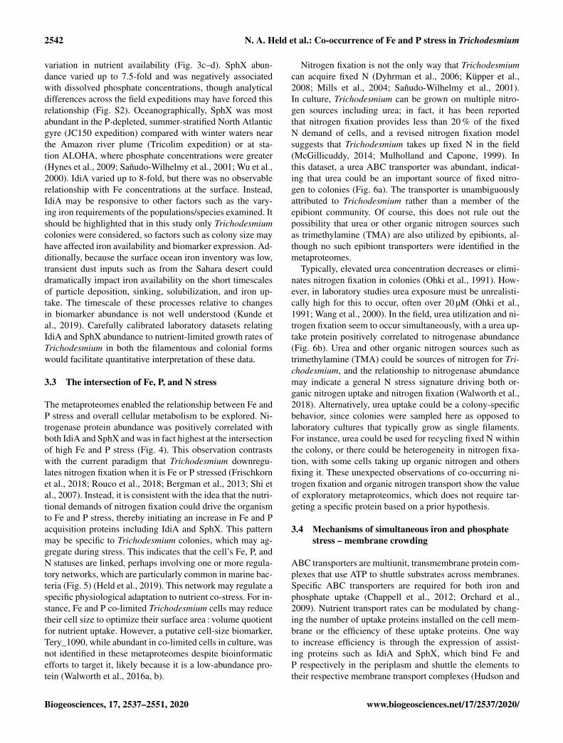
Interestingly, biomarker abundance was not necessarilycorrelated to nutrient concentrations in the surface ocean,suggesting that the colonies were experiencing stress despite
www.biogeosciences.net/17/2537/2020/ Biogeosciences, 17, 2537–2551, 2020
2542 N. A. Held et al.: Co-occurrence of Fe and P stress in Trichodesmium
variation in nutrient availability (Fig. 3c–d). SphX abun-dance varied up to 7.5-fold and was negatively associatedwith dissolved phosphate concentrations, though analyticaldifferences across the field expeditions may have forced thisrelationship (Fig. S2). Oceanographically, SphX was mostabundant in the P-depleted, summer-stratified North Atlanticgyre (JC150 expedition) compared with winter waters nearthe Amazon river plume (Tricolim expedition) or at sta-tion ALOHA, where phosphate concentrations were greater(Hynes et al., 2009; Sañudo-Wilhelmy et al., 2001; Wu et al.,2000). IdiA varied up to 8-fold, but there was no observablerelationship with Fe concentrations at the surface. Instead,IdiA may be responsive to other factors such as the vary-ing iron requirements of the populations/species examined. Itshould be highlighted that in this study only Trichodesmiumcolonies were considered, so factors such as colony size mayhave affected iron availability and biomarker expression. Ad-ditionally, because the surface ocean iron inventory was low,transient dust inputs such as from the Sahara desert coulddramatically impact iron availability on the short timescalesof particle deposition, sinking, solubilization, and iron up-take. The timescale of these processes relative to changesin biomarker abundance is not well understood (Kunde etal., 2019). Carefully calibrated laboratory datasets relatingIdiA and SphX abundance to nutrient-limited growth rates ofTrichodesmium in both the filamentous and colonial formswould facilitate quantitative interpretation of these data.
3.3 The intersection of Fe, P, and N stress
The metaproteomes enabled the relationship between Fe andP stress and overall cellular metabolism to be explored. Ni-trogenase protein abundance was positively correlated withboth IdiA and SphX and was in fact highest at the intersectionof high Fe and P stress (Fig. 4). This observation contrastswith the current paradigm that Trichodesmium downregu-lates nitrogen fixation when it is Fe or P stressed (Frischkornet al., 2018; Rouco et al., 2018; Bergman et al., 2013; Shi etal., 2007). Instead, it is consistent with the idea that the nutri-tional demands of nitrogen fixation could drive the organismto Fe and P stress, thereby initiating an increase in Fe and Pacquisition proteins including IdiA and SphX. This patternmay be specific to Trichodesmium colonies, which may ag-gregate during stress. This indicates that the cell’s Fe, P, andN statuses are linked, perhaps involving one or more regula-tory networks, which are particularly common in marine bac-teria (Fig. 5) (Held et al., 2019). This network may regulate aspecific physiological adaptation to nutrient co-stress. For in-stance, Fe and P co-limited Trichodesmium cells may reducetheir cell size to optimize their surface area : volume quotientfor nutrient uptake. However, a putative cell-size biomarker,Tery_1090, while abundant in co-limited cells in culture, wasnot identified in these metaproteomes despite bioinformaticefforts to target it, likely because it is a low-abundance pro-tein (Walworth et al., 2016a, b).
Nitrogen fixation is not the only way that Trichodesmiumcan acquire fixed N (Dyhrman et al., 2006; Küpper et al.,2008; Mills et al., 2004; Sañudo-Wilhelmy et al., 2001).In culture, Trichodesmium can be grown on multiple nitro-gen sources including urea; in fact, it has been reportedthat nitrogen fixation provides less than 20 % of the fixedN demand of cells, and a revised nitrogen fixation modelsuggests that Trichodesmium takes up fixed N in the field(McGillicuddy, 2014; Mulholland and Capone, 1999). Inthis dataset, a urea ABC transporter was abundant, indicat-ing that urea could be an important source of fixed nitro-gen to colonies (Fig. 6a). The transporter is unambiguouslyattributed to Trichodesmium rather than a member of theepibiont community. Of course, this does not rule out thepossibility that urea or other organic nitrogen sources suchas trimethylamine (TMA) are also utilized by epibionts, al-though no such epibiont transporters were identified in themetaproteomes.
Typically, elevated urea concentration decreases or elimi-nates nitrogen fixation in colonies (Ohki et al., 1991). How-ever, in laboratory studies urea exposure must be unrealisti-cally high for this to occur, often over 20 µM (Ohki et al.,1991; Wang et al., 2000). In the field, urea utilization and ni-trogen fixation seem to occur simultaneously, with a urea up-take protein positively correlated to nitrogenase abundance(Fig. 6b). Urea and other organic nitrogen sources such astrimethylamine (TMA) could be sources of nitrogen for Tri-chodesmium, and the relationship to nitrogenase abundancemay indicate a general N stress signature driving both or-ganic nitrogen uptake and nitrogen fixation (Walworth et al.,2018). Alternatively, urea uptake could be a colony-specificbehavior, since colonies were sampled here as opposed tolaboratory cultures that typically grow as single filaments.For instance, urea could be used for recycling fixed N withinthe colony, or there could be heterogeneity in nitrogen fixa-tion, with some cells taking up organic nitrogen and othersfixing it. These unexpected observations of co-occurring ni-trogen fixation and organic nitrogen transport show the valueof exploratory metaproteomics, which does not require tar-geting a specific protein based on a prior hypothesis.
3.4 Mechanisms of simultaneous iron and phosphatestress – membrane crowding
ABC transporters are multiunit, transmembrane protein com-plexes that use ATP to shuttle substrates across membranes.Specific ABC transporters are required for both iron andphosphate uptake (Chappell et al., 2012; Orchard et al.,2009). Nutrient transport rates can be modulated by chang-ing the number of uptake proteins installed on the cell mem-brane or the efficiency of these uptake proteins. One wayto increase efficiency is through the expression of assist-ing proteins such as IdiA and SphX, which bind Fe andP respectively in the periplasm and shuttle the elements totheir respective membrane transport complexes (Hudson and
Biogeosciences, 17, 2537–2551, 2020 www.biogeosciences.net/17/2537/2020/
N. A. Held et al.: Co-occurrence of Fe and P stress in Trichodesmium 2543
Figure 3. Relative abundance of iron stress protein IdiA (a) and phosphate stress protein SphX (b). IdiA and SphX were among the mostabundant proteins in the entire dataset. Error bars are 1 standard deviation from the mean. Dashed lines represent average values across thedataset. Protein abundances were normalized such that the total MS1 peak area across the entire proteome was the same for each sample. (c,d) Concentrations of dissolved Fe and dissolved phosphate nutrients. (e) Relative abundance of IdiA (orange) and SphX (blue) overlaid onthe sampling locations.
Morel, 1992). The high abundance of proteins involved inABC transport suggested that nutrient transport rates couldlimit the amount of Fe and P Trichodesmium can acquire.Thus, we explored whether membrane crowding, i.e., lackof membrane space, can constrain the nutrient acquisition ofTrichodesmium.
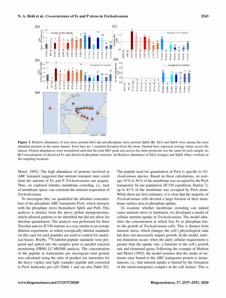
To investigate this, we quantified the absolute concentra-tion of the phosphate ABC transporter PstA, which interactswith the phosphate stress biomarkers SphX and PstS. Thisanalysis is distinct from the above global metaproteomes,which allowed patterns to be identified but did not allow forabsolute quantitation. The analysis was performed for threeTricolim and six JC150 stations in a way similar to an isotopedilution experiment, in which isotopically labeled standards(in this case for each peptide) are used to control for analyt-ical biases. Briefly, 15N-labeled peptide standards were pre-pared and spiked into the samples prior to parallel reactionmonitoring (PRM) LC-MS/MS analysis. The concentrationof the peptide in femtomoles per microgram total proteinwas calculated using the ratio of product ion intensities forthe heavy (spike) and light (sample) peptide and convertedto PstA molecules per cell (Table 1 and see also Table S5).
The peptide used for quantitation of PstA is specific to Tri-chodesmium species. Based on these calculations, on aver-age 19 % to 36 % of the membrane was occupied by the PstAtransporter. In one population (JC150 expedition, Station 7),up to 83 % of the membrane was occupied by PstA alone.While these are first estimates, it is clear that the majority ofTrichodesmium cells devoted a large fraction of their mem-brane surface area to phosphate uptake.
To examine whether membrane crowding can indeedcause nutrient stress or limitation, we developed a model ofcellular nutrient uptake in Trichodesmium. The model iden-tifies the concentration at which free Fe or phosphate lim-its the growth of Trichodesmium cells. This is distinct fromnutrient stress, which changes the cell’s physiological statebut does not necessarily impact growth. In the model, nutri-ent limitation occurs when the daily cellular requirement isgreater than the uptake rate, a function of the cell’s growthrate and elemental quota. Following the example of Hudsonand Morel (1992), the model assumes that the intake of nu-trients once bound to the ABC transporter protein is instan-taneous, i.e., that nutrient uptake is limited by the formationof the metal-transporter complex at the cell surface. This is
www.biogeosciences.net/17/2537/2020/ Biogeosciences, 17, 2537–2551, 2020

2544 N. A. Held et al.: Co-occurrence of Fe and P stress in Trichodesmium
Table 1. Quantification of the ABC transporter PstA and estimation of membrane space occupied.
Station [Pst] in Standard Pst molecules % surface area Pst molecules % surface areafmol µg−1 deviation per cell assuming occupied assuming per cell assuming occupied assuming
total protein replicates 30 % w/w protein 30 % w/wc 55 % w/w protein 55 % w/wc
(replicate average) (if available) contenta contentb
Tricolim_18 13.0 1.8 3.8× 10⁵ 3.6 7.0× 10⁵ 6.6Tricolim_15 11.2 3.4 3.3× 10⁵ 3.1 6.1× 10⁵ 5.7Tricolim_16 89.1 123.1 2.6× 10⁶ 24.5 4.8× 10⁶ 45.0JC150_3 38.7 63.3 1.1× 10⁶ 10.7 2.1× 10⁶ 19.5JC150_4 89.6 14.7 2.6× 10⁶ 24.7 4.9× 10⁶ 45.2JC150_5 74.2 36.4 2.2× 10⁶ 20.4 4.0× 10⁶ 37.5JC150_6 61.6 40.1 1.8× 10⁶ 17.0 3.3× 10⁶ 31.1JC150_7 165.7 4.9× 10⁶ 45.6 9.0× 10⁶ 83.6JC150_1 106.1 3.1× 10⁶ 29.2 5.7× 10⁶ 53.5
Average 19.9 36.4SD 13.4 24.6
a Calculated using Trichodesmium cell volume of 3000 µm3 (Berman-Frank et al., 2001), cell volume to carbon conversion logC = 0.716log(V )− 0.314 (Strathman, 1967), proteincontent of a cyanobacterium 30 % w/w (López et al., 2010), carbon to total protein conversion 0.53 g C g−1 total protein (Rouwenhorst et al., 1991). b Calculated as in footnote a
but with protein content of a cyanobacterium 55 % w/w (López et al., 2010). c Calculated using cross-sectional area of a Ca ATPase of 0.0000167 µm2 (Hudson and Morel, 1992).
Figure 4. Nitrogenase abundance is highest at the intersection ofhigh iron and phosphate stress. (a) IdiA and (b) SphX abundance ispositively related to nitrogenase MoFe (NifK) and Fe (NifH) pro-tein abundance (c represents the Spearman rank-order correlationcoefficient and p represents the Spearman p value). Effects of com-bined iron and phosphate stress biomarkers on nitrogenase abun-dance. Marker colors represent abundance of NifK (c) and NifH (d).
an idealized scenario, because if intake is the slow step, forinstance in a high-affinity transport system, the uptake ratewill be slower and nutrient limitation exacerbated (discussedbelow).
Figure 5. The metaproteomes suggest that there is a currently un-known regulatory link between cellular Fe, P, and N regulation.Fur is the ferric uptake regulator, PhoRB is the phosphate two-component sensory system, Tery_1090 is the putative cell-size reg-ulator, and P-II and NtcA are nitrogen regulatory proteins.
We considered two types of nutrient limitation in themodel (Table S10). First, we considered a diffusion-limitedcase in which the rate of uptake is determined by the dif-fusion of the nutrient to the cell’s boundary layer (µ ·Q=2/3kD [nutrient], where µ represents the cell growth rate, Qrepresents the cell nutrient quota, and kD represents the dif-fusion rate constant, dependent on the surface area and dif-fusion coefficient of the nutrient in seawater). Based on em-pirical evidence provided by Hudson and Morel (1992), lim-itation occurs when the cell quota is greater than two-thirdsthe diffusive-limited flux because beyond this, depletion ofthe nutrient in the boundary layer occurs. In the second case,membrane crowding limitation, the rate of uptake is deter-mined by the rate of transporter-metal complex formation(µ ·Q= kf [transport protein] [nutrient], where kf representsthe rate of ligand-nutrient complex formation). Here, up to50 % of the membrane can be occupied by the transport pro-tein following the example of Hudson and Morel (1992).This is within the range of the above estimates of mem-brane occupation by the phosphate transporter PstA. Themodel uses conservative estimates for diffusion coefficients,cell quotas, growth rates, and membrane space occupation to
Biogeosciences, 17, 2537–2551, 2020 www.biogeosciences.net/17/2537/2020/

N. A. Held et al.: Co-occurrence of Fe and P stress in Trichodesmium 2545
Figure 6. (a) Relative abundance of the Trichodesmium urea ABC transporter. (b) The abundance of the urea ABC transporter is positivelycorrelated with NifH and NifK abundance. Pearson linear correlation coefficients (r values) are provided (p value for NifK= 1.7× 10⁻⁵,NifH= 0.02). Shaded bars indicate 95 % confidence intervals.
identify the lowest concentration threshold for nutrient lim-itation; as a result it is likely that Trichodesmium becomeslimited at higher nutrient concentrations than the model sug-gests. At this time, the model can only consider labile dis-solved Fe and inorganic phosphate, though Trichodesmiumcan also acquire particulate iron, organic phosphorus, phos-phite, and phosphonates (Dyhrman et al., 2006; Frischkorn etal., 2018; Polyviou et al., 2015; Poorvin et al., 2004; Rubinet al., 2011).
We first considered a spherical cell, where the surfacearea : volume quotient decreases as cell radius increases(Fig. 7). As the cell grows in size, higher nutrient concentra-tions are required to sustain growth. This is consistent withthe general understanding that larger microbial cells withlower surface area : volume quotient are less competitive innutrient uptake (Chisholm, 1992; Hudson and Morel, 1992).For a given surface area : volume quotient, the mechanismdriving nutrient limitation is whichever model (diffusion ormembrane crowding) results in a higher minimum nutrientconcentration below which limitation occurs. For a sphericalcell, Fe limitation is driven by diffusion when the cell is largeand the surface area : volume quotient is low (Fig. 7a). How-ever, when cells are smaller and the surface area : volumequotient is high, membrane crowding drives nutrient limi-tation, meaning that the number of ligands – and not diffu-sion from the surrounding environment – is the primary con-trol on nutrient uptake. For phosphate, diffusion is almost al-ways the driver of nutrient limitation owing to the higher rateof transporter-nutrient complex formation (kf) for phosphate,which causes very fast membrane transport rates and relievesmembrane-crowding pressures across all cell sizes (Fig. 7b)(Froelich et al., 1982).
While this model may be directly applicable to some N2-fixing cyanobacteria such as Groups B and C, which haveroughly spherical cells, Trichodesmium cells are not spheresbut rather roughly cylindrical (Hynes et al., 2012). Thus, werepeated the model calculations for cylinders with varyingradii (r) and heights (2r or 10r) based on previous estimatesof Trichodesmium cell sizes (Bergman et al., 2013; Hynes etal., 2012). Cylinders have lower surface area : volume quo-
tient than spheres of similar sizes. In addition, the rate con-stant (kD) for diffusion, which is a function of cell geometry,is greater. This increases the slope of the diffusion limita-tion line such that membrane crowding is important across agreater range of cell sizes (Fig. 7c–d). Trichodesmium cellsizes vary in nature; for instance, the cylinder height canbe elongated, improving the surface area : volume quotient.However, the impact of cell elongation to radius r and height10r on both diffusion limitation and membrane crowding issubtle (Fig. 7e–f). Furthermore, though not explicitly consid-ered here, cylindrical cells living in filaments would have areduced surface area available for nutrient uptake. Thus, weconclude that in certain scenarios, lack of membrane spacecould hypothetically limit Fe and perhaps P acquisition byTrichodesmium, particularly when the cells live in filamentsor colonies as occurs in nature.
A key assumption of the model is that uptake rates are in-stantaneous. In the above calculations, we use the dissocia-tion kinetics of Fe from water and phosphate from commonseawater cations as the best case (i.e., fastest possible) ki-netic scenario for nutrient acquisition. The model does notaccount for delays caused by internalization kinetics, whichwould exacerbate nutrient limitation. The involvement of theperiplasmic binding proteins IdiA and SphX suggests thatuptake is not simultaneous; their participation is likely as-sociated with a kinetic rate of binding and dissociation fromthe periplasmic proteins in addition to any rate of ABC trans-port. Additionally, the model does not consider nutrient spe-ciation, which could affect internalization rates, particularlyfor Fe (Hudson and Morel, 1992).
Membrane crowding could produce real cellular chal-lenges, leading to the observation of Fe and P co-stress acrossthe field populations examined. The above model explicitlyallows 50 % of the cell surface area to be occupied by anyone type of transporter, consistent with our estimate of cellsurface area occupied by the PstA transporter. If 50 % of themembrane is occupied by phosphate transporters and another50 % for Fe transporters, this would leave no room for otheressential membrane proteins and even the membrane lipidsthemselves. The problem is further exacerbated if the cell in-
www.biogeosciences.net/17/2537/2020/ Biogeosciences, 17, 2537–2551, 2020

2546 N. A. Held et al.: Co-occurrence of Fe and P stress in Trichodesmium
Figure 7. Model calculations for membrane space and diffusion-based nutrient limitation reveal that membrane crowding could drive Tri-chodesmium to iron or phosphate stress, particularly when cells are small. Two cell morphologies (sphere and cylinder) were modeled forboth iron and phosphate limitation. Calculations are detailed in Table S10. As the cell radius increases and the surface area : volume quotientdecreases, the limiting concentration increases. This is concurrent with the current understanding that the low surface area : volume quotientof large cells leads to limitation. Green bars represent common SA :V quotients for T. theibautii (Hynes et al., 2012) (a–b). Membranecrowding (purple) occurs if the limiting nutrient concentration is greater than in the diffusion limitation model (blue). Membrane crowdingis more significant for cylindrical cells in particular (c–d); altering the length of the cylinder minimally affects the model (e–f).
stalls transporters for nitrogen compounds such as urea, asthe metaproteomes suggest. Thus, installation of transportersfor any one nutrient must be balanced against transportersfor other nutrients. This interpretation is inconsistent withLiebig’s law of nutrient limitation, which assumes that nu-trients are independent (Liebig, 1855; Saito et al., 2008). Inan oligotrophic environment, membrane crowding could ex-plicitly link cellular Fe, P, and N uptake statuses, driving thecell to be co-stressed for multiple nutrients.
3.5 Advantages of the colonial form for nutrientacquisition
Living in a colony has specific advantages and disadvantagesfor a Trichodesmium cell. Colonies may be able to access nu-trient sources that single cells or filaments could not feasiblyuse. For instance, Trichodesmium colonies have a remark-
able ability to entrain dust particles and can move these par-ticles into the center of said colony (Basu et al., 2019; Basuand Shaked, 2018; Poorvin et al., 2004; Rubin et al., 2011).In this study, which focused on Trichodesmium colonies,the chemotaxis response regulator CheY was very abundant,particularly in populations sampled near the Amazon andOrinoco river plumes. CheY was positively correlated withthe Fe stress biomarker IdiA but not with the phosphate stressbiomarker SphX, suggesting that chemotactic movement isinvolved in the entrainment of trace metals including fromparticulate sources (Fig. 8).
The metaproteomes and nutrient uptake model presentedin this paper support the growing understanding that Tri-chodesmium must be able to access particulate and organicmatter. Living in a colony can be advantageous becausesuch substrates can be concentrated, improving the viabil-ity of extracellular nutrient uptake systems. Trichodesmium’s
Biogeosciences, 17, 2537–2551, 2020 www.biogeosciences.net/17/2537/2020/

N. A. Held et al.: Co-occurrence of Fe and P stress in Trichodesmium 2547
Figure 8. CheY is positively correlated with the iron stressbiomarker IdiA but has a weaker association with the phosphatestress biomarker SphX. This suggests that it might be involved iniron acquisition, for instance by helping colonies to move dust par-ticles to the colony center. Pearson linear correlation coefficients(r values) are provided (p value for IdiA= 6× 10⁻⁴, SphX= 0.1).Shaded bars indicate 95 % confidence intervals.
epibiont community produces siderophores, which assist inFe uptake, particularly from particulate sources (Chappelland Webb, 2010; Lee et al., 2018). Siderophore productionis energetically and nutritionally expensive, so it is most ad-vantageous when resource concentrations are high and lossis low, as would occur in the center of a colony (Leventhal etal., 2019). Colonies may similarly enjoy advantages for phos-phate acquisition, particularly when the excreted enzymealkaline phosphatase is utilized to access organic sources(Frischkorn et al., 2018; Orchard, 2010; Orcutt et al., 2013;Yamaguchi et al., 2016; Yentsch et al., 1970). Additionally,the concentration of cells in a colony means that the productsof nitrogen fixation, including urea, can be recycled and areless likely to be lost to the environment.
A key hallmark of Trichodesmium colony formation is theproduction of mucus, which can capture particulate matterand concentrate it within the colony (Eichner et al., 2019). Inaddition to particle entrainment, the mucus layer can bene-fit cells by inhibiting oxygen diffusion, facilitating epibiontassociations, regulating buoyancy, defending against graz-ers, and helping to “stick” trichomes together (Eichner et al.,2019; Lee et al., 2018; Sheridan, 2002). However, these bene-fits come at a cost because the mucus layer hinders diffusionto the cell surface (Fig. 9), reducing contact with the sur-rounding seawater. Despite this, the benefits of colony forma-tion seem to outweigh the costs since Trichodesmium formscolonies in the field, particularly under stress (Bergman etal., 2013; Capone et al., 1997; Hynes et al., 2012).
4 Conclusions
Trichodesmium’s colonial lifestyle likely produces chal-lenges for dissolved Fe and P acquisition, which must be
Figure 9. Scheme for the effect of a mucus layer on nutrient dif-fusion. h represents the height of the mucus membrane and Dmrepresents the diffusion coefficient of the mucus. Assuming somediffusion constant for the nutrient through the mucus and the samestarting seawater nutrient concentration, a thicker layer of mucussurrounding a cell in a colony would result in a lower concentrationof nutrients experienced at the cell surface.
compensated for by the production of multiple nutrient trans-port systems, such as for particulate iron and organic phos-phorous, at a considerable cost. While laboratory studieshave largely focused on single nutrient stresses in free fila-ments, these metaproteomic observations and the accompa-nying nutrient uptake model demonstrate that Fe and P co-stress may be the norm rather than the exception for coloniesin the North Atlantic Ocean. This means that the emphasison single limiting nutrients in culture studies and biologicalmodels may not capture the complexities of Trichodesmium’sphysiology in situ. Thus, biogeochemical models should con-sider incorporating Fe and P co-stress conditions. Specifi-cally, in this study and in others there is evidence that nitro-gen fixation is optimal under co-limited or co-stressed condi-tions, implying that an input of either Fe or P could counter-intuitively decrease N2-driven new production (Garcia et al.,2015; Walworth et al., 2016a).
These data demonstrate that Trichodesmium cells are con-fronted by the biophysical limits of membrane space and dif-fusion rates for their Fe, P, and possibly urea acquisition sys-tems. This means that there is little room available for sys-tems that interact with other resources such as light, CO2, Ni,and other trace metals, providing a mechanism by which nu-trient stress could compromise the acquisition of other sup-plies. The cell membrane could be a key link allowing Tri-chodesmium to optimize its physiology in response to mul-tiple environmental stimuli. This is particularly important inan ocean where nutrient availability is sporadic and unpre-dictable. Future studies should aim to characterize the spe-cific regulatory systems, chemical species and phases (i.e.,
www.biogeosciences.net/17/2537/2020/ Biogeosciences, 17, 2537–2551, 2020

2548 N. A. Held et al.: Co-occurrence of Fe and P stress in Trichodesmium
dissolved versus particulate nutrient sources), and symbioticinteractions that underlie Trichodesmium’s unique behaviorand lifestyle.
Data availability. All new data are provided in the Supple-ment and are also available at the Biological and ChemicalOceanography Data Management Office (BCO-DMO) with thefollowing DOIs: https://doi.org/10.26008/1912/bco-dmo.787093.1(Held and Saito, 2020a), https://doi.org/10.26008/1912/bco-dmo.787078.1 (Held and Saito, 2020b),https://doi.org/10.26008/1912/bco-dmo.787147.1 (Held andSaito, 2020c), https://doi.org/10.26008/1912/bco-dmo.787168.1(Held and Saito, 2020d), and https://doi.org/10.26008/1912/bco-dmo.787181.1 (Held and Saito, 2020e). The mass spectrometryproteomics data have been deposited in the ProteomeXchangeConsortium via the PRIDE partner repository with the datasetidentifier PXD016225 and https://doi.org/10.6019/PXD016225(Held and Saito, 2019).
Supplement. Supplementary information is provided in a separatefile (Figs. S1 and S2, Tables S1, S2, S5, S6, S7, S8, S9) withTables S3, S4, and S10 provided separately due to their largesizes. The supplement related to this article is available onlineat: https://doi.org/10.5194/bg-17-2537-2020-supplement.
Author contributions. NAH and MAS conceptualized the study.DAH and EAW led the Tricolim expedition. CM and MCL led theJC150 expedition. NRC, EMSW, and KK measured nutrient distri-butions on the Tricolim and JC150 expeditions. DMM and MMMhelped with proteomic analyses. NAH prepared the paper with con-tributions from all co-authors.
Competing interests. The authors declare that they have no conflictof interest.
Acknowledgements. We acknowledge Elena Cerdan Garcia,Asa Conover, Joanna Harley, Despo Polyviou, and Petroc Shelleyfor assistance with sampling and nutrient measurements whileat sea, in addition to the entire crew of the JC150 and Tricolimexpeditions. We thank Ben Van Mooy for insightful discussionsregarding this work. This work was supported by a NationalScience Foundation Graduate Research Fellowship under grant1122274 (Noelle A. Held), Gordon and Betty Moore Foundationgrant number 3782 (Mak A. Saito), and National Science Foun-dation grants OCE-1657755 (Mak A. Saito), EarthCube-1639714(Mak A. Saito), OCE-1657757 (David A. Hutchins), and OCE-1851222 (David A. Hutchins). We also acknowledge funding fromthe UK Natural Environment Research Council (NERC) grantsawarded to CM (NE/N001079/1) and ML (NE/N001125/1). NRCwas supported by grant 544236 from the Simons Foundation.
Financial support. This research has been supported by the Na-tional Science Foundation (Division of Graduate Education (grantnos. 1122274), Division of Ocean Sciences (grant nos. 1657755,1657757, and 1851222), Directorate for Geosciences (grant no.1639714)), the Gordon and Betty Moore Foundation (grant no.3782), and the Natural Environment Research Council (NERC)(grant nos. NE/N001079/1 and NE/N001125/1).
Review statement. This paper was edited by Koji Suzuki and re-viewed by two anonymous referees.
References
Basu, S. and Shaked, Y.: Mineral iron utilization by naturaland cultured Trichodesmium and associated bacteria, Limnol.Oceanogr., 63, 2307–2320, https://doi.org/10.1002/lno.10939,2018.
Basu, S., Gledhill, M., de Beer, D., Matondkar, S. G. P., and Shaked,Y.: Colonies of marine cyanobacteria Trichodesmium interactwith associated bacteria to acquire iron from dust, Communica-tions Biology, 2, 1–8, https://doi.org/10.1038/s42003-019-0534-z, 2019.
Bergman, B., Sandh, G., Lin, S., Larsson, J., and Carpenter, E.J.: Trichodesmium – a widespread marine cyanobacterium withunusual nitrogen fixation properties, FEMS Microbiol. Rev.,37, 286–302, https://doi.org/10.1111/j.1574-6976.2012.00352.x,2013.
Berman-Frank, I., Cullen, J. T., Shaked, Y., Sherrell, R. M., andFalkowski, P. G.: Iron availability, cellular iron quotas, and ni-trogen fixation in Trichodesmium, Limnol. Oceanogr., 46, 1249–1260, https://doi.org/10.4319/lo.2001.46.6.1249, 2001.
Capone, D. G., Zehr, J. P., Paerl, H. W., Bergman, B.,and Carpenter, E. J.: Trichodesmium, a globally signif-icant marine cyanobacterium, Science, 276, 1221–1229,https://doi.org/10.1126/science.276.5316.1221, 1997.
Carpenter, E. J. and Romans, K.: Major Role of the CyanobacteriumTrichodesmium in Nutrient Cycling in the North Atlantic Ocean,Science, 254, 1989–1992, 1991.
Carpenter, E. J., Bergman, B., Dawson, R., Siddiqui, P. J., Söder-bäck, E., and Capone, D. G.: Glutamine synthetase and nitrogencycling in colonies of the marine diazotrophic cyanobacteria Tri-chodesmium spp., Appl. Environ. Microb., 58, 3122–3129, 1992.
Chappell, P. D. and Webb, E. A.: A molecular assessment of the ironstress response in the two phylogenetic clades of Trichodesmium,Environ. Microbiol., 12, 13–27, https://doi.org/10.1111/j.1462-2920.2009.02026.x, 2010.
Chappell, P. D., Moffett, J. W., Hynes, A. M., and Webb, E.A.: Molecular evidence of iron limitation and availability inthe global diazotroph Trichodesmium, ISME J., 6, 1728–1739,https://doi.org/10.1038/ismej.2012.13, 2012.
Chisholm, S. W.: Phytoplankton Size, in: Primary Productivity andBiogeochemical Cycles in the Sea, edited by: Falkowski, P. G.,Woodhead, A. D., and Vivirito, K., Springer, Boston, USA, 213–237, https://doi.org/10.1007/978-1-4899-0762-2_12, 1992.
Coles, V. J., Hood, R. R., Pascual, M., and Capone, D. G.:Modeling the impact of Trichodesmium and nitrogen fixation
Biogeosciences, 17, 2537–2551, 2020 www.biogeosciences.net/17/2537/2020/

N. A. Held et al.: Co-occurrence of Fe and P stress in Trichodesmium 2549
in the Atlantic ocean, J. Geophys. Res.-Oceans, 109, 1–17,https://doi.org/10.1029/2002JC001754, 2004.
Deutsch, C., Sarmiento, J. L., Sigman, D. M., Gruber,N., and Dunne, J. P.: Spatial coupling of nitrogen in-puts and losses in the ocean, Nature, 445, 163–167,https://doi.org/10.1038/nature05392, 2007.
Dyhrman, S. T., Chappell, P. D., Haley, S. T., Moffett,J. W., Orchard, E. D., Waterbury, J. B., and Webb,E. A.: Phosphonate utilization by the globally impor-tant marine diazotroph Trichodesmium, Nature, 439, 68–71,https://doi.org/10.1038/nature04203, 2006.
Eichner, M., Thoms, S., Rost, B., Mohr, W., Ahmerkamp, S., Ploug,H., Kuypers, M. M. M., and de Beer, D.: N2 fixation in free-floating filaments of Trichodesmium is higher than in transientlysuboxic colony microenvironments, New Phytol., 222, 852–863,https://doi.org/10.1111/nph.15621, 2019.
Eng, J. K., Fischer, B., Grossmann, J., and MacCoss, M. J.: Afast SEQUEST cross correlation algorithm, Journal of ProteomeResearch, 7(10), 4598–4602, https://doi.org/10.1021/pr800420s,2008.
Flores, E. and Herrero, A.: Nitrogen assimilation and nitro-gen control in cyanobacteria, Biochem. Soc. T., 33, 164–167,https://doi.org/10.1042/BST0330164, 2005.
Forchhammer, K. and de Marsac, N. T.: The P(II) protein in thecyanobacterium Synechococcus sp. strain PCC 7942 is modifiedby serine phosphorylation and signals the cellular N-status, J.Bacteriol., 176, 84–91, 1994.
Frischkorn, K. R., Krupke, A., Guieu, C., Louis, J., Rouco, M.,Salazar Estrada, A. E., Van Mooy, B. A. S., and Dyhrman, S.T.: Trichodesmium physiological ecology and phosphate reduc-tion in the western tropical South Pacific, Biogeosciences, 15,5761–5778, https://doi.org/10.5194/bg-15-5761-2018, 2018.
Froelich, P. N., Bender, M. L., Luedtke, N. A., Heath, G. R., andDeVries, T.: The Marine Phosphorus Cycle, Am. J. Sci., 282,464–511, 1982.
Fu, Q., Chen, Z., Zhang, S., Parker, S. J., Fu, Z., Tin, A., Liu, X.,and Van Eyk, J. E.: Multiple and Selective Reaction MonitoringUsing Triple Quadrupole Mass Spectrometer: Preclinical LargeCohort Analysis, in: Quantitative Proteomics by Mass Spectrom-etry, edited by: Sechi, S., Springer, New York, USA, 249–264,https://doi.org/10.1007/978-1-4939-3524-6_15, 2016.
Garcia, N. S., Fu, F., Sedwick, P. N., and Hutchins, D. A.:Iron deficiency increases growth and nitrogen-fixation rates ofphosphorus-deficient marine cyanobacteria, ISME J., 9, 238–245, https://doi.org/10.1038/ismej.2014.104, 2015.
Held, N. A. and Saito, M. A.: Trichodesmium field metaproteomes,PRIDE, https://doi.org/10.6019/PXD016225, 2019.
Held, N. A. and Saito, M. A.: Trichodesmium sample prove-nance from samples collected in North Atlantic surface wa-ters, station BATS, and station ALOHA between 2000 and2018, Biological and Chemical Oceanography Data Man-agement Office (BCO-DMO), Dataset version 2020-01-10,https://doi.org/10.26008/1912/bco-dmo.787093.1, 2020a.
Held, N. A. and Saito, M. A.: Metaproteomes of Trichodesmiumfrom samples collected in North Atlantic surface waters,station BATS, and station ALOHA between 2000 and2018, Biological and Chemical Oceanography Data Man-agement Office (BCO-DMO), Dataset version 2020-01-10,https://doi.org/10.26008/1912/bco-dmo.787078.1, 2020b.
Held, N. A. and Saito, M. A.: Net tow metaproteome of Tri-chodesmium species mapped to a Trichodesmium metagenomeplus cyanoGEBA species genomes in units of normalized pro-tein spectral counts from samples collected in the Atlantic andPacific Ocean between 2000 and 2018, Biological and Chem-ical Oceanography Data Management Office (BCO-DMO),Dataset version 2020-04-30, https://doi.org/10.26008/1912/bco-dmo.787147.1, 2020c.
Held, N. A. and Saito, M. A.: Net tow metaproteoome of Tri-chodesmium species mapped to a Trichodesmium metagenomeplus cyanoGEBA species genomes in units of normalized pep-tide spectral counts from samples collected in the Atlantic andPacific Ocean between 2000 and 2018, Biological and Chem-ical Oceanography Data Management Office (BCO-DMO),Dataset version 2020-04-30, https://doi.org/10.26008/1912/bco-dmo.787168.1, 2020d.
Held, N. A. and Saito, M. A.: FASTA file of sequences in Tri-chodesmium field metaproteomes mapped to a Trichodesmiummetagenome plus cyanoGEBA species genomes from sam-ples collected in the Atlantic and Pacific Ocean between2000 and 2018, Biological and Chemical Oceanography DataManagement Office (BCO-DMO), Dataset version 2020-01-14,https://doi.org/10.26008/1912/bco-dmo.787181.1, 2020e.
Held, N. A., McIlvin, M. R., Moran, D. M., Laub, M. T., and Saito,M. A.: Unique Patterns and Biogeochemical Relevance of Two-Component Sensing in Marine Bacteria, MSystems, 4, e00317-18, https://doi.org/10.1128/mSystems.00317-18, 2019.
Hudson, R. J. and Morel, F. M. M.: Trace metal transport by ma-rine microorganisms: implications of metal coordination kinet-ics, Deep-Sea Res. Pt. I, 40, 129–150, 1992.
Hynes, A. M., Chappell, P. D., Dyhrman, S. T., Doney, S. C.,and Webb, E. A.: Cross-basin comparison of phosphorus stressand nitrogen fixation in Trichodesmium, Limnol. Oceanogr., 54,1438–1448, https://doi.org/10.4319/lo.2009.54.5.1438, 2009.
Hynes, A. M., Webb, E. A., Doney, S. C., and Waterbury, J. B.:Comparison of cultured Trichodesmium (Cyanophyceae) withspecies characterized from the field, J. Phycol., 48, 196–210,https://doi.org/10.1111/j.1529-8817.2011.01096.x, 2012.
Karl, D., Michaels, A., Bergman, B., Capone, D., Carpenter, E.,Letelier, R., Lipschultz, F., Paerl, H., Sigman, D., and Stal, L.:Dinitrogen fixation in the world’s oceans, Biogeochemistry, 57–58, 47–98, https://doi.org/10.1023/A:1015798105851, 2002.
Kunde, K., Wyatt, N. J., González-Santana, D., Tagliabue,A., Mahaffey, C., and Lohan, M. C.: Iron Distributionin the Subtropical North Atlantic: The Pivotal Role ofColloidal Iron, Global Biogeochem. Cy., 33, 1532–1547,https://doi.org/10.1029/2019GB006326, 2019.
Küpper, H., Šetlík, I., Seibert, S., Prášil, O., Šetlikova, E., Strittmat-ter, M., Levitan, O., Lohscheider, J., Adamska, I., and Berman-Frank, I.: Iron limitation in the marine cyanobacterium Tri-chodesmium reveals new insights into regulation of photosynthe-sis and nitrogen fixation, New Phytol., 179, 784–798, 2008.
Lee, M. D., Webb, E. A., Walworth, N. G., Fu, F. X., Held, N.A., Saito, M. A., and Hutchins, D. A.: Transcriptional activi-ties of the microbial consortium living with the marine nitrogen-fixing cyanobacterium Trichodesmium reveal potential roles incommunity-level nitrogen cycling, Appl. Environ. Microb., 84,e02026-17 https://doi.org/10.1128/AEM.02026-17, 2018.
www.biogeosciences.net/17/2537/2020/ Biogeosciences, 17, 2537–2551, 2020
2550 N. A. Held et al.: Co-occurrence of Fe and P stress in Trichodesmium
Leventhal, G. E., Ackermann, M., and Schiessl, K. T.: Why mi-crobes secrete molecules to modify their environment: Thecase of iron-chelating siderophores, J. R. Soc. Interface, 16,20180674, https://doi.org/10.1098/rsif.2018.0674, 2019.
Liebig, J. V.: Principles of agricultural chemistry with special refer-ence to the late researches made in England, Dowden, Hutchin-son, and Ross, London, UK, 1855.
López, C. V. G., García, M. d. C., Fernández, F. G. A., Bustos,C. S., Chisti, Y., and Sevilla, J. M. F.: Protein measurementsof microalgal and cyanobacterial biomass, Bioresource Technol.,101, 7587–7591, https://doi.org/10.1016/j.biortech.2010.04.077,2010.
Lu, X. and Zhu, H.: Tube-Gel Digestion: A Novel Pro-teomic Approach for High Throughput Analysis of Mem-brane Proteins, Mol. Cell. Proteomics, 4, 1948–1958,https://doi.org/10.1074/mcp.M500138-MCP200, 2005.
McGillicuddy, D. J.: Do Trichodesmium spp. popula-tions in the North Atlantic export most of the nitro-gen they fix?, Global Biogeochem. Cy., 28, 103–114,https://doi.org/10.1002/2013GB004652, 2014.
Mills, M. M., Ridame, C., Davey, M., La Roche, J., and Gei-der, R. J.: Iron and phosphorus co-limit nitrogen fixation inthe eastern tropical North Atlantic, Nature, 429, 292–294,https://doi.org/10.1038/nature02550, 2004
Milo, R.: What is the total number of protein molecules per cellvolume? A call to rethink some published values, BioEssays, 35,1050–1055, https://doi.org/10.1002/bies.201300066, 2013.
Mulholland, M. R. and Capone, D. G.: Nitrogen utilization andmetabolism relative to patterns of N2 fixation in populations ofTrichodesmium from the North Atlantic Ocean and CaribbeanSea, Mar. Ecol.-Prog. Ser., 188, 33–49, 1999.
Ohki, K., Zehr, J. P., Falkowski, P. G., and Fujita, Y.: Reg-ulation of nitrogen-fixation by different nitrogen sourcesin the marine non-heterocystous cyanobacterium Tri-chodesmium sp. NIBB1067, Arch. Microbiol., 156, 335–337,https://doi.org/10.1007/BF00248706, 1991.
Orchard, E. D.: Phosphorus physiology of the marine Cyanobac-terium Trichodesmium, Massachusetts Institute of Technology,130, 81–122, https://doi.org/10.1575/1912/3366, 2010.
Orchard, E. D., Webb, E. A., and Dyhrman, S. T.: Molec-ular analysis of the phosphorus starvation response inTrichodesmium spp., Environ. Microbiol., 11, 2400–2411,https://doi.org/10.1111/j.1462-2920.2009.01968.x, 2009.
Orcutt, K. M., Gundersen, K., and Ammerman, J. W.: Intenseectoenzyme activities associated with Trichodesmium coloniesin the Sargasso Sea, Mar. Ecol.-Prog. Ser., 478, 101–113,https://doi.org/10.3354/meps10153, 2013.
Perez-Riverol, Y., Csordas, A., Bai, J., Bernal-Llinares, M., Hewap-athirana, S., Kundu, D. J. Inuganti, A., Griss, J., Mayer G., Eise-nacher, M., Perez, E., Uszkoriet, J., Pfeuffer, J., Sachsenberg, T.,Yilmaz, S., Tiwary, S., Cox, J., Audain, E., Walzer, M., Januczak,A. F., Ternent, T., Brazma, A., and Vizcaíno, J. A.: The PRIDEdatabase and related tools and resources in 2019: Improving sup-port for quantification data, Nucleic Acids Res., 47, D442–D450,https://doi.org/10.1093/nar/gky1106, 2019.
Polyviou, D., Hitchcock, A., Baylay, A. J., Moore, C. M., andBibby, T. S.: Phosphite utilization by the globally important ma-rine diazotroph Trichodesmium, Env. Microbiol. Rep., 7, 824–830, https://doi.org/10.1111/1758-2229.12308, 2015.
Poorvin, L., Rinta-kanto, J. M., Hutchins, D. A., and Wilhelm, S.W.: Viral release of iron and its bioavailability to marine plank-ton, Limnol. Oceanogr., 49, 1734–1741, 2004.
Reddy, R. J., Gajadhar, A. S., Swenson, E. J., Rothenberg, D. A.,Curran, T. G., and White, F. M.: Early signaling dynamics of theepidermal growth factor receptor, P. Natl. Acad. Sci. USA, 113,201521288, https://doi.org/10.1073/pnas.1521288113, 2016.
Rouco, M., Frischkorn, K. R., Haley, S. T., and Alexander, H.: Tran-scriptional patterns identify resource controls on the diazotrophTrichodesmium in the Atlantic and Pacific oceans, ISME J.,1486–1495, https://doi.org/10.1038/s41396-018-0087-z, 2018.
Rouwenhorst, R. J., Frank Jzn, J., Scheffers, W. A., and van Di-jken, J. P.: Determination of protein concentration by total or-ganic carbon analysis, J. Biochem. Bioph. Meth., 22, 119–128,https://doi.org/10.1016/0165-022X(91)90024-Q, 1991.
Rubin, M., Berman-Frank, I., and Shaked, Y.: Dust-and mineral-iron utilization by the marine dinitrogen-fixer Trichodesmium,Nat. Geosci., 4, 529–534, https://doi.org/10.1038/ngeo1181,2011.
Saito, M. A., Goepfert, T. J., and Ritt, J. T.: Some thoughtson the concept of colimitation: Three definitions and the im-portance of bioavailability, Limnol. Oceanogr., 53, 276–290,https://doi.org/10.4319/lo.2008.53.1.0276, 2008.
Saito, M. A., McIlvin, M. R., Moran, D. M., Goepfert, T. J.,DiTullio, G. R., Post, A. F., and Lamborg, C. H.: Mul-tiple nutrient stresses at intersecting Pacific Ocean biomesdetected by protein biomarkers, Science, 345, 1173–1177,https://doi.org/10.1126/science.1256450, 2014.
Saito, M. A., Dorsk, A., Post, A. F., McIlvin, M. R., Rappé, M. S.,DiTullio, G. R., and Moran, D. M.: Needles in the blue sea: Sub-species specificity in targeted protein biomarker analyses withinthe vast oceanic microbial metaproteome, Proteomics, 15, 3521–3531, https://doi.org/10.1002/pmic.201400630, 2015.
Sañudo-Wilhelmy, S. A., Kustka, A. B., Gobler, C. J., Hutchins, D.A., Yang, M., Lwiza, K., Burns, J., Capone, D. G., Raven, J. A.,and Carpenter, E. J.: Phosphorus limitation of nitrogen fixationby Trichodesmium in the central Atlantic Ocean, Nature, 411,66–69, https://doi.org/10.1038/35075041, 2001.
Sheridan, C. C.: The microbial and metazoan commu-nity associated with colonies of Trichodesmium spp.:a quantitative survey, J. Plankton Res., 24, 913–922,https://doi.org/10.1093/plankt/24.9.913, 2002.
Shi, T., Sun, Y., and Falkowski, P. G.: Effects of iron limitationon the expression of metabolic genes in the marine cyanobac-terium Trichodesmium erythraeum IMS101, Environ. Micro-biol., 9, 2945–2956, 2007.
Shih, P. M., Wu, D., Latifi, A., Axen, S. D., Fewer, D. P., Talla, E.,Calteau, A., Cai, F., Tandeau de Marsac, N., Rippka, R., Herd-man, M., Sivonen, K., Coursin, T., Laurent, T., Goodwin, L.,Nolan, M., Davenport, K. W., Han, C. S., Rubin, E. M., Eisen,J. A., Woyke, T., Gugger, M., and Kerfeld, C. A.: Improvingthe coverage of the cyanobacterial phylum using diversity-drivengenome sequencing, P. Natl. Acad. Sci. USA, 110, 1053–1058,https://doi.org/10.1073/pnas.1217107110, 2013.
Snow, J. T., Polyviou, D., Skipp, P., Chrismas, N. A. M., Hitch-cock, A., Geider, R., Moore, C. M., and Bibby, T. S.: Quantifyingintegrated proteomic responses to iron stress in the globally im-portant marine diazotroph Trichodesmium, PLoS ONE, 10, 1–24,https://doi.org/10.1371/journal.pone.0142626, 2015.
Biogeosciences, 17, 2537–2551, 2020 www.biogeosciences.net/17/2537/2020/

N. A. Held et al.: Co-occurrence of Fe and P stress in Trichodesmium 2551
Sohm, J. A., Webb, E. A., and Capone, D. G.: Emerging patternsof marine nitrogen fixation, Nat. Rev. Microbiol., 9, 499–508,https://doi.org/10.1038/nrmicro2594, 2011.
Strathmann, R. R.: Estimating Organic Carbon Content of Phy-toplankton from Cell Volume or Plasma Volume, Limnol.Oceanogr., 12, 411–418, 1967.
Sunda, W. G.: Feedback interactions between trace metal nutri-ents and phytoplankton in the ocean, Front. Microbiol., 3, 1–22,https://doi.org/10.3389/fmicb.2012.00204, 2012.
Walworth, N. G., Fu, F.-X., Webb, E. A., Saito, M. A., Moran, D.,Mcllvin, M. R., Lee, M. D., and Hutchins, D. A.: Mechanismsof increased Trichodesmium fitness under iron and phosphorusco-limitation in the present and future ocean, Nat. Commun., 7,1–11, https://doi.org/10.1038/ncomms12081, 2016a.
Walworth, N. G., Lee, M. D., Fu, F.-X., Hutchins, D. A., andWebb, E. A.: Molecular and physiological evidence of ge-netic assimilation to high CO2 in the marine nitrogen fixerTrichodesmium, P. Natl. Acad. Sci. USA, 3, 201605202,https://doi.org/10.1073/pnas.1605202113, 2016b.
Walworth, N. G., Fu, F. X., Lee, M. D., Cai, X., Saito, M. A., Webb,E. A., and Hutchins, D. A.: Nutrient-colimited Trichodesmium asa nitrogen source or sink in a future ocean, Appl. Environ. Mi-crob., 84, 1–14, https://doi.org/10.1128/AEM.02137-17, 2018.
Wang, Q., Li, H., and Post, A. F.: Nitrate assimilation genesof the marine diazotrophic, filamentous cyanobacterium Tri-chodesmium sp. strain WH9601, J. Bacteriol., 182, 1764–1767,https://doi.org/10.1128/JB.182.6.1764-1767.2000, 2000.
Webb, E. A., Moffett, J. W., and Waterbury, J. B.: IronStress in Open-Ocean Cyanobacteria (Synechoccoccus, Tri-chodesmium, and Crocosphaera spp.): Identification ofthe IdiA Protein, Appl. Environ. Microb., 67, 5444–5452,https://doi.org/10.1128/AEM.67.12.5444-5452.2001, 2001.
Webb, E. A., Jakuba, R. W., Moffett, J. W., and Dyhrman, S.T.: Molecular assessment of phosphorus and iron physiologyin Trichodesmium populations from the western Central andwestern South Atlantic, Limnol. Oceanogr., 52, 2221–2232,https://doi.org/10.4319/lo.2007.52.5.2221, 2007.
Wu, J. F., Sunda, W., Boyle, E. A., and Karl, D. M.: Phosphate de-pletion in the western North Atlantic Ocean, Science, 289, 759–762, https://doi.org/10.1126/Science.289.5480.759, 2000.
Yamaguchi, T., Furuya, K., Sato, M., and Takahashi, K.: Phos-phate release due to excess alkaline phosphatase activity in Tri-chodesmium erythraeum, Plankton Benthos Res., 11, 29–36,https://doi.org/10.3800/pbr.11.29, 2016.
Yentsch, C. M., Yentsch, C. S., and Perras, J. P.: Al-kaline phosphatase activity in the tropical marine blue-green alga Trichodesmium, Limnol. Oceanogr., 17, 72–774,https://doi.org/10.4319/lo.1972.17.5.0772, 1972.
www.biogeosciences.net/17/2537/2020/ Biogeosciences, 17, 2537–2551, 2020


![The Stellar Populations Galaxiesrichard/ASTRO620/...The Stellar Populations of Galaxies H.‐W. Rix IMPRS Galaxies Course March 11, 2011 Goal: Determine n (M *,t age,[Fe/H],R) for](https://img.dokumen.tips/doc/110x75/5f744a36325108294830c324/the-stellar-populations-galaxies-richardastro620-the-stellar-populations-of.jpg)