Embed Size (px)
Citation preview
Clues You Can Use:
Core Breast
Breast Imaging Division March 2018

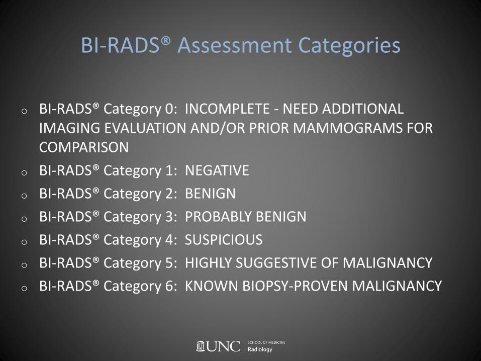
BI-RADS® Assessment Categories
o Assessments are divided into incomplete Category 0 and final assessment categories Categories 1-6
o Overall assessment based on the most worrisome finding or the need for immediate additional evaluation
o Screening mammograms may be assigned Category 0,1,2
BI-RADS® Assessment Categories
o BI-RADS® Category 0: INCOMPLETE - NEED ADDITIONAL IMAGING EVALUATION AND/OR PRIOR MAMMOGRAMS FOR COMPARISON
o BI-RADS® Category 1: NEGATIVE
o BI-RADS® Category 2: BENIGN
o BI-RADS® Category 3: PROBABLY BENIGN
o BI-RADS® Category 4: SUSPICIOUS
o BI-RADS® Category 5: HIGHLY SUGGESTIVE OF MALIGNANCY
o BI-RADS® Category 6: KNOWN BIOPSY-PROVEN MALIGNANCY

Management Recommendations
o Management recommendations on a per Assessment basis
o Wording emphasizes recall, routine mammography screening, tissue diagnosis, surgical excision
o Management recommended wording for tissue diagnosis is: “Biopsy should be performed in the absence of clinical contraindications.”
o Management recommended wording for BI-RADS® 6 is : “Surgical excision when clinically appropriate.”

o Category 0: INCOMPLETE - NEED ADDITIONAL IMAGING EVALUATION AND/OR PRIOR MAMMOGRAMS FOR COMPARISON
Recall for additional imaging and/or comparison with prior examinations
o Category 1: NEGATIVE
Routine mammography screening
o Category 2: BENIGN
Routine mammography screening
o Category 3: PROBABLY BENIGN
Short interval 6 month follow-up OR continued surveillance
o Category 4: SUSPICIOUS ABNORMALITY
Biopsy should be performed in the absence of clinical contraindications
o Category 5: HIGHLY SUGGESTIVE OF MALIGNANCY
Biopsy should be performed in the absence of clinical contraindications
o Category 6: KNOWN BIOPSY-PROVEN MALIGNANCY
Surgical excision when clinically appropriate

BI-RADS® Assessment Categories
o BI-RADS® Category 0: INCOMPLETE - NEED ADDITIONAL IMAGING EVALUATION AND/OR PRIOR MAMMOGRAMS FOR COMPARISON
o BI-RADS® Category 1: NEGATIVE
o BI-RADS® Category 2: BENIGN
o BI-RADS® Category 3: PROBABLY BENIGN <2% chance CA
o BI-RADS® Category 4: SUSPICIOUS 2-95% chance CA
o BI-RADS® Category 5: HIGHLY SUGGESTIVE OF MALIGNANCY >95% chance CA
o BI-RADS® Category 6: KNOWN BIOPSY-PROVEN MALIGNANCY

BI-RADS® Category 3 Assignment
NO !
➢ Screening mammogram
➢ Solitary dilated duct (B-R 4)
➢ BI-RADS® Atlas older edition so-called indeterminate calcifications (B-R 4)
➢ Developing asymmetry
➢ Any mass with mammogram orultrasound features of malignancy (B-R 4)
➢ Masses in BRCA gene mutation pts (B-R 4)
➢ Multiple bilateral circumscribed masses (B-R 2)
MAYBEEE
➢ MUST complete diagnostic workup prior to assigning BI-RADS® 3
➢ Solitary solid mass with benign mammogram and ultrasound features
➢ Focal asymmetry with negative CBE and ultrasound
➢ Fat necrosis calcifications
➢ Young women with palpable mass, imaging = FA

Screening Mammography
o Standard projections MLO and CC
o If Baseline risk pt: Begin age 40 and annual thereafter
o High risk screening pt: test or tests determined by her specific risk parameters ie mammo+US/MR
o SEARCH! Screening mammogram hallmarks of malignancy: suspiciousmass, calcifications, site of architectural distortion, asymmetry

Asymmetries
Unilateral deposits of fibroglandular tissue not conforming to the definition of a radiodense mass. Four types:
1. ASYMMETRY - visible in only one mammographic projection. Typically summation artifact
2. GLOBAL ASYMMETRY - large amount of fibroglandular-density tissue over a substantial portion of breast at least a quadrant compared to contralateral breast. Usually normal variant
3. FOCAL ASYMMETRY - relatively small amount of fibroglandular-density tissue over a confined portion of breast < a quadrant. Concave borders and interspersed fat distinguish from mass. DDx Superimposition of two normal structures, Mass
4. DEVELOPING ASYMMETRY - focal asymmetry that is new, larger, or more conspicuous than previously. 15% are CA

Architectural Distortion defined
o Normal breast architecture is distorted with no definite mass
o Thin straight lines of spiculations radiating from a point; focal retraction, distortion, straightening at the anterior or posterior edge of the parenchyma
o May also be seen in association with asymmetry or calcifications
o Determine if concordant history of trauma or surgery
o DDx = Cancer, Radial scar, Posttraumatic/Surgical scar

The Abnormal Screening Mammogram
Abnormal screening mammogram is assigned
BI-RADS® CATEGORY 0 and is called back for a Diagnostic breast imaging workup
o For mass/asymmetry: compression magnification views in MLO and CC possibly followed by targeted ultrasound
o For architectural distortion: compression magnification views in MLO and CC possibly followed by targeted ultrasound
o For calcifications: compression magnification views in TL 90 and CC
o PPV1 based on abnormal screening exam 3-8%
o PPV2 when biopsy - surgical, FNA, or core - recommended 20-40%

The Abnormal Screening Mammogram
ACR BI-RADS® 5th ed Desirable Medical Audit #s for interpreting radiologists
o Cancer detection rate per 1000 exams >2%
o Abnormal interpretation ie recall rate 5-12%
o Sensitivity >75%
o Specificity 88%-95%
o PPV1 based on abnormal screening exam 3-8%
o PPV2 when biopsy -surgical, FNA, or core - recommended 20-40%

Classic Benign Breast Disease
Fibroadenomao Most common breast mass in
patients <35 yoo Contain stromal tissue and breast
ductuleso Sensitive to hormone changes - eg
pregnancyo Oval parallel circumscribed
hypoechoico May contain calcificationso Biopsy is recommended for newly
palpable or increasing in size or demonstrating suspicious features on exam
Simple Cysto Occur in 10% of all women
o May be palpable, painful, grow/regress quickly
o Anechoic mass, imperceptible wall, posterior enhancement
o Painful cysts can be aspirated under ultrasound - benign type fluid is yellow, green

Classic Benign Breast Disease
Complicated Cyst
o Homogeneous low level internal echoes
o May have a layered appearance
o Fluid-debris levels may shift with pt position
o May also contain brightly echogenic foci that scintillate as they shift
o NOT complex cystic and solid echo mass
Clustered Microcystso Etiologies include fibrocystic
change and apocrine metaplasia
o Lesion consists of cluster of tiny anechoic foci, individually smaller than 3 mm
o Present as asx mammogram mass, occasionally palpable
o Thin intervening septations and no discrete solid component for B-R 3 vs B-R 2 mass
o Complex cystic and solid mass B-R 4 mass

Classic Benign Breast Disease
Fat Necrosiso Benign condition
o Trauma-induced may be surgical, although patient may have no knowledge of precipitating event
o Typically presents as palpable mass - oil cyst
o Occasionally present as irregular mass or calcifications that merit CNB
Papilloma
o Solitary intraductal papillomas usulocated centrally or in the retroareolar region
o Present with bloody or clear nipple discharge
o Most commonly observed in symptomatic perimenopausalpatients in the past
o Now presenting as mass on mammogram in younger asx
o Round or oval circumscribed masses, may be complex, intraductal, calcifications

Classic Benign Breast Disease
Lactating Adenoma
o Unique to pregnant and lactating pt
o Fibroadenoma mimicker
o Oval parallel circumscribed hypoechoic
o <3cm and contain stromal tissue and breast ductules
o May contain calcifications
o May infarct and present with enlarging mass
o Tend to regress after cessation of breastfeeding
Galactocele
o Unique to pregnant and lactating pt
o Typically present as fluctuating mass in pt who recently ceased breastfeeding
o Round or oval parallel circumscribed mass. May be an-hypo- or hyperechoic, depending on fat content. Mammo TL proj for fat fluid level
o Tend to regress spontaneously and most do NOT require aspiration
o May be aspirated Fat fluid levels
true latFA appearance in pregnant? Think LA!

Classic Breast Disease
Phyllodes
o Fibroepithelial - histologically similar to fibroadenoma
o Typically present as rapidly enlarging mass
o Appearance is similar to large fibroadenoma
o May be complex and contain cystic spaces
o 10% malignant - no distinguishing features
o Excision of benignAND malignant
o Recurrence is local. Rare lung, bone, liver mets
DCIS
o Accounts for 20-25% CAo Confined to the ductso Increased risk in pts with family
history, elevated BMI postmenopausal, dense breasts
o Mortality is extremely low and related to IDC 8-10 years post diagnosis of DCIS
o Mammographically detectedo May also present with mass, nipple
discharge, Paget diseaseo On MRI - NME
with delayed peak enhancementkinetic

Mammo Calcifications BI-RADS® AtAWord
BENIGN
o All ‘typically benign’: Round, Rim, Dystrophic, Coarse ie Popcorn, Milk of Calcium, Large Rod-Like ieSecretory, Skin, Vascular, Suture 0% risk
o Diffuse
SUSPICIOUS
o Fine linear and fine linear branching 70% risk
o Fine pleomorphic 29%
o Amorphous 21%
o Coarse heterogeneous 13%
o Segmental 62%
o Linear 60%
o Grouped 31%
o Regional 26%
CA % RISKS reported are in BOLD

Mammo Mass BI-RADS® AtAWord
BENIGN
Mass
Shape: Oval RoundMargin: CircumscribedDensity: Fat Low
SUSPICIOUS
Mass
Shape: IrregularMargin: Spiculated Indistinct
MicrolobulatedDensity: High EqualAssociated Features:
Skin retractionNipple retractionSkin thickeningTrabecular thickeningAxillary adenopathy

US Mass BI-RADS® AtAWord
BENIGN
Mass
Shape: Oval
Orientation: Parallel
Margin: Circumscribed
Echogenicity: Anechoic
Hyperechoic
Posterior Features: Enhanced
SUSPICIOUS
Mass
Shape: IrregularOrientation: Not parallelMargin: Not circumscribedEchogenicity:
HypoechoicIsoechoicComplex cystic and solidHeterogeneous
Posterior Features: NoneShadowingCombined

MR Mass BI-RADS® AtAWord
BENIGN
Mass
Shape: Oval
Margin: Circumscribed
Enhancement:
Homogeneous
Dark internal septations
NME Homogeneous
Multiple
Diffuse
SUSPICIOUS
Mass
Shape: Irregular
Margin: Spiculated
Irregular
Enhancement: Heterogeneous
Rim
NME Heterogeneous
Clumped
Clustered ring
Segmental
Linear

Classic Malignant Breast Disease
Invasive Mammary (Ductal)
o Accounts for 50-75% invasive cancers
o Heterogeneous group of tumors without sufficient histologic features to be classified more specifically
o Ductal origin
o Present as mass, size varies
o Mammogram, US, and MR evident
Invasive Lobular
o Accounts for 10-15% CA
o Lobule rather than duct origin
o Women in early 60s slightly older than IDC
o Present as thickening
o Present as asymmetry, architectural distortion, even calcifications but may be mammographically occult
o Bilateral, multifocal, multicentric,
o Indistinct mass on US
o MRI may be required

Special Types Breast CA
Re Special subtypes - all account for <1-5% of CA:
➢ Asymmetry and architectural distortion - think lobular
➢ Small spiculated mass mimicking radial scar - think tubular
➢ Favorable prognosis round-ish circumscribed-ish mass - think papillary, medullary, mucinous
➢ T2 bright enhancing mass - think mucinous
➢ BRCA1 and BRCA2 patients - think medullary
➢ Large and extremely dense - think metaplastic
➢ Nipple eczematous or erythematous change - think Paget
Tu Mu Med Met Pa

MR Characteristics of Breast Masses
Benign
Mass
Shape: Oval
Margin: Circumscribed
Enhancement:
Homogeneous
Dark internal septations
NME Homogeneous
Multiple
Diffuse
Suspicious
MassShape: IrregularMargin: Spiculated
IrregularEnhancement: Heterogeneous
RimNME Heterogeneous
Clumped Clustered ringSegmentalLinear
Breast Mass
T1 Pre NonFatSat T2 STIR
T1 Pre FatSat
T1 Post FatSat Distinguishing Features
FA dark bright dark bright dark internal septation
Simple Cyst dark bright dark dark imperceptible wall
Complicated Cyst dark bright dark may be bright rim enhancement
LNdark except hilum bright dark bright fatty hilum Type III curve
Papilloma bright bright bright bright homogenous enhancement
Oil Cyst bright dark dark dark
Fat Necrosis bright dark dark may be bright variable enhancement
CA - IBC NST dark dark dark bright Type III or II enhancement
CA - Mucinous dark bright dark bright homogenous enhancement
CA - TNBC dark dark dark brightheterogeneous rim Type III enhancement

From Special Types Breast CA on to . . .
Special Types Clinical PresentationsEach of these are assigned up to 5% on Core but more importantly, are
common scenarios presenting to clinic!
➢ Nipple discharge - distinguish clinical presentation
➢ Male breast - distinguish gynecomastia and breast CA
➢ Axillary lymphadenopathy - distinguish unilateral and bilateral
➢ Implants - distinguish saline and silicone ruptures
➢ Inflammatory disease - distinguish differential diagnoses
➢ Breast conserving therapy - distinguish normal and recurrence
LN Si EIC BCT rim Gynecom

Nipple Discharge
BENIGN
o Usually bilateral, multiductal
o Yellow, orange, green, milky
o Occurs with breast manipulation
o Age<40yo
o Etiologies: pregnancy, lactation, physiologic, drug-related
PATHOLOGIC
o More likely unilateral, uniductal
o Bloody, clear
o Occurs spontaneously
o Age >40yo
o May be associated with mass
o Duct ectasia, abscess, papilloma, high-risk lesion, DCIS, invasive cancer
o Isolated papillomas are usually benign, but can harbor areas of atypia or DCIS
o Solitary dilated duct 9% chance of malignancy ie no longer BI-RADS 3

Male Breast Disease
Gynecomastia Rule of 3s
o Unilateral
o 3 times for gynecomastia: neonate, puberty, senescence
o 3 types gynecomastia: nodular, dendritic, diffuse
o 3+ etiologies gynecomastia: physiologic, drugs, hyperestrogen, systemic diseases cirrhosis, CRF
o Gynecomastia: soft tender mass, mobile, bilateral, central to nipple, typical mammogram flame-shaped appearance with no secondary features, no axillary LN
Male Breast Cancer
o Male breast CA: soft or firm nontender mass, nonmobile or mobile; unilateral; eccentric to nipple, typical mammogram irregular hyperdense mass may have calcifications, skin thickening, nipple retraction, axillary LN, iesecondary features
o Histologies: IDC NOS, DCIS, Invasive papillary
o NOT ILC
o Males > 60 years
o Account for <1% new breast CA diagnoses and may be associated with BRCA2 gene mutation

Male Breast Disease
Pseudogynecomastia
o Usually bilateral
o No palpable mass
o Excessive fat deposition in the breasts
o Results from genetic normal variant, truncal obesity, and occasionally in neurofibromatosis
Other Masseso Epidermal inclusion cyst
o IMLN
o Papilloma
o Mesenchymal tumors including lipoma, granular cell tumor, myofibroblastoma
Abscess
o Male breast abscess - typically present as tender palpable mass with erythema and warmth
o Rx male breast abscess is similar to female
Breast Hematomao Antecedent trauma or
anticoagulant therapy
o Mass or bruise ie skin involved
o Complex cystic and solid mass

Axillary Lymphadenopathy
BI-RADS® Category 4
o Unilateral
o DDx breast carcinoma, metastatic melanoma, ovarian CA, other CA
o Careful eval ipsilateral breast
o Bilateral axillary US to determine if uni/bilateral
o (Clinical eval for mastitis, breast abscess, skin infx, cat scratch fever ie convert to 2)
o Proceed to FNA or CNB
BI-RADS® Category 2
o Bilateral
o Frequently reactive in inflammatory ds and HIV
o Sarcoid, SLE, psoriasis, analogous ds
o Known dx Lymphoma - add wording “known lymphoma”
o When bilateral LN new or increasing - rethink BI-RADS® 4 and include pass for flow cytometry (saline or RPMI)

Breast Implants Rule of 2s
o 2 types Saline, Silicone
o 2 locations Retropectoral, Subglandular
o 2 indications Cosmesis, Reconstruction
o 2 postoperative complications Early: hematoma and infection Late: capsular contraction and implant rupture
o 2 Si ruptures Intracapsular, Extracapsular
o 2 x 2 signs Si rupture Intracapsular - ladder, teardrop, keyhole, salad oil and linguini signs Extracapsular - snowstorm and free silicone/silicone granuloma Also, 2 extracapsular implant rupture are current or prior implant rupture and silicone injections
o 2 MRI frequencies to suppress for silicone Fat suppression and Water suppression
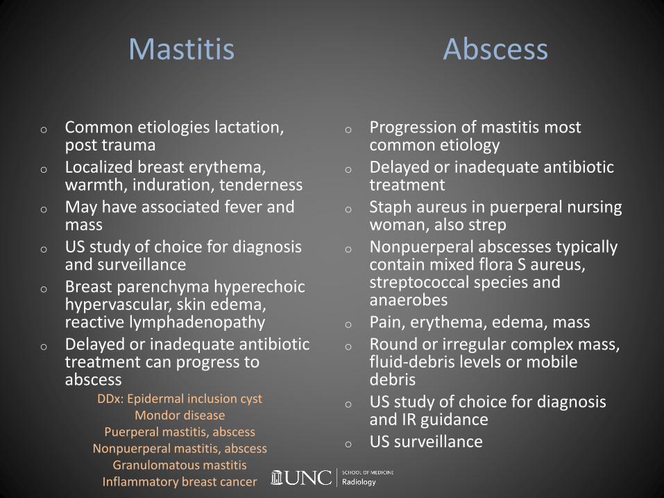
Mastitis Abscess
o Common etiologies lactation, post trauma
o Localized breast erythema, warmth, induration, tenderness
o May have associated fever and mass
o US study of choice for diagnosis and surveillance
o Breast parenchyma hyperechoichypervascular, skin edema, reactive lymphadenopathy
o Delayed or inadequate antibiotic treatment can progress to abscess
DDx: Epidermal inclusion cystMondor disease
Puerperal mastitis, abscessNonpuerperal mastitis, abscess
Granulomatous mastitisInflammatory breast cancer
o Progression of mastitis most common etiology
o Delayed or inadequate antibiotic treatment
o Staph aureus in puerperal nursing woman, also strep
o Nonpuerperal abscesses typically contain mixed flora S aureus, streptococcal species and anaerobes
o Pain, erythema, edema, masso Round or irregular complex mass,
fluid-debris levels or mobile debris
o US study of choice for diagnosis and IR guidance
o US surveillance

Post Breast Conserving Therapy
Normal ie BI-RADS® Category 2,3
o Lumpx or axillary node site complex cystic fluid collection
o Trabecular thickening (breast edema) AND skin thickening peak at 6 months post radiation then improve thereafter and attain stability at 2-3 yrs
o Lumpx and axillary node sites benign architectural distortion (with central lucencies) stabilize by 2 yrs
o Fat necrosis calcifications both dystrophic and rim oil cysts
Abnormal BI-RADS® Category 4,5
o Recurrence - at lumpx site but rare until 2 yr mark
o New or increasing asymmetry, mass, linear pleomorphic amorphous calcifications all concerning
o Metachronous tumor - other quadrants of ipsilateral and in contralateral breast
o Radiation-induced malignancies esp (angio)sarcoma

Breast IR etcetera
1. Sentinel node injection Nonmigration of isotope: injection into malignant mass, injection into hematoma/seroma, lymphatic obstruction or alteration in the setting of bulky axillary LAN or prior lymph node surgery, morbid obesity, CHF/severe CVD, idiopathic
2. US CNB guidance US-guided biopsy preferred offers several advantages over stereotactic and MR imaging-guided CNB
o real-time procedure allows visualization and verification of accurate targeting with faster procedure times
o requires no breast compression and allows more comfortable positioning for the patient
o no ionizing radiation is used compared with stereo CNB and no modality-based contraindications as compared with MR imaging

Stereotactic CNB Tips & Targeting

Stereotactic CNB Tips & Targeting
Stereotactic Tips:o Axes are X (horizontal), Y (vertical), and Z (depth)o Stereotactic are paired 15 degree off midline. Scout image -> X and Y, Stereos -> Zo Stroke margin is distance from postfire needle tip to the distal surface of breasto Negative stroke margin indicates risk postfire needle will strike image receptor distally

Specimen Radiograph - 3 types
1. Core needle biopsy specimen - confirm adequate sampling of the specific calcifications of concern on prebiopsy mammograms. a must for stereotactic biopsy procedures
2. Surgical sample specimen - confirm excision of the localized abnormalities, to include the specific mass/calcifications/architectural distortion/other, metallic localizer clip, and intact localization wire in entirety. also adequacy of surgical margins
3. Paraffin blocks specimen - directs pathologist to detailed review of biopsy or surgical specimen blocks in case of subtle finding

Recommend further tissue sampling due to:
High Risk and Etcetera
o Atypical ductal hyperplasia ADH
o Lobular neoplasia: Atypical lobular hyperplasia ALH Lobular carcinoma in situ LCIS
o Flat epithelial atypia FEA
o Papilloma with atypia
o Radial scar and Complex sclerosing lesion
o Phyllodes
o Locally aggressive benign (mesenchymal)
o Discordance
o Insufficient tissue
If discordant results are returned, arrangements for repeat image guided biopsy or surgical biopsy should be made. If the pathology results are malignant, review of the entire case should be performed, including index and opposite breast, axilla to determine if other findings now require closer scrutiny or bx