Embed Size (px)
Citation preview

Clues for Identifying Unstable Pelvis Fractures
Walter W. Virkus, MD Rush University Medical Center
Chicago, IL

Conflict of Interest
• Consultant- Stryker Orthopedics, Smith & Nephew • Stock- Stryker, Wright Medical

Acknowlegdements
• Andy Burgess, MD • Cliff Turen, MD

Clue #1: HISTORY OF INJURY
• Mechanism – MVC – MCC – Fall from height
• Signs of Shock – Best predictor of mortality
High vs low energy

ASSESSMENT • ABC’S of ATLS • Soft tissue exam- look for open fractures • Neuro exam
– most correlated with long term outcome – Highest % with medial sacral fractures (#2 zone 2)
• Vascular exam • Urogenital exam
– Blood at meatus- retrograde cystourethrogram – Hematuria- bladder injury
• Deformity, asymmetry or instability • Documentation

ASSESSMENT
• 75% Hemorrhage • 12% Urogenital • 8% Lumbosacral plexus • 60-80% Other musculoskeletal • 15-25% Mortality
High energy injuries

RADIOGRAPHY
• 3 trauma X-rays – Lateral C-spine
– AP Chest
– AP Pelvis • Inlet/Outlet

AP VIEW
If evidence of pelvic ring fracture...
INLET VIEW

OUTLET VIEW

CT SCAN

ANATOMY Ligamentous
ASI
ST SS
PSI
ST

ANATOMY Relationships
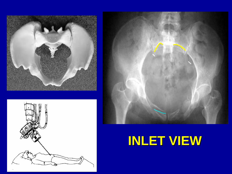
Burgess-Young Classification • Mechanism and direction of injury

LATERAL COMPRESSION
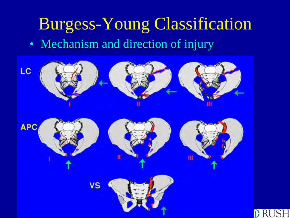
• Three types, increasing in severity • Common anterior fracture pattern • Ligament disruption rare
LATERAL COMPRESSION LC 1: Sacral compression

LATERAL COMPRESSION
LC I: Sacral compression

LATERAL COMPRESSION LC 2: “Crescent fracture”

LATERAL COMPRESSION LC 2: Iliac wing fracture
•Fracture/dislocation of the SI joint
•Internal rotation deformity

LATERAL COMPRESSION LC 3: Windswept pelvis

LC3

LC3

ANTEROPOSTERIOR COMPRESSION
APC The classic “open book” type of pelvic fractures
• 3 types, increasing in severity • Diameter acutely increased • Contents subjected to tensile force • Ligament disruption common • Anterior injury through symphysis or rami • Posterior injury through SI joint or sacrum
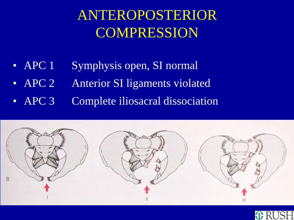
ANTEROPOSTERIOR COMPRESSION
• APC 1 Symphysis open, SI normal • APC 2 Anterior SI ligaments violated • APC 3 Complete iliosacral dissociation

AP 1 • Note that the ligaments are stretched, and not torn

AP 2 • Note: pelvic floor ligaments are violated, as well as
anterior SI ligaments and symphysis
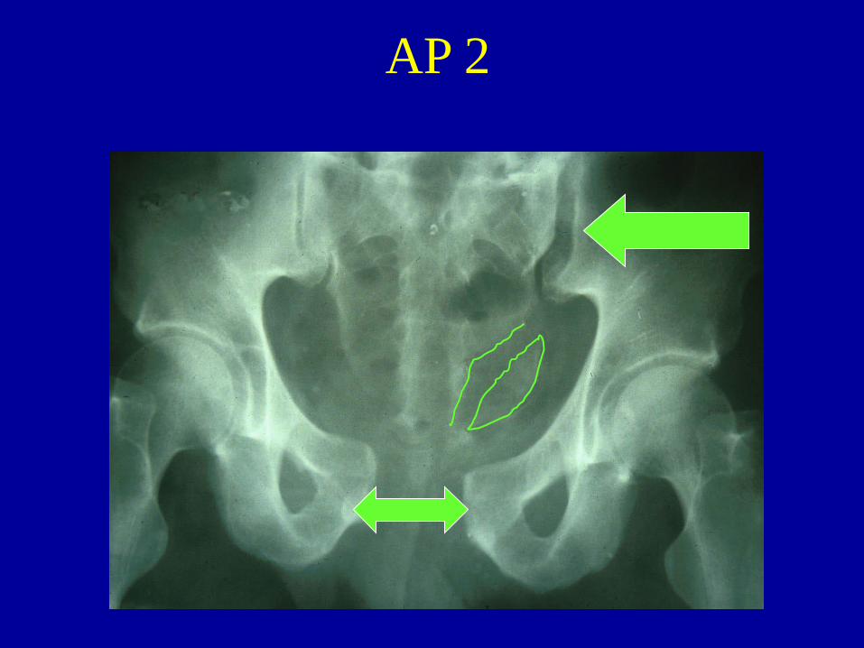
AP 2

AP 2 These anterior SI ligaments are disrupted...
But these posterior SI ligaments remain intact

Complete iliosacral dissociation APC 3

AP 3
a b

AP 3

AP 3

VERTICAL SHEAR

VERTICAL SHEAR

ASSOCIATED INJURIES
Lateral Compression: • Abdominal visceral injury • Head injury • Few pelvic vascular injuries
AP Compression: • Urologic injury • Hemorrhage/pelvic vascular injury:
APCII-10%, APCIII-22%

NEUROLOGIC • Lumbo-sacral plexus • L5, S1 most common • Exploration not indicated • Incomplete lesions may
improve • Often most important factor in
long-term outcome
ASSOCIATED INJURIES

UROLOGIC • Urethra - retrograde urethrogram • Bladder - cystogram
–Extraperitoneal - Foley vs. SP tube
–Intraperitoneal - Repair, SP tube
• Suprapubic tube may complicate surgical treatment
ASSOCIATED INJURIES

Subtle Markers of High Energy
• Lumbar transverse process fractures – Iliac wing attached to
lumber spine by stout iliolumbar ligament
– Sign of vertical instability

Dynamic Instability
• Soft tissue attachments allow pelvis to recoil after initial displacement
• Curved CT table can help reduce pelvis
• Result can be imaging that looks “non displaced”

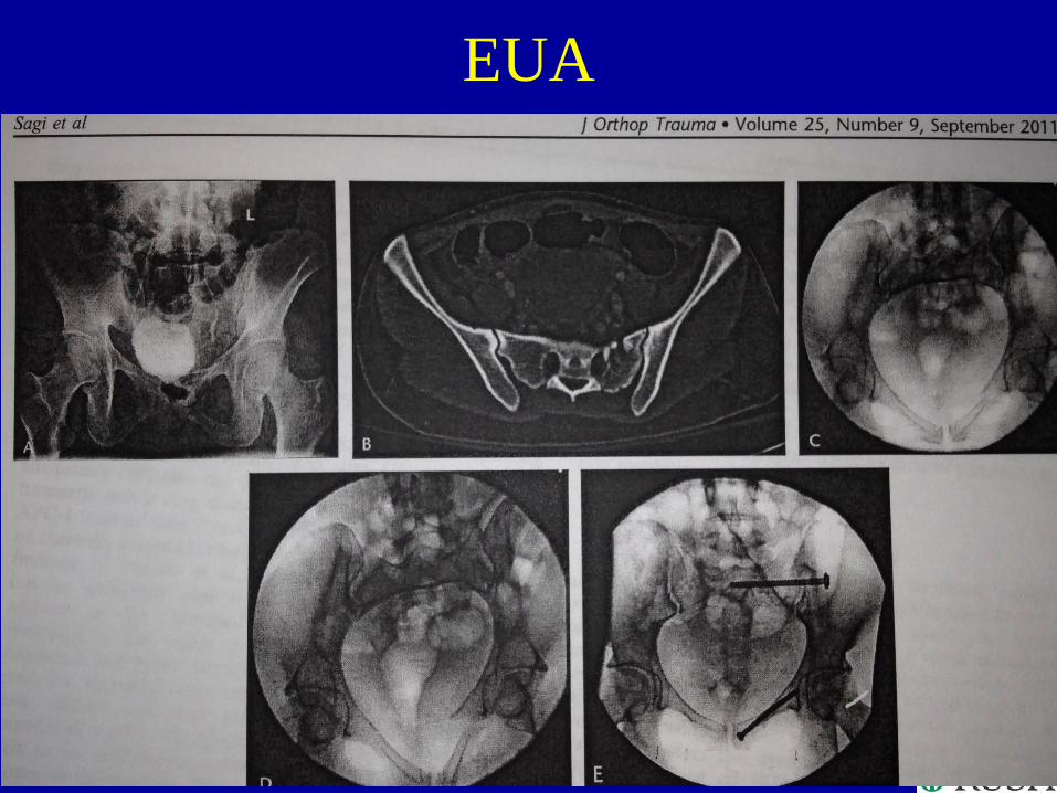
EUA
• Fluoroscopic exam of pelvis under general anesthesia
• Can demonstrate displacement
• Testing – Internal
rotation/compression – External rotation – Push/pull

EUA
• 50% AP1 AP2 (fixation) • 39% AP2 AP2b (posterior
fixation) • 35% LC1 LC1b (fixation)
– Sagi et al, JOT, 2011

EUA
EUA

Management
• ATLS – Airway – Breathing – Circulation
• Early Orthopaedic Involvement – Examine Pelvis Once ? – Examine and Pack Open Wounds – Neuro Exam

Early Management
• Radiographs • ‘Unstable’ Pelvic Ring • Provisional Stabilization
– Sheet – Binder – Caution w LC Injuries

Hemodynamically Stable
• Complete Trauma Workup/Resuscitation • Completion Pelvis Imaging • Watch Vitals Closely • Consider Removing Binder • Elective Stabilization
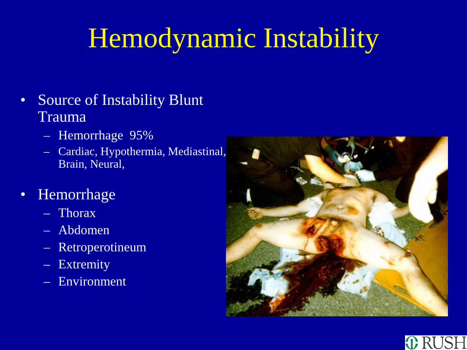
Hemodynamic Instability
• Source of Instability Blunt Trauma – Hemorrhage 95% – Cardiac, Hypothermia, Mediastinal,
Brain, Neural,
• Hemorrhage – Thorax – Abdomen – Retroperotineum – Extremity – Environment

Hemodynamic Instability
• Rapid Assessment of Chest/ Abdomen – Chest Radiograph – FAST – CT – DPL

Instability & CT/Fast Negative
• Continues Resuscitation 1+1+1 – Hypothermia, Coagulopathy, Acidosis
• Provisional Stabilization w Binder • Continued Instability >>> Angio • Definitive Pelvic Ring Stabilization

Instability & CT/Fast Positive
• Laparotomy • Ex Fix prior to Lap • Maintain Binder & Fix After Lap • Be Flexible Depending on Patient Status and
Surgeon Comfort Level • Continued Blood Loss >> Angio

EXTERNAL FIXATION/BINDER
• Immediate application to pt. in extremis
• Controls volume & therefore tamponade
• Stabilizes clots prior to pt. movement

Stabilization Options
• Sheet/Binder/ Ex Fix • ORIF • Percutaneous Fixation
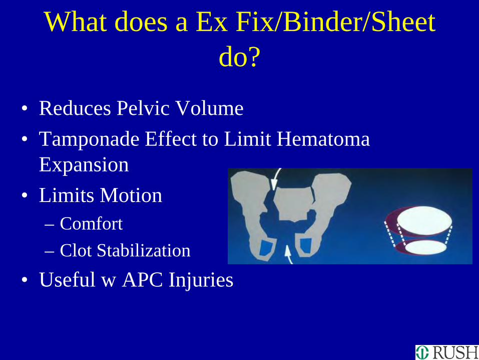
What does a Ex Fix/Binder/Sheet do?
• Reduces Pelvic Volume • Tamponade Effect to Limit Hematoma
Expansion • Limits Motion
– Comfort – Clot Stabilization
• Useful w APC Injuries

Sheet / Binder
• Apply at Greater Trochanter Level
• Allows Access to Abdomen
• Temporary – Access Issues – Soft Tissue Breakdown
• May Modify For Angio Access

Pelvic Binder

• Sheets

INTERVENTIONAL ANGIOGRAPHY
• Much hemorrhage is venous • Timeliness & availability of intervention • May be useful adjunct to other methods • Angiography suite often not optimal for patient
resuscitation • Institution dependent

Angiography
• Allows eval of other organ systems • Embolization
– Selective gelfoam – Multiple Embolization – Proximal Occlusion

Immediate Symphyseal ORIF
• APC, CMI • Laparotomy • Pfannensteil • Avoid Lengthy Surgery

Definitive Treatment Summary
• Rotational and vertically stable injuries – Protected weightbearing
• Rotationally unstable but vertically stable injuries – Protected weightbearing with or without anterior stabilization
• Rotationally and vertically unstable injuries – Posterior stabilization with or without anterior stabilization

Treatment
• LC1 – Protected weightbearing 6 weeks • LC2 – ORIF posterior fracture/dislocation +/- anterior
stabilization • LC3 – Bilateral posterior stabilization with anterior
stabilization

Treatment
• AP1 – Protected weightbearing • AP2 – Controversial – standard treatment is anterior
stabilization, but may not be necessary • AP3 – Posterior stabilization +/- anterior stabilization

Treatment
• Vertical shear – Posterior stabilization, usually with anterior stabilization
• CMI - Treatment directed towards individual injury components

Posterior Fixation
• Open vs. closed reduction
• Percutaneous SI screws • Anterior SI joint plating • Sacral bars • Posterior sacral plating

Thank You


![Pelvis and hip FRACTURES OF THE PELVIS. A) Isolated fractures(stable with no disruption of the pelvic ring ) [1] Fracture of superior ischio- pubic ramus](https://img.dokumen.tips/doc/110x75/56649d9c5503460f94a84d45/pelvis-and-hip-fractures-of-the-pelvis-a-isolated-fracturesstable-with-no.jpg)