Embed Size (px)
Citation preview
Proc. Natl. Acad. Sci. USAVol. 82, pp. 7870-7873, December 1985Biochemistry
Cloning of firefly luciferase cDNA and the expression of activeluciferase in Escherichia coli
(bioluminescence/Photinus pyralis/antibody screening/expression vector/recombinant DNA)
JEFFREY R. DE WET*, KEITH V. WOODt, DONALD R. HELINSKI*, AND MARLENE DELUCAtDepartments of *Biology and tChemistry, University of California, San Diego, La Jolla, CA 92093
Communicated by W. D. McElroy, July 26, 1985
ABSTRACT A cDNA library was constructed from firefly(Photinuspyralis) lantern poly(A)I RNA, using the Escherichiacoli expression vector Xgtll. The library was screened withanti-P. pyralis luciferase (Photinus luciferin:oxygen 4-oxidore-ductase, EC 1.13.12.7) antibody, and several cDNA clonesexpressing luciferase antigens were isolated. One clone, ALucl,contained 1.5 kilobase pairs of cDNA that hybridized to a 1.9-to 2.0-kilobase band on a nitrocellulose blot of electrophoreti-cally fractionated lantern RNA. Hybridization of the clonedcDNA to lantern poly(A)I RNA selected an RNA that directedthe in vitro synthesis of a single polypeptide. This polypeptidecomigrated with luciferase on NaDodSO4/PAGE and producedbioluminescence upon the addition of luciferin and ATP. A1.8-kilobase-pair cDNA was isolated by probing the fireflycDNA library with the cDNA from XLucl. This cDNA con-tained sufficient coding information to direct the synthesis ofactive firefly luciferase in E. coli.
Luciferases are the enzymes that catalyze the light-producingchemical reactions of bioluminescent organisms. Insectluciferases require ATP, an organic molecule called luciferin,and oxygen as substrates (1). The absolute requirement forATP as a substrate is a characteristic unique to insectluciferases, and this property has been used to developbioluminescence assays for ATP (1).The only insect luciferase that has been purified and
extensively characterized was isolated from the North Amer-ican firefly, Photinus pyralis (Photinus luciferin:oxygen 4-oxidoreductase, EC 1.13.12.7) (1, 2). This enzyme catalyzesthe conversion of chemical energy into light with a very highefficiency; the quantum yield (photons emitted per moleculeof luciferin oxidized) is 0.88 (3). Purified P. pyralis luciferasemigrates in NaDodSO4/polyacrylamide gels as a single bandat an apparent molecular weight of 62,000 (4). The luciferasesfrom other firefly species migrate at a similar position, and allshow extensive crossreactivity with antibody raised againstP. pyralis luciferase (26). P. pyralis emits yellow-green lightwith peak emission occurring at 560 nm (3). Other species offireflies emit different colors of light ranging from yellow (582nm) to green (552 nm), the particular color being a charac-teristic of a given species (5). Since all firefly luciferases usethe same substrates (5), these observed differences indicatethat the color of light emission is dependent on enzymestructure. Comparative studies of insect luciferases mayyield information on how changes in protein structure affectthe light-emission properties of these enzymes.
In an earlier paper we showed that poly(A)+ RNA isolatedfrom the lanterns of adult P. pyralis fireflies contained mRNAthat directed the synthesis of luciferase in vitro (4). We haveused this poly(A)+ RNA to construct a cDNA library inXgtll, an Escherichia coli expression vector (6, 7). The
library was screened with anti-P. pyralis luciferase antibody,using a chromogenic detection technique (8), and severalcDNA clones were isolated and characterized. These cloneswere found to be homologous to the mRNA that encodesluciferase. The largest luciferase cDNA clone that wasisolated was able to direct the synthesis of active luciferasein E. coli.
MATERIALS AND METHODSEnzymes and Strains. Restriction endonucleases and E. coli
DNA polymerase I were purchased from New EnglandBiolabs. RNase H and bacteriophage X in vitro packagingextracts were obtained from Bethesda Research Laborato-ries, and avian myeloblastosis virus reverse transcriptasewas from Boehringer Mannheim. XgtJ1 and E. coli strainsY1088 [supE supF metB trpR hsdR hsdM' tonA21 AlacUl69proC:TnS(pMC9)] and Y1090 [AlacUl69 proA' AlonaraD139 strA supF trpC22:TnlO(pMC9)] were obtained fromR. Young (6, 7). E. coli strain TB1 [ara A(lac-proA,B) strAf80dlacZAM15 hsr- hsm'] was obtained from T. Baldwin(Texas A & M University). The plasmid pUC13 has beendescribed (9). The E. coli expression plasmid pKJB824.17,consisting of pBR322 carrying the temperature-sensitive Xrepressor gene c1857 and the X promoter PR was obtainedfrom K. Buckley (10).
Construction and Screening of the Xgtll cDNA Library.Live fireflies (P. pyralis) were obtained from W. Biggley(Johns Hopkins University). Isolated lanterns were homog-enized in guanidinium thiocyanate, and total lantern RNAwas isolated from the homogenate by sedimentation througha CsCl cushion (11). Poly(A)+ RNA was isolated by chro-matography on oligo(dT)-cellulose (12). The construction ofthe Xgtl1 cDNA library was as described (8), except thatRNase H and DNA polymerase I were used to synthesize thesecond strand of the cDNA (13). Packaging of 1 lg of Xgt1lDNA ligated to cDNA yielded =101 recombinant phage, andafter amplification of the library on Y1088, the recombinantsrepresented --10% of the total phage population. The ampli-fied library was screened with rabbit anti-luciferase antibody(2 Ag/ml) as described (8).
Hybridizations. All hybridizations were performed withisolated restriction fragments used as probe. Restrictionfragments were isolated from agarose gels by electrophoresisonto DE81 paper (14), and the DNA was labeled with[a-32P]dCTP (-800 Ci/mmol, Amersham; 1 Ci = 37 GBq) bynick-translation (15) to a specific activity of :-108 cpm/pug.Phage were plated on Y1088 cells and screened by plaquehybridization (16). Southern blots were prepared essentiallyas described (17). RNA samples were electrophoresed informaldehyde/agarose gels and blotted onto nitrocellulose(Schleicher& Schuell, BA85) (18). Hybridizations ofprobe tofilters were at 37°C in 55% (vol/vol) formamide/5x SSPE (lx
Abbreviations: bp, base pair(s); kb, kilobase(s).
7870
The publication costs of this article were defrayed in part by page chargepayment. This article must therefore be hereby marked "advertisement"in accordance with 18 U.S.C. §1734 solely to indicate this fact.

Proc. NatL. Acad. Sci. USA 82 (1985) 7871
SSPE is 0.18M NaC/10mM sodium phosphate, pH 7.7/1 mMEDTA)/heparin (200 pug/ml)/0.1% NaDodSO4 containing 2 x10⁵ cpm of nick-translated probe per ml (19). Filters werewashed in 0.1x SSPE/0.1% NaDodSO4 at 370C and autora-diographed with Kodak XAR-5 film and Cronex Lightning-plus intensifying screens at -70'C.
Hybridization-Selection and in Vitro Translation. Hybrid-ization-selection of mRNA with plasmids was performed asdescribed (20). Twenty micrograms of each plasmid waslinearized with HindIII and bound to 3-mm nitrocellulosesquares. P. pyralis lantern poly(A)+ RNA (30 Ag) washybridized to the filters in one tube at a final concentration of200 1Lg of RNA/ml. Bound RNA was eluted from the filtersby boiling. Nuclease-treated rabbit reticulocyte lysates werepurchased from Bethesda Research Laboratories and wereused according to the supplier's recommended conditions foroptimal protein synthesis. One microgram of total lanternpoly(A)+ RNA and 1/10th of the hybridization-selectedRNAs each were translated in vitro in a final volume of 20 Aul.Translation mixtures contained L-[35S]methionine (600Ci/mmol, Amersham) at 2 mCi/ml. Five-microliter samplesof the translation products from hybridization-selectedRNAs and a 1-1.l sample of the translated lantern poly(A)+RNA were analyzed by NaDodSO4/PAGE (21) followed byfluorography (22). Four microliters of each translation mix-ture was assayed for the presence of active firefly luciferase.Expression of Firefly Luciferase in E. coli. TB 1,
TB1(pKJB824.17), and TB1(pKW101) cells were grown in 10ml of Luria-Bertani medium (10 g of tryptone, 5 g of yeastextract, and 5 g of NaCl per liter, pH 7.4) at 30'C to OD650 =0.5. The cells were heat-induced at 450C for 30 min and thenshifted to 370C for 1 hr. Cells were pelleted for 5 min at 3000x g, and the supernatant was decanted. Pellets were resus-pended in 200 ,ul of 10 mM Tris Cl, pH 8.0/1 mMEDTA/lysozyme (1 mg/ml) and were incubated on ice for 10min. The cells were then frozen on dry ice and thawed. Fiftymicroliters of each cell lysate was assayed for luciferaseactivity.
Assay of Luciferase Activity. Samples to be assayed forluciferase activity were added to 300 ,ul of 25 mMglycylglycine, pH 7.8/5 mM MgCl2/0.1 mM luciferin. Eachtest tube was placed in an LKB luminometer equipped witha chart recorder, and 100 ,l of 20 mM ATP (pH 7.0) wasinjected. The time course of light emission was recorded.
RESULTS
Isolation of Luciferase cDNA Clones. We screened-150,000 phage (=15,000 recombinant phage) from the Xgtllfirefly lantern cDNA library with anti-P. pyralis luciferaseantibody. Sixteen clones expressing luciferase antigens weredetected, and eight of these were purified to homogeneitythrough repeated rounds of screening. All of the clonescontained inserts that were released upon digestion of iso-lated phage DNA with EcoRI, and the inserts ranged from 400base pairs (bp) to 1200 bp in length. Careful analysis of oneclone, XLucl, showed that it contained two EcoRl fragments:LuclA (1200 bp) and LuclB (270 bp). The 1200-bp LuclAfragment hybridized to the other seven clones but was nothomologous to the 270-bp LuclB cDNA fragment.The coding sequence required for the =62-kDa luciferase
protein was estimated to be 1.6 kilobases (kb), whereas thetotal length of the cDNA in XLucl was 1.5 kb. To be certainthat the LuclA and LuclB fragments represented contiguoussequences in the luciferase mRNA, isolated LuclA andLuclB restriction fragments were labeled with [a-32P]dCTPby nick-translation and then used to probe identical lanes ofa blot of P. pyralis poly(A)+ RNA after electrophoresis in adenaturing gel (Fig. 1). LuclA and LuclB both hybridized to
1 2
28S-
23S-
18S-16S -
FIG. 1. Blot hybridization analysis of firefly lantern poly(A)+RNA. Total P. pyralis lantern poly(A)+ RNA (1 1Ag) was electro-phoresed in each lane of a formaldehyde/1.3% agarose gel and thenblotted onto a nitrocellulose filter. The individual lanes were cutapart before hybridization. After hybridization to 32P-labeled DNAprobe, the filters were autoradiographed. Ribosomal RNAs fromhuman (HeLa) and E. coli cells were run as size standards in anadjacent lane, and their positions are indicated at left. Lane 1:Hybridization with LuclA cDNA, the 1200-bp EcoRI fragmentisolated from XLucl. Lane 2: hybridization of LuclB cDNA, the270-bp EcoRI fragment isolated from XLucl.
a single species ofRNA that was 1.9-2.0 kb long; thus, bothof the EcoRI cDNA fragments in XLucl most likely resultedfrom the reverse transcription of a single species of mRNA.LuclA and LuclB Hybrid-Select Luciferase mRNA. The
EcoRI cDNA fragments LuclA and LuclB were inserted intothe EcoRI site of the plasmid pUC13 to generate pLuclA andpLuclB, respectively. Plasmids pLuclA, pLuclB, andpUC13 were used to hybrid-select mRNA from total fireflylantern poly(A)+ RNA. The selected RNAs then were trans-lated in vitro, and the translation products were analyzed bygel electrophoresis (Fig. 2) and assayed for luciferase activ-ity. When translated in vitro, the mRNA selected by pLuclAand pLuclB produced a single species of protein that comi-grated with P. pyralis luciferase in a NaDodSO4/poly-acrylamide gel. No protein bands other than those endoge-nous to the rabbit reticulocyte extract were detected whenpUC13-selected RNA was translated. The in vitro translationof total P. pyralis poly(A)+ RNA and of pLuclA- andpLuclB-hybrid-selected RNA produced active fireflyluciferase as assayed by the production of light in thepresence of luciferin and ATP (data not shown). No activeluciferase could be detected in the reticulocyte translationextract or an extract that contained RNA selected by pUC13alone.
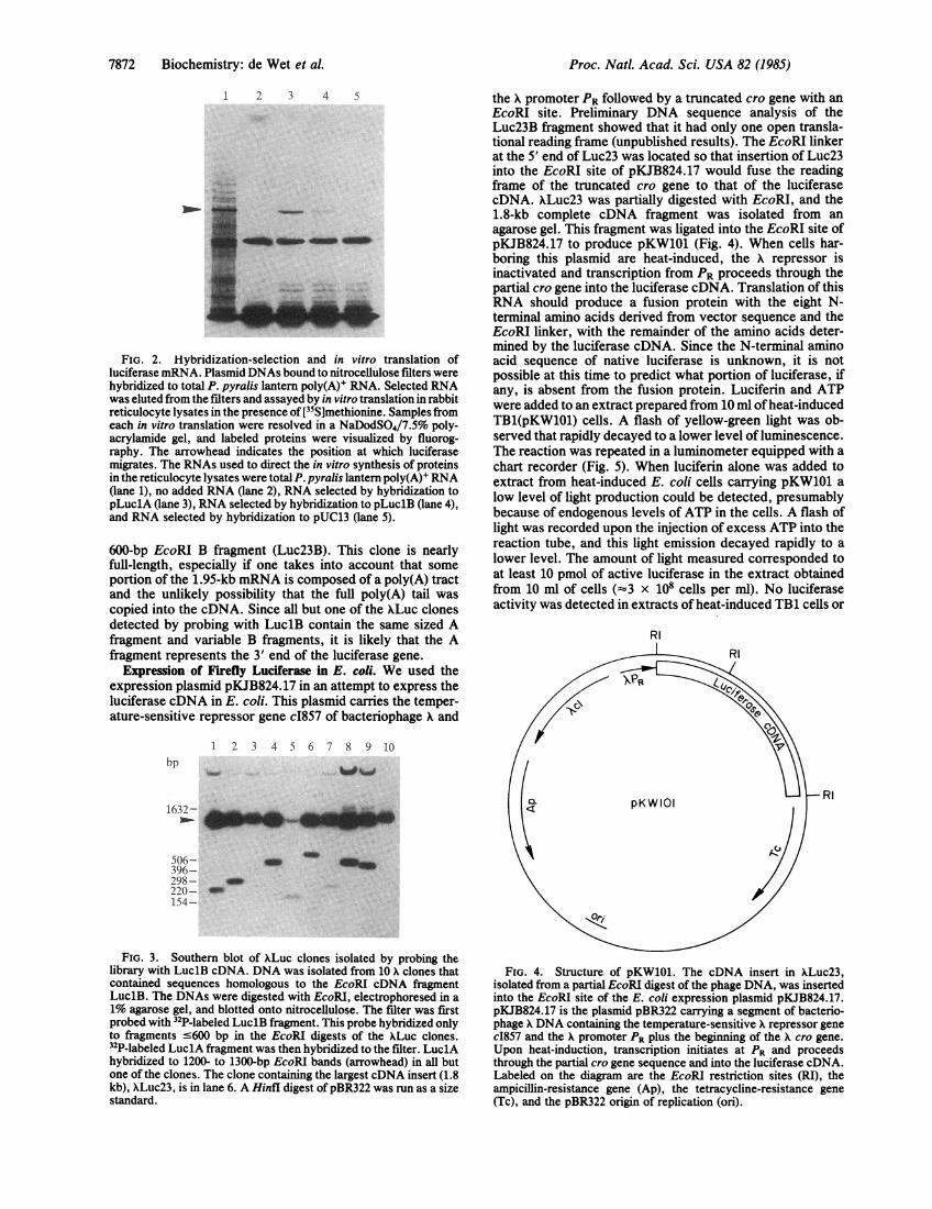
Isolation of Larger Luciferase cDNA Clones. The sum of thetwo cDNA fragments from XLucl (1.5 kb) was less than thesize of the mRNA detected by LuclA and LuclB (1.9-2.0kb). Therefore, the Xgtll lantern cDNA library wasrescreened, using the LuclB fragment labeled with [a-32P]dCTP by nick-translation as probe, to isolate a longercDNA clone. Ten clones were purified to homogeneity, andthe DNA isolated from these phage was cut by EcoRIendonuclease and examined by DNA blot analysis (Fig. 3).All of the clones contained an EcoRI fragment that wasdetectable by hybridization to nick-translated LuclB cDNA.These fragments ranged in size from 200 bp to 600 bp.Subsequent hybridization of the blot to nick-translatedLuclA fragment showed that all but one of these clones alsocontained the large EcoRI A fragment (1.2-1.3 kb). The clonewith the largest insert, XLuc23, carried 1.8 kb of cDNAcomposed of a 1.2-kb EcoRl A fragment (Luc23A) and a
Biochemistry: de Wet et al.
Proc. Natl. Acad. Sci. USA 82 (1985)
1 2 3 4 5
FIG. 2. Hybridization-selection and in vitro translation ofluciferase mRNA. Plasmid DNAs bound to nitrocellulose filters werehybridized to total P. pyralis lantern poly(A)' RNA. Selected RNAwas eluted from the filters and assayed by in vitro translation in rabbitreticulocyte lysates in the presence of[35S]methionine. Samples fromeach in vitro translation were resolved in a NaDodSO4/7.5% poly-acrylamide gel, and labeled proteins were visualized by fluorog-raphy. The arrowhead indicates the position at which luciferasemigrates. The RNAs used to direct the in vitro synthesis of proteinsin the reticulocyte lysates were total P. pyralis lantern poly(A)l RNA(lane 1), no added RNA (lane 2), RNA selected by hybridization topLuc1A (lane 3), RNA selected by hybridization to pLuc1B (lane 4),and RNA selected by hybridization to pUC13 (lane 5).
600-bp EcoRI B fragment (Luc23B). This clone is nearlyfull-length, especially if one takes into account that someportion of the 1.95-kb mRNA is composed of a poly(A) tractand the unlikely possibility that the full poly(A) tail wascopied into the cDNA. Since all but one of the XLuc clonesdetected by probing with LuciB contain the same sized Afragment and variable B fragments, it is likely that the Afragment represents the 3' end of the luciferase gene.
Expression of Firefly Luciferase in E. cofi. We used theexpression plasmid pKJB824.17 in an attempt to express theluciferase cDNA in E. coli. This plasmid carries the temper-ature-sensitive repressor gene c1857 of bacteriophage X and
the X promoter PR followed by a truncated cro gene with anEcoRI site. Preliminary DNA sequence analysis of theLuc23B fragment showed that it had only one open transla-tional reading frame (unpublished results). The EcoRI linkerat the 5' end of Luc23 was located so that insertion of Luc23into the EcoRI site of pKJB824.17 would fuse the readingframe of the truncated cro gene to that of the luciferasecDNA. XLuc23 was partially digested with EcoRI, and the1.8-kb complete cDNA fragment was isolated from anagarose gel. This fragment was ligated into the EcoRI site ofpKJB824.17 to produce pKW101 (Fig. 4). When cells har-boring this plasmid are heat-induced, the X repressor isinactivated and transcription from PR proceeds through thepartial cro gene into the luciferase cDNA. Translation of thisRNA should produce a fusion protein with the eight N-terminal amino acids derived from vector sequence and theEcoRI linker, with the remainder of the amino acids deter-mined by the luciferase cDNA. Since the N-terminal aminoacid sequence of native luciferase is unknown, it is notpossible at this time to predict what portion of luciferase, ifany, is absent from the fusion protein. Luciferin and ATPwere added to an extract prepared from 10 ml ofheat-inducedTB1(pKW101) cells. A flash of yellow-green light was ob-served that rapidly decayed to a lower level of luminescence.The reaction was repeated in a luminometer equipped with achart recorder (Fig. 5). When luciferin alone was added toextract from heat-induced E. coli cells carrying pKW101 alow level of light production could be detected, presumablybecause of endogenous levels of ATP in the cells. A flash oflight was recorded upon the injection of excess ATP into thereaction tube, and this light emission decayed rapidly to alower level. The amount of light measured corresponded toat least 10 pmol of active luciferase in the extract obtainedfrom 10 ml of cells (-3 x 10⁸ cells per ml). No luciferaseactivity was detected in extracts of heat-induced TB1 cells or
RI
1 2 3 4 5 6 7 8 9 10bp
1632- d_ .
506- -396-298-220- _154-
FIG. 3. Southern blot of XLuc clones isolated by probing thelibrary with LuclB cDNA. DNA was isolated from 10 X clones thatcontained sequences homologous to the EcoRI cDNA fragmentLuclB. The DNAs were digested with EcoRI, electrophoresed in a1% agarose gel, and blotted onto nitrocellulose. The filter was firstprobed with 32P-labeled LuclB fragment. This probe hybridized onlyto fragments s600 bp in the EcoRI digests of the XLuc clones.32P-labeled LuclA fragment was then hybridized to the filter. LuclAhybridized to 1200- to 1300-bp EcoRI bands (arrowhead) in all butone of the clones. The clone containing the largest cDNA insert (1.8kb), XLuc23, is in lane 6. A Hinfl digest of pBR322 was run as a sizestandard.
FIG. 4. Structure of pKW101. The cDNA insert in XLuc23,isolated from a partial EcoRI digest of the phage DNA, was insertedinto the EcoRI site of the E. coli expression plasmid pKJB824.17.pKJB824.17 is the plasmid pBR322 carrying a segment of bacterio-phage X DNA containing the temperature-sensitive X repressor genecI857 and the X promoter PR plus the beginning of the X cro gene.Upon heat-induction, transcription initiates at PR and proceedsthrough the partial cro gene sequence and into the luciferase cDNA.Labeled on the diagram are the EcoRI restriction sites (RI), theampicillin-resistance gene (Ap), the tetracycline-resistance gene(Tc), and the pBR322 origin of replication (ori).
7872 Biochemistry: de Wet et al.

Proc. NatL. Acad. Sci. USA 82 (1985) 7873
30
220
10
20 40 60Time. sec
FIG. 5. Active firefly luciferase is synthesized in E. coli cells.TB1 cells containing the plasmid pKW101 were heat-induced andincubated to allow expression of the luciferase cDNA under thecontrol of X PR. Cells were pelleted and lysed, and the lysate was
assayed for luciferase activity in the presence of luciferin and ATP.Light emitted by the luciferase was monitored in an LKBluminometer equipped with a chart recorder. The time course oflightemission is shown. The arrow indicates the time at which ATP was
injected into the reaction mixture.
TB1 cells carrying the vector pKJB824.17, even with thesensitivity of the luminometer set 500 times greater than thesensitivity used to obtain the recording shown in Fig. 5.
DISCUSSIONWe have isolated several cDNA clones that are homologousto the mRNA encoding the luciferase ofthe firefly P. pyralis.The longest of these cDNA clones, Luc23, is 1.8 kb long,which is greater than the 1.6 kb required to encode a 62-kDaprotein. This cDNA is, however, shorter than the 1.95-kbluciferase mRNA that is detected on RNA blots. Furtheranalysis is necessary to determine whether or not Luc23contains the entire coding sequence for luciferase. Luc23does contain the coding information necessary for catalyticactivity of luciferase: when inserted in an E. coli expressionplasmid, this cDNA directs the in vivo synthesis ofan enzymethat produces light in the presence of luciferin and ATP. Onthe basis of nucleotide-sequence information the luciferaseproduced in E. coli is a fusion protein in which the eightN-terminal amino acids are determined by the expressionvector and synthetic restriction-site sequences. The activityof the fusion protein indicates that the N-terminus of P.pyralis luciferase can be altered without complete loss of itscatalytic activity.
Insect luciferases have great potential as an experimentalsystem for investigating the structural basis of enzyme-catalyzed light emission. The demonstration of the synthesisof active firefly luciferase in E. coli will greatly facilitate theisolation of mutant enzymes that may have altered lightemission properties. Although the colors of the light emittedby firefly luciferases range from yellow to green, it should bepossible to modify luciferase so that light of even longerwavelengths will be produced, particularly in view of the
observation that treating P. pyralis luciferase with heat,Zn2+, or low pH causes it to emit red (610 nm) light (5, 23).Furthermore, the railroad worm, which is the larva ofa SouthAmerican beetle (Phrixothrix sp.), has anterior light organsthat naturally produce red light in addition to abdominalyellow-green light organs (24).
In a recent paper, the use ofthe cloned lux operon of Vibriofischeri as an indicator of promoter activity in bacteria wasdescribed (25). The luciferase gene of the firefly, P. pyralis,could be used in a similar fashion as an indicator gene andmay have more extensive applicability. Unlike the bacterialluciferase, the firefly enzyme requires a single subunit foractivity, and its synthesis could therefore be placed under thecontrol of a single eukaryotic promoter. In addition, theexceptionally high quantum yield oflight characteristic ofthefirefly luciferase may make this luciferase gene particularlysuitable for use as an indicator of transcriptional activity.
This work was supported by National Science Foundation GrantPCM8305446 and National Institutes of Health Grant A107194.
1. DeLuca, M. & McElroy, W. D. (1978) Methods Enzymol. 57,3-15.
2. Green, A. A. & McElroy, W. D. (1956) Biochim. Biophys.Acta 20, 170-178.
3. Seliger, H. H. & McElroy, W. D. (1960) Arch. Biochem.Biophys. 88, 136-141.
4. Wood, K. V., de Wet, J. R., Devji, N. & DeLuca, M. (1984)Biochem. Biophys. Res. Commun. 124, 592-596.
5. Seliger, H. H. & McElroy, W. D. (1964) Proc. Natl. Acad.Sci. USA 52, 75-81.
6. Young, R. A. & Davis, R. W. (1983) Proc. Natl. Acad. Sci.USA 80, 1194-1198.
7. Young, R. A. & Davis, R. W. (1983) Science 222, 778-782.8. de Wet, J. R., Fukushima, H., Dewji, N. N., Wilcox, E.,
O'Brien, J. S. & Helinski, D. R. (1984) DNA 3, 437-447.9. Messing, J. (1985) Methods Enzymol. 101, 20-78.
10. Buckley, K. J. (1985) Dissertation (Univ. of California, SanDiego).
11. Chirgwin, J. M., Przybyla, A. E., MacDonald, R. J. & Rutter,W. J. (1979) Biochemistry 18, 5294-5299.
12. Aviv, H. & Leder, P. (1972) Proc. Natl. Acad. Sci. USA 69,1408-1412.
13. Gubler, U. & Hoffman, B. J. (1983) Gene 25, 263-269.14. Dretzen, G., Bellard, M., Sassone-Corsi, P. & Chambon, P.
(1981) Anal. Biochem. 112, 295-298.15. Rigby, P. W. J., Dieckman, M., Rhodes, C. & Berg, P. (1977)
J. Mol. Biol. 113, 237-251.16. Benton, W. D. & Davis, R. W. (1977) Science 196, 180-182.17. Southern, E. (1975) J. Mol. Biol. 98, 503-517.18. Maniatis, T., Fritsch, E. F. & Sambrook, J. (1982) Molecular
Cloning: A Laboratory Manual (Cold Spring Harbor Labora-tory, Cold Spring Harbor, NY), pp. 202-203.
19. Singh, L. & Jones, K. W. (1984) Nucleic Acids Res. 12,5627-5638.
20. Parnes, J. R., Velan, B., Felsenfeld, A., Ramanathan, L.,Ferrini, U., Appella, E. & Seidman, J. G. (1981) Proc. Natl.Acad. Sci. USA 78, 2253-2257.
21. Laemmli, U. K. (1970) Nature (London) 227, 680-685.22. Bonner, W. M. & Laskey, R. A. (1974) Eur. J. Biochem. 46,
83-88.23. McElroy, W. D. & DeLuca, M. (1978) in Bioluminescence in
Action, ed. Herring, P. J. (Academic, London), pp. 109-127.24. Lloyd, J. E. (1978) in Bioluminescence in Action, ed. Herring,
P. J. (Academic, London), p. 256.25. Engebrecht, J. M., Simon, M. & Silverman, M. (1985) Science
227, 1345-1347.26. Weinhausen, G. & DeLuca, M. (1985) Photochem. Photobiol.,
in press.
Biochemistry: de Wet et al.