Embed Size (px)
Citation preview
Proc. Natl. Acad. Sci. USAVol. 88, pp. 9127-9131, October 1991Genetics
Cloning and characterization of a 3-methyladenine DNA glycosylasecDNA from human cells whose gene maps to chromosome 16
(human DNA alkylation repair gene)
LEONA SAMSON*, BRUCE DERFLER, MICHAEL BOOSALIS, AND KATHERINE CALLMolecular and Cellular Toxicology, Harvard School of Public Health, 665 Huntington Avenue, Boston, MA 02115
Communicated by Elkan R. Blout, July 1, 1991 (receivedfor review April 25, 1991)
ABSTRACT We described previously the isolation of aSaceharomyces cerevisiae 3-methyladenine (3-MeAde) DNAglycosylase repair gene (MAG) by its expression in glycosylase-deficient Escherichia coli alkA tag mutant cells and its ability torescue these cells from the toxic effects of alkylating agents.Here we extend this cross-species functional complementationapproach to the isolation of a full-length human 3-MeAde DNAglycosylase cDNA that rescues alkA tag E. coli from killing bymethyl methanesulfonate, and we have mapped the gene tohuman chromosome 16. The cloned cDNA, expressed from thepBR322 /-lactamase promoter, contains an 894-base-pair openreading frame encoding a 32,894-Da protein able to release3-MeAde, but not 7-methylguanine, from alkylated DNA.Surprisingly, the predicted human protein does not sharesignificant amino acid sequence homology with the bacterialAlkA and Tag glycosylases or the yeast MAG glycosylase, butit does share extensive amino acid sequence homology with a rat3-MeAde DNA glycosylase and significant DNA sequence ho-mology with genes from several mammalian species. Thecloning of a human 3-MeAde DNA glycosylase cDNA repre-sents a key step in generating 3-MeAde repair-deficient cellsand the determination of the in vivo role of this DNA repairenzyme in protecting against the toxic and carcinogenic effectsof alkylating agents.
The genome of every organism continually sustains DNAdamage which, if left unrepaired, contributes to cell death,mutation, chromosome damage, ageing, and carcinogenesis.Thus, a number ofDNA repair pathways have evolved, andthese appear to be highly conserved among bacteria, yeast,insect, and mammalian cells. One of the pathways for DNArepair is initiated by the action of certain glycosylases thatexcise abnormal bases from DNA, leaving behind apurinic orapyrimidinic sites that then trigger nucleotide excision repair(1). Abnormal DNA bases are known to be continuallyproduced by uracil misincorporation, spontaneous bondbreakages, and reactions with normal cellular metabolitesand environmental DNA-damaging agents (2). At least eighttypes of glycosylase have been identified, each of which isspecific for the removal of one or more abnormal bases (2).3-Methyladenine (3-MeAde) is one of the major lethal lesionsproduced by agents like methyl methanesulfonate (MMS) andN-methyl-N'-nitro-N-nitrosoguanidine (MNNG) in Esche-richia coli, and this lesion is also believed to be produced bythe normal cellular metabolite S-adenosylmethionine; E. coliis protected against such alkylating agents by the constitu-tively expressed tag gene and the alkylation-inducible alkAgene, which encode two 3-MeAde DNA glycosylases (3).Whether the 3-MeAde lesion is lethal or mutagenic in humancells is not yet known because mutant human cells deficientin 3-MeAde DNA glycosylase have not yet been identified.
Further, it remains to be determined whether the 3-MeAdeDNA glycosylase provides any protection against the carci-nogenic effects of alkylating agents in our environment. Herewe describe a key step towards defining the in vivo role ofthehuman 3-MeAde DNA glycosylase repair enzyme-namely,the cloning of a cDNA coding for this DNA repair function.A number of human DNA repair genes have been cloned
by their ability to rescue DNA damage-sensitive rodent cellsfrom the toxic effects of the appropriate DNA-damagingagent (4, 5). However, this approach is limited by the fact thatthe specific DNA repair defects in the rodent cell lines areunknown (5), and this has made the in vivo role of the clonedgene products difficult to determine. We recently developeda method for cloning eukaryotic DNA repair genes thatdetects expression of the cloned genes by their ability torescue well-characterized DNA repair-deficient strains of E.coli from the toxic effects of DNA damage (6). Using thisprocedure, we cloned a 3-MeAde DNA glycosylase gene(MAG) from Saccharomyces cerevisiae by its ability torescue a 3-MeAde DNA glycosylase-deficient E. coli alkAtag double mutant from the killing effects of MMS. The S.cerevisiae MAG glycosylase shares significant sequence ho-mology with the E. coli AlkA glycosylase, and its expression,like the transcription of alkA, is dramatically increased whenthe yeast is exposed to alkylating agents (7). To determine thein vivo role of3-MeAde repair, we used the clonedMAG geneto produce mag- yeast cells and showed that the reductionin 3-MeAde repair resulted in increased sensitivity to alkyl-ation-induced cell death but not mutation in S. cerevisiae (6,7). We also found that mag- cells express another 3-MeAdeDNA glycosylase (7), but it remains to be determinedwhether this second glycosylase represents a Tag homo-logue.We have now extended this eukaryote/prokaryote func-
tional complementation approach to the isolation of a human3-MeAde DNA glycosylase cDNA. It was not previouslypossible to clone this human gene by conventional methodsbecause 3-MeAde DNA glycosylase-deficient mammaliancell lines are not currently available. Here we describe thecharacterization of a full-length human 3-MeAde DNA gly-cosylase cDNAt isolated by its ability to rescue E. coli alkAtag mutants from killing by MMS, and we have mapped itsgene to human chromosome 16. The predicted human3-MeAde DNA glycosylase enzyme shares no amino acidhomology with the E. coli AlkA and Tag glycosylases or theS. cerevisiae MAG glycosylase, but it does share significanthomology with the rat 3-MeAde DNA glycosylase enzyme(8).
Abbreviations: 3-MeAde, 3-methyladenine; MMS, methyl methane-sulfonate; MNNG, N-methyl-N'-nitro-N-nitrosoguanidine;7-MeGua, 7-methylguanine.*To whom reprint requests should be addressed.tThe sequence reported in this paper has been deposited in theGenBank data base (accession no. M74905).
9127
The publication costs of this article were defrayed in part by page chargepayment. This article must therefore be hereby marked "advertisement"in accordance with 18 U.S.C. §1734 solely to indicate this fact.
Proc. Natl. Acad. Sci. USA 88 (1991)
MATERIALS AND METHODS
Cells, Plasmids, and Enzymes. E. coli AB1157 (F- thr-1leu-6 proA2 thi-1 argE lacY) galK ara-14 xyl-5 mly-1 tsx-33strA sup-37) was the strain used as the wild type for alkyla-tion. MV1932 and MV1902 (gifts from M. Volkert, Universityof Massachusetts, Worcester, MA) are alkylation-sensitivederivatives ofAB1157 and are alkl tag and alkAlOS::ApSG1(9), respectively. The human cDNA expression library wasconstructed by Prochownik etal. (10) by cloning human livercDNAs under the -lactamase promoter of the pBR322derivative pKT218 (11) and was a gift from Stuart Orkin(Harvard Medical School, Boston). Restriction endonu-cleases were from New England Biolabs, and digestions werecarried out by standard protocols (12).
Screen for Alkylation-Resistant Transformants. Two hun-dred nanograms ofthe cDNA library was transformed into E.coli alkA tag (MV1932); at least 107 independent transform-ants were incubated for 3.5 hr at 370C to allow cDNA insertexpression and then were plated on six large Luria-Bertoni(LB) ampicillin plates containing 0.005-0.01% MMS. Ap-proximately 8000 surviving colonies were scraped from theplates, plasmid DNA (pool 1) was isolated and reintroducedinto MV1932, and the process was repeated to generate DNApool 2. Pool 2 DNA was reintroduced into MV1932, andtransformants were plated on LB ampicillin plates withoutMMS; 300 transformants were individually screened forMMS resistance as described (6), and several transformantswere found to be MMS resistant. Each plasmid in theresistant transformants contained the same cDNA insert(data not shown). One plasmid, called pP5-3, was used fordetailed analysis.
Survival Curves and DNA Glycosylase Activity. For survivalmeasurements, bacteria were grown in LB medium to 108cells per ml; MMS was added to 0.05%, and aliquots wereremoved at various times, diluted in M9 salts, and spread onLB plates to estimate cell survival. For measurements of3-MeAde DNA glycosylase activity, cell extracts were pre-pared as described (13) in 50 mM Hepes adjusted with KOHto pH 7.6/100 mM KCl/1 mM EDTA/5 mM dithiothreitol.Extract protein was incubated for 1 hr at 370C with 33,000cpm of di[3H]methyl sulfate-treated calf thymus DNA (250cpm/,ug) prepared according to Samson and Linn (14), andthe release of 3-MeAde and 7-methylguanine (7-MeGua) wasmeasured by paper chromatography as described (6).
Southern and Northern Blot Analysis. Bacterial (15), mam-malian (12), and yeast (16) DNAs were isolated as described,and the corresponding RNAs were isolated as described (12).A "zoo blot" purchased from Clontech contained the EcoRI-digested human, monkey, rat, mouse, dog, cow, rabbit,chicken, and yeast DNAs (8 ,ug per lane). A mapping panelblot, purchased from Bios (New Haven, CT), consisted ofBamHI-digested DNA of human, hamster, and 25 human-hamster hybrids. DNA from human-mouse hybrid GM10567(containing human chromosome 16) was purchased from theNIGMS Human Genetic Cell Repository, Camden, NJ.Northern blot analysis was as described (12). The blots wereprobed with a 32P-labeled 0.5-kilobase (kb) Pst I fragmentfrom the pP5-3 cDNA insert, and the final filter washes wereat high stringency.DNA Sequence Analysis. Three Pst I fragments of 0.3, 0.4,
and 0.5 kb were subcloned from the pP5-3 cDNA insert intopTZ18R and pTZ19R (Pharmacia, LKB), transformed into E.coliNM522, and sequenced in both directions. We confirmedthe fragment order by sequencing across the Pst I sites inpP5-3, using the appropriate oligonucleotide primers to se-quence from the double-stranded pP5-3 plasmid.
RESULTS AND DISCUSSIONIsolation ofa Human 3-MeAdeDNA Glycosylase cDNA. Our
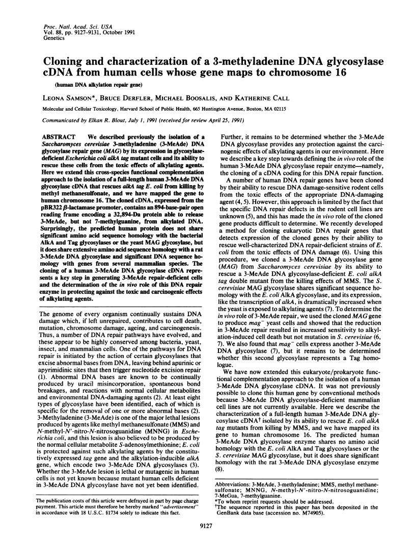
aim was to isolate a human cDNA whose expression rescues3-MeAde-DNA-glycosylase-deficient alkA tag E. coli fromkilling by MMS. The expression library was constructed byProchownik et al. (10) by cloning human liver cDNAs underthe J3-lactamase promoter of the pBR322 derivative pKT218(11); each cDNA insert replaced most of the 3-lactamasecoding sequence and could potentially be expressed either asa fusion protein or from its own ATG translation initiationcodon. The library was transformed into the E. coli alkA tagstrain MV1932 (9), and a population of 10W independenttransformants was enriched for alkylation-resistant cells byrepeated challenges with MMS. Individual colonies from theenriched population were streaked onto MMS plates andcontrol plates; several transformants formed colonies on theMMS plates, and the responsible plasmids were isolated fromthe control plate bacteria and checked for their ability totransmit MMS resistance. In this way we identified plasmidpP5-3 with a 1.2-kb cDNA insert that rescued alkA tag E. colifrom the killing effects ofMMS (Fig. LA) and MNNG (datanot shown). Although alkA tag/pP5-3 cells were consider-ably more resistant than alkA tag cells, they were not asresistant as wild-type AB1157 cells. We eliminated the pos-
1000 1000A B
100
10 100
CO) CO)
Minutes in
E 20000-
X0cJ 10
Protein, yug
FIG. 1. Characterization ofthe phenotype conferred by pP5-3. (Aand B) Bacteria were grown in LB broth to 108 cells per ml, MMS wasadded to 0.05%, and aliquots were removed at various times, diluted,and plated on LB agar to estimate cell survival. The strains are: o,AB1157 (wild type); o, MV1932 (alkA tag); *, MV1932 (alkAtag/pP5-3); A, MV1902 (alkAlOS::ApSG1); and A, MV1902(alkAlO5::ApSG1/pP5-3). (C and D) Extract protein was incubatedfor 1 hr at 370C with di[3H]methyl sulfate-treated calf thymus DNAas described, and the release of 3-MeAde (C) and 7-MeGua (D) wasmeasured by paper chromatography. Extracts were from AB1157(o), MV1932 (alkA tag) (o), and MV1932 (alkA tag/pP5-3) (-).
9128 Genetics: Samson et al.
-1
Proc. Natl. Acad. Sci. USA 88 (1991) 9129
sibility that pP5-3 conferred resistance to MV1932 by sup-pression of the point mutation in alkA or tag by showing thatpP5-3 also conferred alkylation resistance to a nonsuppress-ible E. coli strain bearing a A phage insertion in the alkA gene(Fig. 1B). Further experiments showed that pP5-3 encodes a3-MeAde DNA glycosylase. Fig. 1C shows that alkA tag!pP5-3 cells contained 3-MeAde DNA glycosylase activity,although not as much as in wild-type AB1157 cells, whichagrees with the observation that alkA tag/pP5-3 are not asalkylation-resistant as AB1157 (Fig. 1C). The pP5-3-encodedglycosylase in crude E. coli cell extracts did not release7-MeGua from alkylated DNA (Fig. 1D), and it remains to bedetermined whether it releases 3-methylguanine (3-MeGua)or 02-methylpyrimidines as the E. coli AlkA glycosylasedoes (3). It was previously shown that the partially purifiedhuman 3-MeAde DNA glycosylase releases 7-MeGua and3-MeGua from alkylated DNA, in addition to 3-MeAde, albeitat a slow rate (17, 18); although our human glycosylasepreparations did not release any 7-MeGua in crude E. coli cellextracts, this activity may be detectable when the enzyme ispurified.
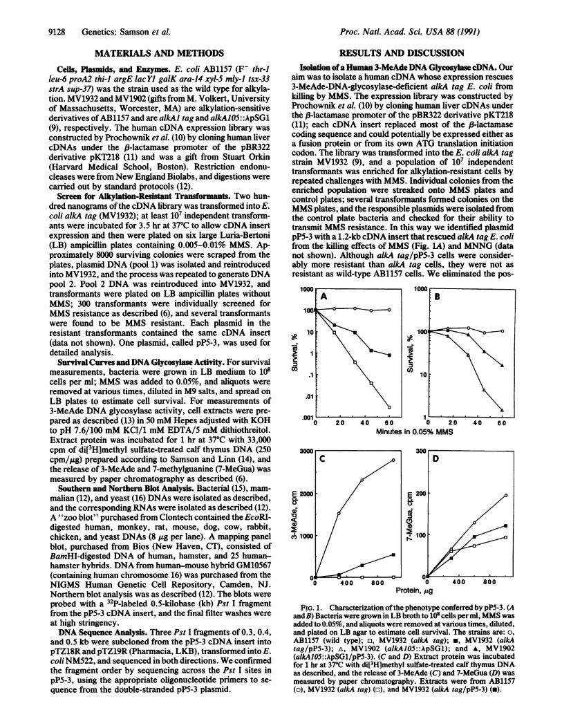
Characterization of the Human 3-MeAde DNA GlycosylasecDNA. A pP5-3 cDNA insert fragment hybridized to humangenomic DNA (Fig. 2A) and to a 1.2- to 1.3-kb human mRNA(Fig. 2B), indicating that we had cloned a virtually full-lengthhuman cDNA encoding 3-MeAde DNA glycosylase. Hybrid-
A
1 2 3
..
944-
4A-**
2.3 -
2.0
B
4 5
4.4 -
7-2
72--
2-4 -
1 4 -
c1 2 3 4 5 6 7 8 9
143-5
4.46.6 - I - ' 1
2.3-2.0 -
FIG. 2. Hybridization analysis of the pP5-3 cDNA insert. (A)Southern blot analysis. DNAs [10 /Lg of human (lanes 1 and 4), 10 jigof hamster (lane 5), 2.5 Ag of S. cerevisiae (lane 2), and 5.0 Ag of E.coli (lane 3)] were digested with BamHI and probed with a 32P-labeled0.5-kb Pst I fragment from the pP5-3 cDNA insert. Final filter washeswere at high stringency. (B) Northern blot analysis. RNA from HeLaCCL2 and HeLa S3 cells (30 jig of total RNA), separated informaldehyde/1% agarose (12), was probed with a 0.5-kb Pst Ifragment from the pP5-3 cDNA insert and washed at high stringency.(C) Southern zoo blot analysis. A "zoo blot", purchased fromClontech, was probed with a 0.5-kb Pst I fragment of the pP5-3 cDNAinsert and was washed at high stringency. The EcoRI-digested DNAs(8 ,.g per lane) were from the following species: human (lane 1);monkey (lane 2); rat (lane 3); mouse (lane 4); dog (lane 5); cow (lane6); rabbit (lane 7); chicken (lane 8); and yeast (lane 9).
ization of the pP5-3 cDNA also detected a 1.2-kb mRNA inbaboon, which was expressed in all tissues examined (datanot shown). The abundance of the mRNA varied as follows:spleen > muscle, liver and heart > kidney and brain.3-MeAde DNA glycosylase activity also varies in mousetissues, with spleen expressing up to 10-fold more activitythan brain tissue (19). 06-methylguanine DNA methyltrans-ferase is the only other alkylation-specific DNA repair en-zyme known in mammalian tissues, but it does not appear tobe coordinately expressed with the 3-MeAde DNA glyco-sylase (at least in mice) because, while spleen has the highestlevel of glycosylase activity relative to other tissues, it hasone of the lowest levels of 06-methylguanine DNA methyl-transferase activity (19-21).The human glycosylase cDNA did not hybridize to S.
cerevisiae or E. coli DNA (Fig. 2 A and C), but it did exhibitcross-species hybridization to six of eight vertebrate DNAstested-namely, hamster (Fig. 2A), monkey, rat, mouse,cow, and chicken; surprisingly, it did not hybridize to dog orrabbitDNA (Fig. 2C). The cloned human cDNA detected thesame size EcoRI band (7 kb) reported to hybridize to the rat3-MeAde DNA glycosylase cDNA (8), suggesting that theDNA fragments hybridizing to the human glycosylase cDNAin the six positive species (Fig. 2A) probably representhomologous glycosylase genes.The sequence of the cloned cDNA showed an 894-base-
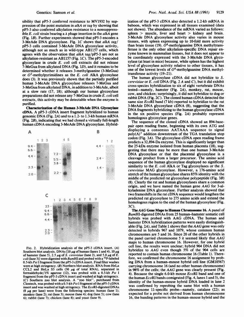
pair open reading frame, beginning with its own ATG anddisplaying a consensus AATAAA sequence to signalpoly(A)+ addition downstream of the TGA translation stopcodon (Fig. 3A). The glycosylase cDNA open reading framepredicts a 32,894-Da enzyme. This is significantly larger thanthe 25-kDa enzyme isolated from human placenta (18), sug-gesting that there may be more than one human 3-MeAdeDNA glycosylase or that the placental glycosylase is acleavage product from a larger precursor. The amino acidsequence of the human glycosylase displayed no significantsimilarity to the E. coli AlkA or Tag glycosylases or the S.cerevisiae MAG glycosylase. However, a 176-amino acidstretch ofthe human glycosylase shares 85% identity with themiddle of the predicted rat glycosylase polypeptide (Fig. 3B)(8). Clearly the rat and human glycosylases share a commonorigin, and we have named the human gene AAG for 3-al-kyladenine DNA glycosylase. Further analysis showed thattwo frameshifts in the ratcDNA sequence would lengthen thepredicted rat glycosylase to 275 amino acids and extend thehomologous region to the end of the human glycosylase (Fig.3C).TheAAG Gene Maps to Human Chromosome 16. A panel of
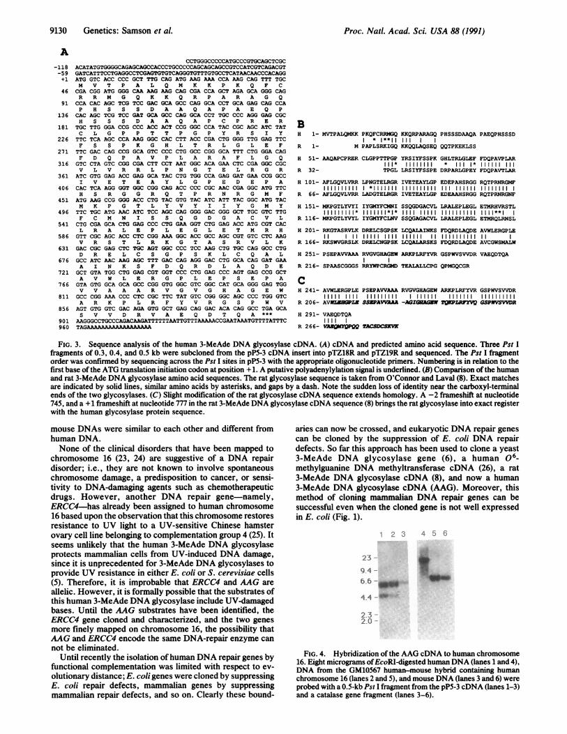
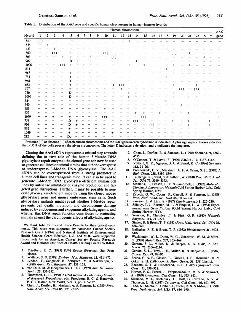
BamHI-digested DNAs from 25 human-hamster somatic cellhybrids was probed with AAG cDNA. The human andhamster DNA hybridization patterns were easily distinguish-able (Fig. 2A), and Table 1 shows that theAAG gene was onlydetected in hybrids 967 and 1079, whose common humanchromosomes are 5 and 16. Since 20 of the other hybrids inthe panel carried chromosome 5 it seemed likely that AAGmaps to human chromosome 16. However, for one hybridcell line, the results were unclear; hybrid 904 DNA did nothybridize to AAG even though 5% of the 904 cells arereported to contain human chromosome 16 (Table 1). There-fore, we confirmed the chromosome 16 assignment by prob-ing DNA from a human-mouse hybrid cell line (GM10567)carrying chromosome 16 (and no other human chromosome)in 98% of the cells; the AAG gene was clearly present (Fig.4). Because the single 6.0-kb mouse EcoRI band and one ofthe human EcoRI bands comigrated (Fig. 4, lanes 1 and 3), theidentity of the human-mouse hybrid DNA loaded in lane 2was confirmed by reprobing the same blot with a humanchromosome 11-specific probe-namely, catalase (22); asexpected for a probe not derived from human chromosome16, the banding patterns in the human-mouse hybrid and the
Genetics: Samson et al.
2. l
9130 Genetics: Samson et al. Proc. Natl. Acad. Sci. USA 88 (1991)
CCTGGGCCCCCATGCCCGTGCAGCTCGCACATATGTGGGGCAGAGCAGCCACCCTGCCCCAGCAGCAGCCGTCCATCGTCAGACGTGATCATTTCCTGAGGCCTCGAGTGTGTCAGGGTGTTTGTGCCTCATAACAACCCACAGGATG GTC ACC CCC GCT TTG CAG ATG AAG AAA CCA AAG CAG TTT TGCM V T P A L Q M K K P K Q F CCGA CGG ATG GGG CAA AAG AAG CAG CGA CCA GCT AGA GCA GGG CAGR R M G Q K K Q R P A R A G Q
CCA CAC AGC TCG TCC GAC GCA GCC CAG GCA CCT GCA GAG CAG CCAP H S S S D A A Q A P A E Q P
CAC AGC TCG TCC GAT GCA GCC GAG GCA CCT TGC CCC AGG GAG CGCH S S S D A A Q A P C P R E R
TGC TTG GGA CCG CCC ACC ACT CCG GGC CCA TAC CGC AGC ATC TATC L G P P T T P G P Y R S I YTTC TCA AGC CCA AAG GGC CAC CTT ACC CGA CTG GGG TTG GAG TTCF S S P K G H L T R L G L E F
TTC GAC CAG CCG GCA GTC CCC CTG GCC CGG GCA TTT CTG GGA CAGF D Q P A V P L A R A F L G Q
GTC CTA GTC CGG CGA CTT CCT AAT GGC ACA GAA CTC CGA GGC CGCV L V R R L P N G T E L R G R
ATC GTG GAG ACC GAG GCA TAC CTG TGG CCA GAG GAT GAA CCG GCCI V E T E A Y L G P E D E P A
CAC TCA AGG GGT GGC CGG GAG ACC CCC CGC AAC CGA GGC ATG TTCH S R G G R Q T P R N R G M FATG AAG CCG GGG ACC CTG TAC GTG TAC ATC ATT TAC GGC ATG TACM K P G T L Y V Y I I Y G M YTTC TGC ATG AAC ATC TCC AGC CAG GGG GAC GGG GCT TGC GTC TTGF C M N I S S Q G D G A C V L
CTG CGA GCA CTG GAG CCC CTG GAA GGT CTG GAG ACC ATG CGT CACL R A L E P L E G L E T M R H
GTT CGC AGC ACC CTC CGG AAA GGC ACC GCC AGC CGT GTC CTC AAGV R S T L R K G T A S R V L KGAC CGC GAG CTC TGC AGT GGC CCC TCC AAG CTG TGC CAG GCC CTGD R E L C S G P S K L C Q A LGCC ATC AAC AAG AGC TTT GAC GAG AGG GAC CTG GCA CAG GAT GAAA I N K S F D Q R D L A Q D E
GCT GTA TGG CTG GAG CGT GGT CCC CTG GAG CCC AGT GAG CCG GCTA V W L E R G P L E P S E P AGTA GTG GCA GCA GCC CGG GTG GGC GTC GGC CAT GCA GGG GAG TGGV V A A A R V G V G H A G E W
GCC CGG AAA CCC CTC CGC TTC TAT GTC CGG GGC AGC CCC TGG GTCA R K P L R F Y V R G S P W V
AGT GTG GTC GAC AGA GTG GCT GAG CAG GAC ACA CAG GCC TGA GCAS V V D R V A E Q D T Q A ***
AAGGGCCTGCCCAGACAAGATTTTTTAATTGTTTAAAAACCGAATAAATGTTTTATTTCTAGAAAAAAAAAAAAAA
BH 1- MVTPALQMKK PKQFCRRMGQ KKQRPARAGQ PHSSSDAAQA PAEQPHSSSD
* 1**I1 III
R 1- M PAPLSRKIGQ KKQQLAQSEQ QQTPKEKLSS
H 51- AAQAPCPRER CLGPPTTPGP YRSIYFSSPK GHLTRLGLEF FDQPAVPLAR* * 1* I II
R 32- TPGL LRSIYFSSPE DRPARLGPEY FDQPAVTLAR
H 101- AFLGQVLVRR LPNGTELRGR IVETEAYLGP EDEPAHSRGG RQTPRNRGMFllll *1111111 IIIIIIIIII III
R 66- AFLGQVLVRR LADGTELRGR IVETF.AYLGP EDEAAHSRGG RQTPRNRGNF
H 151- MKPGTLYVYI IYGMYFCMNI SSQGDGACVL LRALEPLEGL ETMRHVRSTLIIIIIIII1* 11111 1 *1* 11 1 11111H1i 111 1111 1I 1 I**I
R 116- MKPGTLYVYL IYGMYFCLNV SSQGAGACVL LRALEPLEGL ETMRQLRNSL
H 201- RKGTASRVLK DRELCSGPSK LCQALAINKS FDQRDLAQDE AVWLERGPLE11 11 11111 1111 111111 11 1111111111 11
R 166- RKSWVGRSLK DRELCNGPSK LCQALARSKS FDQRDLAQDE AVCGWSMALW
H 251- PSEPAVVAAA RVGVGHAGEW ARKPLRFYVR GSPWVSVVDR VAEQDTQA
R 216- SPAASCGGGS RRYWPCRGMD TEALALLCPG QPMGQCGR
CH 241- AVWLERGPLE PSEPAVVAAA RVGVGHAGEW ARKPLRFYVR GSPWVSVVDR
11111 III 111111111 1 111111 1111111 1111111111R 206- AVIIIGP SWAVAAA -AGIGHAGW TQQLR GSPPSVVDRH 291- VAEQDTQAR I
R 266- VMfQMYQPQSDSW
FIG. 3. Sequence analysis of the human 3-MeAde DNA glycosylase cDNA. (A) cDNA and predicted amino acid sequence. Three Pst Ifragments of 0.3, 0.4, and 0.5 kb were subcloned from the pP5-3 cDNA insert into pTZ18R and pTZ19R and sequenced. The Pst I fragmentorder was confirmed by sequencing across the Pst I sites in pP5-3 with the appropriate oligonucleotide primers. Numbering is in relation to thefirst base of the ATG translation initiation codon at position +1. A putative polyadenylylation signal is underlined. (B) Comparison ofthe humanand rat 3-MeAde DNA glycosylase amino acid sequences. The rat glycosylase sequence is taken from O'Connor and Laval (8). Exact matchesare indicated by solid lines, similar amino acids by asterisks, and gaps by a dash. Note the sudden loss of identity near the carboxyl-terminalends of the two glycosylases. (C) Slight modification of the rat glycosylase cDNA sequence extends homology. A -2 frameshift at nucleotide745, and a + 1 frameshift at nucleotide 777 in the rat 3-MeAde DNA glycosylase cDNA sequence (8) brings the rat glycosylase into exact registerwith the human glycosylase protein sequence.
mouse DNAs were similar to each other and different fromhuman DNA.None of the clinical disorders that have been mapped to
chromosome 16 (23, 24) are suggestive of a DNA repairdisorder; i.e., they are not known to involve spontaneouschromosome damage, a predisposition to cancer, or sensi-tivity to DNA-damaging agents such as chemotherapeuticdrugs. However, another DNA repair gene-namely,ERCC4-has already been assigned to human chromosome16 based upon the observation that this chromosome restoresresistance to UV light to a UV-sensitive Chinese hamsterovary cell line belonging to complementation group 4 (25). Itseems unlikely that the human 3-MeAde DNA glycosylaseprotects mammalian cells from UV-induced DNA damage,since it is unprecedented for 3-MeAde DNA glycosylases toprovide UV resistance in either E. coli or S. cerevisiae cells(5). Therefore, it is improbable that ERCC4 and AAG areallelic. However, it is formally possible that the substrates ofthis human 3-MeAde DNA glycosylase include UV-damagedbases. Until the AAG substrates have been identified, theERCC4 gene cloned and characterized, and the two genesmore finely mapped on chromosome 16, the possibility thatAAG and ERCC4 encode the same DNA-repair enzyme cannot be eliminated.
Until recently the isolation of human DNA repair genes byfunctional complementation was limited with respect to ev-
olutionary distance; E. coli genes were cloned by suppressingE. coli repair defects, mammalian genes by suppressingmammalian repair defects, and so on. Clearly these bound-
aries can now be crossed, and eukaryotic DNA repair genescan be cloned by the suppression of E. coli DNA repalrdefects. So far this approach has been used to clone a yeast3-MeAde DNA glycosylase gene (6), a human 06_methylguanine DNA methyltransferase cDNA (26), a rat3-MeAde DNA glycosylase cDNA (8), and now a human3-MeAde DNA glycosylase cDNA (AAG). Moreover, thismethod of cloning mammalian DNA repair genes can besuccessful even when the cloned gene is not well expressedin E. coli (Fig. 1).
I 2 3 4 5 6
23
9-4 -
6.64--m
4.4 -
2-3 -
2.0 --
FIG. 4. Hybridization of the AAG cDNA to human chromosome16. Eight micrograms ofEcoRI-digested human DNA (lanes 1 and 4),DNA from the GM10567 human-mouse hybrid containing humanchromosome 16 (lanes 2 and 5), and mouse DNA (lanes 3 and 6) wereprobed with a 0.5-kb Pst I fragment from the pP5-3 cDNA (lanes 1-3)and a catalase gene fragment (lanes 3-6).
A-118-59+1
46
91
136
181
226
271
316
361
406
451
496
541
586
631
676
721
766
811
856
901960
Proc. Natl. Acad. Sci. USA 88 (1991) 9131
Table 1. Distribution of the AAG gene and specific human chromosome in human-hamster hybridsHuman chromosome AAG
Hybrid 1 2 3 4 5 6 7 8 9 10 11 12 13 14 15 16 17 18 19 20 21 22 X Y gene
867 (+) - - + - - - - - - - + + - - - + + - - -
854 + - - + - - - - - - -
423 +860 - -(+) - + +-- - ( -803 - - - + + - -+ - (+).+ + -909 - - - - D + -+.+.+ - -1006 - - - (+) + - + + - - - - + - + - - - + - + - - -811. +.++967 - + - - +
734 - + ---+.+.968 - - - - + - - +
683 - - - - +.+ (+) - + - - - - + - + + - - -507 - + ++ - +.(+)-(+)-+ -750 D. . . . . .+ + + +1099 + D. . . . . . . . . .+ + + - -324.+.940 -.-.-.-.+.-.-983 + +937 + +. . . . . .+ + - +.1079 - + + - - (+) - (+) +. . . ..+756 - - D ++ + +(+) + + + - + -904 - - D + . . . + - - (+). . . ..+ -862 - - + +1049 - - ++212 - - Dq. . . . . . . . . . . . . .+ -Presence (+) or absence (-) ofeach human chromosome and theAAG gene in each hybrid line is indicated. A plus sign in parentheses indicates
that <55% of the cells possess the given chromosome. The letter D indicates a deletion, and q indicates the long arm.
Cloning the AAG cDNA represents a critical step towardsdefining the in vivo role of the human 3-MeAde DNAglycosylase repair enzyme; the cloned gene can now be usedto generate cell lines or animal strains that either overexpressor underexpress 3-MeAde DNA glycosylase. The AAGcDNA can be overexpressed from a strong promoter inhuman cell lines and transgenic mice. It can also be used togenerate 3-MeAde DNA glycosylase-deficient human celllines by antisense inhibition of enzyme production and tar-geted gene disruptions. Further, it may be possible to gen-erate glycosylase-deficient mice by using the cloned mouseglycosylase gene and mouse embryonic stem cells. Suchglycosylase mutants might reveal whether 3-MeAde repairprevents cell death, mutation, and chromosome damageinduced by endogenous and exogenous alkylating agents, andwhether this DNA repair function contributes to protectinganimals against the carcinogenic effects of alkylating agents.
We thank John Cairns and Bruce Demple for their critical com-ments. This work was supported by American Cancer SocietyResearch Grant NP448 and National Institute of EnvironmentalHealth Science Grant ES03926. L.S. and M.B. were supportedrespectively by an American Cancer Society Faculty ResearchAward and National Institutes of Health Training Grant CA 09078.
1. Friedberg, E. C. (1985) DNA Repair (Freeman, San Fran-cisco).
2. Wallace, S. S. (1988) Environ. Mol. Mutagen. 12, 431-477.3. Lindahl, T., Sedgwick, B., Sekiguchi, M. & Nakabeppu, Y.
(1988) Annu. Rev. Biochem. 57, 133-158.4. van Duin, M. & Hoeijmakers, J. H. J. (1989) Ann. Ist. Super.
Sanita 25, 131-142.5. Thompson, L. H. (1988) inDNA Repair, A Laboratory Manual
of Research Procedures, eds. Friedberg, E. C. & Hanawalt,P. C. (Dekker, New York), Vol. 3, pp. 115-132.
6. Chen, J., Derfler, B., Maskati, A. & Samson, L. (1989) Proc.Natl. Acad. Sci. USA 86, 7961-7965.
7. Chen, J., Derfler, B. & Samson, L. (1990) EMBO J. 9, 4569-4575.
8. O'Connor, T. & Laval, F. (1990) EMBO J. 9, 3337-3342.9. Volkert, M. R., Nguyen, D. C. & Beard, K. C. (1986) Genetics
112, 11-26.10. Prochownik, E. V., Markham, A. F. & Orkin, S. H. (1983) J.
Biol. Chem. 258, 8389-8394.11. Talmadge, K., Stahl, S. & Gilbert, W. (1980) Proc. Nat!. Acad.
Sci. USA 77, 3369-3373.12. Maniatis, T., Fritsch, E. F. & Sambrook, J. (1982) Molecular
Cloning, A Laboratory Manual (Cold Spring Harbor Lab., ColdSpring Harbor, NY).
13. Rebeck, G. W., Coons, S., Carroll, P. & Samson, L. (1988)Proc. Nat!. Acad. Sci. USA 85, 3039-3043.
14. Samson, L. & Linn, S. (1987) Carcinogenesis 8, 227-230.15. Silhavy, T. J., Berman, M. L. & Enquist, L. W. (1984) Exper-
iments with Gene Fusions (Cold Spring Harbor Lab., ColdSpring Harbor, NY).
16. Winston, F., Chumley, F. & Fink, G. R. (1983) MethodsEnzymol. 101, 211-227.
17. Singer, B. & Brent, T. P. (1981) Proc. Nat!. Acad. Sci. USA 78,856-860.
18. Gallagher, P. E. & Brent, T. P. (1982) Biochemistry 21, 6404-6409.
19. Washington, W. J., Dunn, W. C., Generoso, W. M. & Mitra,S. (1988) Mutat. Res. 207, 165-169.
20. Gerson, S. L., Miller, K. & Berger, N. A. (1985) J. Clin.Invest. 76, 2106-2114.
21. Gerson, S. L., Trey, J. E., Miller, K. & Benjamin, E. (1987)Cancer Res. 47, 89-95.
22. Bruns, G. A. P., Glaser, T., Gusella, J. F., Housman, D. &Orkin, S. H. (1984) Am. J. Hum. Genet. 36, 25S (abstr.).
23. Reeders, S. T. & Hildebrand, C. E. (1989) Cytogenet. CellGenet. 51, 299-318.
24. Harper, P. S., Frezal, J., Ferguson-Smith, M. A. & Schinzel,A. (1989) Cytogenet. Cell Genet. 51, 563-611.
25. Siciliano, M. J., Bachinsky, L., Dolf, G. Carrano, A. V. &Thomson, L. H. (1987) Cytogenet. Cell Genet. 46, 691-692.
26. Tano, K., Shiota, S., Collier, J., Foote, R. S. & Mitra, S. (1990)Proc. Nat!. Acad. Sci. USA 87, 686-690.
Genetics: Samson et A