Embed Size (px)
Citation preview
Vol. 50, No. 1INFECTION AND IMMUNITY, OCt. 1985, p. 236-2420019-9567/85/100236-07$02.00/0Copyright © 1985, American Society for Microbiology
Cloning and Surface Expression in Escherichia coli of a StructuralGene Encoding a Surface Protein of Haemophilus influenzae Type b
PRISCILLA L. HOLMANS, THERESA A. LOFTUS, AND ERIC J. HANSEN*
Department of Microbiology, Southwestern Graduate School of Biomedical Sciences, University of Texas Health ScienceCenter at Dallas, Dallas, Texas 75235
Received 12 April 1985/Accepted 10 July 1985
Recombinant DNA technology was used to clone a gene coding for a surface protein of Haemophilusinfluenzae type b (Hib) into Escherichia coli. Chromosomal DNA from a clinical isolate of Hib was cleaved withEcoRI and ligated into plasmid vectors containing three different translational reading frames. E. coli carryingrecombinant plasmids were screened in a colony blot-radioimmunoassay system by using murine monoclonalantibodies (mabs) directed against cell surface-exposed proteins of Hib. mab 7B2, which is specific for a Hibsurface protein with an apparent molecular weight of 27,000 (27K), reacted with several recombinant strainsof E. coli. Restriction analysis revealed the presence of a 9. 1-kilobase DNA insert in each of these recombinantplasmids and also determined that both transcription and translation of the Hib gene(s) coding for the7B2-reactive antigen were not dependent on the lac operator and promoter of the vectors. Radioim-munoprecipitation and Western blot analyses showed that the antigenic determinant recognized by mab 7B2 inthese recombinant E. coli was present in a 27K protein. In addition, this 27K protein was shown to be bothlocalized on the surface of these E. coli cells and accessible to antibody.
The development of an efficacious vaccine for the protec-tion of infants against systemic disease caused by Haemo-philus influenzae type b (Hib) is the subject of intenseresearch efforts in many laboratories (2, 8, 25-27, 33, 36).We have concentrated on identifying vaccine candidatesamong the outer membrane proteins of this pathogen. Pre-vious studies from this laboratory have established thatcertain Hib outer membrane proteins are immunogenic inhuman infants as well as being both exposed on the Hib cellsurface and accessible to antibodies (16-19).
Molecular cloning into Escherichia coli may provide auseful system for the production of protective Hib antigensand especially for genetic analysis of these antigens. Specif-ically, we wished to isolate recombinant clones which ex-press Hib surface antigens recognized by monoclonal anti-bodies (mabs) which we have previously characterized asbeing directed against cell surface-exposed proteins. Herewe describe the construction of recombinant clones whichsatisfy this criterion. Moreover, we present data whichindicate that the Hib protein encoded by the cloned gene issynthesized in E. coli accurately with respect to size andantigenicity and is localized on the surface of E. coli cells inwhich it is expressed.(A preliminary account of these findings was presented at
the 84th Annual Meeting of the American Society for Micro-biology [P. L. Holmans, T. A. Loftus, and E. J. Hansen,Abstr. Annu. Meet. Am. Soc. Microbiol. 1984, D95, p. 66]1)
MATERIALS AND METHODSBacterial strains, plasmid vectors, and growth conditions.
The clinical isolate of Hib (strain DL41) used in this study asthe source of chromosomal Hib DNA was obtained fromGeorge H. McCracken, Jr., Southwestern Medical School,Dallas, Tex., and has been described previously (14). HibDL41 was grown at 37°C in brain heart infusion broth (DifcoLaboratories, Detroit, Mich.) supplemented with Levinthalbase (1) as a source of hemin and NAD. Solid medium was
* Corresponding author.
prepared by incorporating 1.5% (wt/vol) agar (Difco) in brainheart infusion supplemented with Levinthal base. Agar platecultures were incubated at 37°C in a candle extinction jar for18 to 24 h.The E. coli K-12 derivative HB101 [F- hsdS20(r-Bm B)
recA13 ara-14 proA2 lac Yl galK2 rpsL20 xyl-5 mtl-l supE44X-] (5) was the recipient for all transformation experimentsand the host for the vector plasmids pPC401, pPC42, andpPC+3 (7). Each of these plasmids possesses a differenttranslational reading frame linked to the lac operator andpromoter, such that all three possible reading frames ex-pressed from the lac UV5 promoter are represented by thisset of plasmids. E. coli HB101 was grown in L broth (LB) (1liter contained 10 g of tryptone, 10 g of NaCl, and 5 g of yeastextract) or on LB agar. Plasmid-containing derivatives weregrown in LB or on LB agar supplemented with 100 p.g ofampicillin trihydrate ml-' (Sigma Chemical Co., St. Louis,Mo.). For large-scale plasmid preparations, E. coli HB101derivatives were grown in medium C (29) supplemented with0.2% (wtlvol) glucose, 0.1% (wt/vol) Casamino Acids, 0.5 ,ugof thiamine ml-', and 100 ,ug of ampicillin ml-'.
Preparation of DNAs. Plasmid DNA was prepared by themethod of Guerry et al. (13). Chromosomal DNA fromn Hibwas prepared from 1 liter of stationary-phase cells by themethod of Marmur (30), followed by phenol extraction anddialysis against 50 mM Tris hydrochloride (pH 8.0) contain-ing 5 mM EDTA.
Production of mab 7B2. Eight-week-old female BALB/cmice (Cumberland Laboratories, Clinton, Tenn.) were im-munized by intraperitoneal injection with 107 CFU of HibDL41. One month later, these animals received a secondinjection of 108 CFU of this same strain, and 3 weeks laterthey received a third and final injection identical to thesecond injection. Three days after this last injection, thespleens were removed from two animals and used in thestandard hybridoma production system described in detailpreviously (35). The whole-cell radioimmunoprecipitation(WC-RIP) system (18) was used to screen culture superna-tant fluids (500 [lI) from the resultant hybridomas for the
236

SURFACE PROTEIN OF H. INFLUENZAE TYPE b 237
presence of mabs directed against cell surface-exposed andantibody-accessible Hib proteins as previously described(25). Murine lymphocyte hybridoma 7B2 was identified asproducing an immunoglobulin G mab directed against a cellsurface-exposed outer membrane protein of Hib with anapparent molecular weight of 27,000 (27K) and was clonedby limiting dilution analysis as previously described (35).
Antisera. Immune rat serum was produced in adultSprague-Dawley rats (Charles River Breeding Laboratories,Inc., Wilmington, Mass.) by immunization of these animalswith viable Hib cells. The rats received intraperitonealinjections with 107 CFU of Hib DL41 at 3- to 4-weekintervals for 4 months. Immune serum was prepared fromthe blood of animals exsanguinated by cardiac puncture.AA-RIA. The antibody-accessibility (AA)-radioimmuno-
assay (RIA) procedure was used to assess cell surfaceexposure and AA of the antigenic determinant recognized bymab 7B2 in different strains of bacteria. E. coli strains grownovernight on LB agar were suspended in phosphate-bufferedsaline (PBS) at 4°C to a final concentration of 108 CFU/ml. A1-ml portion of the E. coli suspension was incubated with 500,ul of hybridoma culture supernatant containing mab 7B2 for2 h at 4°C with gentle agitation. The cells were centrifuged at12,000 x g for 2 min and then suspended in 1 ml of PBScontaining 10% fetal calf serum (PBS-FCS). The cells werethen centrifuged again and suspended in 1 ml of the samebuffer. To detect mab 7B2 bound to the bacterial cells, 5 x10⁵ cpm of affinity-purified and radioiodinated rabbit anti-mouse immunoglobulin (specific activity, 107 cpm per ,ug ofprotein) was added to the cell suspension. After incubationfor 1 h at 4°C with gentle agitation, the bacterial suspensionwas washed five times with 1-ml quantitites of PBS-FCS andsuspended in 500 ILI of solubilization buffer (18). Radioactiv-ity in the final washed cell pellet was measured with a Searlemodel 1195 gamma counter (Searle Analytic Inc., Chicago,Ill.). The results are expressed as counts per minute ofantibody probe bound to the test cells. All experimentsincluded a negative control with PBS in place of the 7B2hybridoma culture supernatant. All data represent the aver-age of duplicate samples.
Construction and screening of genomic libraries. The vec-tor DNAs pPC¢1, pPC¢2, and pPC43 were digested withEcoRI (Bethesda Research Laboratories, Inc., Gaith-ersburg, Md.) as described previously (22). Partial EcoRIdigests of chromosomal DNA from Hib DL41 were alsoprepared. The vector DNAs were then treated with calfintestine alkaline phosphatase (Boehringer MannheimBiochemicals, Indianapolis, Ind.), followed by phenol andether extractions (29). The phosphatase-treated, EcoRI-digested plasmid DNAs were individually combined with thecleaved Hib DL41 DNA, and T4 DNA ligase (BoehringerMannheim Biochemicals) was added, along with ATP anddithiothreitol to final concentrations of 0.1 and 1 mM,respectively. The ratio of phosphatase-treated vector DNAto Hib DNA was 20:1, and the ligation reactions wereperformed at 4°C overnight. The ligated DNA was used totransform E. coli HB101 made competent with calciumchloride treatment (28). After incubation at 37°C for 40 minto allow recovery and expression, the transformed cells wereplated on LB agar containing ampicillin and incubatedovernight at 37°C. The resultant colonies were collectivelysuspended in LB containing ampicillin and 20% glycerol andstored at -20°C in multiple portions.Immunoscreening of genomic libraries with mab probes.
The genomic libraries were plated at 2 x 10² to 5 x 10²colonies plate-' and grown overnight to prepare them for
screening with specific mabs directed against cell surface-exposed proteins. Clones which produced antigens recog-nized by the mabs were detected by a modification of thecolony blot RIA of Henning et al. (21). Colonies were liftedon sterile Whatman no. 40 (ashless) filter paper (8.26-cmdiameter). The filters were placed in chloroform for 5 min,dried at 37°C for 30 min, and then probed with a pool of mabsas previously described (14). mab 4C11, which is directedagainst the 100K cell surface-exposed protein of Hib (17),and mab 6G12, which is specific for the 98K cell surface-exposed protein of Hib (25), were used together with mab7B2 in this screening system. The presence of mabs attachedto the filter-bound lysed cells was detected with the affinity-purified and radioiodinated rabbit anti-mouse immunoglob-ulin. All incubations were performed with gentle agitation at4°C. Clones which reacted with the mabs were identified byautoradiographic analysis with Fuji RX safety film and aDuPont Cronex intensifying screen. The original plates wereincubated 4 to 6 h at 37°C, and colony blot-positive colonieswere then picked and rescreened with the individual mabs.
Characterization of recombinant plasmids. Plasmid DNAfrom the clones of interest was isolated from individualcolonies or from 1-ml liquid cultures (medium C) by themethod of Holmes and Quigley (23). The use of supple-mented minimal medium rather than LB resulted in a betterplasmid yield with much less rRNA contamination (P. L.Holmans, unpublished data). The resultant plasmid DNAwas used for restriction endonuclease analysis and subse-quent transformations.Immunochemical identification of cloned gene products.
Identification of gene products recognized by specific mabswas performed by use of the WC-RIP system, as describedpreviously (15, 17), or by Western blot analysis, by amodification of the method of Towbin et al. (41). In the latterprocedure, proteins resolved by sodium dodecyl sulfate(SDS)-polyacrylamide gel electrophoresis in a 10% (wt/vol)polyacrylamide separating gel were electrophoreticallytransferred to nitrocellulose strips overnight at 4°C, by usingeither 165 mA or 75V, in 20 mM Tris containing 150 mMglycine and 20% (vol/vol) methanol. The nitrocellulose stripswere then probed with mab 7B2 by a procedure describedpreviously (25). Detection of antigen-mab complexes on thenitrocellulose strips was achieved by using horseradishperoxidase-conjugated goat anti-mouse immunoglobulin G(diluted 10-3 in PBS containing 0.1% [vol/vol] Tween 20),sometimes followed by peroxidase-conjugated rabbit anti-goat immunoglobulin G (both from Cappel Laboratories,Cochranville, Pa.) and subsequent reaction with 4-chloro-1-naphthol (Sigma) as described by Hawkes et al. (20).
Electron microscopy. Visualization of immune complexeson bacterial cell surfaces was performed as follows. Cellsfrom a freshly grown agar plate were suspended to approx-imately 109 ml-' in PBS-FCS, pelleted by centrifugation, andresuspended in PBS-FCS. A 0.25-ml portion of the sus-pended cells was mixed with 1.0 ml of hybridoma culturesupernatant containing mab 7B2 and was rotated at 4°C for60 to 120 min. After three washes in PBS-FCS, the cells weresuspended in PBS-FCS (1.0 ml), and a suspension of staph-ylococcal protein A-colloidal gold particles prepared by themethod of Geohegan and Ackerman (11) was added suchthat the final mixture was light pink. This suspension wasrotated at 4°C for 60 to 120 min, after which the cells werewashed three times in PBS-FCS and once in PBS and finallysuspended in PBS (0.5 ml). Formaldehyde was added to afinal concentration of 1.2%; after 10 min, the cells wereapplied to carbon-coated grids (10 to 15 ,ul grid-'). Excess
VOL. 50, 1985

238 HOLMANS ET AL.
A
*w:a 0;. S
to,^* *S-S
A - .1
4 * -w * 4
* . Oj.
FIG. 1. Colony blot-RIA of E. coli HB101 recombinant clones. Colonies were transferred to Whatman no. 40 paper and probed with mab7B2 in the colony blot-RIA described in Materials and Methods. Panel A shows a filter to which colonies were transferred and assayed bycolony blot-RIA, followed by staining with methylene blue to facilitate localization of reactive colonies. Panel B is an autoradiograph of thesame filter after colony blot-RIA.
liquid was removed with a wedge of filter paper, and a dropof 2% (wt/vol) neutral aqueous phosphotungstic acid wasapplied to each grid. The phosphotungstic acid was removedwith a wedge of filter paper, and a drop of distilled H20 wasapplied to each grid and then removed. The grids wereexamined in a Philips 301 electron microscope at 60 kVaccelerating voltage and photographed at a magnification ofx 34,000.
RESULTSConstruction and screening of genomic libraries. A total of
6,700 pPC4d clones, 1,870 pPC42 clones, and 2,000 pPC4)3clones were obtained in this study. The three libraries wereplated at 200 to 500 colonies plate-' and screened with amixture of mabs previously found to be specific for Hibsurface proteins. A representative filter from an experimentutilizing mab 7B2 that was stained with methylene blue toreveal the position of the bacterial colonies and the matchingautoradiograph with two positive reactions is shown in Fig.1. Of the colonies tested in one experiment, 4/2,477, 6/749,and 3/990 from the pPC4i1, pPC4+2, and pPC4¢3 libraries,respectively, reacted with mab 7B2. Assuming that eachclone was an independent isolate, mab 7B2-positive recom-binant clones were obtained at a frequency of approximately3 x 10-3. No recombinant clones reactive with mab 4C11 or6G12 were obtained in these experiments.
Characterization of recombinant plasmids. Because mab7B2-positive clones were found in each library, the expres-sion of the mab 7B2-reactive antigen was apparently inde-pendent of the lac operator and promoter of the vectorplasmids for transcription. We therefore addressed the pos-sibility that transcription initiated within the cloned segmentof Hib DNA. Four clones were picked at random and
analyzed to determine whether the DNA insert encoding themab 7B2-reactive antigen was present in only one orienta-tion or in both orientations, the latter situation being evi-dence for expression from a promoter within the cloned HibDNA segment. The recombinant plasmids were cleaved withPstI (which does not cut within the DNA inserts) and XbaI(which does not cut within the vectors) (Fig. 2). The result-ing asymmetry of the cleavage products was used in theanalysis of the restriction profiles. EcoRI cleavage of thefour recombinant plasmids examined in this experimentyielded an insert of 9.1 kilobases (Fig. 3, lanes a to d).However, based on the sizes of the restriction fragmentsresulting from cleavage with PstI and XbaI, clones pPLH401(Fig. 3, lane f) and pPLH402 (lane g) contained inserts whosepolarity is directly opposite that of the inserts in clonespPLH403 and pPLH404 (lanes h and i, respectively). Be-cause cleavage of the plasmids with the combination ofEcoRI, PstI, and XbaI resulted in an identical restrictionpattern from all four plasmids (Fig. 3, lanes k to n), theinserts were assumed to be identical, with either orientationsufficient for expression of the mab 7B2-reactive antigen(Fig. 2). Therefore, both transcription and translation pre-sumably initiated within the cloned Hib DNA and from theauthentic (Hib) promoter.
Characterization of the 7B2 gene product. We wished todetermine whether the cloned gene product which reactedwith mab 7B2 was identical to the protein recognized by mab7B2 in Hib. We addressed this question first with RIPanalysis of one of the recombinant clones, by using aWC-RIP method which has been shown to be specific for theidentification of surface antigens (15, 18). The protein whichwas immunoprecipitated from radiolabeled Hib cells by mab7B2 (Fig. 4, lane c) has an apparent molecular weight of
INFECT. IMMUN.

SURFACE PROTEIN OF H. INFLUENZAE TYPE b 239
LORI -jac
'-tpPC1am1pPC,21J
Chromosomal DNA from DL41;.,
EcoRI
1. Ligate, transform into HB101
2. Screen transformants withmab 7B2
-EggoRI -EcoRIXbaI
FIG. 2. Schematic diagram of the cloning approach used to generate the recombinant plasmids pPLH401, pPLH402, pPLH403, andpPLH404. The arrows on the Hib DNA inserts reflect the relative orientation of each insert and do not necessarily indicate the direction oftranscription. The cleavage sites for EcoRI, PstI, and XbaI are indicated on each recombinant plasmid.
FIG. 3. Restriction endonuclease analysis of recombinant plas-mids. Plasmid DNAs were isolated by a minipreparation method(23) and cleaved with the restriction enzymes indicated, followed byelectrophoretic separation of the products in 1% agarose containing0.5 ug of ethidium bromide ml-'. Electrophoresis was performed ina Bio-Rad minisubmarine apparatus. The samples were as follows.EcoRI digests: pPLH401 (lane a), pPLH402 (lane b), pPLH403 (lanec), pPLH404 (lane d). PstI and XbaI digests: pPLH401 (lane f),pPLH402 (lane g), pPLH403 (lane h), pPLH404 (lane i). EcoRI,PstI, and XbaI digests: pPLH401 (lane k), pPLH402 (lane 1),pPLH403 (lane m), pPLH404 (lane n). HindIll fragments (23.1, 9.6,6.6, 4.4, 2.3, and 2.01 kilobases) of bacteriophage lambda DNA(lanes e and o) and EcoRI fragments (19.4, 9.15, 7.16, 3.98, and 2.39kilobases) of bacteriophage P22 DNA (lane j) were used as molec-ular mass markers. The upper band in lanes a to d represents thecloned Hib DNA insert. The very small DNA fragment resultingfrom cleavage by EcoRI and PstI at two closely adjacent sitescannot be seen in lanes k, 1, m, and n in this particular gel becauseof the relatively small quantity of plasmid DNA used in these lanes.
27,000 (27K). A negative control utilizing E. coli HB101containing the vector (pPC&j1) showed that mab 7B2 did notimmunoprecipitate any radiolabeled native E. coli proteins(lane e). In contrast, when E. coli HB101 containing recom-binant plasmid pPLH401 was assayed in the WC-RIP sys-tem, a 27K protein was immunoprecipitated by mab 7B2(lane g). Thus, the Hib protein recognized by mab 7B2 isapparently synthesized as the authentic polypeptide in E.coli.Western blot analysis was used to determine whether the
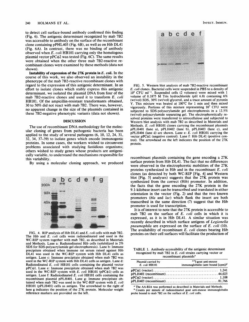
other three mab 7B2-reactive recombinant clones carryingplasmids pPLH402, pPLH403, and pPLH404 all synthesizeda 27K protein. All three of these recombinant clones synthe-sized 27K proteins which reacted with mab 7B2 and whichexhibited electrophoretic mobilities (Fig. 5, lanes b to d)identical to the 27K protein synthesized by Hib DL41 (lanef).The Hib protein recognized by mab 7B2 is a surface
antigen of Hib DL41, and the results of the WC-RIP assayindicated that it was also localized on the surface of therecombinant clones. To confirm this finding, two differentapproaches were used, including an RIA method whichdetermines the accessibility to antibody of a given antigen inwhole bacterial cells and electron microscopy of immunecomplexes on the bacterial cell surface.The results obtained with the AA-RIA are shown in Table
1. E. coli HB101 containing the recombinant plasmidspPLH401 and pPLH403 bound a significantly greater amountof mab 7B2 than E. coli HB101 containing the vectorplasmids pPC4l and pPC,02. Electron microscopic exami-nation of whole cells of these strains treated with mab 7B2followed by reaction with protein A-colloidal gold particles
VOL. 50, 1985
240 HOLMANS ET AL.
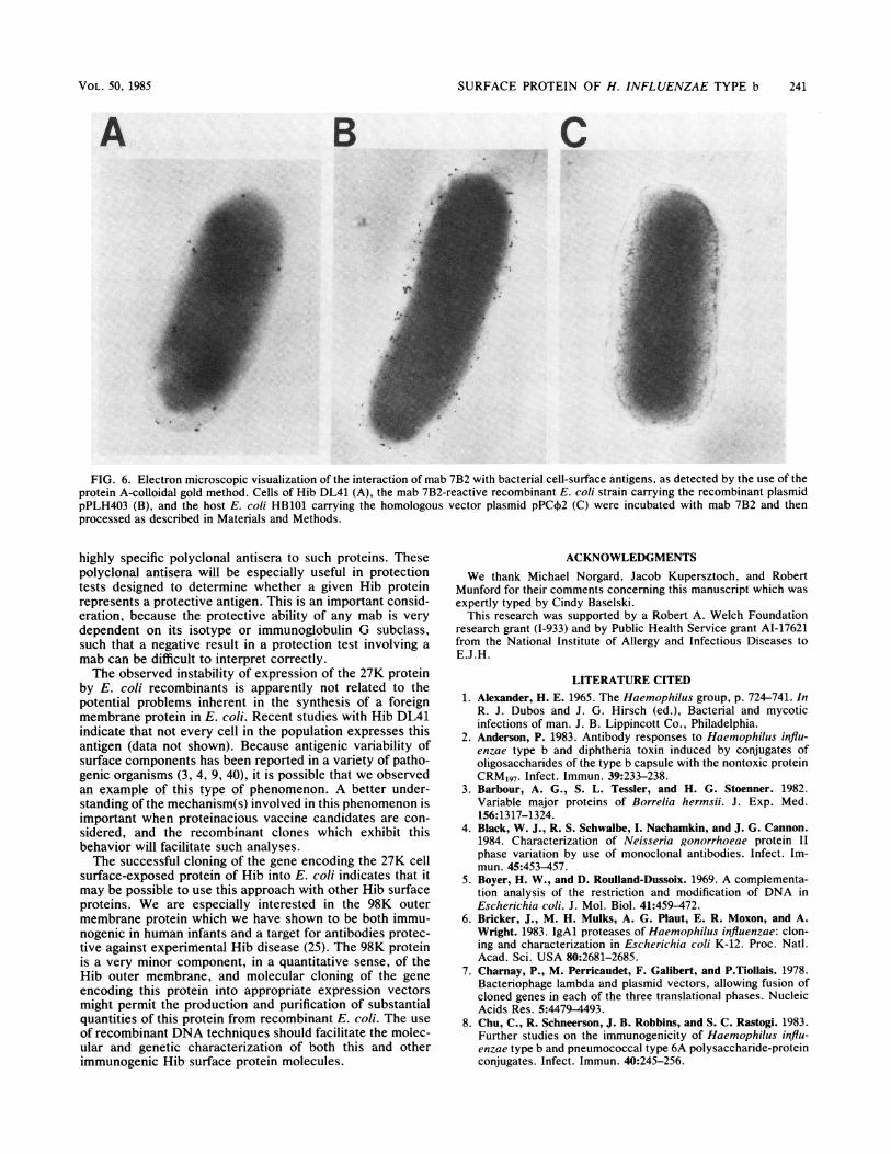
to detect cell surface-bound antibody confirmed this finding(Fig. 6). The antigenic determinant recognized by mab 7B2was accessible to antibody on the surface of the recombinantclone containing pPHL403 (Fig. 6B), as well as on Hib DL41(Fig. 6A). In contrast, there was no binding of antibodyobserved when E. coli HB101 carrying only the homologousplasmid vector pPC¢2 was tested (Fig. 6C). The same resultswere obtained when the other three mab 7B2-reactive re-
combinant clones were examined by these methods (data notshown).
Instability of expression of the 27K protein in E. coli. In thecourse of this work, we also observed an instability in thephenotype of the mab 7B2-reactive recombinant clones withregard to the expression of this antigenic determinant. In an
effort to isolate clones which stably express this antigenicdeterminant, we isolated the plasmid DNA from four of themab 7B2-reactive clones and used it to transform E. coliHB101. Of the ampicillin-resistant transformants obtained,30 to 50% did not react with mab 7B2. There was, however,no apparent change in the size of the plasmids contained inthese 7B2-negative phenotypic variants (data not shown).
DISCUSSIONThe use of recombinant DNA methodology for the molec-
ular cloning of genes from pathogenic bacteria has beenapplied to the study of several pathogens (6, 10, 12, 24, 31,32, 34, 37-39) to isolate genes which encode immunogenicproteins. In some cases, the workers wished to circumventproblems associated with studying fastidious organisms;others wished to study genes whose products are antigeni-cally variable, to understand the mechanisms responsible forthis variability.By using a molecular cloning approach, we produced
200ka bc d e f g
*'I
_
FIG. 4. RIP analysis of Hib DL41 and E. coli cells with mab 7B2.The Hib and E. coli cells were radioiodinated and used in theWC-RIP system together with mab 7B2, as described in Materialsand Methods. Lane a: Radioiodinated Hib cells (solubilized in 2%SDS for SDS-polyacrylamide gel electrophoresis). Lane b: Immuneprecipitate obtained when immune rat serum raised against HibDL41 was used in the WC-RIP system with Hib DL41 cells as
antigen. Lane c: Immune precipitate obtained when mab 7B2 wasused in the WC-RIP system with Hib DL41 cells as antigen. Lane d:Radioiodinated E. coli HB101 cells containing the plasmid vectorpPC4i1. Lane e: Immune precipitate obtained when mab 7B2 wasused in the WC-RIP system with E. coli HB101 (pPC41) cells as
antigen. Lane f: Radioiodinated E. coli HB101 cells containing therecombinant plasmid pPLH401. Lane g: Immune precipitate ob-tained when mab 7B2 was used in the WC-RIP system with E. coliHB101 (pPLH401) cells as antigen. The arrowhead to the right oflane g indicates the position of the 27K protein. Molecular weightreference markers are provided on the left.
a bc d e f
FIG. 5. Western blot analysis of mab 7B2-reactive recombinantE. coli clones. Bacterial cells were suspended in PBS to a density of109 CFU ml-'. Suspended cells (2 volumes) were mixed with 1volume of 0.1875 M Tris hydrochloride (pH 6.8) containing 6%(wt/vol) SDS, 30% (wt/vol) glycerol, and a trace amount of pyroninY. This mixture was heated at 100°C for 1 min and then mixedvigorously. Portions of this mixture representing 10' CFU weresubjected to SDS-polyacrylamide gel electrophoresis in a 12.5%(wt/vol) polyacrylamide separating gel. The electrophoretically re-solved proteins were transferred to nitrocellulose and subjected toWestern blot analysis with mab 7B2 as described in Materials andMethods. E. coli HB101 clones carrying the recombinant plasmidspPLH401 (lane a), pPLH402 (lane b), pPLH403 (lane c), andpPLH404 (lane d) are shown. Lane e: E. coli HB101 carrying thevector pPC4A (negative control). Lane f: Hib DL41 (positive con-trol). The arrowhead on the left indicates the position of the 27Kprotein.
recombinant plasmids containing the gene encoding a 27Ksurface protein from Hib DL41. The fact that no differenceswere observed in the electrophoretic mobilities of the 27Kproteins synthesized in Hib and in the recombinant E. coliclones (as detected by both WC-RIP [Fig. 4] and Westernblot [Fig. 5] analyses) suggests that the 27K protein wassynthesized from the correct (Hib) promoter. In addition,the facts that the gene encoding the 27K protein in the9.1-kilobase insert can be transcribed and translated in eitherorientation in the vector (Fig. 2) and that the two knownpromoters (bla and lac) which flank the insert are bothtranscribed in the same direction (7) suggest that the Hibpromoter is used for transcription.
It is of interest to note that the 27K protein is accessible tomab 7B2 on the surface of E. coli cells in which it isexpressed, as it is in Hib DL41. A similar situation wasrecently described in which surface antigens of Legionellapneumophila are expressed on the surface of E. coli (10).The availability of recombinant E. coli clones bearing Hibproteins on their cell surfaces will facilitate the production of
TABLE 1. Antibody-accessibility of the antigenic determinantrecognized by mab 7B2 in E. coli strains carrying vector or
recombinant plasmidsaPlasmid carried by 1251I-goat anti-mouse
E. coli HB101 immunoglobulin (amt bound [cpmI)bpPC4A (vector)................ 1,241pPLH401 (recombinant) ................ 44,025pPC02 (vector)................ 1,108pPLH403 (recombinant) ................ 81,766
a The AA-RIA was performed as described in Materials and Methods.b Counts per minute of radioiodinated goat anti-mouse immunoglobulin
probe bound to mab 7B2 on the surface of E. coli cells.
INFECT. IMMUN.
SURFACE PROTEIN OF H. INFLUENZAE TYPE b 241
A B C0
FIG. 6. Electron microscopic visualization of the interaction of mab 7B2 with bacterial cell-surface antigens, as detected by the use of theprotein A-colloidal gold method. Cells of Hib DL41 (A), the mab 7B2-reactive recombinant E. coli strain carrying the recombinant plasmidpPLH403 (B), and the host E. coli HB101 carrying the homologous vector plasmid pPC+2 (C) were incubated with mab 7B2 and thenprocessed as described in Materials and Methods.
highly specific polyclonal antisera to such proteins. Thesepolyclonal antisera will be especially useful in protectiontests designed to determine whether a given Hib proteinrepresents a protective antigen. This is an important consid-eration, because the protective ability of any mab is verydependent on its isotype or immunoglobulin G subclass,such that a negative result in a protection test involving amab can be difficult to interpret correctly.The observed instability of expression of the 27K protein
by E. coli recombinants is apparently not related to thepotential problems inherent in the synthesis of a foreignmembrane protein in E. coli. Recent studies with Hib DL41indicate that not every cell in the population expresses thisantigen (data not shown). Because antigenic variability ofsurface components has been reported in a variety of patho-genic organisms (3, 4, 9, 40), it is possible that we observedan example of this type of phenomenon. A better under-standing of the mechanism(s) involved in this phenomenon isimportant when proteinacious vaccine candidates are con-sidered, and the recombinant clones which exhibit thisbehavior will facilitate such analyses.The successful cloning of the gene encoding the 27K cell
surface-exposed protein of Hib into E. coli indicates that itmay be possible to use this approach with other Hib surfaceproteins. We are especially interested in the 98K outermembrane protein which we have shown to be both immu-nogenic in human infants and a target for antibodies protec-tive against expenmental Hib disease (25). The 98K proteinis a very minor component, in a quantitative sense, of theHib outer membrane, and molecular cloning of the geneencoding this protein into appropriate expression vectorsmight permit the production and purification of substantialquantities of this protein from recombinant E. coli. The useof recombinant DNA techniques should facilitate the molec-ular and genetic characterization of both this and otherimmunogenic Hib surface protein molecules.
ACKNOWLEDGMENTSWe thank Michael Norgard, Jacob Kupersztoch, and Robert
Munford for their comments concerning this manuscript which wasexpertly typed by Cindy Baselski.
This research was supported by a Robert A. Welch Foundationresearch grant (1-933) and by Public Health Service grant AI-17621from the National Institute of Allergy and Infectious Diseases toE.J.H.
LITERATURE CITED1. Alexander, H. E. 1965. The Haemophilus group, p. 724-741. In
R. J. Dubos and J. G. Hirsch (ed.), Bacterial and mycoticinfections of man. J. B. Lippincott Co., Philadelphia.
2. Anderson, P. 1983. Antibody responses to Haemophilus influ-enzae type b and diphtheria toxin induced by conjugates ofoligosaccharides of the type b capsule with the nontoxic proteinCRM197. Infect. Immun. 39:233-238.
3. Barbour, A. G., S. L. Tessler, and H. G. Stoenner. 1982.Variable major proteins of Borrelia hermsii. J. Exp. Med.156:1317-1324.
4. Black, W. J., R. S. Schwalbe, I. Nachamkin, and J. G. Cannon.1984. Characterization of Neisseria gonorrhoeae protein IIphase variation by use of monoclonal antibodies. Infect. Im-mun. 45:453-457.
5. Boyer, H. W., and D. Roulland-Dussoix. 1969. A complementa-tion analysis of the restriction and modification of DNA inEscherichia coli. J. Mol. Biol. 41:459-472.
6. Bricker, J., M. H. Mulks, A. G. Plaut, E. R. Moxon, and A.Wright. 1983. IgAl proteases of Haemophilus influenzae: clon-ing and characterization in Escherichia coli K-12. Proc. Natl.Acad. Sci. USA 80:2681-2685.
7. Charnay, P., M. Perricaudet, F. Galibert, and P.Tiollais. 1978.Bacteriophage lambda and plasmid vectors, allowing fusion ofcloned genes in each of the three translational phases. NucleicAcids Res. 5:4479-4493.
8. Chu, C., R. Schneerson, J. B. Robbins, and S. C. Rastogi. 1983.Further studies on the immunogenicity of Haemophilus influ-enzae type b and pneumococcal type 6A polysaccharide-proteinconjugates. Infect. Immun. 40:245-256.
VOL. 50, 1985

242 HOLMANS ET AL.
9. Diaz, J. L., and J. E. Heckels. 1982. Antigenic variation of outermembrane protein II in colonial variants of Neisseria gonor-rhoeae P9. J. Gen. Microbiol. 128:585-591.
10. Engleberg, N. C., E. Pearlman, and B. I. Eisenstein. 1984.Legionella pneumophila surface antigens cloned and expressedin Escherichia coli are translocated to the host cell surface andinteract with specific anti-Legionella antibodies. J. Bacteriol.160:199-203.
11. Geohegan, W. D., and G. A. Ackerman. 1977. Adsorption ofhorseradish peroxidase, ovmucoid and immunoglobulin to col-loidal gold for the indirect detection of concanavalin A, wheatgerm agglutinin and goat anti-human immunoglobulin G on cellsurfaces at the electron microscopic level: a new method,thepry and applications. J. Histochem. Cytochem. 25:1187-1200.
12. Gray, G. L., D. H. Smith, J. S. Baldridge, R. N. Harkins, M. L.Vasil, E. Y. Chen, and H. L. Heyneker. 1984. Cloning, nucleo-tide sequence, and expressioh in Escherichia coli of theexotoxin A structural gene of Pseudomonas aeruginosa. Proc.Natl. Acad. Sci. USA 81:2645-2649.
13. Guerry, P., D. J. LeBianc, and S. Falkow. 1973. General methodfor the isolation of plasmid deoxyribonucleic acid. J. Bacteriol.116:1064-1066.
14. Gulig, P. A., C. F. Frisch, and E. J. Hansen. 1983. A set of twomonoclonal antibodies specific for the cell surface-exposed 39Kmajor outer membrane protein of Haemophilus influenzae typeb defines all straips of this pathogen. Infect. Immun. 42:516-524.
15. Gulig, P. A., and E. J. Hansen. 1985. Coprecipitation of lipo-polysaccharide and the 39,000-molecular-weight major outermembrane protein of Haemophilus influenzae type b by lipo-polysaccharide-directed monoclonal antibody. Infect. Immun.49:819-827.
16. Gulig, P. A., G. H. McCracken, Jr., C. F. Frisch, K. H.Johnston, and E. J. Hansen. 1982. Antibody response of infantsto cell surface-exposed outer membrane proteins of Haemo-philus influenzae type b after systemic Haemophilus disease.Infect. Immun. 37:82-88.
17. Gulig, P. A., G. H. McCracken, Jr., P. L. Holmans, and E. J.Hansen. 1984. Immunogenic proteins in cell[free culture super-natants of Haemophilus influenzae type b. Infect. Immun.44:41-48.
18. Hansen, E. J., C. F. Frisch, and K. H. Johnston. 1981. Detectionof antibody-accessible proteins on the cell surface of Haemo-philus influenzae type b. Infect. Immun. 33:950-953.
19. Hansen, E. J., C. F. Frisch, and K. H. Johnston. 1982. Cellenvelope 'proteins of Haemophilus influenzae type b: potentialvaccinogen candidates, p. 197-206. In S. H. Sell and P. F.Wright (ed.), Haemophilus influenzae: epidemiology, immunol-ogy, and prevention of disease. Elsevier/North-Holland Pub-lishing Co., New York.
20. Hawkes, R., E. Niday, and J. Gordon. 1982. A dot immunobind-ing assay' for monoclonal and other antibodies. Anal. Biochem.119:142-147.
21. Henning, U., H. Schwarz, and R. Chen. 1979. Radiochemicalscreening method for specific membrane proteins. Anal.Biochem. 97:153-157.
22. Holmans, P. L., and R. C. Clowes. 1979. Transposition of aduplicate antibiotic resistance gene and generation of deletionsin plasmid R6K. J. Bacteriol. 137:977-989.
23. Holmes, D. S., and M. Quigley. 1981. A rapid boiling method for
the preparation of bacterial plasmids. Anal. Biochem.114:193-197.
24. Holt, R. G., Y. Abiko, S. Saito, M. Smorawinska, J. B. Hansen,and R. Curtiss III. 1982. Streptococcus mutans genes that codefor extracellular proteins in Escherichia coli K-12. Infect. Im-mun. 38:147-156.
25. Kimura, A., P. A. Gulig, G. H. McCracken, Jr., T. A. Loftus,and E. J. Hansen. 1985. A minor high-molecular-weight outermembrane protein of Haemophilus influenzae type b is a pro-tective antigen. Infect. Immun. 47:253-259.
26. King, S. D., H. Wynter, A. Ramal, K. Moodie, D. Castle, J. S. C.Kuo, L. Barnes, and C. L. Williams. 1981. Safety and im-munogenicity of a new Haemophilus influenzae type b vaccinein infants under one year of age. Lancet ii:705-709.
27. Loeb, M. R., and D. H. Smith. 1982. Human antibody responseto individual outer membrane proteins of Haemophilus influ-enzae type b. Infect. Immun. 37:1032-1036.
28. Mandel, M., and A. Higa. 1970. Calcium-dependent bacterio-phage DNA infection. J. Mol. Biol. 53:159-162.
29. Maniatis, T., E. F. Fritsch, and J. Sambrook. 1982. Molecularcloning: a laboratory manual. Cold Spring Harbor Laboratory,Cold Spring Harbor, N.Y.
30. Marmur, J. 1961. A procedure for the isolation of deoxyribo-nucleic acid from microorganisms. J. Mol. Biol. 3:208-218.
31. Meyer, T. F., N. Mlawer, and M. So. 1982. Pilus expression inNeisseria gonorrhoeae involves chromosomal rearrangement.Cell 30:45-52.
32. Moxon, E. R., R. A. Deich, and C. Connelly. 1984. Cloning ofchromosomal DNA from Haemophilus influenzae: its use forstudying the expression of the type b capsule and virulence. J.Clin. Invest. 73:298-306.
33. Munson, R. S., Jr., J. L. Shenep, S. J. Barenkamp, and D. M.Granoff. 1983. Purification and comparison of outer membraneP2 from Haemophilus influenzae type b isolates. J. Clin. Invest.72:677-684.
34. Norgard, M. V., and J. N. Miller. 1983. Cloning and expressionof Treponema pallidum (Nichols) antigen genes in Escherichiacoli. Infect. Immun. 42:435-445.
35. Robertson, S. M., C. F. Frisch, P. A. Gulig, J. R. Kettman, K. H.Johnston, and E. J. Hansen. 1982. Monoclonal antibodies di-rected against a cell surface-exposed outer membrane protein ofHaemophilus influenzae type b. Infect. Immun. 36:80-88.
36. Schneerson, R., 0. Barrera, A. Sutton, and J. B. Robbins. 1980.Preparation, characterization, and immunogenicity of Haemo-philus influenzae type b polysaccharide-protein conjugates. J.Exp. Med. 152:361-376.
37. Scott, J. R., and V. A. Fischetti. 1983. Expression of strepto-coccal M protein in Escherichia coli. Science 221:758-760.
38. So, M., F. Heffron, and B. J. McCarthy. 1979. The E. coliencoding a heat stable toxin (ST) in a bacterial transposonflanked by inverted repeats of IS1. Nature (London) 277:453-456.
39. Stamm, L. V., J. D. Folds, and P. J. Bassford, Jr. 1982.Expression of Treponema pallidum antigens in Escherichia coli.DNA 1:329-333.
40. Stoenner, H. G., T. Dodd, and C. Larsen. 1982. Antigenicvariation of Borrelia hermsii. J. Exp. Med. 156:1297-1311.
41. Towbin, H., T. Stahelin, and J. Gordon. 1979. Electrophoretictransfer of proteins from polyacrylamide gels to nitrocellulosesheets. Proc. Natl. Acad. Sci. USA 76:4350-4354.
INFECT. IMMUN.