Embed Size (px)
Citation preview

698
Cloning and Nucleotide Sequence of Type 3 M Protein Gene (emm3)Consisting of an N-Terminal Variable Portion and
C-Terminal Conserved C Repeat Regions:Relation to Other Genes of
Streptococcus pyogenes
Streptococcal Diseases Study Group (Chief: Kiyoshi HOSHINA)Chihiro KATSUKAWA
Laboratory of Microbiology, Osaka Prefectural Institute of Public Health(Received: November 12, 1993)(Accepted: February 7, 1994)
Key words: Streptococcus pyogenes, M proteingenes, PCR, emm3, emm12
Abstract
The structural gene for type 3 M protein of Streptococcus pyogenes, which consists of an N-terminalvariable portion and C-terminal conserved repeat regions, has been cloned by the polymerase chain reaction
(PCR) with two primers (K-1 and K-2). They were selected from the best conserved region of the leadersequences and of the C-terminal portion near the Hexapeptide (LPSTGE) sequence found in different M
proteins. From the nucleotide sequence of the product, 1645 base pairs were determined, including 32amino acids of the leader sequences, the complete N-terminal variable region and the conserved C repeatregions. Analysis of the deduced amino acids of the sequence revealed the existence of two major repeatregions, the B and C repeat regions. Comparison of the C-repeat regions among M3 and other M proteinsshowed them to be more than 90% identical. The two B repeat blocks in M3 protein are also similar to thosein M12 protein. Predictive secondary structure analysis of M3 protein reveals a strong alpha-helical
potential. The algorithm also shows that the beta-sheet and turn potential for region 23-42 in M3 proteinare similar to those for region 28-50 in M12 protein. The results indicate that M3 protein is closely relatedto M12 protein.
Introduction
Streptococcus pyogenes is responsible for a wide variety of human diseases, the most common of which
are nasopharyngitis and impetigol). Moreover, streptococcal pharyngeal infection in humans may develop
into rheumatic fever or glomerulonephritisl). The principal virulence factor of S. pyogenes is a cell wall
constituent known as M protein that gives the organism the ability to resist phagocytosis2). This virulence
factor displays antigenic diversity within its amino terminal region2). The highly variable portions of M
proteins form the basis of a serological typing scheme, and only antibodies directed to type-specific epitopes
are capable of circumventing the antiphagocytic effect2). M protein is thought to inhibit alternative C3
convertase formation to restrict deposition of C3 on the streptococci and also to inhibit the classical C5
convertase formation in order to interfere with efficient complement receptor-mediated phagocytosis4).
However, the relationship between M protein antigenic diversity and its antiphagocytic activity is not
understood, and neither is the genetic basis for M protein antigenic diversity or the structural basis for
別刷請求先: (〒537)大 阪市東成区中道1-3-69
大阪府立公衆衛生研究所 勝川 千尋
感染症学雑誌 第68巻 第5号

Cloning and Nucleotide Sequence of emm3 Gene 699
functions common to all M protein serotypes. In order to characterize them in more detail, the nucleotide
sequences of genes encoding a number of different M protein serotypes need to be cloned, sequenced and
compared. In this report, we describe the cloning and sequencing of emm3 from the S. pyogenes type 3 M
protein gene which has not been known to date and compare this gene with others reported previously.
Materials and Methods
Bacterial strains, plasmid and media: S. pyogenes, the M+ (type 3) strain C203 (ATCC 12384) was usedin this study. Escherichia coli SJ2, harboring plasmid pJRS42.50, consists of an Xba I-Pvu II fragmentincluding the emm6 gene from S. pyogenes D471 cloned into pUC19; it was a gift from om Dr , June R. Scott5). E.coli JM109 was used as the recipient for plasmid transformation and for phage M13 propagation. The
plasmid vector was pUC118 obtained from Takara Shuzo Co., Ltd, Kyoto, Japan. S. pyogenes was grown inTodd-Hewitt broth (Difco Laboratories, Detroit, MI, U.S.A.); E. coli strains were grown in LB broth.
Isolation of DNA: Chromosomal DNA from S. pyogenes strain C203 was prepared according to a
procedure reported previously6). Briefly, the cultured strain C203 (500 ml) was centrifuged at 8,000 rpm for
30 min and the resulting pellet was lysed in 25 ml of 10 mM Tris-HC1, 1 mM EDTA, pH 8.0 (TE), 1.25 ml of
10% SDS and 0.125 ml of a 20 mg/ml solution of proteinase K. The resulting mixture was incubated at 37•Ž
for 45 min. To the lysate, 4.74 ml of 5 M NaCl was added with thorough mixing, and then 4 ml of
CTAB/NaC1 solution (10% hexadecyl trimethyl ammonium bromide in 0.7 M NaC1) was added to the mixed
lysate and this mixture was incubated at 65•Ž for 20 min. The CTAB-treated lysate was extracted with an
equal volume of phenol/chloroform/isoamyl alcohol to removeCTAB-protein/polysaccharide complexes.
The aqueous phase was transferred to a fresh tube and then 0.6 volume of isopropanol was added. The
resulting precipitate was washed once with 70% ethanol. The washed DNA was suspended at 100 ng of
genomic DNA per ml in TE.
E. coli SJ2 cultured in LB broth containing 50p of ampicillin per ml was collected by centrifugation at8,000 rpm for 30 min, and the plasmid in the bacteria was isolated with a Qiagen column (QIAGEN-tip 100)
(Diagen Inc. Chatsworth, CA, U.S.A.), according to the protocol of the manufacturer. The isolated plasmidwas digested with Msp I and Pvu II restriction enzymes. After electrophoresis of the restricted plasmid in a0.8% gel, the fragment, which contained only the region encoding the M6 protein and lacked 32 bases at the5' end and 38 bases at the 3' end of the gene, was purfied with a Gene Clean Kit (Bio 101, Inc., La Jolla, CA,U.S.A.).
Oligonucleotides: Two oligonucleotides were synthesized with a 380 B automatic DNA synthesizer
(Applied Biosystems Inc., Foster City, CA, U.S.A.) and used as specific primers for the required extensionand amplification reactions. They were 31-mer (specific primer K-1; 5'-CCGGGATCCTATTCGCTTA-GAAAATTAAAAA-3') and 30-mer (specific primer K-2; 5'-CCGGTCGACAAGTTCTTCAGCTTGTT-TCGC-3'). The K-1 and K-2 primers contained Bam HI and Sal I recognition sequences at the 5' endrespectively. This made it easy to insert the amplified fragment into the vector.
Cloning of emm3 gene by the polymerase chain reaction: PCR was performed in a Hybaid Thermal
Reactor (Hybaid Ltd., U.K.). The reaction mixture contained 10 ,ul of 100 ng/,u1 genomic DNA, 25 pmol of
each phosphorylated primer, 0.2 mM deoxynucleotide triphosphate mix, 0.01% (w/v) gelatin, 10 mM Tris-
HC1, pH 8.3, 50 mM KC1, 1.5 mM MgC12 and 2 U of Taq polymerase (Takara) in a total volume of 100 ,u1, and
the reaction mixture was overlaid with 2 drops of mineral oil (Sigma Chemical Co., St. Louis, MO, U.S.A.).
PCRs (30 cycles) were performed with each cycle consisting of denaturation (94•Ž, 1 min), annealing (55•Ž,
2 min) and extension (72•Ž, 2 min). The PCR product was precipitated with ethanol and digested with Bam
HI and Sal I restriction enzymes. After electrophoresis, it was extracted from the preparative agarose gel
and cloned into the cloning vector pUC118 which had been digested with Bam HI and Sal I.
平成6年5月20日
700 Chihiro KATSUKAWA
DNA sequence: Plasmid pUC118 harboring the emm3 gene transformed in E. coli JM109 was grown inL-broth containing 50 ,ug of ampicillin per ml and purified with Econopack Q (Pharmacia LKBBiotechnology Inc., Piscataway, N.J., U.S.A.). The purified emm3 gene inserted into the vector was digestedwith Barn HI, Sal I and Bgl II (fragments of about 440 and 1030 base pairs), with Barn HI, Sal I and Sca I
(fragments of about 880 and 590 base pairs) and with Barn HI, Sal I and Stu I (fragments of about 1200 and270 base pairs). Each fragment was purified with a Sephaglas(R) Band Prep Kit (Pharmacia) and insertedinto M13 phage. Single-strand DNA was purified from the supernatant of the cultured phage with aSephaglas(R) Phage Prep Kit (Pharmacia). The purified single-strand DNA was sequenced with an AutoRead(R) Sequencing Kit (Pharmacia) by A. L. F. DNA Sequencer (Pharmacia).
Hybridization: DNA samples were denatured by heating for 10 min at 95•Ž and then chilled rapidly on
ice for dot blot hybridization. DNA samples also were denatured in NaOH for Southern hybridization before
application to membrane filters (Schleicher and Schuell, Dassel, Germany) which had been soaked in
distilled water and then in 20 •~ SSC buffer (1 •~ SSC is 0.15 M sodium chloride, 0.15 M sodium citrate, pH
7.0). Hybridization of the restriction fragments was conducted by the method of Southern. The DNA
samples were bound to a nitrocellulose membrane by baking for 2 hr in a vacuum at 80•Ž. The DNA bound
to the filter was incubated for 2 hr at 68•Ž in prehybridization solution (0.25% powdered skim milk in 2 •~
SSC). Hybridization was carried out by overnight incubation at 65•Ž with labeled probe DNA diluted to a 5
ml final volume of prehybridization solution. The hybridized filters were washed twice in 2 •~ SSC, 0.1%
SDS for 15 min at room temperature and twice at 68•Ž in 0.1 •~ SSC, 0.1% SDS for 15 min 68•Ž. Probes
were labeled with digoxigenin-11-UTP (Boehringer Corp. Ltd., Sussex, U.K.) by random hexanucleotide
primers according to the protocol supplied by the manufacturer. Digoxigenin-labeled probes were detected
by using an anti-digoxigenin antibody alkaline phosphatase conjugate (Boehringer) and the substrates of
BCIP (5-bromo-4-chloro-3-indolyl phosphate) and NBT (nitroblue tetrazolium).
Nucleotide sequence accession number. The 1465 base pair nucleotide sequence of emm3 gene is
available from DDBJ, EMBL and GenBank Nucleotide Sequence Databases under accession number
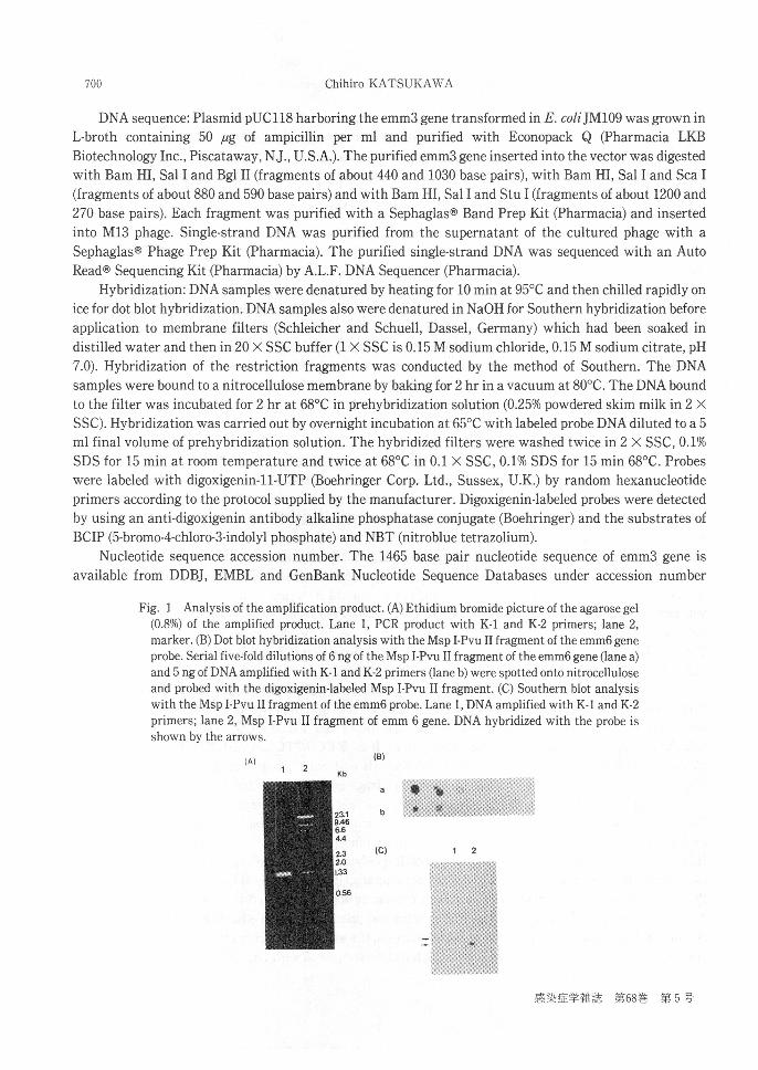
Fig. 1 Analysis of the amplification product. (A) Ethidium bromide picture of the agarose gel
(0.8%) of the amplified product. Lane 1, PCR product with K-1 and K-2 primers; lane 2,marker. (B) Dot blot hybridization analysis with the Msp I-Pvu II fragment of the emm6 gene
probe. Serial five-fold dilutions of 6 ng of the Msp I-Pvu II fragment of the emm6 gene (lane a)and 5 ng of DNA amplified with K-1 and K-2 primers (lane b) were spotted onto nitrocelluloseand probed with the digoxigenin-labeled Msp I-Pvu II fragment. (C) Southern blot analysiswith the Msp I-Pvu II fragment of the emm6 probe. Lane 1, DNA amplified with K-1 and K-2
primers; lane 2, Msp I-Pvu II fragment of emm 6 gene. DNA hybridized with the probe isshown by the arrows.
(A)(B)
(C)
感染症学雑誌 第68巻 第5号

Cloning and Nucleotide Sequence of emm3 Gene 701
D14415.
Results
Cloning and nucleotide sequence of the emm3 gene from S.pyogenes type 3 strain C203 with PCR:
Much information is available on nucleotide sequences from various strains7)-12).We tried to obtain the
Fig.2 Nucleotide and deduced amino acid sequences of the emm3 gene.The DNA strand is
located at 5' to 3' and its nucleotides are numbered above each line.Amino acid residues are
presented as single letters below each line.B repeat blocks and C repeat blocks are indicatedby underlining.
平成6年5月20日
702 Chihiro KATSUKAWA
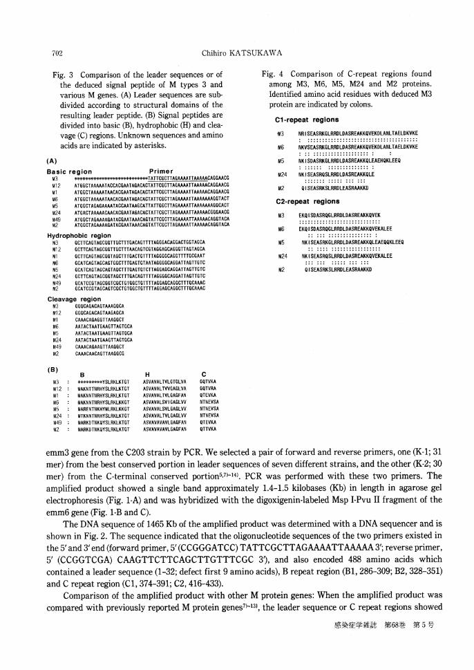
Fig.3 Comparison of the leader sequences or ofthe deduced signal peptide of M types 3 andvarious M genes.(A)Leader sequences are sub-divided according to structural domains of theresulting leader peptide.(B)Signal peptides aredivided into basic(B),hydrophobic(H)and clea-vage(C)regions.Unknown sequences and aminoacids are indicated by asterisks.
(A)
Basic region Primer
Hydrophobic region
Cleavage region
(B)B H C
Fig.4 Comparison of C-repeat regions found
among M3, M6, M5, M24 and M2 proteins.
Identified amino acid residues with deduced M3
protein are indicated by colons.
Cl-repeat regions
C2-repeat regions
emm3 gene from the C203 strain by PCR. We selected a pair of forward and reverse primers,one (K-1;31mer)from the best conserved portion in leader sequences of seven different strains,and the other(K-2;30mer)from the C-terminal conserved portion5,7)-14).PCR was performed with these two primers.The
amplified product showed a single band approximately 1.4-1.5 kilobases(Kb)in length in agarose gelelectrophoresis (Fig.1-A)and was hybridized with the digoxigenin-labeled Msp I-Pvu II fragment of theemm6 gene (Fig.1-B and C).
The DNA sequence of 1465 Kb of the amplified product was determined with a DNA sequencer and isshown in Fig.2.The sequence indicated that the oligonucleotide sequences of the two primers existed inthe 5' and 3' end (forward primer,5' (CCGGGATCC) TATTCGCTTAGAAAATTAAAAA 3'; reverse primer,
5' (CCGGTCGA)CAAGTTCTTCAGCTTGTTTCGC 3D,and also encoded 488 amino acids whichcontained a leader sequence (1-32;defect first 9 amino acids),B repeat region (B1,286-309;B2,328-351)and C repeat region (C1,374-391;C2,416-433).
Comparison of the amplified product with other M protein genes:When the amplified product wascompared with previously reported M protein genes7a-'3),the leader sequence or C repeat regions showed
感染症学雑誌 第68巻 第5号

Cloning and Nucleotide Sequence of emm3 Gene 703
Fig.5 Comparison of homologous regions in M3
and M12 proteins,and relationship between B
and C repeats in the predicted amino acid se-
quences of M3 and M12 proteins.The residuesfrom each protein being compared are indicated
by the numbering system of Robbins et al.12) for
M12 protein. B repeat blocks and C repeat blocks
are indicated by underlining,and identified
amino acid residues are indicated by colons.The
numbers enclosed in parentheses indicate %
homology.
high homology with the other emm genes(Fig.3 and 4).While the N-terminal amino acid portion of theamplified product was variable,it was found to be identical to 96 of 98 nucleotides downstream of theleader peptide sequence of another emm3 gene15).Therefore,the amplified product was characterized as theemm3 gene.
Comparison of homologous regions in M3 and M12 proteins:When the region of 252-488 deducedamino acids in M3 protein was compared with the region of 256-355 and 357-493 deduced amino acids inM12 protein, 96.6% homology was found between them (Fig.5).Interestingly,the B repeat region showedhigh homology with only that of M12 protein(91.7 and 100%,respectively). However,the A repeat region inM12 protein was not present in the M3 protein.Predictive secondary structure analysis of M3 protein:From analysis of the predictive secondary structure of the amplified product by the algorithm of Robson),most of the product was found to exhibit strong alpha-helical potential.In addition,the beta-sheet and turn
potential seen for region 23 to 42 in the M protein was similar to that seen forregion 28 to 50 in M12protein.The results suggest that M3 protein may be closely related to M12 protein.
Discussion
DNA sequence analysis has made it clear that all M proteins studied to date7)-12) are structurally
related and are therefore encoded by a family of genes.The regions of amino acid sequence homology in the
protein include the signal sequences,the C repeat region in the central part of the protein chain and the
carboxyl-terminal part. On the basis of available genetic information,we cloned the emm3 gene by using
PCR and sequenced its DNA.The primers prepared originated from the best conserved leader sequence or
the C-terminal portion of the emm genes7)-12).The amplified product was hybridized with an emm6 gene
probe (Fig. 1).Sequence analysis of the amplified product identified sequences complementary to both
oligonucleotide primers (Fig.2).In addition,the product had both B and C repeat regions.By comparing the
amplified product with known emm genes,we found not only that the N-terminal portion is very variable,
but also that the C-terminal region is conserved7)•`12),14).
Several streptococcal immunoglobulin-binding proteins have also been characterized as members of
the M protein family and as having C repeats17,18).Our selected primers have similar regions which can be
平成6年5月20日

704 Chihiro KATSUKAWA
screened.However,our product amplified by PCR from the type 3 strain C203 of S.pyogenes was a singleDNA and had no homology with amino terminal regions of the streptococcal immunoglobulin-binding
proteins.Furthermore,96 of 98 nucleotides downstream of the leader peptide sequence of the amplifiedproduct were found to be identical to the corresponding sequence of another emm3 gene of type 3 M strain3-3/31715).The evidence shows that the amplified product is the emm3 gene.
B and C repeat blocks that exist in M3 protein are similar to those in M12 protein (Fig. 5).Furthermore, predictive secondary structure analysis of M3 protein revealed that the majority of the
products exhibit strong alpha-helical potential as found with other M protein structures19). The algorithmalso showed that region 23-42 exhibits beta-sheet and turn potential with a pattern similar to that forregion 28-50 found by predictive secondary analysis of M12 protein.
S.pyogenes can be divided into two major classes on the basis of their immune reactivity withmonoclonal antibodies(mAbs)directed against epitopes which lie within the conserved half of M
proteins20).Class I serotype are defined as those which bind their mAbs,whereas class II isolates do not.Mainly the class I-specific mAb binding sites map to a region of C repeats within M proteins.Inasmuch asthe C repeat region of our emm 3 gene represents more than 90% homology with the known emm genes,itbelongs to the class I serotype.This agrees with the report of Bessen et al.who decided that M type 3 S.
pyogenes had a class I protein20).Furthermore,we found similarity between emm 3 and emm 12 genes intheir B repeat regions and predictive secondary structure.Thus,there may exist a subclass of class I M
proteins.Bessen et al.20)discriminated between serotypes sharing both B and C repeat region epitopes andthose sharing only C repeat region epitopes by using only antibody probes directed to antigenic sites withinthe B and C repeat regions of the M protein molecules in class I serotype.We suggest that M3 and M12
proteins belong to a subclass of class I M proteins.
Acknowledgements
I am grateful to Dr. Kyonsgu Hong of the Department of Bacteriology,Osaka University MedicalSchool,Dr.Ikuya Yano of the Department of Bacteriology,Osaka City University Medical School,Dr.Masanao Makino and Dr.Yasuhiko Suzuki of Osaka Prefectural Institute of Public Health for technical
advice and helpful discussions.In addition,I thank Dr.June R.Scott(Emory University)for providing the
pUC19: M6 plasmid and Dr.Tatsuya Tanaka of the Central Laboratoey for Research and Education,OsakaUniversity,Faculty of Medicine,for synthesizing the oligonucleotides.This work was supported by a
grant-in-aid from the Ministry of Education,Science and Culture of Japan (C63570192).
References
1) Bisno, A.: Group A streptococcal infections and acute rheumatic fever. N. Engl. J. Med. 325: 783-793 , 1991.2) Lancefield, R. C.: Current knowledge of the type-specific M antigens of group A streptococci. J. Immunol . 89:
307-313, 1962.3) Beachey, E. H., Seyer, J. M., Dale, J. B., Simpson, W. A. & Kang, A. H.: Type-specific protective immunity evoked by
synthetic peptide of Streptococcus pyogenes M protein. Nature (London) 292: 457-459, 1981.4) Hong, K., Kinoshita, T., Takeda, J., Kozono, H., Pramoonjago, P., Kim, Y. U. & Inoue, K.: Inhibition of the alternative
C3 convertase and classical C5 convertase of complement by group A streptococcal M protein. Infect. Immun. 58:2535-2541, 1990.
5) Scott, J. R., Pulliam, W. M., Hollingshead, S. K. & Fischetti, V. A.: Relationship of M protein genes in group Astreptococci. Proc. Natl. Acad. Sci. USA 82: 1822-1826, 1985.
6) Murry, M. G. & Thompson, W. F.: Rapid isolation of high-molecular-weight plant DNA. Nucleic Acids Res. 8: 4321-4325, 1988.
7) Bessen, D. E. & Fischetti, V. A.: Nucleotide sequence of two adjacent M or M-like protein genes of group Astreptococci: Different RNA transcript levels and identification of a unique immunoglobulin A-binding protein.Infect. Immun. 60: 124-135, 1992.
8) Haanes, E J. & Cleary, P. P.: Identification of a divergent M protein gene and an M protein-related gene family in
感染症学雑誌 第68巻 第5号

Cloning and Nucleotide Sequence of emm3 Gene 705
Streptococcus pyogenes serotype 49. J. Bacteriol. 171: 6397-6408, 1989.9) Hollingshead, S. K., Fischetti, V. A. & Scott, J. R.: Complete nucleotide sequence of type 6 M protein of the group A
streptococcus: Repetitive structure and membrane anchor. J. Biol. Chem. 261: 1677-1686, 1986.10) Miller, L., Gray, L., Beachey, E. & Kehoe, M.: Antigenic variation among group A streptococcal M proteins:
Nucleotide sequence of the serotype 5 M protein gene and its relationship with genes encoding type 6 and 24 Mproteins. J. Biol. Chem. 263: 5668-5673, 1988.
11) Mouw, A. R., Beachey, E. H. & Burdett, V.: Molecular evolution ofstreptococcal M protein: Cloning and nucleotidesequence of type 24 M protein gene and relation to other genes of Streptococcus pyogenes. J. Bacteriol. 170: 676-684,1988.
12) Robbins, J. C., Spanier, J. G., Jones, S. J., Simpson, W. J. & Cleary, P. P.: Streptococcus pyogenes type 12 M protein generegulation by upstream sequences, J. Bacteriol. 169: 5633-5640, 1987.
13) Haanes-Fritz, E., Kraus, W., Burdett, V., Dale, J. B. & Beachey, E. H.: Comparison of the leader sequences of fourgroup A streptococcal M protein genes. Nucleic Acids Res. 16: 4667-4677, 1988.
14) Hollingshead, S. K., Fischetti, V. A. & Scott, J. R.: A highly conserved region present in transcripts encodingheterologous M proteins of group A streptococci. Infect. Immun. 55: 3237-3239, 1987.
15) Podbielski, A., Baird, R. & Kaufhold, A.: The group A streptococcal M-type 3 protein gene exhibits a C terminustypical for class I M proteins. Med. Microbiol. Immunol. 181: 209-213, 1992.
16) Garnier, J., Osguthorpe, D. J. & Robson, B.: Analysis of the accuracy and implications of simple methods forpredicting the secondary structure of globular proteins. J. Mol. Biol. 120: 97-120, 1978.
17) Gomi, H., Hozumi, T., Hattori, S., Tagawa, C., Kishimoto, F. & Bjorck, L.: The gene sequence and some properties ofprotein H. J. Immunol. 144: 4046-4052, 1990.
18) Heath, D. G. & Cleary, P. P.: Fc-receptor and M-protein genes of group A streptococci are products of geneduplication. Proc. Natl. Acad. Sci. USA 86: 4741-4745, 1989.
19) Phillips, G. N., Flicker, P. F., Cohen, C., Manjula, B. N. & Fischetti, V. A.: Streptococcal M protein: a-helical coiled-coilstructure and arrangement on the cell surface. Proc. Natl. Acad. Sci. USA 78: 4689-4693, 1981.
20) Bessen, D., Jones, K. F. & Fischetti, V. A.: Evidence for two distinct classes of streptococcal M protein and theirrelationship to rheumatic fever. J. Exp. Med. 169: 269-283, 1989.
PCR法 を用いてクローニングしたA群 溶血 レンサ球菌
M3蛋 白遺伝子の解析および他菌型 との比較
(レンサ球菌感染症研究会:会 長 保科 清)
大阪府立公衆衛生研究所
勝 川 千 尋
(平成5年11月12日 受付)
(平成6年2月7日 受理)
要 旨
A群 溶血 レンサ球菌M3蛋 白遺伝子 のN末 端
の高度型特異領域からC未 端 の保存領域までの
部分をPCR法 を用いてクローニングを行った.
各菌型 に共通なN末 端の リーダーシクエンス
部分 とC末 端 の保存領域部分を プライマーとし
て用い,1465bpの 遺伝子配列を決定 し,他のM蛋
白遺伝子 と比較検討した.そ の結果,N末 端側の
100塩 基から750塩 基の範囲にM3型 特異的な領域
を見いだすことができた.ま た,ア ミノ酸配列を
検討した ところ,2つ の繰 り返 し配列を見いだし
た(Bリ ピー トおよびCリ ピー ト).Cリ ピー トは
現在知 られている他のM蛋 白遺伝子の塩基配列
と非常に高い相同性を示 した.こ れに対 して,Bリ
ピー トはM12蛋 白遺伝子のBリ ピー ト配列 との
み高い相同性を示 し,ま た二次構造の解析結果で
もこの2菌 型は構造が類似していた.こ れ らの結
果よりM3蛋 白とM12蛋 白は遺伝学的に非常に近
い蛋白であることが示唆された.
平成6年5月20日