Embed Size (px)
Citation preview

1
Journal of Neuropsychology (2012)C© 2012 The British Psychological Society
TheBritishPsychologicalSociety
www.wileyonlinelibrary.com
Clock drawing in spatial neglect: Acomprehensive analysis of clock perimeter,placement, and accuracy
Peii Chen1,2,∗ and Kelly M. Goedert31Kessler Foundation Research Center, West Orange, New Jersey, USA2Department of Physical Medicine and Rehabilitation, The University of Medicineand Dentistry of New Jersey, Newark, New Jersey, USA
3Department of Psychology, Seton Hall University, South Orange, New Jersey, USA
Clock drawings produced by right-brain-damaged (RBD) individuals with spatial neglectoften contain an abundance of empty space on the left while numbers and hands areplaced on the right. However, the clock perimeter is rarely compromised in neglectpatients’ drawings. By analysing clock drawings produced by 71 RBD and 40 healthyadults, this study investigated whether the geometric characteristics of the clockperimeter reveal novel insights to understanding spatial neglect. Neglect participantsdrew smaller clocks than either healthy or non-neglect RBD participants. While healthyparticipants’ clock perimeter was close to circular, RBD participants drew radiallyextended ellipses. The mechanisms for these phenomena were investigated by examiningthe relation between clock-drawing characteristics and performance on six subtests ofthe Behavioral Inattention Test (BIT). The findings indicated that the clock shape wasindependent of any BIT subtest or the drawing placement on the test sheet and thatthe clock size was significantly predicted by one BIT subtest: the poorer the figure andshape copying, the smaller the clock perimeter. Further analyses revealed that in allparticipants, clocks decreased in size as they were placed farther from the centre ofthe paper. However, even when neglect participants placed their clocks towards thecentre of the page, they were smaller than those produced by healthy or non-neglectRBD participants. These results suggest a neglect-specific reduction in the subjectivelyavailable workspace for graphic production from memory, consistent with the hypothesisthat neglect patients are impaired in the ability to enlarge the attentional aperture.
Textbook descriptions of spatial neglect are usually accompanied by patients’ drawings.Right-brain-damaged (RBD) individuals with spatial neglect often depict an object withleft-sided features absent. As neglect symptoms cannot be explained by sensory or motordefects (Heilman, Watson, & Valenstein, 2003), omissions in drawing may result fromthe degradation of left-sided representational imaging (Bartolomeo, 2007; Berti, 2004;
∗Correspondence should be addressed to Peii Chen, PhD, Kessler Foundation Research Center, 1199 Pleasant Valley Way,
West Orange, NJ 07052, USA (e-mail: [email protected]).
DOI:10.1111/j.1748-6653.2012.02028.x

2 Peii Chen and Kelly M. Goedert
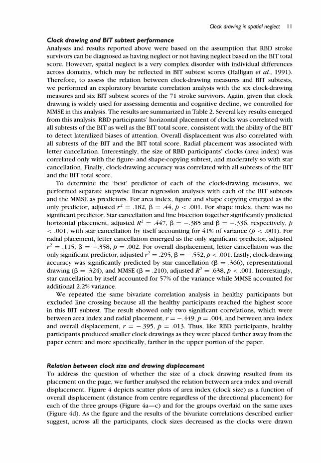
Figure 1. Examples of clock drawings produced by right-brain-damaged stroke survivors with spatialneglect. The reference scale of 10 mm is noted.
Bisiach, Luzzatti, & Perani, 1979), or the inability to deploy attention leftward in suchmental imagery (Hillis, 2006), thus fading the left-sided figures into the background(Marshall & Halligan, 1994). Many have documented this phenomenon in non-artistsas well as professional artists with spatial neglect (Apfeldorf, 1962; Chatterjee, 2004;Chokron, Colliot, & Bartolomeo, 2004; Halligan & Marshall, 2001; Poole, Sadek, &Haaland, 2011). Some drawings appear as having a clear and complete cut betweenemptiness on the left and abundance on the right as if an opaque blank covered the leftside of the object; others show an absence of details or parts on the left side as if thedrawing were unfinished.
Similarly, clock drawings produced by neglect patients often present a left–rightasymmetry. Specifically, the asymmetrical layout of clock numerals signals the mani-festation of spatial neglect (Agrell & Dehlin, 1998). Typically, these drawings containempty space on the left, as if the clock were covered by an opaque semicircle (seeexamples in Figure 1). However, this metaphoric occlusion frequently leaves the rimof the clock unaffected. The circular outline is rarely compromised in neglect patients’clock drawings and is almost always the first feature produced in the task. To commenton this seemingly preserved ability to produce the clock circumference, Halligan andMarshall (1993b) suggested that ‘the optimal gestalt of the circle precludes the omissionof parts thereof’ (p. 15).
However, there is little to no evidence as to whether the clock perimeters producedby neglect patients are normal. The standardized scoring methods for clock drawing arefocused on the clock face: number sequencing and allocating, and hand placement(Cohen, Ricci, Kibby, & Edmonds, 2000; Lessig, Scanlan, Nazemi, & Borson, 2008;Mendez, Ala, & Underwood, 1992; Shulman, Gold, Cohen, & Zucchero, 1993; Tuokko,Hadjistavropoulos, Miller, & Beattie, 1992). Indeed, patients may not even be asked to

Clock drawing in spatial neglect 3
draw the clock perimeter, which is often provided and seldom taken into account inthe scoring method. For instance, Ishiai and colleagues investigated whether neglectseverity was correlated with clock-drawing accuracy (Ishiai, Sugishita, Ichikawa, Gono,& Watabiki, 1993), and addressed this question by asking neglect patients to produce aclock face within a printed circle that was provided. Another example is the BehavioralInattention Test (BIT; Wilson, Cockburn, & Halligan, 1987), a widely used screening toolfor spatial neglect. Clock drawing is part of the BIT, and examinees are instructed todraw a clock face with no circle provided. However, the scoring method emphasizes theleft–right symmetry of the drawing in general, and a response with ‘omission or grossdistortion of any major contralesional component of the drawing’ is considered incorrect(Halligan, Cockburn, & Wilson, 1991). This all-or-none scoring method is unlikely tocapture any abnormality of the clock perimeter. In one of the few studies examiningclock perimeters, Suhr, Grace, Allen, Nadler, and McKenna (1998) compared differentscoring methods for the clock-drawing test and found that RBD stroke survivors producedsmaller clock drawings than left-brain-damaged stroke survivors and healthy individuals.However, Suhr et al. (1998) did not provide a mechanistic explanation for their findingor whether such finding would be related to spatial neglect.
In an investigation of neglect patients’ graphic production of circles, Smith andcolleagues asked RBD stroke survivors to make copies of circles of various sizes,and neglect patients consistently produced circles that were smaller than the model(Smith, Gilchrist, Butler, & Harvey, 2006; Smith et al., 2007). In addition, the size ofdrawings decreased with increasing neglect severity (Smith et al., 2006). Surprisingly,size distortion in circle copying was not related to the laterality of the start position, norwas the clockwise or counterclockwise direction of the drawing movement (Smith et al.,2007), suggesting that this abnormality in graphic production of circles may not beassociated with the lateralized bias that is the hallmark of spatial neglect. However,with only six patients with spatial neglect (based on the BIT cut-off score), Smithet al.’s studies may underestimate the extent of graphic distortions, and their resultsonly indirectly address the possibility of abnormal perimeters in clock drawing: theirparticipants copied circles rather than producing them from memory. Thus, it remainsunknown whether persons with spatial neglect produce abnormal clock perimeterswhen drawing clocks from memory.
Potential mechanisms of small clock drawingIn this paper, we consider several possible mechanisms of small clock drawing in spatialneglect. The possibility that we consider most plausible is that neglect patients drawsmall clocks because of a reduction in the available workspace for producing the clock.The clock perimeter defines the workspace for placing the other features inside theperimeter to form a clock face, but the size of the workspace for producing theclock perimeter may be limited for neglect patients due to one or both of the twoseparable mechanisms of spatial attention: Using the metaphor of a floodlight (Barrett,Beversdorf, Crucian, & Heilman, 1998), spatial attention can be spread over a broad areaor focused on a specific location, which is the ability to scale the attentional aperture(Barriopedro & Botella, 1998); spatial attention can also be shifted from location tolocation, which is the ability to move attention (Posner, 1980). Abnormality in the abilityto enlarge the attentional aperture may reduce the subjectively available workspace (e.g.,spontaneously producing a small circular outline as the foundation for the clock face);

4 Peii Chen and Kelly M. Goedert
abnormality in the ability to move attention in space may constrain the amount of spaceavailable for creating the drawing.
In a task that required participants to judge whether two separate line segments werecollinear, Barrett et al. (1998) found that RBD stroke survivors had difficulty in wideningtheir attentional aperture to perform the task normally. Independent of the distancebetween the line segments, both RBD and left-brain-damaged stroke survivors were ableto indicate the locations of the line segments in the right and left hemispaces. However,in comparison to healthy controls, RBD individuals produced significantly greater errorsin collinearity judgement as the gap between the line segments increased while left-brain-damaged individuals’ performance was poorer than healthy controls only in thecondition with the smallest gap between the line segments (Barrett et al., 1998). Suchfindings suggest that RBD stroke survivors may have difficulty enlarging the attentionalaperture to sufficiently circumscribe both line segments with large gaps and thereforefail to judge whether the lines were collinear. Consistent with this interpretation, afunctional imaging study using the collinearity judgement task to investigate the abilityto scale the attentional aperture in healthy adults showed that the right inferior frontalgyrus was differentially activated when the gap between the line segments was increasingrather than decreasing, and that the right posterior temporal parietal areas were criticallyassociated with switching size of the attentional aperture (i.e., zoom-in to zoom-out orzoom-out to zoom-in; Chen, Marshall, Weidner, & Fink, 2009). Thus, the phenomena ofsmall clock drawings in spatial neglect may be related to right-brain damage impairingthe ability to widen attention for determining the subjectively available workspace forcreating the clock perimeter.
Alternatively, the reduced size of clock drawing in spatial neglect may result fromthe limited ability to move attention around. It is known that persons with left-sidedneglect typically start exploring a predetermined workspace (e.g., a page) from theright hemispace (Chen et al., 2008; Fong et al., 2007; Nurmi et al., 2010) as well asfrom the distal/upper portion of the entire workspace (Chatterjee, Thompson, & Ricci,1999). This spatial bias may be due to deficits in the perceptual–attentional or themotor-intentional spatial systems (Barrett & Burkholder, 2006; Heilman et al., 2003; Lau,Rogers, Haggard, & Passingham, 2004; Na et al., 1998). Therefore, when exploring theavailable workspace to initiate the graphic production of a clock face, neglect patientsmay be abnormally constrained to the upper right portion of the page. Constraining theclock to the upper-right portion of the page may limit the amount of space available forproducing the clock drawing and thus neglect patients may draw a small clock.
In addition to a potential reduction in the available workspace, we consider threeother mechanisms for the small clock drawings produced in spatial neglect. However,these three accounts are less plausible explanations for the small clock perimeters. First,it is possible that neglect patients produce smaller clocks because of hypometria (i.e.,an abnormally reduced amplitude of manual movements towards contralesional space).However, no evidence supports that the initial direction (leftward or rightward) affectsthe amplitude of a circular movement (Smith et al., 2007). Furthermore, hypometriain neglect patients is observed mostly with the contralesional limb (left) movingin the contralesional space without visual feedback (Meador et al., 2000; Meador,Watson, Bowers, & Heilman, 1986). Second, making a graphic production, by drawingfrom memory or copying from a model, is related to constructional abilities, whichis vulnerable to right-brain damage (Laeng, 2006). Thus, it is possible that reducedconstructional abilities in neglect patients affect the size of the clock drawing. However,in Smith et al.’s study (2007), the size of circles copied was predicted by neglect severity,

Clock drawing in spatial neglect 5
but it was independent of constructional abnormalities. This suggests that graphicallyproducing a stand-alone circle may not be related to constructional abilities. A thirdpossibility is that small clock drawing is due to representational neglect. That is, it isthe mental imagery of the clock, rather than the attentional aperture, that is reduced insize and reflected in the drawing. Given, however, that Smith et al. (2006) found thatneglect patients produced smaller drawings even when copying circles, we considerthis alternative unlikely too.
Is the clock perimeter in normal shape?In addition to size, spatial neglect may alter the shape of clock perimeter as well.Literature on representational neglect does not indicate whether the contour of anobject image would be affected. Since the drawing is produced from memory, and thecircular outline is the first component produced, it may be least affected by immediatevisual input, and thus may reflect how the stereotypical image of a clock is representedin the mind (Anderson, 1993; Chokron et al., 2004). Alternatively, converging evidencesuggests that in comparison to the radial dimension, the horizontal dimension is morelikely to be underestimated, and thus misperceived or misrepresented in spatial neglect(Guariglia & Piccardi, 2010; Halligan & Marshall, 1993a, 1995; Harvey, Gilchrist, Olk,& Muir, 2003; Heilman & Valenstein, 1979; Irving-Bell, Small, & Cowey, 1999; Milner& Harvey, 1995). Therefore, underestimation of the horizontal dimension may result inthe imprecise production of a circle as a radially extended ellipse, namely an oval shapewith the height longer than the width (see Figure 1a–f).
Current studyFew studies have comprehensively analysed the perimeter of clock drawings in spatialneglect. Suhr et al.’s report (1998) suggested that right-brain stroke may be relatedto small clock drawings but treated it as an ‘error’ in clock drawing without muchelaboration. Smith et al. (2006, 2007) studied neglect patients’ graphic productionof circular shapes via copying models, but the results may not be directly related tographic production of clock perimeters from memory and they had a small sample ofneglect patients (n = 6). With a large sample of RBD neglect, RBD non-neglect, andhealthy participants in the present study, we addressed the question of whether the sizeand shape of the clock perimeter were abnormal in spatial neglect, and whether anydistortions were associated with known characteristics of spatial neglect. We performeda comprehensive analysis of the clock-drawing performance, assessing the horizontal andradial extensions of the drawing, the overall size of the drawing, and placement of thedrawing on the page. We also performed a comprehensive assessment of clock-drawingaccuracy, as typically employed for dementia screening (Lessig et al., 2008). Lastly, tounderstand the potential mechanisms that may underlie abnormal clock perimeters, weinvestigated the relation between participants’ clock-drawing characteristics and theirperformance on the BIT subtests, each of which may detect different manifestations andmechanisms of spatial neglect (Halligan et al., 1991; Halligan, Marshall, & Wade, 1989).

6 Peii Chen and Kelly M. Goedert
Table 1. Characteristics of RBD neglect participants (BIT < 129), RBD non-neglect participants, andhealthy participants
RBD neglect RBD non-neglect Healthy(n = 31) (n = 40) (n = 40)
Men/women 15/16 23/17 14/26Age, years 68.1 ± 15.8 62.7 ± 13.5 65.0 ± 9.9Education years∗∗∗ 12.9 ± 2.9 14.0 ± 2.9 16.3 ± 3.1Post-stroke days 21.9 ± 9.3 26.2 ± 16.9 NAMMSE (out of 30)∗∗∗ 23.1 ± 3.9 28.5 ± 2.1 29.7 ± 0.8BIT total score (out of 146)∗∗∗ 67.6 ± 39.8 139.4 ± 5.5 144.9 ± 1.2BIT subtests
Line crossing (out of 36)∗∗∗ 21.1 ± 12.3 35.8 ± 0.7 36.0 ± 0.0Letter cancellation (out of 40)∗∗∗ 17.0 ± 12.3 37.4 ± 2.6 39.4 ± 0.9Star cancellation (out of 54)∗∗∗ 24.9 ± 16.1 52.6 ± 1.7 53.8 ± 0.5Figure and shape copying (out of 4)∗∗∗ 0.9 ± 1.0 3.2 ± 0.7 3.9 ± 0.4Line bisection (out of 9)∗∗∗ 2.7 ± 2.3 7.7 ± 1.8 8.9 ± 0.3Representational drawing (out of 3)∗∗∗ 1.1 ± 1.0 2.7 ± 0.6 3.0 ± 0.2
Stroke etiology (Hem/Isc)a 7/22 14/25 NARight-brain lesion locationb
Frontal cortex 15 6 NAParietal cortex 13 7Temporal cortex 7 6Occipital cortex 1 1Insular cortex 5 5Basal ganglia 11 23Thalamus 3 8Subcortical WM 13 14
Note. RBD, right-brain-damaged; MMSE, Mini-Mental State Examination; BIT, Behavioral InattentionTest; NA, not applicable; Hem, haemorrhage or haemorrhagic conversion; Isc, ischaemic; WM, whitematter.aTwo neglect patients and one non-neglect participant’s radiology records were unavailable, but right-brain strokes were noted in their charts.bMultiple cytoarchitectonic sites may be involved in individual participants.∗∗∗ p � .001 from one-way ANOVA.
MethodParticipantsParticipant characteristics are summarized in Table 1. All information was obtainedin compliance with the regulations of the Institutional Review Board (IRB) of theauthors’ organization. Seventy-one consecutive stroke survivors, admitted to one of thecollaborating rehabilitation hospitals after an ischaemic or haemorrhage stroke in theright cerebral hemisphere, were included. All were right-hand dominant pre- and post-stroke (determined by a 17-item handedness questionnaire; Raczkowski, Kalat, & Nebes,1974); had no history of a stroke in the left hemisphere; and completed the Mini-MentalState Examination (MMSE; Folstein, Folstein, & McHugh, 1975) and the conventionalsubtests of the BIT within 100 days post-stroke (M = 24.3; SD = 14.2). There were38 men and 33 women, between 34 and 91 years of age (M = 65.1; SD = 14.7) whohad 6–20 years of education (M = 13.5; SD = 2.9). MMSE scores ranged from 15 to

Clock drawing in spatial neglect 7
30 of 30 (M = 26.1; SD = 4.0), and BIT scores from 11 to 146 of 146 (M = 108.0;SD = 44.5). Because the BIT total score is frequently used to diagnose spatial neglect inclinical practices and research (e.g., Cherney, Halper, Kwasnica, Harvey, & Zhang, 2001;Fong et al., 2007; Hartman-Maeir & Katz, 1995; Luukkainen-Markkula, Tarkka, Pitkanen,Sivenius, & Hamalainen, 2009), we used the standardized cut-off (Halligan et al., 1991) togroup the stroke survivors into neglect and non-neglect RBD participants. Thirty-one ofthe RBD participants met the criterion for having spatial neglect (BIT total score <129).
Healthy participants consisted of 40 right-handed individuals with no history of braininjury or neurological disease. They were recruited via flyers and e-mails in the authors’organization and collaborating rehabilitation hospitals. Healthy participants (14 males;26 females) aged from 50 to 86 years (M = 65.0; SD = 9.9) with 12–26 years of education(M = 16.3; SD = 3.1). Their MMSE and BIT total scores were 29.7 ± 0.8 and 144.9 ±1.2, respectively. Between healthy and stroke participants, there were no group-leveldifferences in sex (Fisher’s exact: p = .076) or age (independent samples t-test: p =.975). Healthy participants had more years of education than stroke participants andbetter scores on the MMSE and BIT (all p < .001).
Procedure for clock drawingParticipants sat at a desk and used their right hand for the task. Following thestandard procedure for administering the BIT (Wilson et al., 1987), the examiner askedparticipants to draw a clock, while presenting participants with a blank letter-sizedsheet of paper (215.9 mm × 279.4 mm), of which the shorter edge was parallel toparticipants’ coronal plane and centred at body midline. Head and eye movements werenot constrained. Moving the sheet was not permitted. Drawing was untimed, and notime limit for completion was imposed.
MeasurementsThe height (radial diameter; H) and width (horizontal diameter; W ) of the clock drawingwere measured in millimeters (mm) to determine drawing distortion in terms of size andshape. We created an area index by multiplying height by width (H × W ) to convey thesize of the clock perimeter. We created a shape index that was the height-to-width ratio(H/W ). A shape index of 1 indicates a perfect circle, greater than 1 indicates a radiallyextended ellipse, and less than 1 a horizontally extended ellipse. Both area and shapeindices were values with no unit.
Placement bias was measured horizontally and radially from the paper centre tothe clock centre in millimeters. Horizontal placement was the distance right (codedpositive) or left (coded negative) to the paper centre. Radial placement was the distanceabove (coded positive) or below (coded negative) the paper centre. We also calculatedthe overall displacement, which was the distance, regardless of the directional bias,between the clock centre and the paper centre.
Lastly, to quantify accuracy in clock drawing, 19 items (scored as correct = 1 orincorrect = 0) were selected from the clock scoring technique described by Lessiget al. (2008), incorporating three popular methods (Mendez et al., 1992; Shulman et al.,1993; Tuokko et al., 1992). Five error types from Lessig et al.’s method were excludedbecause they were irrelevant to the standard BIT instruction, which did not requireparticipants to set a particular time or provide a second try. An independent rater scoredall clock drawings. The Appendix lists the 19 scoring items for clock-drawing accuracy.The clock-drawing accuracy score was the total number of items correct.

8 Peii Chen and Kelly M. Goedert
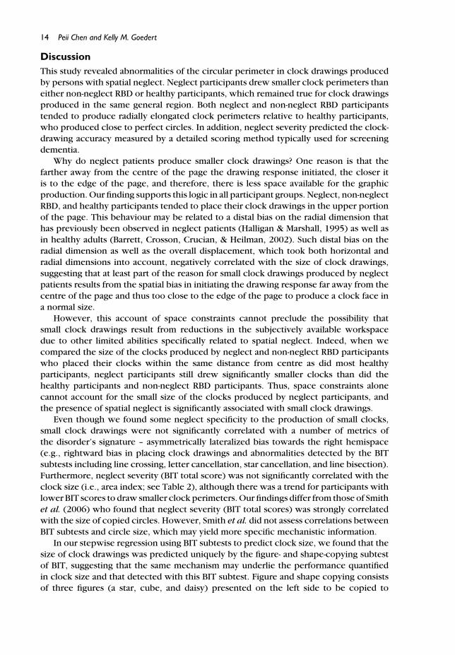
Figure 2. Means and standard errors (presented in error bars) of clock-drawing geometry in right-brain-damaged (RBD) participants with or without spatial neglect, and healthy participants. (a) Areaindex (denoted on the y-axis). (b) Shape index (denoted on the y-axis): horizontal line at 1 indicates theshape of a circle; values greater than 1 indicate shapes of vertical ovals. (c) Horizontal placement(denoted on the x-axis): vertical line at zero indicates clock centre overlapping paper centre onthe horizontal dimension; positive values denote clock centres on the right side of paper. (d) Radialplacement (denoted on the y-axis): zero indicates clock centre overlapping paper centre on the radialdimension; positive values denote clock centres on the upper part of paper. (e) Overall displacement(denoted on the y-axis): zero indicates clock centre overlapping paper centre. (f) Drawing accuracy.Asterisks (∗p < .05; ∗∗p < .01; ∗∗∗p < .001) indicate significant differences in pairwise comparisons(Tukey’s HSD tests) post the one-way ANOVA.
Analyses and ResultsClock drawings characteristicsThroughout, we used an alpha of .05 and performed post hoc pairwise comparisonsusing Tukey’s Honestly Significant Difference (HSD) after significant one-way ANOVAs.

Clock drawing in spatial neglect 9
Clock sizeThe ANOVA showed a significant difference among the three groups of participants, F(2,108) = 14.03, p < .001. As seen in Figure 2a, and consistent with our prediction, thedrawings of neglect participants (mean area index = 2,988.4 ± 3,834.2) were smallerthan those of non-neglect RBD (7,187.9 ± 6,230.9; p = .006) and healthy participants(10,044.5 ± 5,981.9; p < .001), who did not differ from each other.
Clock shapeAll participants drew the clock with an elliptical contour rather than any other shapesuch as rectangle or triangle. However, the ANOVA, revealed a significant differencein the shape index of the three groups, F(2, 108) = 6.44, p = .002 (see Figure 2b).Both neglect and non-neglect RBD participants had shape indices greater than that ofthe healthy participants (p = .025 for neglect vs. healthy; p = .003 for non-neglect vs.healthy). However, there was no statistical difference between neglect and non-neglectRBD groups on the measure of clock shape (p = .870). The healthy participants’ clockperimeter was very close to a circle with a shape index of 1.01 ± .09 (one-sample t-testcomparing to value of 1: t(39) = .82, p = .418). However, both neglect and non-neglectRBD participants’ clock perimeters were radially extended ellipses with shape indices of1.12 ± .22, t(30) = 3.07, p = .005, and 1.14 ± .19, t(39) = 4.69, p < .001, respectively.
Horizontal placementAs illustrated in Figure 2c, neglect participants’ clock drawings were placed morerightward than those of non-neglect participants’ and healthy participants’. Theseimpressions were confirmed with the one-way ANOVA, F(2, 108) = 15.45, p < .001. Onaverage, neglect participants placed clock drawings more rightward (23.6 ± 31.2 mm)than those of non-neglect RBD (1.8 ± 9.3 mm) and healthy participants (0.4 ± 13.6 mm;p < .001 for both comparisons). Neglect participants also placed the clock right ofthe paper centre (one-sample t-test: t(30) = 4.21, p < .001). Both non-neglect RBDparticipants’ and healthy participants’ clock drawings were not significantly deviatedfrom the paper centre (p = .407 and p = .234, respectively).
Radial placementThe ANOVA indicated a significant difference among the groups, F(2, 108) = 3.64,p = .030: As illustrated in Figure 2d, neglect participants drew clocks more distally thandid healthy participants (p = .044), with a trend towards the same difference whencompared to non-neglect RBD participants (p = .055). There was no difference betweennon-neglect RBD participants and healthy participants (p = .995) in radial placement.However, all three groups placed the clock centre in the upper portion of the paper,confirmed by one-sample t-tests: neglect participants: 34.4 ± 33.7 mm, t(30) = 5.68,p < .001; non-neglect RBD participants: 19.4 ± 25.3 mm, t(39) = 4.84, p < .001; healthyparticipants: 18.8 ± 21.7 mm, t(39) = 5.48, p < .001.
Overall displacementAs would be expected from the results of the radial and horizontal placement biases, theone-way ANOVA yielded a significant difference among the groups in the distance of the

10 Peii Chen and Kelly M. Goedert
Figure 3. Error rates in clock-drawing items (see Appendix for scoring description). Asterisks (∗p< .05; ∗∗p < .01) indicate significant differences in the error rates of the neglect and non-neglectright-brain-damaged (RBD) groups, controlling for Mini-Mental State Examination (MMSE).
clock from the paper centre, F(2, 108) = 10.54, p < .001. Shown in Figure 2e, neglectparticipants drew their clocks farther from centre (50.03 ± 36.22 mm) than healthy(25.26 ± 16.37 mm; p = .001) and non-neglect RBD participants (28.33 ± 18.23 mm;p = .001), who did not differ from each other (p = .836). All three groups displacedtheir clocks significantly away from the centre of the page, confirmed by one-samplet-tests: neglect, t(30) = 7.69, p < .001; non-neglect RBD, t(39) = 9.83, p < .001; healthyparticipants, t(39) = 9.76, p < .001.
Clock-drawing accuracyThe one-way ANOVA revealed a significant difference in the clock-drawing accuracy ofthe groups, F(2, 108) = 38.34, p < .001. Neglect participants had lower accuracy (12.0± 4.4) than both non-neglect RBD (16.7 ± 1.8; p < .001) and healthy participants (17.3± 1.4; p < .001), who did not differ from each other (Figure 2f). Figure 3 depicts theerror rate of each group on each item.
To examine whether any of the errors were neglect specific, we performed thefollowing analysis. Given that this assessment of clock-drawing accuracy is sensitive todementia and general cognitive decline (Lessig et al., 2008), we controlled for MMSEin separate logistic regression analyses assessing the ability of the presence of neglectamong the RBD participants to predict accuracy on each item. Controlling for MMSE,the presence of spatial neglect was associated with a significantly increased error rate(over non-neglect RBD) on three of the 19 items: ‘All numbers present’, b = −3.27, SE= .96, p = .001; ‘No empty quadrant’, b = −3.29, SE = 1.19, p = .006; and ‘Two hands’,b = −1.90, SE = .88, p = .030. Analysis of the item ‘Equal spacing between numbers’approached significance, b = −2.17, SE = 1.19, p = .068. These items all refer to errorsof omission, spatial allocation, or spatial relations among numbers.
Clock drawing in spatial neglect 11
Clock drawing and BIT subtest performanceAnalyses and results reported above were based on the assumption that RBD strokesurvivors can be diagnosed as having neglect or not having neglect based on the BIT totalscore. However, spatial neglect is a very complex disorder with individual differencesacross domains, which may be reflected in BIT subtest scores (Halligan et al., 1991).Therefore, to assess the relation between clock-drawing measures and BIT subtests,we performed an exploratory bivariate correlation analysis with the six clock-drawingmeasures and six BIT subtest scores of the 71 stroke survivors. Again, given that clockdrawing is widely used for assessing dementia and cognitive decline, we controlled forMMSE in this analysis. The results are summarized in Table 2. Several key results emergedfrom this analysis: RBD participants’ horizontal placement of clocks was correlated withall subtests of the BIT as well as the BIT total score, consistent with the ability of the BITto detect lateralized biases of attention. Overall displacement was also correlated withall subtests of the BIT and the BIT total score. Radial placement was associated withletter cancellation. Interestingly, the size of RBD participants’ clocks (area index) wascorrelated only with the figure- and shape-copying subtest, and moderately so with starcancellation. Finally, clock-drawing accuracy was correlated with all subtests of the BITand the BIT total score.
To determine the ‘best’ predictor of each of the clock-drawing measures, weperformed separate stepwise linear regression analyses with each of the BIT subtestsand the MMSE as predictors. For area index, figure and shape copying emerged as theonly predictor, adjusted r2 = .182, � = .44, p < .001. For shape index, there was nosignificant predictor. Star cancellation and line bisection together significantly predictedhorizontal placement, adjusted R2 = .447, � = −.385 and � = −.336, respectively, p< .001, with star cancellation by itself accounting for 41% of variance (p < .001). Forradial placement, letter cancellation emerged as the only significant predictor, adjustedr2 = .115, � = −.358, p = .002. For overall displacement, letter cancellation was theonly significant predictor, adjusted r2 = .295, � = −.552, p < .001. Lastly, clock-drawingaccuracy was significantly predicted by star cancellation (� = .366), representationaldrawing (� = .324), and MMSE (� = .210), adjusted R2 = .638, p < .001. Interestingly,star cancellation by itself accounted for 57% of the variance while MMSE accounted foradditional 2.2% variance.
We repeated the same bivariate correlation analysis in healthy participants butexcluded line crossing because all the healthy participants reached the highest scorein this BIT subtest. The result showed only two significant correlations, which werebetween area index and radial placement, r = −.449, p = .004, and between area indexand overall displacement, r = −.395, p = .013. Thus, like RBD participants, healthyparticipants produced smaller clock drawings as they were placed farther away from thepaper centre and more specifically, farther in the upper portion of the paper.
Relation between clock size and drawing displacementTo address the question of whether the size of a clock drawing resulted from itsplacement on the page, we further analysed the relation between area index and overalldisplacement. Figure 4 depicts scatter plots of area index (clock size) as a function ofoverall displacement (distance from centre regardless of the directional placement) foreach of the three groups (Figure 4a—c) and for the groups overlaid on the same axes(Figure 4d). As the figure and the results of the bivariate correlations described earliersuggest, across all the participants, clock sizes decreased as the clocks were drawn

12 Peii Chen and Kelly M. Goedert
Tabl
e2.
Cor
rela
tion
mat
rix
ofBI
Tan
dcl
ock
draw
ing,
cont
rolli
ngfo
rM
MSE
,in
allR
BDpa
rtic
ipan
ts
Are
ain
dex
Shap
ein
dex
Hor
izon
tal
Rad
ial
Ove
rall
Clo
ck-d
raw
ing
RBD
part
icip
ants
(N=
71)
(H×
W)
(H/W
)pl
acem
ent
plac
emen
tdi
spla
cem
ent
accu
racy
Beha
vior
alIn
atte
ntio
nTe
stLi
necr
ossi
ng.1
36−.
040
−.44
0∗∗∗
−.17
3−.
345∗∗
.431
∗∗∗
Lett
erca
ncel
latio
n.1
94−.
053
−.46
8∗∗∗
−.26
9∗−.
427∗∗
∗.5
32∗∗
∗
Star
canc
ella
tion
.244
∗−.
024
−.54
1∗∗∗
−.19
2−.
347∗∗
.567
∗∗∗
Figu
rean
dsh
ape
copy
ing
.347
∗∗.0
04−.
446∗∗
∗−.
067
−.24
8∗.4
31∗∗
∗
Line
bise
ctio
n.1
54−.
008
−.52
5∗∗∗
−.19
0−.
383∗∗
.359
∗∗
Rep
rese
ntat
iona
ldra
win
g.1
29−.
091
−.44
3∗∗∗
−.12
1−.
242∗
.549
∗∗∗
BIT
tota
lsco
re.2
23−.
040
−.55
2∗∗∗
−.22
8−.
411∗∗
∗.5
69∗∗
∗
Clo
ck-d
raw
ing
mea
sure
sA
rea
inde
x(H
×W
)Sh
ape
inde
x(H
/W)
.182
Hor
izon
talp
lace
men
t(m
m)
−.16
9−.
123
Rad
ialp
lace
men
t(m
m)
−.41
6∗∗∗
−.08
0.1
96O
vera
lldi
spla
cem
ent
(mm
)−.
434∗∗
∗−.
150
.543
∗∗∗
.834
∗∗∗
Clo
ck-d
raw
ing
accu
racy
.194
−.01
7−.
446∗∗
∗.0
06−.
145
Not
e.BI
T,Be
havi
oral
Inat
tent
ion
Test
;MM
SE,M
ini-M
enta
lSta
teEx
amin
atio
n;R
BD,r
ight
-bra
in-d
amag
ed.
∗p
�.0
5;∗∗
p�
.01;
∗∗∗ p
�.0
01.

Clock drawing in spatial neglect 13
Figure 4. Scatter plots of clock size (area index) as a function of distance between the centres of pageand clock drawing (overall displacement) for each of the groups individually (a–c) and overlaid on thesame axes (d).
farther from the centre of the page, r(109) = −.514, p < .001. The magnitude of thisrelation was similar within each of the groups, although its significance varied, perhapsdue to differences in sample size and range of data along the axes: r = −.415, p =.020 for neglect; r = −.562, p < .001 for non-neglect RBD; r = −.482, p = .002 forhealthy participants. Collectively, these results suggest that mechanistic constraints maycontribute to the size of the clock created by all participants, regardless of their neglectstatus.
However, we pursued the hypothesis that neglect patients may also have a reducedsubjectively available workspace, in addition to any space constraint due to drawingdisplacement, affecting the size of clock drawing. We selected subsets of RBD partici-pants and healthy participants who placed their clocks within one standard deviationof the mean of healthy participants’ overall displacement (25.26 ± 16.37 mm). Thus,neglect (n = 16), non-neglect RBD (n = 30) and healthy (n = 35) participants who hadoverall displacement of less than 41.63 mm were included in the one-way ANOVA withpost hoc Tukey’s HSD for pairwise comparisons. Among this subset of participants, whodrew their clocks in the same general region, there was a significant difference in clocksize, F(2, 78) = 6.47, p = .003. Neglect participants drew smaller clocks (area index= 4,252.94 ± 4,911.23) than did non-neglect RBD participants (8,652.77 ± 6,465.74;p = .052) and healthy participants (10,755.23 ± 6,004.55; p = .002); the differencebetween non-neglect RBD and healthy participants did not reach significance, p = .341.Therefore, space constraint due to drawing displacement by itself cannot account forthe small clocks produced by neglect participants.
14 Peii Chen and Kelly M. Goedert
DiscussionThis study revealed abnormalities of the circular perimeter in clock drawings producedby persons with spatial neglect. Neglect participants drew smaller clock perimeters thaneither non-neglect RBD or healthy participants, which remained true for clock drawingsproduced in the same general region. Both neglect and non-neglect RBD participantstended to produce radially elongated clock perimeters relative to healthy participants,who produced close to perfect circles. In addition, neglect severity predicted the clock-drawing accuracy measured by a detailed scoring method typically used for screeningdementia.
Why do neglect patients produce smaller clock drawings? One reason is that thefarther away from the centre of the page the drawing response initiated, the closer itis to the edge of the page, and therefore, there is less space available for the graphicproduction. Our finding supports this logic in all participant groups. Neglect, non-neglectRBD, and healthy participants tended to place their clock drawings in the upper portionof the page. This behaviour may be related to a distal bias on the radial dimension thathas previously been observed in neglect patients (Halligan & Marshall, 1995) as well asin healthy adults (Barrett, Crosson, Crucian, & Heilman, 2002). Such distal bias on theradial dimension as well as the overall displacement, which took both horizontal andradial dimensions into account, negatively correlated with the size of clock drawings,suggesting that at least part of the reason for small clock drawings produced by neglectpatients results from the spatial bias in initiating the drawing response far away from thecentre of the page and thus too close to the edge of the page to produce a clock face ina normal size.
However, this account of space constraints cannot preclude the possibility thatsmall clock drawings result from reductions in the subjectively available workspacedue to other limited abilities specifically related to spatial neglect. Indeed, when wecompared the size of the clocks produced by neglect and non-neglect RBD participantswho placed their clocks within the same distance from centre as did most healthyparticipants, neglect participants still drew significantly smaller clocks than did thehealthy participants and non-neglect RBD participants. Thus, space constraints alonecannot account for the small size of the clocks produced by neglect participants, andthe presence of spatial neglect is significantly associated with small clock drawings.
Even though we found some neglect specificity to the production of small clocks,small clock drawings were not significantly correlated with a number of metrics ofthe disorder’s signature – asymmetrically lateralized bias towards the right hemispace(e.g., rightward bias in placing clock drawings and abnormalities detected by the BITsubtests including line crossing, letter cancellation, star cancellation, and line bisection).Furthermore, neglect severity (BIT total score) was not significantly correlated with theclock size (i.e., area index; see Table 2), although there was a trend for participants withlower BIT scores to draw smaller clock perimeters. Our findings differ from those of Smithet al. (2006) who found that neglect severity (BIT total scores) was strongly correlatedwith the size of copied circles. However, Smith et al. did not assess correlations betweenBIT subtests and circle size, which may yield more specific mechanistic information.
In our stepwise regression using BIT subtests to predict clock size, we found that thesize of clock drawings was predicted uniquely by the figure- and shape-copying subtestof BIT, suggesting that the same mechanism may underlie the performance quantifiedin clock size and that detected with this BIT subtest. Figure and shape copying consistsof three figures (a star, cube, and daisy) presented on the left side to be copied to

Clock drawing in spatial neglect 15
the right side of a portrait-oriented page, as well as three triangle-based shapes on onelandscape-oriented page to be copied to another blank page. Previous research suggeststhat neglect patients may be able to deploy attention to the left side of the figure-copyingpage but are unable to encompass each figure in its entirety, resulting in incorrect objectperception and consequently half-figure production (Ishiai, Seki, Koyama, & Yokota,1996); in the shape-copying task, neglect patients may not be able to attend to theentire page at once and fail to self-evaluate whether they copied all the shapes. If themechanism for scaling the attentional aperture, rather than shifting it, is defective, thenone would produce a clock drawing small in size regardless of where on the page itwas placed and would also produce asymmetrical copies of figures. This is exactly whatthe present study found: Neglect participants produced smaller clocks than non-neglectRBD and healthy participants, even when those clocks were placed more centrally onthe page, in the same general region as clocks drawn by most healthy participants.
An alternative explanation for the relation between participants’ clock-drawing sizesand figure- and shape-copying performance is that both tasks rely on constructionalabilities, which are impaired in neglect. Given that the scoring method of this BIT subtestis focused on whether the models are copied symmetrically instead of on whether theyare produced in a well-constructed process (Halligan et al., 1991), the present studyproduced limited evidence on participants’ constructional abilities. However, Smithet al. (2007) found that performance on constructional subtests of the WAIS (picturecompletion, block design, and object assembly) were independent of the drawing sizein neglect patients. Thus, a reduction in the attentional aperture may be the most likelyexplanation for neglect patients’ reduced clock sizes. Admittedly, while extant evidencesuggests the attentional aperture explanation more likely, more research is necessary todefinitively decide between these alternative explanations.
Independent of size distortion, we also observed consistent shape distortions of theclock perimeters in RBD stroke survivors. Unlike previous studies that investigated spatialdistortion with horizontally oriented stimuli (Harvey et al., 2003; Irving-Bell et al., 1999;Milner & Harvey, 1995) or radially oriented stimuli (Halligan & Marshall, 1993a, 1995),clock drawing allowed the current study to examine both extents in one response. Inthis task, healthy participants were able to produce a circle perimeter with the horizontaldiameter (width) and the radial diameter (height) statistically equal, but RBD participantswith or without spatial neglect drew circular shapes with the width shorter than theheight, i.e., radially extended ellipses. Apart from right-brain damage, no other variablein the current study predicted shape index, suggesting that drawing a radially extendedellipse to represent a clock may be an independent phenomenon following damage tothe right brain.
Collectively considering the results of our study and that of Smith et al. (2006),it is possible that the shape distortion we observed is the result of a representationaldeficit. Smith et al. failed to find any systematic distortion in the shape of copied circles,suggesting that when the image was provided, its shape was veridically reproduced.However, when drawing a clock perimeter from memory, we observed that RBDparticipants produced radially extended ellipses. Therefore, a shape distortion may occureither in the memory itself, or in the process of transferring the memory representationto the page. If the observed shape distortion were the result of a representational deficit,however, it is not quite clear why we would observe this same shape distortion in bothneglect and non-neglect RBD participants. Regardless, future work is needed to addressthe source of the shape distortion that we observed.

16 Peii Chen and Kelly M. Goedert
Our discussion thus far has focused on aspects of the clock perimeter, but it iswidely accepted that neglect patients demonstrate abnormality in producing a clockface (consisting of numbers and hands). However, the focus of evaluating clock drawingin spatial neglect is almost always on lateralized symmetry, ignoring other details (e.g.,BIT). One exception is the work of Ishiai et al. (1993), who emphasized the spatialrelationship between anchor numbers (12, 6, 3, and 9) and found this accuracy measurein clock drawing to be uncorrelated with the severity of spatial neglect. However, spatialneglect is associated with other errors including numbers and hands being clustered,repeatedly produced, omitted, obscured by the intrusion of irrelevant features, placedwith numeral values increasing counterclockwise, or transposed from the left to the rightside (Chokron et al., 2004; Di Pellegrino, 1995; Lepore, Conson, Grossi, & Trojano, 2003).Using a detailed scoring method (such as those typically used for screening dementia,cognitive decline, and child development), we found that neglect participants madesignificantly more errors than non-neglect RBD participants and healthy individuals. Moreimportantly, neglect severity was associated with clock-drawing accuracy (total score),which was correlated with all BIT subtests and especially predicted by star cancellation,even when controlling for MMSE. Therefore, accuracy evaluated with a detailed scoringmethod is sensitive to spatial neglect, and taking the size of clock drawing into accountmay further increase the sensitivity of this screening assessment for spatial neglect orother diseases (Rouleau, Salmon, Butters, Kennedy, & McGuire, 1992).
To conclude, RBD individuals with spatial neglect do indeed produce smaller clockperimeters than non-neglect RBD and healthy people. Our novel findings on shape dis-tortion in clock drawing after right-brain stroke require further investigation. Our resultssupport the hypothesis that small clock drawings in spatial neglect reflect a reductionin both objective and subjective workspaces. Furthermore, our correlational analysessupport the hypothesis that the reduction in the subjectively available workspace forclock drawing is due to the limited ability to enlarge the attentional aperture afterright-brain stroke, but this hypothesis requires additional investigation. The presentfindings also suggest that accuracy measures typically used for screening dementia maybe sensitive to neglect-specific deficits.
AcknowledgementsThe authors thank the participants who took part in the experiment. The authors also thanksthe two anonymous reviewers for comments, Milda Woods and Jenny Masmela for laboratorymanagement, Anna Barrett for theoretical advice, and Shaista Jabeen, Nicole Anastasides,James Maniscalco, Mahesh Lellella, Naureen Zaidi, Kara Pigott, Nadia Senmartin, C. PriscillaGalarza, Paola Fortis, Franco Amati, and Yamini Lad for participant recruitment and datacollection. This work was supported by the Kessler Foundation, the Eunice Kennedy ShriverNational Institute of Child Health & Human Development (1R03HD063177, PI: Chen) andthe National Institute of Neurological Disorders and Stroke (R01 NS055808, PI: Barrett; K02NS47099, PI: Barrett).
ReferencesAgrell, B., & Dehlin, O. (1998). The clock-drawing test. Age and Ageing, 27(3), 399–403.
doi:10.1093/ageing/27.3.399Anderson, B. (1993). Spared awareness for the left side of internal visual images in patients with
left-sided extrapersonal neglect. Neurology, 43(1), 213–216.

Clock drawing in spatial neglect 17
Apfeldorf, M. (1962). Perceptual and conceptual processes in a case of left-sided spatial inattention.Perceptual and Motor Skills, 14, 419–423.
Barrett, A. M., Beversdorf, D. Q., Crucian, G. P., & Heilman, K. M. (1998). Neglect after righthemisphere stroke – A smaller floodlight for distributed attention. Neurology, 51(4), 972–978.
Barrett, A. M., & Burkholder, S. (2006). Monocular patching in subjects with right-hemispherestroke affects perceptual-attentional bias. Journal of Rehabilitation Research and Develop-ment, 43(3), 337–345. doi:10.1682/JRRD.2005.01.0015
Barrett, A. M., Crosson, J. B., Crucian, G. P., & Heilman, K. M. (2002). Far bias on the radial linebisection task: Measuring perceptual-attentional and motor-intentional bias in normal subjects.Cortex, 38(5), 769–778. doi:10.1016/S0010-9452(08)70043-1
Barriopedro, M. I., & Botella, J. (1998). New evidence for the zoom lens model using the RSVPtechnique. Perception & Psychophysics, 60(8), 1406–1414. doi:10.3758/BF03208001
Bartolomeo, P. (2007). Visual neglect. Current Opinion in Neurology, 20(4), 381–386.doi:10.1097/WCO.0b013e32816aa3a3
Berti, A. (2004). Cognition in dyschiria: Edoardo Bisiach’s theory of spatial disorders andconsciousness. Cortex, 40(2), 275–280. doi:10.1016/S0010-9452(08)70122-9
Bisiach, E., Luzzatti, C., & Perani, D. (1979). Unilateral neglect, representational schema andconsciousness. Brain, 102(3), 609–618. doi:10.1093/brain/102.3.609
Chatterjee, A. (2004). The neuropsychology of visual artistic production. Neuropsychologia,42(11), 1568–1583. doi:10.1016/j.neuropsychologia.2004.03.011
Chatterjee, A., Thompson, K. A., & Ricci, R. (1999). Quantitative analysis of cancellation tasks inneglect. Cortex, 35(2), 253–262. doi:10.1016/S0010-9452(08)70798-6
Chen, C. C., Hsieh, J. C., Wu, Y. Z., Lee, P. L., Chen, S. S., Niddam, D. M., . . . Wu, Y. T. (2008).Mutual-information-based approach for neural connectivity during self-paced finger lifting task.Human Brain Mapping, 29(3), 265–280. doi:10.1002/hbm.20386
Chen, Q., Marshall, J. C., Weidner, R., & Fink, G. R. (2009). Zooming in and zooming out of theattentional focus: An fMRI study. Cerebral Cortex, 19(4), 805–819. doi:10.1093/cercor/bhn128
Cherney, L. R., Halper, A. S., Kwasnica, C. M., Harvey, R. L., & Zhang, M. (2001). Recovery offunctional status after right hemisphere stroke: Relationship with unilateral neglect. Archivesof Physical Medicine and Rehabilitation, 82(3), 322–328. doi:10.1053/apmr.2001.21511
Chokron, S., Colliot, P., & Bartolomeo, P. (2004). The role of vision in spatial representation.Cortex, 40(2), 281–290. doi:10.1016/S0010-9452(08)70123-0
Cohen, M. J., Ricci, C. A., Kibby, M. Y., & Edmonds, J. E. (2000). Developmental progressionof clock face drawing in children. Child Neuropsychology, 6(1), 64–76. doi:10.1076/0929-7049(200003)6:1;1-B;FT064
Di Pellegrino, G. (1995). Clock-drawing in a case of left visuospatial neglect: A deficit ofdisengagement? Neuropsychologia, 33(3), 353–358. doi:10.1016/0028-3932(94)00106-Y
Folstein, M. F., Folstein, S. E., & McHugh, P. R. (1975). Mini-mental state – Practical methodfor grading cognitive state of patients for clinician. Journal of Psychiatric Research, 12(3),189–198. doi:10.1016/0022-3956(75)90026-6
Fong, K. N. K., Chan, M. K. L., Ng, P. P. K., Tsang, M. H. M., Chow, K. K. Y., Lau, C. W. L., . . . Chan,C. C. H. (2007). The effect of voluntary trunk rotation and half-field eye-patching for patientswith unilateral neglect in stroke: A randomized controlled trial. Clinical Rehabilitation, 21(8),729–741. doi:10.1177/0269215507076391
Guariglia, C., & Piccardi, L. (2010). Environmental orientation and navigation in differenttypes of unilateral neglect. Experimental Brain Research, 206(2), 163–169. doi:10.1007/s00221-010-2310-7
Halligan, P. W., Cockburn, J., & Wilson, B. A. (1991). The behavioural assessment of visual neglect.Neuropsychological Rehabilitation, 1(1), 5–32.
Halligan, P. W., & Marshall, J. C. (1993a). The bisection of horizontal and radial lines: A case studyof normal controls and 10 patients with left visuospatial neglect. International Journal ofNeuroscience, 70(3–4), 149–167. doi:10.3109/00207459309000571

18 Peii Chen and Kelly M. Goedert
Halligan, P. W., & Marshall, J. C. (1993b). The history and clinical presentation of neglect. In I. H.Robertson & J. C. Marshall (Eds.), Unilateral neglect: Clinical and experimental studies (pp.3–25). Hillsdale: Lawrence Erlbaum Associates.
Halligan, P. W., & Marshall, J. C. (1995). Lateral and radial neglect as a function of spatial position:A case study. Neuropsychologia, 33(12), 1697–1702. doi:10.1016/0028-3932(95)00041-0
Halligan, P. W., & Marshall, J. C. (2001). Graphic neglect – More than the sum of the parts.Neuroimage, 14(1), S91–S97. doi:10.1006/nimg.2001.0821
Halligan, P. W., Marshall, J. C., & Wade, D. T. (1989). Visuospatial neglect: Underlying factors andtest sensitivity. Lancet, 2(8668), 908–911.
Hartman-Maeir, A., & Katz, N. (1995). Validity of the Behavioral Inattention Test (BIT): Relation-ships with functional tasks. American Journal of Occupational Therapy, 49(6), 507–516.
Harvey, M., Gilchrist, I. D., Olk, B., & Muir, K. (2003). Eye-movement patterns do not mediate sizedistortion effects in hemispatial neglect: Looking without seeing. Neuropsychologia, 41(8),1114–1121.
Heilman, K. M., & Valenstein, E. (1979). Mechanisms underlying hemispatial neglect. Annals ofNeurology, 5(2), 166–170. doi:10.1002/ana.410050210
Heilman, K. M., Watson, R. T., & Valenstein, E. (2003). Neglect and related disorders. In K. M.Heilman & E. Valenstein (Eds.), Clinical neuropsychology (4th ed., pp. 296–346). New York:Oxford University.
Hillis, A. E. (2006). Neurobiology of unilateral spatial neglect. Neuroscientist, 12(2), 153–163.doi:10.1177/1073858405284257
Irving-Bell, L., Small, M., & Cowey, A. (1999). A distortion of perceived space in patientswith right-hemisphere lesions and visual hemineglect. Neuropsychologia, 37(8), 919–925.doi:10.1016/S0028-3932(98)00147-X
Ishiai, S., Seki, K., Koyama, Y., & Yokota, T. (1996). Mechanisms of unilateral spatial neglect in copy-ing a single object. Neuropsychologia, 34(10), 965–971. doi:10.1016/0028-3932(96)00017-6
Ishiai, S., Sugishita, M., Ichikawa, T., Gono, S., & Watabiki, S. (1993). Clock-drawing test andunilateral spatial neglect. Neurology, 43(1), 106–110.
Laeng, B. (2006). Constructional apraxia after left or right unilateral stroke. Neuropsychologia,44(9), 1595–1606. doi:10.1016/j.neuropsychologia.2006.01.023
Lau, H., Rogers, R. D., Haggard, P., & Passingham, R. E. (2004). Attention to intention. Science,303, 1208–1210. doi:10.1126/science.1090973
Lepore, M., Conson, M., Grossi, D., & Trojano, L. (2003). On the different mechanisms of spatialtranspositions: A case of representational allochiria in clock drawing. Neuropsychologia,41(10), 1290–1295. doi:10.1016/s0028-3932(03)00062-9
Lessig, M. C., Scanlan, J. M., Nazemi, H., & Borson, S. (2008). Time that tells: Critical clock-drawingerrors for dementia screening. International Psychogeriatrics, 20(3), 459–470. doi:10.1017/s1041610207006035
Luukkainen-Markkula, R., Tarkka, I. M., Pitkanen, K., Sivenius, J., & Hamalainen, H. (2009).Rehabilitation of hemispatial neglect: A randomized study using either arm activation or visualscanning training. Restorative Neurology and Neuroscience, 27(6), 663–672. doi:10.3233/rnn-2009-0520
Marshall, J. C., & Halligan, P. W. (1994). The yin and the yang of visuo-spatial neglect: A case study.Neuropsychologia, 32(9), 1037–1057. doi:10.1016/0028-3932(94)90151-1
Meador, K. J., Moore, E. E., Martin, R. C., Loring, D. W., Hess, D. C., & Heilman, K. M. (2000). Limband hemispatial hypometria. Journal of the International Neuropsychological Society, 6(1),71–75.
Meador, K. J., Watson, R. T., Bowers, D., & Heilman, K. M. (1986). Hypometria with hemispatialand limb motor neglect. Brain, 109, 293–305. doi:10.1093/brain/109.2.293
Mendez, M. F., Ala, T., & Underwood, K. L. (1992). Development of scoring criteria for the clockdrawing task in Alzheimer’s disease. Journal of the American Geriatrics Society, 40(11),1095–1099.

Clock drawing in spatial neglect 19
Milner, A. D., & Harvey, M. (1995). Distortion of size perception in visuospatial neglect. CurrentBiology, 5(1), 85–89. doi:10.1016/S0960-9822(95)00020-0
Na, D. L., Adair, J. C., Williamson, D. J. G., Schwartz, R. L., Haws, B., & Heilman, K. M. (1998).Dissociation of sensory-attentional from motor-intentional neglect. Journal of NeurologyNeurosurgery and Psychiatry, 64(3), 331–338. doi:10.1136/jnnp.64.3.331
Nurmi, L., Kettunen, J., Laihosalo, M., Ruuskanen, E. I., Koivisto, A. M., & Jehkonen, M. (2010).Right hemisphere infarct patients and healthy controls: Evaluation of starting points incancellation tasks. Journal of the International Neuropsychological Society, 16(5), 902–909.doi:10.1017/s1355617710000792
Poole, J. L., Sadek, J., & Haaland, K. Y. (2011). Meal preparation abilities after left or righthemisphere stroke. Archives of Physical Medicine and Rehabilitation, 92(4), 590–596.doi:10.1016/j.apmr.2010.11.021
Posner, M. I. (1980). Orienting of attention. Quarterly Journal Of Experimental Psychology,32(February), 3–25. doi:10.1080/00335558008248231
Raczkowski, D., Kalat, J. W., & Nebes, R. (1974). Reliability and validity of some handednessquestionnaire items. Neuropsychologia, 12(1), 43–47. doi:10.1016/0028-3932(74)90025-6
Rouleau, I., Salmon, D. P., Butters, N., Kennedy, C., & McGuire, K. (1992). Quantitative andqualitative analyses of clock drawings in Alzheimer’s and Huntington’s disease. Brain AndCognition, 18, 70–87. doi:10.1016/0278-2626(92)90112-Y
Shulman, K. I., Gold, D. P., Cohen, C. A., & Zucchero, C. A. (1993). Clock drawing and dementiain the community – A longitudinal study. International Journal of Geriatric Psychiatry, 8(6),487–496. doi:10.1002/gps.930080606
Smith, A. D., Gilchrist, I. D., Butler, S. H., & Harvey, M. (2006). Around the clock surveillance:Simple graphic disturbance in patients with hemispatial neglect carries implications for theclock drawing task. Journal of Neurology Neurosurgery and Psychiatry, 77(3), 407–409.doi:10.1136/jnnp.2005.071472.
Smith, A. D., Gilchrist, L. D., Butler, S. H., Muir, K., Bone, I., Reeves, I., & Harvey, M. (2007).Non-lateralised deficits of drawing production in hemispatial neglect. Brain And Cognition,64(2), 150–157. doi:10.1016/j.bandc.2007.02.001
Suhr, J., Grace, J., Allen, J., Nadler, J., & McKenna, M. (1998). Quantitative and qualitativeperformance of stroke versus normal elderly on six clock drawing systems. Archives Of ClinicalNeuropsychology, 13(6), 495–502. doi:10.1016/S0887-6177(97)00039-5
Tuokko, H., Hadjistavropoulos, T., Miller, J. A., & Beattie, B. L. (1992). The clock test: A sensitivemeasure to differentiate normal elderly from those with Alzheimer’s disease. Journal of theAmerican Geriatrics Society, 40(6), 579–584.
Wilson, B., Cockburn, J., & Halligan, P. (1987). Behavioural Inattention Test. Titchfield, Hants:Thames Valley Test Company.
Received 24 August 2011; revised version received 04 January 2012
Appendix: Scoring Items for Clock-Drawing AccuracyModified from Lessig et al.’s method (2008), with five items excluded (see detail in‘Method’).
(1) Hands present: One or more hands are present.(2) All numbers present: Each and every number from 1 to 12 is present.(3) No number repeated: No number appears more than once.(4) No substitution: No placeholder (e.g., tick mark) other than numbers for repre-
senting hours is present.(5) Number orientation: Numbers are increasing in the normal clockwise fashion.

20 Peii Chen and Kelly M. Goedert
(6) Number order: The sequence of numbers is accurate. If one number is repeatedtwice (violating scoring item 3) but next number’s order is correct (e.g., 1, 2, 3,4, 4, 5, . . . ), this item is not violated.
(7) All number inside circle: No number overlaps the perimeter or clearly outside theperimeter.
(8) No empty quadrant: No quadrant is empty. Clocks with anchoring numbers onlyare considered incorrect on this item.
(9) No extra marks inside circle: Within the perimeter, no clock-irrelevant symbol orfigure is present.
(10) No number greater than 12: No number greater than 12 (e.g., 13, 14, . . . ) ispresent.
(11) Hands connected: Hands are clearly connected.(12) Hands radiated from centre: Hands are radiated from the clock centre.(13) Two hands: The number of hands is two.(14) One hand longer than the other: One of the two hands (hour and minute) is longer
than the other.(15) No number orientation rotated: Each number is written right-side up as opposed
to upside down or rotated.(16) Number-to-edge distance: The distance between a number to the perimeter is
constant.(17) Equal spacing between numbers: No obvious gap or uneven spacing between
numbers.(18) No military time: The drawing represents a common use of a clock (1–12) instead
of military expression (1–24).(19) Anchoring numbers present: All the four anchoring numbers (12, 3, 6, 9) are
present.


![!Chen Taijiquan 2]. chen 38](https://img.dokumen.tips/doc/110x75/5535fa5b550346640d8b4777/chen-taijiquan-2-chen-38.jpg)


