Embed Size (px)
Citation preview

Egyptian Journal of Chest Diseases and Tuberculosis (2013) 62, 705–712
The Egyptian Society of Chest Diseases and Tuberculosis
Egyptian Journal of Chest Diseases and Tuberculosis
www.elsevier.com/locate/ejcdtwww.sciencedirect.com
REVIEW
Clinico-pathological profile of bronchogenic carcinoma
cases presented to Chest Department, Cairo University
in the last 10 years
Yosri M. Akl, Raef H. Emam, Irene M. Sabry *, Abdullah A. Ali
Chest Department, Faculty of Medicine, Cairo University, Egypt
Received 22 September 2013; accepted 24 September 2013Available online 14 October 2013
*
E-
Pe
D
04
Op
KEYWORDS
Clinico-pathological;
Bronchogenic carcinoma;
Adenocarcinoma;
Squamous Cell Carcinoma
Corresponding author. Tel.:
mail address: irene.sabry@y
er review under responsibil
iseases and Tuberculosis.
Production an
22-7638 ª 2013 Production
en access under CC BY-NC-ND li
+20 012
ahoo.com
ity of Th
d hostin
and hosti
httpcense.
Abstract Introduction: Lung cancer was the most commonly diagnosed cancer as well as the lead-
ing cause of cancer death in males in 2008 globally.
Aim of the work: To evaluate the clinico-pathological profile of the bronchogenic carcinoma
cases in the Chest Department, Cairo University.
Patients and methods: Retrospective study was carried out in the Chest Department, Cairo Uni-
versity, in which four hundred and four confirmed cases of bronchogenic carcinoma were admitted
during July 2002 till July 2012. Data regarding demographics, smoking, histology, clinical presen-
tation, radiographic findings are reported.
Results: Our study included 404 confirmed cases of bronchogenic carcinoma. Male to female
ratio was 4.6:1. The highest incidence was in the sixth and seventh decades of life (63.6%). Smoking
was found to be the main risk factor in 75.7% of patients. Cough was the most common symptom
found in 347 patients (85.9%), followed by dyspnea in 276 patients (68.3%). Most common radio-
logical finding was mass lesion (49.8%). Majority of cases were diagnosed by bronchoscopy
(68.1%). Four types of bronchogenic carcinoma were found: squamous cell carcinoma 37.4% ade-
nocarcinoma 29.5%, small cell carcinoma 14.9%, large cell carcinoma 7.2% and undifferentiated
carcinoma 11.1%. In females, adenocarcinoma was the predominant cell type (54.2%) while in
males, squamous cell carcinoma was the predominant cell type (42.5%).
24109776.
(I.M. Sabry).
e Egyptian Society of Chest
g by Elsevier
ng by Elsevier B.V. on behalf of The Egyptian Society of Chest Diseases and Tuberculosis.
://dx.doi.org/10.1016/j.ejcdt.2013.09.019

706 Y.M. Akl et al.
Conclusion: Bronchogenic carcinoma is more frequent beyond the middle age. Smoking is still
the major risk factor. Adenocarcinoma is more common in females and was the most frequent
tumor in non-smokers, while in males, squamous cell carcinoma is still the predominant cell type.
ª 2013 Production and hosting by Elsevier B.V. on behalf of The Egyptian Society of Chest Diseases and
Tuberculosis. Open access under CC BY-NC-ND license.
Contents
Introduction . . . . . . . . . . . . . . . . . . . . . . . . . . . . . . . . . . . . . . . . . . . . . . . . . . . . . . . . . . . . . . . . . . . . . . . . . . . . 706Patients and methods . . . . . . . . . . . . . . . . . . . . . . . . . . . . . . . . . . . . . . . . . . . . . . . . . . . . . . . . . . . . . . . . . . . . . . 706Results . . . . . . . . . . . . . . . . . . . . . . . . . . . . . . . . . . . . . . . . . . . . . . . . . . . . . . . . . . . . . . . . . . . . . . . . . . . . . . . . 706
Discussion. . . . . . . . . . . . . . . . . . . . . . . . . . . . . . . . . . . . . . . . . . . . . . . . . . . . . . . . . . . . . . . . . . . . . . . . . . . . . . 706Conclusion . . . . . . . . . . . . . . . . . . . . . . . . . . . . . . . . . . . . . . . . . . . . . . . . . . . . . . . . . . . . . . . . . . . . . . . . . . . . . 711References. . . . . . . . . . . . . . . . . . . . . . . . . . . . . . . . . . . . . . . . . . . . . . . . . . . . . . . . . . . . . . . . . . . . . . . . . . . . . . 711
Table 1 Sex distribution of the bronchogenic carcinoma
cases.
Sex Frequency (No.) Percentage Ratio
Male 332 82.2 4.6
Female 72 17.8 1.0
Total 404 100.0 4.6:1
Introduction
At the end of the 20th century, Bronchogenic Carcinoma hadbecome one of the leading causes of preventable death. It was arare disease at the start of that century, but exposures to new
etiologic agents and an increasing life span combined to makelung cancer a scourge of the 20th century. Lung cancer is themost common malignancy worldwide and is the leading causeof cancer deaths in men and women [1].
Lung cancer was the most commonly diagnosed cancer aswell as the leading cause of cancer death in males in 2008globally. Among females, it was the fourth most common
diagnosed cancer and the second leading cause of cancerdeath. Lung cancer accounted for 13% (1.6 million) of thetotal cases and 18% (1.4 million) of the death in 2008 [2].
Patients and methods
This retrospective study was carried out in the Department of
Chest Medicine of Cairo University Hospital, in which fourhundred and four (404 cases) histopathologically and/orcytologically confirmed cases of bronchogenic carcinoma were
included in the study. These patients were admitted during thelast 10 years (from 2001 to 2010).
Data regarding demographics (age of the patients, sex),smoking status, histopathological type, clinical presentation,
radiographic findings, the method of diagnosis and clinicalstage of the disease were obtained from the files of confirmedcases of bronchogenic carcinoma.
Radiological assessment was done in all cases. Thediagnosis of bronchogenic carcinoma was based on positivehistopathological or cytological examination. Patients without
histopathological confirmation were excluded from this study.For confirmation of diagnosis of bronchogenic carcinoma,majority of patients were subjected to fibro-optic bronchos-
copy and/or CT-guided biopsy. Other diagnostic methods wereopen biopsy, thoracoscopy, pleural fluid and supraclavicularlymph node biopsy.
All case data were tabulated. Statistical analysis was
performed using descriptive statistics of the collected data.
Results
The results are shown in Tables 1–10 and Figs. 1–9.
Discussion
Accurate epidemiological data on lung cancer in Egypt are notavailable since a comprehensive national population-based
cancer registry is lacking. However, official statistics as wellas institution and hospital-based studies show that it is thesecond most common cancer in men and second leading cause
of cancer death, after bladder cancer [3].As regards to the sex distribution of bronchogenic
carcinoma cases, Table 1 shows that 82.2% of cases were malesand 17.8% of cases were females with a male to female ratio of
4.6:1 which is near to that reported by Elattar from theNational Cancer Institute (NCI) in Egypt [3]. According toBahader and Jazieh study the male:female ratio was 3:1 [4].
In many of the more developed countries, the incidence oflung cancer in men has reached a plateau and is nowdecreasing, whereas the number of new cases in women contin-
ues to increase [5,6].According to Devesa et al., male:female rate ratios varied
from less than 2 in Iceland, US whites, Canada, Denmark
and Sweden to more than 6 in Slovenia, Italy, and Franceand more than 10 in Spain [7].
Generally, lung cancer trends among females lag behindmales lung cancer rates in females are increasing in many
countries [2]. In recent years, adenocarcinoma has increasedas a proportion of all lung cancers diagnosed [8]. Women are
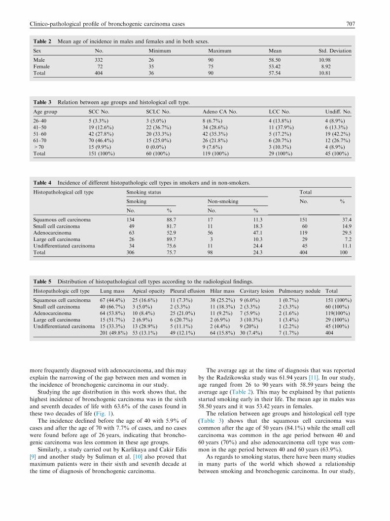
Table 2 Mean age of incidence in males and females and in both sexes.
Sex No. Minimum Maximum Mean Std. Deviation
Male 332 26 90 58.50 10.98
Female 72 35 75 53.42 8.92
Total 404 36 90 57.54 10.81
Table 3 Relation between age groups and histological cell type.
Age group SCC No. SCLC No. Adeno CA No. LCC No. Undiff. No.
26–40 5 (3.3%) 3 (5.0%) 8 (6.7%) 4 (13.8%) 4 (8.9%)
41–50 19 (12.6%) 22 (36.7%) 34 (28.6%) 11 (37.9%) 6 (13.3%)
51–60 42 (27.8%) 20 (33.3%) 42 (35.3%) 5 (17.2%) 19 (42.2%)
61–70 70 (46.4%) 15 (25.0%) 26 (21.8%) 6 (20.7%) 12 (26.7%)
>70 15 (9.9%) 0 (0.0%) 9 (7.6%) 3 (10.3%) 4 (8.9%)
Total 151 (100%) 60 (100%) 119 (100%) 29 (100%) 45 (100%)
Table 4 Incidence of different histopathologic cell types in smokers and in non-smokers.
Histopathological cell type Smoking status Total
Smoking Non-smoking No. %
No. % No. %
Squamous cell carcinoma 134 88.7 17 11.3 151 37.4
Small cell carcinoma 49 81.7 11 18.3 60 14.9
Adenocarcinoma 63 52.9 56 47.1 119 29.5
Large cell carcinoma 26 89.7 3 10.3 29 7.2
Undifferentiated carcinoma 34 75.6 11 24.4 45 11.1
Total 306 75.7 98 24.3 404 100
Table 5 Distribution of histopathological cell types according to the radiological findings.
Histopathologic cell type Lung mass Apical opacity Pleural effusion Hilar mass Cavitary lesion Pulmonary nodule Total
Squamous cell carcinoma 67 (44.4%) 25 (16.6%) 11 (7.3%) 38 (25.2%) 9 (6.0%) 1 (0.7%) 151 (100%)
Small cell carcinoma 40 (66.7%) 3 (5.0%) 2 (3.3%) 11 (18.3%) 2 (3.3%) 2 (3.3%) 60 (100%)
Adenocarcinoma 64 (53.8%) 10 (8.4%) 25 (21.0%) 11 (9.2%) 7 (5.9%) 2 (1.6%) 119(100%)
Large cell carcinoma 15 (51.7%) 2 (6.9%) 6 (20.7%) 2 (6.9%) 3 (10.3%) 1 (3.4%) 29 (100%)
Undifferentiated carcinoma 15 (33.3%) 13 (28.9%) 5 (11.1%) 2 (4.4%) 9 (20%) 1 (2.2%) 45 (100%)
201 (49.8%) 53 (13.1%) 49 (12.1%) 64 (15.8%) 30 (7.4%) 7 (1.7%) 404
Clinico-pathological profile of bronchogenic carcinoma cases 707
more frequently diagnosed with adenocarcinoma, and this may
explain the narrowing of the gap between men and women inthe incidence of bronchogenic carcinoma in our study.
Studying the age distribution in this work shows that, the
highest incidence of bronchogenic carcinoma was in the sixthand seventh decades of life with 63.6% of the cases found inthese two decades of life (Fig. 1).
The incidence declined before the age of 40 with 5.9% ofcases and after the age of 70 with 7.7% of cases, and no caseswere found before age of 26 years, indicating that broncho-genic carcinoma was less common in these age groups.
Similarly, a study carried out by Karlikaya and Cakir Edis[9] and another study by Suliman et al. [10] also proved thatmaximum patients were in their sixth and seventh decade at
the time of diagnosis of bronchogenic carcinoma.
The average age at the time of diagnosis that was reported
by the Radzikowska study was 61.94 years [11]. In our study,age ranged from 26 to 90 years with 58.59 years being theaverage age (Table 2). This may be explained by that patients
started smoking early in their life. The mean age in males was58.50 years and it was 53.42 years in females.
The relation between age groups and histological cell type
(Table 3) shows that the squamous cell carcinoma wascommon after the age of 50 years (84.1%) while the small cellcarcinoma was common in the age period between 40 and60 years (70%) and also adenocarcinoma cell type was com-
mon in the age period between 40 and 60 years (63.9%).As regards to smoking status, there have been many studies
in many parts of the world which showed a relationship
between smoking and bronchogenic carcinoma. In our study,

Table 6 Different cell types of cases that present with pleural
effusion.
Cell type No. and % of cases present
with pleural effusion
Squamous cell carcinoma 11 22.4%
Small cell carcinoma 2 4.1%
Adenocarcinoma 25 51%
Large cell carcinoma 6 12.2%
Undifferentiated carcinoma 5 10.2%
Total 49 100%
Table 7 Different cell types of cases that present with hilar
shadow.
Cell type No. and % of cases present
with hilar shadow
Squamous cell carcinoma 38 59.4%
Small cell carcinoma 11 17.2%
Adenocarcinoma 11 17.2%
Large cell carcinoma 2 3.1%
Undifferentiated carcinoma 2 3.1%
Total 64 100%
Table 8 Different methods of diagnosis of bronchogenic
carcinoma and their percentages.
Methods of diagnosis No. %
FOB 275 68.1
CT guided biopsy 86 21.3
Supraclavicular LN biopsy 2 0.5
Pleural fluid 9 2.2
Thoracoscopy 15 3.7
Open biopsy 17 4.2
Total 404 100
708 Y.M. Akl et al.
smoking habit of the patient (Fig. 2), revealed that, the numberof smokers (75.7%) is 3 folds that of non-smokers (24.3%).
Study of Rawat et al., reported that smoking was the main riskfactor in 81.77% patients [12].
In our study of 72 females with bronchogenic carcinoma, a
high percentage of them were non-smokers (97.25% non-smokers versus 2.8% smokers). And this explains the relativelyhigh percentage (42.3%) of non-smokers in our study.
In male cases, the percentage of smokers was 91.6% while
the percentage of non-smokers was 8.4% of male (Fig. 3).In our study, the percentage of non-smoking females was
97.2% versus only 2.8% of smoking females, so other risk fac-
tors other than smoking, should be considered that includeexposure to environmental tobacco smoke (ETS), fumes andsmoke from certain cooking fuels and environmental
pollution.In our study smoking is associated with all types of bron-
chogenic carcinoma. The connection between smoking and
bronchogenic carcinoma was most evident among patientswith squamous cell carcinoma, small cell carcinoma and largecell carcinoma and less evident among patients with adenocar-cinoma (Fig. 4).
The strength of the association between cigarette smokingand bronchogenic carcinoma varies by cell type, with thelargest for squamous and small cell carcinomas and somewhat
smaller for adenocarcinoma. This result is similar to thatreported in another study [13].
The different clinical presentations of the patients (Fig. 5)
revealed that cough was the most frequent symptom in thewhole group, as 347 patients (85.9%) had this complaint.According to Rawat et al. study cough was the common
symptom (72.90%) [12].Cough is present in >65% of patients at the time lung
cancer is diagnosed [14].In this study cough was the most common symptom (347
patients; 85.9%) and was followed by dyspnea (276 patients;68.3%), expectoration (270 patients; 66.8%), chest pain (241patients; 59.7%), hemoptysis (142 patients; 35.1%) weight loss
(115 patients; 28.5%), and hoarseness of voice (85 patients;21%) was also a frequent symptom. Other symptoms, as fever(16.3%), dysphagia (9.2%) and supraclavicular lymph node
(1%) were not so frequent clinical presentations. In our studythe incidence of symptoms was high compared to thosedescribed in the literature (Fig. 5) and this explained by that
our patients presented late in their disease.The radiological findings (Fig. 6), showed that, lung mass
was the commonest radiological picture, as it occurred in201 cases (49.8%) near to that percentage (46.31%) that was
reported by Rawat et al. study [12] followed by hilar shadow(15.8%), apical opacity (13.1%),and pleural effusion (12.1%).
Cavitary lesion was found in 7.4% of cases. A prospective
study by Shetty et al., found that cavitary lesion incidence was8.6% of lung cancer [15]. Pulmonary nodule in our studyoccurred in 1.7% of cases, this may be explained by that most
cases were presented in late stages.Distribution of histopathologic cell types according to the
radiological findings (Table 5), showed that 44.4% of cases
of squamous cell carcinoma presented with mass lesionfollowed by 25.2% of cases presented with hilar shadow and16.6% presented radiologically with apical opacity whilepleural effusion and cavitary lesion presented in 7.3% and
6% of the squamous cell carcinoma cases, respectively.In our study, 53.8% of cases of adenocarcinoma presented
with mass lesion followed by 21% of cases of adenocarci-
noma presented with pleural effusion while hilar shadowincidence was 9.2% of adenocarcinoma cases. Most of casesof small cell carcinoma presented radiologically with mass
lesion 66.7% followed by hilar shadow in 18.3% of small cellcarcinoma cases. 51.7% of large cell carcinoma presentedwith mass lesion and 20.7% of large cell carcinoma withpleural effusion.
In all cell types of bronchogenic carcinoma, the mass lesionwas the major radiological picture, in this study (Fig. 8).
Table 6 shows that pleural effusion was more frequent with
adenocarcinoma, as 51% of cases that presented with pleuraleffusion were adenocarcinoma and this may be due to thatmost of adenocarcinoma located peripherally and it can cause
pleural invasion.Table 7 shows that hilar shadows were common with
squamous cell carcinoma, as 59.4% of cases presented with
hilar shadows were squamous cell carcinoma followed by smallcell carcinoma and adenocarcinoma (17.2%).
Apical opacity incidence was more frequent with squamouscell carcinoma, 47.2% of cases that presented with apical

Table 10 Stage of disease.
Stage Frequency (No.) Percentage
Operable stage 11 9.8
Inoperable stage 101 90.2
Total 112 100.0
Figure 1 Age distribution of the bronchogenic carcinoma cases.
Figure 2 Incidence of bronchogenic carcinoma in smokers and
in non-smokers.
Table 9 Distribution of bronchogenic carcinoma cell types according to the method used for diagnosis.
Diagnosis method Histopathology Total
SCC SCLC Adeno-CA LCC Undiff
FOB Count 107 52 60 21 35 275
% within diagnosis method 38.9 18.9 21.8 7.6 12.7 100.0
% within histopathology 70.9 86.7 50.4 72.4 77.8 68.1
CT guided biopsy Count 36 5 38 3 4 86
% within diagnosis method 41.9 5.8 44.2 3.5 4.7 100.0
% within histopathology 23.8 8.3 31.9 10.3 8.9 21.3
Supraclavicular LN biopsy Count 0 1 0 0 1 2
% within diagnosis method 0.0 50.0 0.0 0.0 50.0 100.0
% within histopathology 0.0 1.7 0.0 0.0 2.2 0.5
Pleural fluid Count 2 0 3 4 0 9
% within diagnosis method 22.2 0.0 33.3 44.4 0.0 100.0
% within histopathology 1.3 0.0 2.5 13.8 0.0 2.2
Thoracoscopy Count 2 0 9 1 3 15
% within diagnosis method 13.3 0.0 60.0 6.7 20.0 100.0
% within histopathology 1.3 0.0 7.6 3.4 6.7 3.7
Open biopsy Count 4 2 9 0 2 17
% within diagnosis method 23.5 11.8 52.9 0.0 11.8 100.0
% within histopathology 2.6 3.3 7.6 0.0 4.4 4.2
Total Count 151 60 119 29 45 404
% within diagnosis method 37.4 14.9 29.5 7.2 11.1 100.0
% within histopathology 100.0 100.0 100.0 100.0 100.0 100.0
Clinico-pathological profile of bronchogenic carcinoma cases 709
opacity were squamous cell carcinoma, followed by 24.4%
undifferentiated carcinoma.Table 8 and Fig. 7 illustrate different methods of diagnosis
that had been performed in this study, in our study, diagnosis
of bronchogenic carcinoma was made by fibreoptic bronchos-copy in 275 patients (68.1%), and this percentage is near tothat reported in another study which was 61.7% [16]. An
important role in improving the diagnostic relevance of thefibreoptic bronchoscopy was assumed by the more frequentadoption of biopsies and trans-bronchial needle aspirationon parenchyma and mediastinal lymph nodes.
Table 9 shows that bronchoscopic biopsy was positive in
107 of 151 patients (70.9%) of the squamous cell carcinomacases, also in 52 of 60 patients (86.7%) of cases of the small cellcarcinoma, in 60 of 119 patients (50.4%) of cases of adenocar-
cinoma, in 21 of 29 patients (72.4%) of cases of the large cellcarcinoma and. in 35 of 45 patients (68.1%) of cases ofundifferentiated carcinoma.
The cases of bronchogenic carcinoma that was diagnosedby CT guided biopsy were 86 cases (21.3% of all cases). Table 9shows that most cases that were diagnosed by FNAB (FNABdone under CT) was adenocarcinoma (38 patients; 44.2%),
followed by squamous cell carcinoma (36 patients; 41.9%),while only 5 cases (5.8%) of the small cell carcinoma, 3 cases(3.5%) of the large cell carcinoma and 4 cases (4.7%) of the
undifferentiated carcinoma were diagnosed by CT guidedbiopsy.
Supraclavicular lymph node biopsy was performed in 2
cases (0.5% of all cases), 1 of these cases proved to be

Figure 3 Smoking status distribution in males and females.
Figure 4 Incidence of different histopathological cell types in
smokers and in non-smokers.
Figure 5 Percentages of different clinical presentations.
Figure 6 Radiological findings of bronchogenic carcinoma.
Figure 7 Methods of diagnosis and their percentages.
Figure 8 Percentage of different pathologic cell types in this
study.
Figure 9 Distribution of histopathologic cell type in males and
females.
710 Y.M. Akl et al.
squamous cell carcinoma, while other case was undifferenti-ated carcinoma. Pleural fluid cytology was positive in 9 cases
(2.2% of all cases), 3 cases were adenocarcinoma, 4 cases werelarge cell carcinoma and 2 cases were squamous cellcarcinoma.
Thoracoscopy was the diagnostic procedure in 15 cases(3.7% of all cases), 60% of them were adenocarcinoma,13.3% were squamous cell carcinoma and 20% were undiffer-entiated carcinoma.
Open biopsy was performed in l7cases (4.2% of all cases),as regards the histologic cell type, that most cases were adeno-carcinoma (52.9%) followed by squamous cell carcinoma
(23.5%).In our study the most common histopathological cell type
was squamous cell carcinoma (37.4%), followed by adenocar-
cinoma (29.5%), small cell carcinoma (14.9%), large cell

Clinico-pathological profile of bronchogenic carcinoma cases 711
carcinoma (7.2%) and undifferentiated carcinoma (11.1%)(Fig. 8).
In the cell type distribution reported by Radzikowska,
squamous cell carcinoma had the highest cell type incidence(52.1%) followed by small cell carcinoma (20.8%) whileadenocarcinoma represented only 11.3% of the cases [11].
According to Shetty et al., study, squamous cell carcinomaalso presented 44.5% of cases followed by adenocarcinoma(18.5%) and small cell carcinoma (17.2%) [15].
In another study by Rawat et al., they reported that 44.8%of cases were squamous cell carcinoma followed by adenocar-cinoma (19.7%) and small cell carcinoma (16.75%) [12].
In our study the incidence of squamous cell carcinoma
(37.4%) is less than that reported in a previous study and closeto percentage reported in study by Lam et al., (39%) [17].
The incidence of adenocarcinoma in our study (29.5%) is
higher than that reported in other studies 11.3%, 18.5% and19.7% by Radzikowska et al. [11], Shetty et al. [15] and Rawatet al. [12], respectively.
There was a shift in the incidence of squamous cellcarcinoma and adenocarcinoma, up to the late 1980s,squamous cell carcinoma was the most common subtype,
which was then surpassed by adenocarcinoma. In recent yearsadenocarcinoma is the most common histologic subtype oflung cancer in most countries, accounting for almost half ofall lung cancers [18].
The shift in the incidence of squamous cell carcinoma andadenocarcinoma may be associated with the switch fromnon-filtered to filtered cigarettes, the depth of inhalation had
been altered [19].In particular, smoke from unfiltered strong cigarettes may
be shallowly inhaled, resulting in chemical carcinogen deposi-
tion centrally in the bronchial area and giving rise to squamouscell carcinomas. Smoke from filtered milder cigarettes may bemore deeply inhaled, resulting in carcinogen deposition more
peripherally and giving rise to adenocarcinomas. Reducingthe nicotine content may also promote deeper inhalation assmokers attempt to compensate [20]. The changes in cigarettecomposition reduced the yield of carcinogenic polycyclic
aromatic hydrocarbons (PAHs), inducers of squamous cellcarcinomas, while increasing the yields of carcinogenictobacco-specific N-nitrosamines (TSNAs), inducers of adeno-
carcinomas [21].Other possible explanations that the increasing use of
mucin stains and immunocytochemical staining for antibodies
to CEA has contributed to enhanced recognition of adenocar-cinomas [15].
These factors have contributed to the emerging predomi-nance of adenocarcinoma in the lung cancer rates.
Fig. 9 shows that adenocarcinoma was the predominanttype (54.2%) in females followed by small cell carcinoma(16.7%) and squamous cell carcinoma (13.9%). In males the
predominant type was squamous cell carcinoma (42.5%)followed by adenocarcinoma (14.5%).
The criteria of inoperability in our cases included: X-ray
finding of chest wall invasion, finding of bilateral hilar ormediastinal enlargement suggestive of lymph node involve-ment, pleural effusion, mediastinal invasion and signs of vital
structures involvement, presence of extrathoracic metastasis,bronchoscopic finding as vocal cord paralysis and tracheal orcarinal involvement and the presence of supraclavicular lymphnodes metastasis.
The high percentage of inoperable cases in our study maybe attributed to that some of the cases were misdiagnosed astuberculosis and treated by anti-tuberculous treatment at other
centers and after long time, the patients were referred to ourchest department, thereby causing a delay in diagnosis, thesmokers often explain any cough they experience as smoker’s
cough and this wrong belief may mask presence of tumor fora long time before diagnosis and thus delay diagnosis untilthe disease is at a more advanced. Lack of diagnostic facilities
at peripheral health centers could be another cause for delay indiagnosis of bronchogenic carcinoma.
Conclusion
We can conclude from the study:
1. In this work, the highest incidence of bronchogeniccarcinoma was in the sixth and seventh decades of life.
The incidence declined before the age of 40 years and afterthe age of 70 years.
2. Smoking still remains the major risk factor in the pathogen-esis of bronchogenic carcinoma.
3. On histological examination, squamous cell carcinoma wasrelatively more frequent than any other tumor typefollowed by adenocarcinoma and then by small cell
carcinoma.4. In all cell types of bronchogenic carcinoma, the mass lesion
was the major radiological finding and the majority of cases
were diagnosed by fibro-optic bronchoscopy.
References
[1] A.J. Alberg, J.G. Ford, J.M. Samet, Epidemiology of lung
ACCP evidence-based clinical practice guidelines cancer, Chest
132 (2007) 29S–55S.
[2] A. Jemal, F. Bray, M. Melissa, et al, Global cancer statistics:
CA, Cancer J. Clin. 61 (2011) 69–90.
[3] I. Elattar, Lung cancer in Egypt and neighbouring Arab
countries: magnitude of the problem, NCI, Egypt, 2005.
Available from: <http://www.nci.cu.edu.eg>.
[4] Y. Bahader, A. Jazieh, Epidemiology of lung cancer, Ann.
Thorac. Med. 3 (2008) 365–367.
[5] F. Bray, R. Sankila, J. Ferlay, et al, Estimates of cancer
incidence and mortality in Europe in 1995, Eur. J. Cancer 38
(2002) 99–166.
[6] A. Jemal, W.E. Travis, R.E. Tarone, et al, Lung cancer rates
convergence in young men and women in the United States:
analysis by birth cohort and histologic type, Int. J. Cancer 105
(2003) 101–107.
[7] S. Devesa, F. Bray, P. Vizcaino, et al, International lung cancer
trends by histologic type: male:female differences diminishing
and adenocarcinoma rates rising diminishing and
adenocarcinoma rates rising, Int. J. Cancer 117 (2005) 294–299.
[8] L. Blizzard, T. Dwyer, Case-control study of lung cancer during
1994–1997 in the birth cohort in Tasmania, Australia, with an
excess of female cases during 1983–1992, Cancer Causes Control
14 (2003) 123–129.
[9] C. Karlikaya, E. Cakir Edis, Lung cancer histopathology in the
Thrace region of Turkey and comparison with national data,
Tuberk Torkas 53 (2005) 132–138.
[10] M.I. Suliman, R. Jibran, M.Z. Majeed, Demographics of
bronchogenic carcinoma patients and frequency of cell types,
Gomal J. Med. Sci. 4 (1) (2006) 2–6.

712 Y.M. Akl et al.
[11] E. Radzikowska, P. Glaz, K. Roszkowski, Lung cancer in
women: age, smoking, histology, performance status, stage,
initial treatment and survival. Population-based study of 20 561
cases, Ann. Oncol. 13 (2002) 1087–1093.
[12] J. Rawat, G. Sindhwani, D. Gaur, et al, Clinico-pathological
profile of lung cancer, Lung India 26 (3) (2009) 74–76.
[13] S.A. Khuder, Effect of cigarette smoking on major histological
types of lung cancer: a meta-analysis, Lung Cancer 31 (2001)
139–148.
[14] P.A. Kvale, Chronic cough due to lung tumors: ACCP evidence-
based clinical practice guidelines, Chest 129 (2006) 147–153.
[15] C. Shetty, B. Lakhkar, V. Gangadhar, et al, Changing pattern
of bronchogenic carcinoma, Indian J. Radiol. Imaging 15 (2005)
233–238.
[16] M. Filippetti, P. Visca, V. Bordignon, et al, Fiberoptic
bronchoscopy as diagnostic tool in primitive lung cancer, Clin.
Ter. 160 (2009) 43–46.
[17] W. Lam, N. White, M. Chan-Yeung, Lung cancer epidemiology
and risk factors in Asia and Africa, Int. J. Tuberc. Lung Dis. 8
(2004) 1045–1057.
[18] W. Travis, E. Brambilla, M. Noguchi, et al, International
multidisciplinary classification of lung adenocarcinoma, J.
Thorac. Oncol. 6 (2) (2011) 244–285.
[19] E. Wynder, E. Hoffmann, Cigarette smoking and the
histopathology of lung cancer, J. Natl. Cancer Inst. 90 (1998)
1486–1488.
[20] M. Djordjevic, D. Hoffmann, I. Hoffman, Nicotine regulates
smoking patterns, Prev. Med. 26 (1997) 435–440.
[21] D. Hoffmann, M. Djordjevic, I. Hoffmann, The changing
cigarette, Prev. Med. 26 (1997) 427–434.


![Clinico-pathological case 1 [Trinity College Dublin] Prof T Rogers Prof O Sheils Dr C D’Adhemar](https://img.dokumen.tips/doc/110x75/56649f355503460f94c53b07/clinico-pathological-case-1-trinity-college-dublin-prof-t-rogers-prof-o-sheils.jpg)


