Embed Size (px)
Citation preview
Hindawi Publishing CorporationObstetrics and Gynecology InternationalVolume 2013, Article ID 931318, 8 pageshttp://dx.doi.org/10.1155/2013/931318
Clinical StudyRobotic-Assisted Transperitoneal Aortic Lymphadenectomyas Part of Staging Procedure for Gynaecological Malignancies:Single Institution Experience
V. Zanagnolo,1 D. Rollo,1 T. Tomaselli,1 P. G. Rosenberg,2 L. Bocciolone,1 F. Landoni,1
G. Aletti,1 M. Peiretti,1 F. Sanguineti,3 and A. Maggioni1
1 Department of Gynecology, Cervical Cancer Center, European Institute of Oncology, Milan 20141, Italy2 Department of Obstetrics and Gynaecology, Ospedale San Bortolo, Vicenza 36010, Italy3 Department of Obstetrics and Gynaecology, Niguarda Ca’ Granda Hospital, Milan 20162, Italy
Correspondence should be addressed to V. Zanagnolo; [email protected]
Received 18 December 2012; Revised 24 May 2013; Accepted 15 June 2013
Academic Editor: Curt W. Burger
Copyright © 2013 V. Zanagnolo et al. This is an open access article distributed under the Creative Commons Attribution License,which permits unrestricted use, distribution, and reproduction in any medium, provided the original work is properly cited.
Introduction. This study was designed to confirm the feasibility and safety of robotic-assisted transperitoneal aortic lymphadenec-tomy as part of staging procedure for gynecologic malignancies.Methods. Chart review of 51 patients who had undergone roboticstaging with aortic lymphadenectomy for different gynaecologic malignancies was performed. Results. The primary diagnosiswas as follows: 6 cases of endometrial cancer, 31 epithelial ovarian cancer, 9 nonepithelial ovarian cancer, 4 tubal cancer, and1 cervical cancer. Median BMI was 23 kg/m2. Except for a single case of aortic lymphadenectomy only, both aortic and pelviclymphadenectomies were performed at the time of the staging procedure. All the para-aortic lymphadenectomies were carried outto the level of the renal veinl but 6 cases were carried out to the level of the inferior mesenteric artery. Hysterectomy was performedin 24 patiens (47%).There was no conversion to LPT.Themedian console time was 285 (range 195–402) with a significant differencebetween patients who underwent hysterectomy and those who did not.Themedian estimated blood loss was 50mL (range 20–200).Themean number of removed nodes was 29±9.6.Themean number of pelvic nodes was 15±7.6, whereas themean number of para-aortic nodes was 14 ± 6.6. Conclusions. Robotic transperitoneal infrarenal aortic lymphadenectomy as part of staging procedure isfeasible and can be safely performed. Additional trocars are needed when pelvic surgery is also performed.
1. Introduction
The feasibility and safety of robotically assisted para-aorticlymphadenectomy (PAL) have been already well reported,both with the robotic setup for pelvic surgery or with thesovrapubic approach [1, 2]. However, the upper limit, up tothe left renal vein, is still debated, and technical aspects ofPAL may differ depending on whether this procedure is theonly one performed, or it is combined with other stagingprocedures for gynaecologic malignancies, such as pelviclymphadenectomy, hysterectomy, omentectomy, and randomperitoneal sampling.
The inframesenteric aortic nodes in most patients canbe accessed and removed with the robotic setup for pelvicsurgery. However, removal of the infrarenal aortic nodes up
to the renal veins and, in particular, the left group can be verychallenging.
The infrarenal nodes have been reported as one of themost common site of nodal metastases in epithelial ovariancancer, and recently they have been shown to be positivenodes in the absence of metastases in the ipsilateral inframe-senteric nodes in endometrial cancer [3].
One of the major limitations of the current da Vincirobotic systems (Intuitive Surgical Inc., Sunnyvale, CA. USA)is its inability to provide access to the entire abdomenwithout relocating the robotic column. When removal of theinfrarenal aortic nodes is required in case of full staging orexcision, or both, of early or localized relapses of gynecologicmalignancies, relocation of the robotic column may need tobe performed.
2 Obstetrics and Gynecology International
Magrina has shown, in his series of 33 patients, thatrobotic transperitoneal infrarenal aortic lymphadenectomycan be performed adequately and safely with the roboticcolumn at the patient’s head. Operating table/robotic columnrotation and additional trocar sites are needed when used inconjunction with robotic pelvic surgery [2].
2. Material and Methods
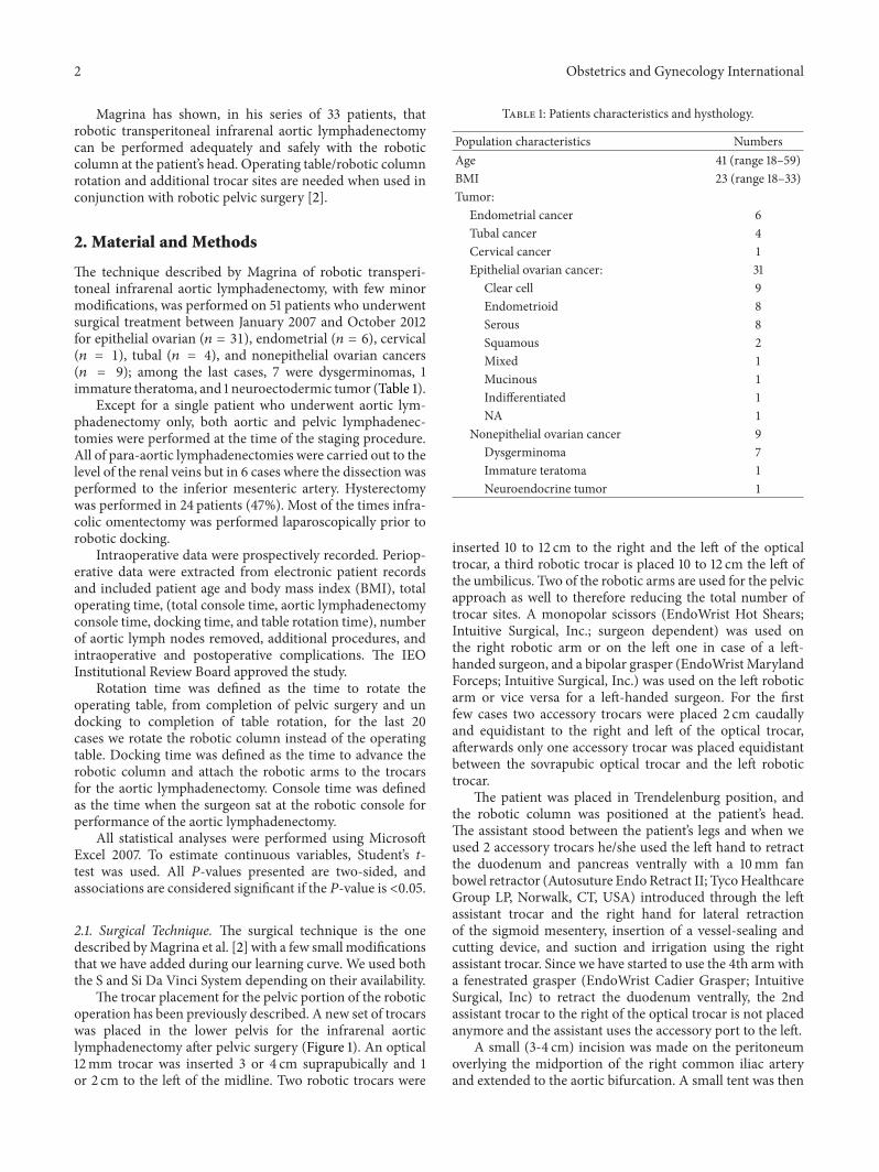
The technique described by Magrina of robotic transperi-toneal infrarenal aortic lymphadenectomy, with few minormodifications, was performed on 51 patients who underwentsurgical treatment between January 2007 and October 2012for epithelial ovarian (𝑛 = 31), endometrial (𝑛 = 6), cervical(𝑛 = 1), tubal (𝑛 = 4), and nonepithelial ovarian cancers(𝑛 = 9); among the last cases, 7 were dysgerminomas, 1immature theratoma, and 1 neuroectodermic tumor (Table 1).
Except for a single patient who underwent aortic lym-phadenectomy only, both aortic and pelvic lymphadenec-tomies were performed at the time of the staging procedure.All of para-aortic lymphadenectomies were carried out to thelevel of the renal veins but in 6 cases where the dissection wasperformed to the inferior mesenteric artery. Hysterectomywas performed in 24 patients (47%). Most of the times infra-colic omentectomy was performed laparoscopically prior torobotic docking.
Intraoperative data were prospectively recorded. Periop-erative data were extracted from electronic patient recordsand included patient age and body mass index (BMI), totaloperating time, (total console time, aortic lymphadenectomyconsole time, docking time, and table rotation time), numberof aortic lymph nodes removed, additional procedures, andintraoperative and postoperative complications. The IEOInstitutional Review Board approved the study.
Rotation time was defined as the time to rotate theoperating table, from completion of pelvic surgery and undocking to completion of table rotation, for the last 20cases we rotate the robotic column instead of the operatingtable. Docking time was defined as the time to advance therobotic column and attach the robotic arms to the trocarsfor the aortic lymphadenectomy. Console time was definedas the time when the surgeon sat at the robotic console forperformance of the aortic lymphadenectomy.
All statistical analyses were performed using MicrosoftExcel 2007. To estimate continuous variables, Student’s t-test was used. All 𝑃-values presented are two-sided, andassociations are considered significant if the 𝑃-value is <0.05.
2.1. Surgical Technique. The surgical technique is the onedescribed byMagrina et al. [2] with a few small modificationsthat we have added during our learning curve. We used boththe S and Si Da Vinci System depending on their availability.
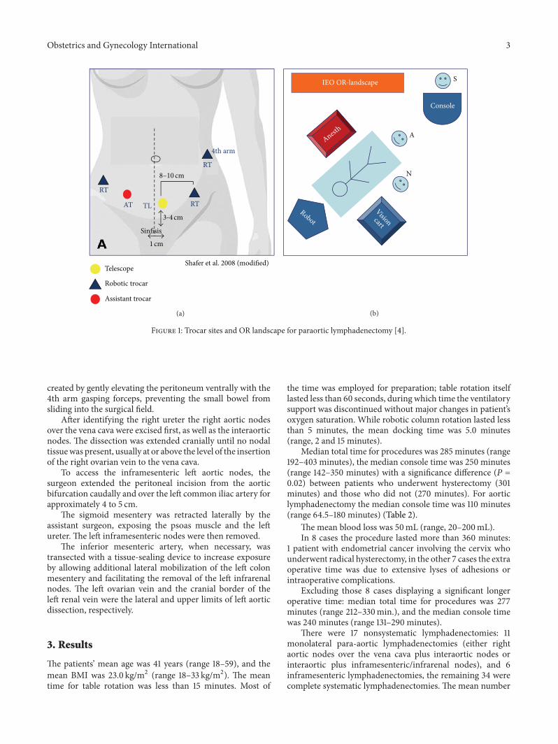
The trocar placement for the pelvic portion of the roboticoperation has been previously described. A new set of trocarswas placed in the lower pelvis for the infrarenal aorticlymphadenectomy after pelvic surgery (Figure 1). An optical12mm trocar was inserted 3 or 4 cm suprapubically and 1or 2 cm to the left of the midline. Two robotic trocars were
Table 1: Patients characteristics and hysthology.
Population characteristics NumbersAge 41 (range 18–59)BMI 23 (range 18–33)Tumor:
Endometrial cancer 6Tubal cancer 4Cervical cancer 1Epithelial ovarian cancer: 31
Clear cell 9Endometrioid 8Serous 8Squamous 2Mixed 1Mucinous 1Indifferentiated 1NA 1
Nonepithelial ovarian cancer 9Dysgerminoma 7Immature teratoma 1Neuroendocrine tumor 1
inserted 10 to 12 cm to the right and the left of the opticaltrocar, a third robotic trocar is placed 10 to 12 cm the left ofthe umbilicus. Two of the robotic arms are used for the pelvicapproach as well to therefore reducing the total number oftrocar sites. A monopolar scissors (EndoWrist Hot Shears;Intuitive Surgical, Inc.; surgeon dependent) was used onthe right robotic arm or on the left one in case of a left-handed surgeon, and a bipolar grasper (EndoWrist MarylandForceps; Intuitive Surgical, Inc.) was used on the left roboticarm or vice versa for a left-handed surgeon. For the firstfew cases two accessory trocars were placed 2 cm caudallyand equidistant to the right and left of the optical trocar,afterwards only one accessory trocar was placed equidistantbetween the sovrapubic optical trocar and the left robotictrocar.
The patient was placed in Trendelenburg position, andthe robotic column was positioned at the patient’s head.The assistant stood between the patient’s legs and when weused 2 accessory trocars he/she used the left hand to retractthe duodenum and pancreas ventrally with a 10mm fanbowel retractor (Autosuture EndoRetract II; TycoHealthcareGroup LP, Norwalk, CT, USA) introduced through the leftassistant trocar and the right hand for lateral retractionof the sigmoid mesentery, insertion of a vessel-sealing andcutting device, and suction and irrigation using the rightassistant trocar. Since we have started to use the 4th arm witha fenestrated grasper (EndoWrist Cadier Grasper; IntuitiveSurgical, Inc) to retract the duodenum ventrally, the 2ndassistant trocar to the right of the optical trocar is not placedanymore and the assistant uses the accessory port to the left.
A small (3-4 cm) incision was made on the peritoneumoverlying the midportion of the right common iliac arteryand extended to the aortic bifurcation. A small tent was then
Obstetrics and Gynecology International 3
Shafer et al. 2008 (modified)
8–10 cm
RT
RT
RTTLAT
Sinfisis
3-4 cm
1 cm
4th arm
Telescope
Robotic trocar
Assistant trocar
(a)
Console
A
N
SIEO OR-landscape
Anesth
Robot
Visioncart
(b)
Figure 1: Trocar sites and OR landscape for paraortic lymphadenectomy [4].
created by gently elevating the peritoneum ventrally with the4th arm gasping forceps, preventing the small bowel fromsliding into the surgical field.
After identifying the right ureter the right aortic nodesover the vena cava were excised first, as well as the interaorticnodes. The dissection was extended cranially until no nodaltissuewas present, usually at or above the level of the insertionof the right ovarian vein to the vena cava.
To access the inframesenteric left aortic nodes, thesurgeon extended the peritoneal incision from the aorticbifurcation caudally and over the left common iliac artery forapproximately 4 to 5 cm.
The sigmoid mesentery was retracted laterally by theassistant surgeon, exposing the psoas muscle and the leftureter. The left inframesenteric nodes were then removed.
The inferior mesenteric artery, when necessary, wastransected with a tissue-sealing device to increase exposureby allowing additional lateral mobilization of the left colonmesentery and facilitating the removal of the left infrarenalnodes. The left ovarian vein and the cranial border of theleft renal vein were the lateral and upper limits of left aorticdissection, respectively.
3. Results
The patients’ mean age was 41 years (range 18–59), and themean BMI was 23.0 kg/m2 (range 18–33 kg/m2). The meantime for table rotation was less than 15 minutes. Most of
the time was employed for preparation; table rotation itselflasted less than 60 seconds, during which time the ventilatorysupport was discontinued without major changes in patient’soxygen saturation. While robotic column rotation lasted lessthan 5 minutes, the mean docking time was 5.0 minutes(range, 2 and 15 minutes).
Median total time for procedures was 285 minutes (range192–403 minutes), the median console time was 250 minutes(range 142–350 minutes) with a significance difference (P =0.02) between patients who underwent hysterectomy (301minutes) and those who did not (270 minutes). For aorticlymphadenectomy the median console time was 110 minutes(range 64.5–180 minutes) (Table 2).
The mean blood loss was 50mL (range, 20–200mL).In 8 cases the procedure lasted more than 360 minutes:
1 patient with endometrial cancer involving the cervix whounderwent radical hysterectomy, in the other 7 cases the extraoperative time was due to extensive lyses of adhesions orintraoperative complications.
Excluding those 8 cases displaying a significant longeroperative time: median total time for procedures was 277minutes (range 212–330min.), and the median console timewas 240 minutes (range 131–290 minutes).
There were 17 nonsystematic lymphadenectomies: 11monolateral para-aortic lymphadenectomies (either rightaortic nodes over the vena cava plus interaortic nodes orinteraortic plus inframesenteric/infrarenal nodes), and 6inframesenteric lymphadenectomies, the remaining 34 werecomplete systematic lymphadenectomies. The mean number
4 Obstetrics and Gynecology International
Table 2: Operation and console time.
Procedure Total operative time (min) 𝑃 value Console time (min) 𝑃 valuePelvic + LA plus hysterectomy 301 0.02 270 0.12Pelvic + LA without hysterectomy 270 240
of nodes was 29.2 ± 9.6 with median number of pelvic nodesof 15 ± 7.6, whereas the mean number of para-aortic nodeswas 14 ± 6.6. In the group of systematic lymphadenectomiesthe mean number of pelvic lymph nodes is 20 ± 5.5 and ofpara-aortic is 15±5.5 (Table 3).Themean number of positivenodes was 0.37 ± 1.13.
The mean number of aortic nodes collected with system-atic pelvic and aortic lymphadenectomy comparing our first10 and final 10 patients was 15 (±4.9) in first group and 16(±5.5) in second group (𝑃 = 0.5).
In 8 patients with a BMI ≥28 kg/m2 the mean number ofaortic nodes was 13±7.4. Due to the small number of cases wedid not compare this group of patients with those with BMI<25 kg/m2.
Table 4 summarizes intraoperative and post-operativecomplications: 2 patients had significant intraoperative bleed-ing (>500mL) one from the vena cava and the other one froma lumbar artery, both controlled robotically. We did not haveany conversion to laparotomy. Seven cases of chylous asciteswere observed in the immediate post-operative course: 4 ofthe cases improved with low-fat diet only, on the contrary theother 3 patients required total parenteral nutrition for a fewdays while fasting.
The mean length of hospital stay for all surgical proce-dures was 3.0 days (range 2–7.5 days).
Late postoperative complications that could be relatedto pelvic and aortic lymphadenectomy included pelvic andaortic lymph cyst formation in 3 patients, which resolvedconservatively. Other postoperative complications were: onecase of ureteral fistula which was treated by ureteral stentplacement, two cases of port-site hernia, of which onerequired a reintervention, and mild to moderate legs edemain three cases.
Median followup was 26 months (range 1–56 months).During followup the six cases of recurrence were observed:one port-site recurrence in a patient with epithelial ovariancancer FIGO stage IC after 30 months, this patient had asecond recurrence (carcinomatosis) after 22months; a secondpatient with ovarian cancer FIGO stage IIB had a recurrence(carcinomatosis) after 6 months, and she died of the diseaseafter 8 months. A third patient with ovarian cancer FIGOstage IB had a spleen recurrence, treated by splenectomy. Afourth patient with ovarian cancer FIGO stage IIC had a liverrecurrence after 33months, and at then a patient with IA highgrade ovarian cancer presented with lymphnode recurrence.
4. Discussion
Gynecology oncologists still have some disagreements con-cerning Para-Aortic Lymphadenectomy (PAL) for gyneco-logicmalignancies such as its therapeutic role, the upper limit
Table 3: Mean number of lymph nodes in different groups.
Pelvic lymph nodesmean (sd)
LA lymph nodesmean (sd)
Totallymphadenectomies(Pts = 51)
15 (±7) 14 (±6)
Systematiclymphadenectomies(Pts = 34)
20 (±5) 15 (±5.5)
Nonsystematiclymphadenectomies(Pts = 31)
11.3 (±4) (Pts = 28) 12 (±6) (Pts = 14)
Obese pts (Pts = 8) 17 (±10) 13 (±7)
Table 4: Intraoperative and postoperative complications.
Complications No. pts (%)Intraoperative complications
Significant Bleeding (>500mL) 2 (3.9)Conversion rate 0
Postoperative complicationsTrasfusion rate 3 (5.8)Chylous ascites 7 (13.7)Vaginal leakage 2 (3.9)Ureteral fistula 1 (1.9)Femoral nerve injury 1 (1.9)Legs edema G1-G2 4 (7.8)Port-site hernia 2 (3.9)Lymphocele 4 (7.8)Lymphatic ascites 1 (1.9)Total 25
of dissection: inferior mesenteric artery versus left renal vein,and indications of different surgical approaches: traditionalversus minimally invasive one.
The inframesenteric aortic nodes in most patients canbe accessed and removed with the robotic setup for pelvicsurgery, by placing the trocars higher than usual and leavingthe robot column between the patient’s legs at all times [14].However, removal of the infrarenal aortic nodes up to therenal vein and, in particular, the left group is difficult, can beincomplete, or can be unsafe due to the steep orientation ofthe robotic instruments in such a setting and the proximitybetween the optic and the renal vein (just beneath the cameraport site).
Therefore, exposure of the upper limit (left renal veinin our practice) as described by Lambaudie et al. [1] wassometimes difficult, particularly in case of high BMI. Thisdifficulty in exposing the higher part of the dissection
Obstetrics and Gynecology International 5
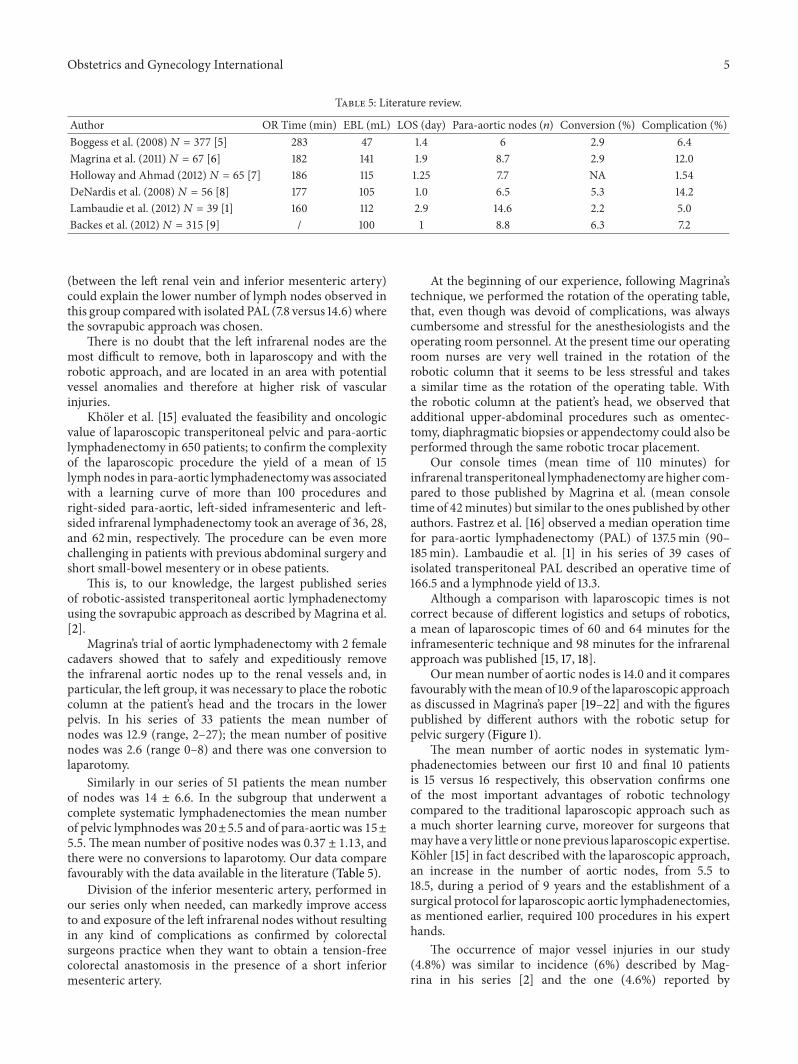
Table 5: Literature review.
Author OR Time (min) EBL (mL) LOS (day) Para-aortic nodes (𝑛) Conversion (%) Complication (%)Boggess et al. (2008)𝑁 = 377 [5] 283 47 1.4 6 2.9 6.4Magrina et al. (2011)𝑁 = 67 [6] 182 141 1.9 8.7 2.9 12.0Holloway and Ahmad (2012)𝑁 = 65 [7] 186 115 1.25 7.7 NA 1.54DeNardis et al. (2008)𝑁 = 56 [8] 177 105 1.0 6.5 5.3 14.2Lambaudie et al. (2012)𝑁 = 39 [1] 160 112 2.9 14.6 2.2 5.0Backes et al. (2012)𝑁 = 315 [9] / 100 1 8.8 6.3 7.2
(between the left renal vein and inferior mesenteric artery)could explain the lower number of lymph nodes observed inthis group comparedwith isolated PAL (7.8 versus 14.6) wherethe sovrapubic approach was chosen.
There is no doubt that the left infrarenal nodes are themost difficult to remove, both in laparoscopy and with therobotic approach, and are located in an area with potentialvessel anomalies and therefore at higher risk of vascularinjuries.
Kholer et al. [15] evaluated the feasibility and oncologicvalue of laparoscopic transperitoneal pelvic and para-aorticlymphadenectomy in 650 patients; to confirm the complexityof the laparoscopic procedure the yield of a mean of 15lymphnodes in para-aortic lymphadenectomywas associatedwith a learning curve of more than 100 procedures andright-sided para-aortic, left-sided inframesenteric and left-sided infrarenal lymphadenectomy took an average of 36, 28,and 62min, respectively. The procedure can be even morechallenging in patients with previous abdominal surgery andshort small-bowel mesentery or in obese patients.
This is, to our knowledge, the largest published seriesof robotic-assisted transperitoneal aortic lymphadenectomyusing the sovrapubic approach as described by Magrina et al.[2].
Magrina’s trial of aortic lymphadenectomy with 2 femalecadavers showed that to safely and expeditiously removethe infrarenal aortic nodes up to the renal vessels and, inparticular, the left group, it was necessary to place the roboticcolumn at the patient’s head and the trocars in the lowerpelvis. In his series of 33 patients the mean number ofnodes was 12.9 (range, 2–27); the mean number of positivenodes was 2.6 (range 0–8) and there was one conversion tolaparotomy.
Similarly in our series of 51 patients the mean numberof nodes was 14 ± 6.6. In the subgroup that underwent acomplete systematic lymphadenectomies the mean numberof pelvic lymphnodes was 20±5.5 and of para-aortic was 15±5.5. The mean number of positive nodes was 0.37 ± 1.13, andthere were no conversions to laparotomy. Our data comparefavourably with the data available in the literature (Table 5).
Division of the inferior mesenteric artery, performed inour series only when needed, can markedly improve accessto and exposure of the left infrarenal nodes without resultingin any kind of complications as confirmed by colorectalsurgeons practice when they want to obtain a tension-freecolorectal anastomosis in the presence of a short inferiormesenteric artery.
At the beginning of our experience, following Magrina’stechnique, we performed the rotation of the operating table,that, even though was devoid of complications, was alwayscumbersome and stressful for the anesthesiologists and theoperating room personnel. At the present time our operatingroom nurses are very well trained in the rotation of therobotic column that it seems to be less stressful and takesa similar time as the rotation of the operating table. Withthe robotic column at the patient’s head, we observed thatadditional upper-abdominal procedures such as omentec-tomy, diaphragmatic biopsies or appendectomy could also beperformed through the same robotic trocar placement.
Our console times (mean time of 110 minutes) forinfrarenal transperitoneal lymphadenectomy are higher com-pared to those published by Magrina et al. (mean consoletime of 42minutes) but similar to the ones published by otherauthors. Fastrez et al. [16] observed a median operation timefor para-aortic lymphadenectomy (PAL) of 137.5min (90–185min). Lambaudie et al. [1] in his series of 39 cases ofisolated transperitoneal PAL described an operative time of166.5 and a lymphnode yield of 13.3.
Although a comparison with laparoscopic times is notcorrect because of different logistics and setups of robotics,a mean of laparoscopic times of 60 and 64 minutes for theinframesenteric technique and 98 minutes for the infrarenalapproach was published [15, 17, 18].
Our mean number of aortic nodes is 14.0 and it comparesfavourably with themean of 10.9 of the laparoscopic approachas discussed in Magrina’s paper [19–22] and with the figurespublished by different authors with the robotic setup forpelvic surgery (Figure 1).
The mean number of aortic nodes in systematic lym-phadenectomies between our first 10 and final 10 patientsis 15 versus 16 respectively, this observation confirms oneof the most important advantages of robotic technologycompared to the traditional laparoscopic approach such asa much shorter learning curve, moreover for surgeons thatmay have a very little or none previous laparoscopic expertise.Kohler [15] in fact described with the laparoscopic approach,an increase in the number of aortic nodes, from 5.5 to18.5, during a period of 9 years and the establishment of asurgical protocol for laparoscopic aortic lymphadenectomies,as mentioned earlier, required 100 procedures in his experthands.
The occurrence of major vessel injuries in our study(4.8%) was similar to incidence (6%) described by Mag-rina in his series [2] and the one (4.6%) reported by
6 Obstetrics and Gynecology International
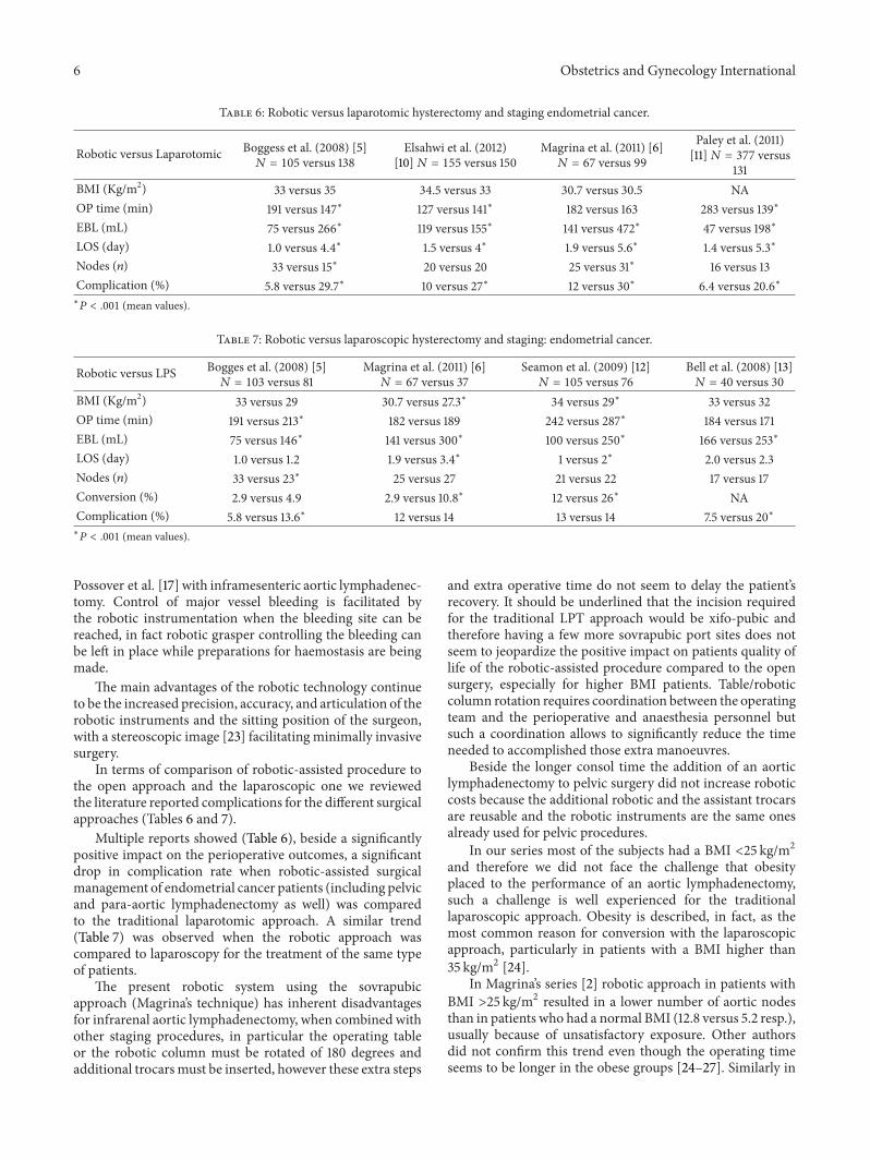
Table 6: Robotic versus laparotomic hysterectomy and staging endometrial cancer.
Robotic versus Laparotomic Boggess et al. (2008) [5]𝑁 = 105 versus 138
Elsahwi et al. (2012)[10]𝑁 = 155 versus 150
Magrina et al. (2011) [6]𝑁 = 67 versus 99
Paley et al. (2011)[11]𝑁 = 377 versus
131BMI (Kg/m2) 33 versus 35 34.5 versus 33 30.7 versus 30.5 NAOP time (min) 191 versus 147∗ 127 versus 141∗ 182 versus 163 283 versus 139∗
EBL (mL) 75 versus 266∗ 119 versus 155∗ 141 versus 472∗ 47 versus 198∗
LOS (day) 1.0 versus 4.4∗ 1.5 versus 4∗ 1.9 versus 5.6∗ 1.4 versus 5.3∗
Nodes (𝑛) 33 versus 15∗ 20 versus 20 25 versus 31∗ 16 versus 13Complication (%) 5.8 versus 29.7∗ 10 versus 27∗ 12 versus 30∗ 6.4 versus 20.6∗∗𝑃 < .001 (mean values).
Table 7: Robotic versus laparoscopic hysterectomy and staging: endometrial cancer.
Robotic versus LPS Bogges et al. (2008) [5]𝑁 = 103 versus 81
Magrina et al. (2011) [6]𝑁 = 67 versus 37
Seamon et al. (2009) [12]𝑁 = 105 versus 76
Bell et al. (2008) [13]𝑁 = 40 versus 30
BMI (Kg/m2) 33 versus 29 30.7 versus 27.3∗ 34 versus 29∗ 33 versus 32OP time (min) 191 versus 213∗ 182 versus 189 242 versus 287∗ 184 versus 171EBL (mL) 75 versus 146∗ 141 versus 300∗ 100 versus 250∗ 166 versus 253∗
LOS (day) 1.0 versus 1.2 1.9 versus 3.4∗ 1 versus 2∗ 2.0 versus 2.3Nodes (𝑛) 33 versus 23∗ 25 versus 27 21 versus 22 17 versus 17Conversion (%) 2.9 versus 4.9 2.9 versus 10.8∗ 12 versus 26∗ NAComplication (%) 5.8 versus 13.6∗ 12 versus 14 13 versus 14 7.5 versus 20∗∗𝑃 < .001 (mean values).
Possover et al. [17] with inframesenteric aortic lymphadenec-tomy. Control of major vessel bleeding is facilitated bythe robotic instrumentation when the bleeding site can bereached, in fact robotic grasper controlling the bleeding canbe left in place while preparations for haemostasis are beingmade.
The main advantages of the robotic technology continueto be the increased precision, accuracy, and articulation of therobotic instruments and the sitting position of the surgeon,with a stereoscopic image [23] facilitating minimally invasivesurgery.
In terms of comparison of robotic-assisted procedure tothe open approach and the laparoscopic one we reviewedthe literature reported complications for the different surgicalapproaches (Tables 6 and 7).
Multiple reports showed (Table 6), beside a significantlypositive impact on the perioperative outcomes, a significantdrop in complication rate when robotic-assisted surgicalmanagement of endometrial cancer patients (including pelvicand para-aortic lymphadenectomy as well) was comparedto the traditional laparotomic approach. A similar trend(Table 7) was observed when the robotic approach wascompared to laparoscopy for the treatment of the same typeof patients.
The present robotic system using the sovrapubicapproach (Magrina’s technique) has inherent disadvantagesfor infrarenal aortic lymphadenectomy, when combined withother staging procedures, in particular the operating tableor the robotic column must be rotated of 180 degrees andadditional trocarsmust be inserted, however these extra steps
and extra operative time do not seem to delay the patient’srecovery. It should be underlined that the incision requiredfor the traditional LPT approach would be xifo-pubic andtherefore having a few more sovrapubic port sites does notseem to jeopardize the positive impact on patients quality oflife of the robotic-assisted procedure compared to the opensurgery, especially for higher BMI patients. Table/roboticcolumn rotation requires coordination between the operatingteam and the perioperative and anaesthesia personnel butsuch a coordination allows to significantly reduce the timeneeded to accomplished those extra manoeuvres.
Beside the longer consol time the addition of an aorticlymphadenectomy to pelvic surgery did not increase roboticcosts because the additional robotic and the assistant trocarsare reusable and the robotic instruments are the same onesalready used for pelvic procedures.
In our series most of the subjects had a BMI <25 kg/m2and therefore we did not face the challenge that obesityplaced to the performance of an aortic lymphadenectomy,such a challenge is well experienced for the traditionallaparoscopic approach. Obesity is described, in fact, as themost common reason for conversion with the laparoscopicapproach, particularly in patients with a BMI higher than35 kg/m2 [24].
In Magrina’s series [2] robotic approach in patients withBMI >25 kg/m2 resulted in a lower number of aortic nodesthan in patients who had a normal BMI (12.8 versus 5.2 resp.),usually because of unsatisfactory exposure. Other authorsdid not confirm this trend even though the operating timeseems to be longer in the obese groups [24–27]. Similarly in
Obstetrics and Gynecology International 7
our series in the overweight group (BMI ≥ 28 kg/m2), eventhough small, therewere no conversions to LPT and themeannumber of aortic nodes was not significantly smaller than thenumber yielded in patients with BMI <28 kg/m2, confirmingthe positive impact of the robotic approach in obese subjectswho are the ones that may benefit the most from minimallyinvasive surgery.
In conclusion, our data confirm that robotic infrarenalaortic lymphadenectomy performed with the sovrapubicapproach is feasible, safe, and oncologically adequate whenperformed with the robotic column at the patient’s head.The operating time was acceptable and comparable withpreviously published data, and the number of aortic nodeswas similar or slightly better for sovra-pubic robotic surgeryapproach compared to robotic setup for pelvic surgery andlaparoscopy. The major limitation of the study is due tothe retrospective nature of the data collection that, as it iswell known, may hold a very high bias for under reportingcomplications especially minor complications, however thedata available in the literature were of the same nature.
Major disadvantages to this approach are the need foroperative table/robotic column rotation and additional trocarsites placement when the infrarenal aortic lymphadenectomyis performed as a part of staging procedures. Future robotictechnology should facilitate operations in all 4 abdominalquadrants therefore avoiding table/robotic column rotation,additional trocars placement, shortening operative time andfarther improving patients’ quality of life.
References
[1] E. Lambaudie, F. Narducci, E. Leblanc et al., “Roboticallyassisted laparoscopy for paraaortic lymphadenectomy: techni-cal description and results of an initial experience,” SurgicalEndoscopy, vol. 26, no. 9, pp. 2430–2435, 2012.
[2] J. F. Magrina, J. B. Long, R. M. Kho, D. L. Giles, R. P. Montero,and P. M. Magtibay, “Robotic transperitoneal infrarenal aorticlymphadenectomy technique and results,” International Journalof Gynecological Cancer, vol. 20, no. 1, pp. 184–187, 2010.
[3] A. Mariani, S. C. Dowdy, W. A. Cliby et al., “Prospectiveassessment of lymphatic dissemination in endometrial cancer:a paradigm shift in surgical staging,” Gynecologic Oncology, vol.109, no. 1, pp. 11–18, 2008.
[4] A. Shafer and J. F. Boggess, “Robotic-assisted endometrialcancer staging and radical hysterectomy with the da Vincisurgical system,” Gynecologic Oncology, vol. 111, supplement 2,pp. S18–S23, 2008.
[5] J. F. Boggess, P. A. Gehrig, L. Cantrell et al., “A comparativestudy of 3 surgical methods for hysterectomy with stagingfor endometrial cancer: robotic assistance, laparoscopy, laparo-tomy,” American Journal of Obstetrics and Gynecology, vol. 199,no. 4, pp. 360.e1–360.e9, 2008.
[6] J. F. Magrina, V. Zanagnolo, D. Giles, B. N. Noble, R. M. C. Kho,and P. M. Magtibay, “Robotic surgery for endometrial cancer:comparison of perioperative outcomes and recurrence withlaparoscopy, vaginal/laparoscopy and laparotomy,” EuropeanJournal of Gynaecological Oncology, vol. 32, no. 5, pp. 476–480,2011.
[7] R. W. Holloway and S. Ahmad, “Robotic-assisted surgery in themanagement of endometrial cancer,” Journal of Obstetrics and
Gynaecology Research, vol. 38, no. 1, pp. 1–8, 2012.[8] S. A. DeNardis, R. W. Holloway, G. E. Bigsby IV, D. P. Pikaart,
S. Ahmad, and N. J. Finkler, “Robotically assisted laparoscopichysterectomy versus total abdominal hysterectomy and lym-phadenectomy for endometrial cancer,” Gynecologic Oncology,vol. 111, no. 3, pp. 412–417, 2008.
[9] F. J. Backes, L. A. Brudie, M. R. Farrell et al., “Short- and long-termmorbidity and outcomes after robotic surgery for compre-hensive endometrial cancer staging,”Gynecologic Oncology, vol.125, no. 3, pp. 546–551, 2012.
[10] K. S. Elsahwi, C. Hooper, M. C. De Leon et al., “Comparisonbetween 155 cases of robotic vs. 150 cases of open surgicalstaging for endometrial cancer,” Gynecologic Oncology, vol. 124,no. 2, pp. 260–264, 2012.
[11] P. J. Paley, D. S. Veljovich, C. A. Shah et al., “Surgical outcomesin gynecologic oncology in the era of robotics: analysis of first1000 cases,” American Journal of Obstetrics and Gynecology, vol.204, no. 6, pp. 551.e1–551.e9, 2011.
[12] L. G. Seamon, J. M. Fowler, D. L. Richardson et al., “A detailedanalysis of the learning curve: robotic hysterectomy and pelvic-aortic lymphadenectomy for endometrial cancer,” GynecologicOncology, vol. 114, no. 2, pp. 162–167, 2009.
[13] M. C. Bell, J. Torgerson, U. Seshadri-Kreaden, A. W. Suttle, andS. Hunt, “Comparison of outcomes and cost for endometrialcancer staging via traditional laparotomy, standard laparoscopyand robotic techniques,” Gynecologic Oncology, vol. 111, no. 3,pp. 407–411, 2008.
[14] A. Shafer and J. F. Boggess, “Robotic-assisted endometrialcancer staging and radical hysterectomy with the da Vincisurgical system,” Gynecologic Oncology, vol. 111, supplement 2,pp. S18–S23, 2008.
[15] C. Kohler, P. Klemm, A. Schau et al., “Introduction of transperi-toneal lymphadenectomy in a gynecologic oncology center:analysis of 650 laparoscopic pelvic and/or paraaortic transperi-toneal lymphadenectomies,” Gynecologic Oncology, vol. 95, no.1, pp. 52–61, 2004.
[16] M. Fastrez, J. Vandromme, P. George, S. Rozenberg, and M.Degueldre, “Robot assisted laparoscopic transperitoneal para-aortic lymphadenectomy in the management of advancedcervical carcinoma,” European Journal of Obstetrics Gynecologyand Reproductive Biology, vol. 147, no. 2, pp. 226–229, 2009.
[17] M. Possover, N. Krause, K. Plaul, R. Kuhne-Heid, and A.Schneider, “Laparoscopic para-aortic and pelvic lymphadenec-tomy: experience with 150 patients and review of the literature,”Gynecologic Oncology, vol. 71, no. 1, pp. 19–28, 1998.
[18] I. Vergote, F. Amant, P. Berteloot, and M. Van Gramberen,“Laparoscopic lower para-aortic staging lymphadenectomy instage IB2, II, and III cervical cancer,” International Journal ofGynecological Cancer, vol. 12, no. 1, pp. 22–26, 2002.
[19] C. Kohler, R. Tozzi, P. Klemm, and A. Schneider, “Laparoscopicparaaortic left-sided transperitoneal infrarenal lymphadenec-tomy in patients with gynecologic malignancies: technique andresults,” Gynecologic Oncology, vol. 91, no. 1, pp. 139–148, 2003.
[20] N. R. Abu-Rustum,D. S. Chi, Y. Sonoda et al., “9Transperitoneallaparoscopic pelvic and para-aortic lymph node dissectionusing the argon-beam coagulator and monopolar instruments:an 8-year study and description of technique,” GynecologicOncology, vol. 89, no. 3, pp. 504–513, 2003.
[21] F. Ghezzi, A. Cromi, S. Uccella et al., “Laparoscopy versuslaparotomy for the surgical management of apparent early stageovarian cancer,” Gynecologic Oncology, vol. 105, no. 2, pp. 409–413, 2007.
8 Obstetrics and Gynecology International
[22] J. B. Schlaerth, N. M. Spirtos, L. F. Carson, G. Boike,T. Adamec, and B. Stonebraker, “Laparoscopic retroperi-toneal lymphadenectomy followed by immediate laparotomyin women with cervical cancer: a gynecologic oncology groupstudy,” Gynecologic Oncology, vol. 85, no. 1, pp. 81–88, 2002.
[23] J. F.Magrina, “Robotic surgery in gynecology,”European Journalof Gynaecological Oncology, vol. 28, no. 2, pp. 77–82, 2007.
[24] P. A. Gehrig, L. A. Cantrell, A. Shafer, L. N. Abaid, A. Mendivil,and J. F. Boggess, “What is the optimal minimally invasivesurgical procedure for endometrial cancer staging in the obeseandmorbidly obesewoman?”Gynecologic Oncology, vol. 111, no.1, pp. 41–45, 2008.
[25] D. E. Cohn, L. G. Seamon, S. A. Bryant et al., “Comprehensivesurgical staging for endometrial cancer in obese patients,”Obstetrics and Gynecology, vol. 114, no. 1, pp. 16–21, 2009.
[26] M. Q. Bernardini, L. T. Gien, H. Tipping, J. Murphy, and B. P.Rosen, “Surgical outcome of robotic surgery in morbidly obesepatient with endometrial cancer compared to laparotomy,”International Journal of Gynecological Cancer, vol. 22, no. 1, pp.76–81, 2012.
[27] K. Y. Tang, S. K. Gardiner, C. Gould, B. Osmundsen,M. Collins,andW.E.Winter III, “Robotic surgical staging for obese patientswith endometrial cancer,” American Journal of Obstetrics &Gynecology, vol. 206, no. 6, pp. 513.e1–513.e6, 2012.

Submit your manuscripts athttp://www.hindawi.com
Stem CellsInternational
Hindawi Publishing Corporationhttp://www.hindawi.com Volume 2014
Hindawi Publishing Corporationhttp://www.hindawi.com Volume 2014
MEDIATORSINFLAMMATION
of
Hindawi Publishing Corporationhttp://www.hindawi.com Volume 2014
Behavioural Neurology
EndocrinologyInternational Journal of
Hindawi Publishing Corporationhttp://www.hindawi.com Volume 2014
Hindawi Publishing Corporationhttp://www.hindawi.com Volume 2014
Disease Markers
Hindawi Publishing Corporationhttp://www.hindawi.com Volume 2014
BioMed Research International
OncologyJournal of
Hindawi Publishing Corporationhttp://www.hindawi.com Volume 2014
Hindawi Publishing Corporationhttp://www.hindawi.com Volume 2014
Oxidative Medicine and Cellular Longevity
Hindawi Publishing Corporationhttp://www.hindawi.com Volume 2014
PPAR Research
The Scientific World JournalHindawi Publishing Corporation http://www.hindawi.com Volume 2014
Immunology ResearchHindawi Publishing Corporationhttp://www.hindawi.com Volume 2014
Journal of
ObesityJournal of
Hindawi Publishing Corporationhttp://www.hindawi.com Volume 2014
Hindawi Publishing Corporationhttp://www.hindawi.com Volume 2014
Computational and Mathematical Methods in Medicine
OphthalmologyJournal of
Hindawi Publishing Corporationhttp://www.hindawi.com Volume 2014
Diabetes ResearchJournal of
Hindawi Publishing Corporationhttp://www.hindawi.com Volume 2014
Hindawi Publishing Corporationhttp://www.hindawi.com Volume 2014
Research and TreatmentAIDS
Hindawi Publishing Corporationhttp://www.hindawi.com Volume 2014
Gastroenterology Research and Practice
Hindawi Publishing Corporationhttp://www.hindawi.com Volume 2014
Parkinson’s Disease
Evidence-Based Complementary and Alternative Medicine
Volume 2014Hindawi Publishing Corporationhttp://www.hindawi.com


![Original Article The extent of aortic lymphadenectomy in ... · computed tomography (PET-CT), are less invasive but have low sensitivity for evaluation of aortic LN [5]. On the other](https://img.dokumen.tips/doc/110x75/6113eed7b1e18257fb339f86/original-article-the-extent-of-aortic-lymphadenectomy-in-computed-tomography.jpg)