Embed Size (px)
Citation preview
Pm. 27 (19X6) 277-290
Ehsevier
PA1 00971
277
Clinical Section Compulsive Thalamic Self-Stimulation:
a Case with Metabolic, Electrophysiologic and Behavioral Correlates ’
Russell K. Portenoy l, Jens 0. Jarden, John J. Sidtis, Richard B. Lipton, Kathleen M. Foley and David A. Rottenberg
U~Gfiecl Pui~ Sertvce urtd Depurtnterlt of Neurohgv. Albert Eimteirt College oj Medicitte. Rrort.~ N Y IOJ6I.
urd Puirl Service ud Depurtnte~tt of Neurolo~, Mentonol Sloutz-Kettering Cuwer Cettter uttd Cornell
Uttwersr(~~ Medicul College, New York. NY (U.S.A.)
(Received 6 March 1986, revised received 8 May 1986, accepted 22 May 19X6)
Summary
A 48-year-old woman with a stimulating electrode implanted in the right thalamic nucleus ventralis posterolateralis developed compulsive self-stimulation associated with erotic sensations and changes in autonomic and neurologic function. Stimula- tion effects were evaluated by neuropsychologic testing, endocrine studies, positron emission tomographic measurements of regional cerebral metabolic rate for glucose, EEG and evoked potentials. During stimulation, vital signs and pupillary diameter increased and a left hemiparesis and left hemisensory loss developed. Verbal
functions deteriorated and visuospatial processing improved. Plasma growth hormone concentrations decreased, and adrenocorticotrophic hormone and cortisol
levels rose. With stimulation, glucose metabolism increased in both thalami and
both hemispheres, reversing baseline right-sided hypometabolism and right-left
asymmetries. EEG and both somatosensory and brain-stem auditory evoked poten- tials remained unchanged during stimulation, while visual evoked potentials re-
vealed evidence of anterior visual pathway dysfunction in the left eye. This case
establishes the potential for addiction to deep brain stimulation and demonstrates that widespread behavioral and physiological changes, with concomitant alteration
in the regional cerebral metabolic rate for glucose, may accompany unilateral thalamic stimulation.
Key words: thalamus; self-stimulation; deep brain stimulation
i Supported in part by NINCDS Grants NS03346, NS23473 and NC1 Grants CA32897 and CA09461.
’ Address correspondence and reprint requests to: Dr. R.K. Portenoy, Department of Neurology, Albert
Einstein College of Medicine, 1300 Morris Park Avenue, Bronx, NY 10461, U.S.A.
0304.3959/86/$03.50 cc’ 1986 Elsevier Science Publishers B.V. (Biomedical Division)
2:x
Introduction
Therapeutic deep brain stin~ul~~tioll (DBS) w-as first described mart’ than 30 vc:trl ago [321. The use of DBS for the managelllent of pain was spurred by the discctverv of stimulation-induced analgesia in animals 1361 and the cluci~~~~tiotl of the multiple e~ld~~gel~~?us pain-~n~~dulating systems [l J. In recent years. ilun~er~~us series of pn-
tients undergo~l~~ chronic st~rnui~ti~l~ for pain have been reported. varying widely ill
site and technique of stit~ulati~~n~ clinical jndicati~~ns. extent of evaluation. and results [6.9,14.15.19,20.22.31,34.37.3~,4~.43.~2~.
The low incidence of adverse or associated reactions to anaigesic DBS suggests that it functions through activation of discrete neural pathways [I 3.1437.38.51 J. With stimulation in sensory thalamus. reactions other than analgesia have been limited almost exclusively to paresthesias and occasional minor motor effects [13.19.46,48,51.52]. Two patients have been reported who exhibited excessive self- stimulation of implanted electrodes: me was described as ‘addicted’ to the device [6]. In these cases. however. the site of stimulation was not precisely defined and clinical details were lacking.
We describe a patient wfith an electrode implanted in nucleus ventralis posterola- teralis (nVPL) for pain management who developed compulsive stimulation associ- ated with erotic sensations and a variety of abnormal motor. sensory and autonomic responses. Behavioral and physiologic measures, including positron emission tomo- graphic (PET) measurements of regional cerebral metabolic rate for gtucose (rCMRGlu). were evaluated in the unstimulated and stimulated states.
Case report
A 4%year-old, right-handed alcoholic woman developed a chronic pain syndrome following an LS-Sl herniated nucleus pulposus 10 years prior to presentation. Conservative treatments during the ensuing years included a variety of antide- pressant and analgesic drugs, acupuncture, transcutaneous nerve stimulation. and cognitive behavioral therapies; all consistently failed to provide lasting benefit despite numerous trials. Opioid drugs were prescribed throughout her course, despite occasional problems with unsanctioned dose escalation. Surgical therapies also provided only transient relief of pain. These included 4 laminectomies during the 2 years after pain onset: unilateral, then bilateral, facet denervations; 2 trials of spinal epidural stimulation; multi-level hemilaminectomy; and L5 and Sl dorsal rhizotomies. Five years prior to presentation, a right posterior medial thalamic electrode was inserted, with the tip lateral to the posterior aspect of the third ventricle. Stimulation elicited a flush and a warm sensation in the left hemibody which was associated with analgesia for less than 6 months. The ineffective electrode was left in situ and a low cervical percutaneous right anterolateral cordotomy was performed, which relieved pain for only 6 weeks. Approximately 4 years prior to presentation, a second electrode was implanted in the right nVPL. Stimulation here elicited tingling paresthesias in the Left side of the body associated with several

279
months of adequate analgesia. Pain then recurred, and though slight improvement with stimulation was thereafter reported by the patient, pain remained generally
intractable from that time on. Soon after insertion of the nVPL electrode, the patient noted that stimulation
also produced erotic sensations. This pleasurable response was heightened by
continuous stimulation at 75% maximal amplitude, frequently augmented by short bursts at maximal amplitude. Though sexual arousal was prominent, no orgasm occurred with these brief increases in stimulation intensity. Despite several episodes of paroxysmal atria1 tachycardia and the development of adverse behavioral and
neurological symptoms during maximal stimulation, compulsive use of the stimula-
tor developed. At its most frequent, the patient self-stimulated throughout the day, neglecting personal hygiene and family commitments. A chronic ulceration devel- oped at the tip of the finger used to adjust the amplitude dial and she frequently
tampered with the device in an effort to increase the stimulation amplitude. At times, she implored her family to limit her access to the stimulator, each time
demanding its return after a short hiatus. During the past 2 years, compulsive use
has become associated with frequent attacks of anxiety, depersonalization, periods of psychogenic polydipsia, and virtually complete inactivity.
Methods
The patient reported that medication intake during all study periods was con- stant, including methadone 30 mg 4 times daily and the tricyclic antidepressant,
doxepin, 250 mg nightly. A computerized tomographic scan prior to the neurophysi-
ologic studies confirmed the presence of two electrodes in the right thalamus, one abutting the wall of the third ventricle and the other, currently active electrode situated in the lateral thalamus.
Study I Da): 1. The patient had not stimulated for 2 months. A baseline neurological
examination was obtained and supine and standing pulse and blood pressure,
temperature, respiratory rate and pupillary diameter were determined 3 times at 4 h intervals. Blood was taken at 8 a.m. for determination of plasma levels of thyroid-
stimulating hormone (TSH), luteinizing hormone (LH), follicle-stimulating hormone
(FSH), growth hormone (GH), adrenocorticotrophic hormone (ACTH), prolactin and cortisol. A plasma concentration of methadone was measured concurrently. Neuropsychological testing was performed during the morning and comprised
alternate items from the Revised Wechsler Adult Intelligence Scale (WAIS-R), form II of the Wechsler Memory Scale, Temporal Orientation and alternate i.tems of the
Visual Form Discrimination Test [2], alternate items of the Hooper Visual Organiza- tion Test, Benton Visual Retention Test (form C), the Rey Auditory Verbal Learning Test, and several subtests of the Boston Diagnostic Aphasia Exam (left/right discrimination, commands, recitation, and repetition). An g-channel EEG was obtained at noon.
Dq ,7. The patient began stimulating nVPL, at 6 a.m. using a Medtronic Model 3522 stimulator, which produced a 120 Hz signal at 1 msec pulse width. 0 IO V
amplitude. Stimulation simulated home use, with continuous middle-range ampli- tude stimulation punctuated by bursts at maximum amplitude lusting bever-al seconds and occurring approximately every 5 min. This pattern of stimulation
continued throughout the day and was altered only during EEG and PET studies. when high amplitude bursts were omitted.
Neurological examinations were repeated with the stimulator set at mid-range amplitudes and again at near-maximal values. Pulse, blood pressure, temperature,
respiratory rate and pupillary diameter were determined as before. Blood was taken for determination of plasma concentrations of TSH. LH. FSH, GH. ACTH. prolactin. and cortisol at 8 a.m. (2 h after stimulation began) and 4 p.m. and a
plasma methadone level was repeated. As on day 1, neuropsychologic testing
occurred in the morning and consisted of a repetition of the previous examination. To avoid a practice effect on specific test items. the remaining alternate items were used for the WAIS-R, Hooper Visual Organization Test, and Visual Form Dis-
crimination Tests. Similarly, form I of the Wechsler Memory Scale, form D of the Benton Visual Retention Test, and a different word list for the Rey Auditory Verbal
Learning Test were used. A repeat 8-channel EEG was obtained after 6 h of
stimulation. PET .rtudie.s. rCMRGlu was determined using the ‘autoradiographic’ 2-deoxy-I,-
[lXF]glucose (FDG)/PET method of Phelps et al. [30]. FDG. produced by a
modification of Tewson’s synthesis [45], was > 97% radiochemically pure (specific activity 15 mCi/mmole). Approximately 8 mCi FDG was injected as an in-
travenous bolus, and a 10 min PET scan was acquired 45-55 min later using the PC 4600 Positron Camera [17]. Accurate patient repositioning was facilitated by a custom-molded poly-urethane head holder-immobilizer [18] and crossed Gammex
lasers. The patient lay quietly in the scanner gantry, blindfolded and listening to light music through acoustically isolated earphones. A transmission scan. obtained before each emission study, served to confirm the anatomical level of section and to
correct for tissue attenuation. Region-of-interest (ROI) analysis was performed on 128 X 128 PET reconstruc-
tions, which were appropriately corrected for random coincidence. tissue attenua-
tion and electronic dead time. A thresholding strategy was used to obtain regional estimates of ‘*F concentration from reconstructed images: frontal, temporal, parietal
and occipital cortex, hippocampus, thalamus, lentiform nucleus and cerebellum were outlined by irregular ROIs, the upper 10% of ROI pixel values were high-
lighted and the mean of these pixel values calculated. This characteristic mean value was then used to calculate rCMRGlu according to Huang et al. [16] using a lumped constant of 0.418. Hemispheral glucose metabolic rates were calculated as the weighted means of frontal, temporal, parietal and occipital rCMRGlu values [7].
After PET scans were obtained in the unstimulated and stimulated states. the effect on metabolic rate was assessed by calculating ratios A and B:
Ratio A = [ (R,)2 - R,l)/R$/(H, - H,)/H,]

2x1
where R,l and R,2 are rCMRGlu values for ROI, in the unstimulated and
stimulated states, respectively, and H, and H, are the corresponding hemisphere
means; and
Ratio B = Rr/Rl,
where Rr and Rl refer to rCMRGlu values from right- and left-sided ROIs,
respectively. Day 3. Stimulation ceased at 5 p.m. on day 2; the patient was not stimulated on
day 3. As on prior days, a neurological examination, vital signs and pupillary diameter were repeated. A blood sample was taken at 8 a.m. for cortisol. and a
trough plasma methadone level was repeated. Neuropsychological testing consisted of Temporal Orientation, Benton Visual Retention (form E), and a third version of
the Rey Auditory Verbal Learning Test. An g-channel EEG was repeated and
rCMRG1u was determined by PET scan, as described above.
Study 2 The patient did not stimulate during a 6 week interim. On day 1, visual evoked
potentials (VEPs), brain-stem auditory EPs (BAEPs), and somatosensory EPs (SEPs)
were performed using standard clinical protocols and a 20-channel EEG was
obtained in the unstimulated state. SEPs were recorded to unilateral stimulation of the median nerve at the wrist from electrodes at Erb’s point, the cervical spine (C7), the mid-inion, and contralateral sensorimotor cortex. BAEPs were recorded monau-
rally to ll/sec clicks at 85 dB SPL and 65 dB HL. Pattern reversal VEPs were recorded monocularly to full-field stimulation with 28 min checks. On day 2, stimulation was initiated and continued throughout the day as described for study 1.
Repeat studies were done in the same order as before beginning 2 h after the start of stimulation. During each study, the stimulator was maintained at middle-range
intensities; between studies, the patient returned to intermittent bursts of high intensity stimulation.
Results
Clinical status In the unstimulated site, neurological examination revealed saccadic visual pursuit
(more apparent on gaze to the right), evidence of a left hemiparesis (flattening of the
left nasolabial fold and minimal pronator drift), and a mild postural tremor. After 2
h of stimulation, examination with the stimulator set at mid-range amplitude revealed persistence of saccadic visual pursuit and postural tremor; worsening of the left hemiparesis, with a mild supranuclear facial palsy, moderate pronator drift and
left-sided reflex preponderance; and mild loss of proprioception in the left toes. Sensory examination was otherwise normal. At high amplitude stimulation, a mild tlexion dystonia was superimposed upon an obvious hemiparesis, with left hemibody hypertonus, an unequivocal left facial palsy and marked reflex asymmetry. Pro-

prioceptivc loss in the toes wc~sened. though this remained the only sensory ~CI’ICII. and both abnormal visual pursuit movements and postural tremor were unchanged.
Maximal stimulation was described as ‘pleasurable discomfort’ and could hc maintained for several seconds. During this period. severe left-sided posturing occurred. with blrpharospasm and contraction of left lower facial m~~sclea. head turning to the left. ftexion at the elbow and wrist. extension at all joints of the left
leg. and less severe torsion of the upper body to the left.
During the stimulation session, the patient expressed an irresistible urge to momentarily maximize stimulation every 5-- 10 min. She described erotic sensations often intermixed with an undercurrent of anxiety. She also noted extreme thirst.
drinking copiously during the session. and alternating generalized hot and cold sensations.
In the unstimulated state, mean pulse and blood pressure were respectively 89
beats/min (bpm) and 119/76 mm Hg while supine and 103 bpm and 121/X1 mm Hg while standing. After several hours of stimulation. mean pulse rate increased to 106 bpm supine and 116 standing, while blood pressure rose to 144/90 mm Hg
supine and 126/90 mm Hg standing. From the unstimulated to the stimulated state, mean temperature increased 1” C (36.4-37.4”C). respiratory rate rose 4 breaths/ min
(1 I-15). and pupillary size with ambient light controlled increased 1.9 mm (5.1-7
mm).
Endocrine studies
ACTH concentration increased 20-fold ( < lo-199 pg/ml) and cortisol rose more than 3-fold (15.4-49.8 pg/dl) with stimulation. These levels were lower the day following stimulation (70 pg/ml and 39.4 pgg/dl respectively), while those obtained
during the afternoon on the day of stimulation were similar to baseline values. Other endocrine changes associated with stimulation included a 90% decrease in
GH level (7.8-0.8 ng/ml) and a consistent decline in prolactin levels (30.7, 15.1 and 5.5 ng/ml on days 1 through 3 of study 1, respectively). Neither TSH, LH or FSH
were altered by stimulation. Though the patient reported that plasma methadone concentration was de-
termined at the same interval after an equal dose on each day, this was considered
to be unreliable. Levels were 419. 146 and 983 ng/ml on the morning of each day of
study 1, respectively.
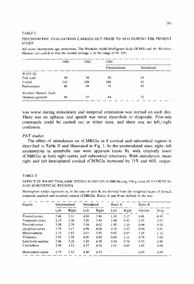
Neuropsychological evaluation
Verbal and performance IQ and memory function were decreased when com- pared to prior testing in 1980 and 1982. With stimulation during study 1, verbal IQ
decreased further, while performance IQ and memory quotient improved, the latter due largely to faster performance on the mental control subtest and improved visual reproduction. The results are summarized in Table I.
Differences between stimulated and unstimulated states were less significant on other tests. Visual reproduction memory was poor on day 1, but within normal limits on day 2 (both unstimulated days), and intermediate on day 2 (during stimulation). Performance on the visual organization test was uniformly poor, but
2x3
TABLE I
PSYCHOMETRIC EVALUATIONS CARRIED OUT PRIOR TO AND DURING; THE PRESENT
STUDY
All scores incorporate age corrections. The Wechsler Adult lntelligencc Scale (WAIS) and the Wcchslcr
Memory are scaled so that the normal average is in the range of 90-109.
1980 1982 19x5
Unstimulated Stimulated
WAIS-IQ Full scale 99 99 xx X3
Verbal 110 10X 101 x2
Performance 86 89 75 x5
Wec~ltsler Memq Sule
Memory quotient 96 97 64 73
was worse during stimulation and temporal orientation was normal on each day. There was no aphasia, and speech was never dysarthric or dysprodic. Five-step commands could be carried out in either state, and there was no left/right
confusion.
PET studies
The effect of stimulation on rCMRGlu in 8 cortical and subcortical regions is
described in Table II and illustrated in Fig. 1. In the unstimulated state, right-left
asymmetries in metabolic rate were apparent (ratio B), with relatively lower rCMRGlu in both right cortex and subcortical structures. With stimulation. mean right and left hemispheral cortical rCMRGlu increased by 71% and 60%, respec-
TABLE II
EFFECT OF RIGHT THALAMIC STIMULATION ON rCMRGlu (mg/lOO g/min) IN 8 CORTICAL
AND SUBCORTICAL REGIONS
Hemisphere values represent or, in the case of ratio B, are derived from the weighted means of frontal,
temporal, parietsl and occipital cortical rCMRGlu. Ratios A and B are defined in the text.
Region Unstimulated Stimulated Ratio A Ratio B
Frontal cortex
Temporal cortex
Parietal cortex
Occipital cortex
Hippocampus
Thalamus
Lentiform nucleus
Cerebellum
Left Right Left Right
2.46 2.11 4.08 3.88
2.i9 2.30 3.56 3.80
3.09 2.59 5.04 4.62
3.39 3.17 4.94 4.00
1.75 1.83 2.61 2.92
2.93 2.28 4.06 4.08
3.46 3.20 5.20 4.98
2.98 3.12 4.87 4.56
Left Right
1.10 1.17
1.04 0.91
1.05 1.10
0.76 0.37
0.x2 0.83
0.64 1.11
0.x4 0.7x
1.05 0.65
0.X6
1.05 0.x4
0.94
1.05
0.7x
0.92
1.05
Hemisphere mean 2.75 2.52 4.40 4.32 _ 0.93
Unstim Stim.
0.95
1.07
O.Y2
0.x1
1.12
1 .oo
0.96
0.94
0.99

Fig. 1. FDG/PET images of regional cerebral metabolic rate for glucose (rCMRGlu) in the unstimulated
(left tier) and stimulated (right tier) states. Images in the upper and lower rows correspond to PET brain
slices at the level of the thalami and the semioval centers. respectively. The global increase in ccrchral
metabolic rate for glucose associated with right thalamic stimulation becomes apparent H hen the Icft tier
images are compared with the co-planar images to the right. The right side of the scan is the paticnt‘h
right.
tively. Relative changes in metabolic rate with stimulation were evaluated by
indexing the change in rCMRGlu of specific structures to that of the ipsilateral hemisphere (ratio A). These changes were most marked in left thalamus, right
occipital cortex and right cerebellum. Ratio B values increased with stimulation, with the greatest change in thalamus, where stimulation reversed the relative hypometabolism of the right thalamus. Overall, ratio B values for mean hemisphere cortex increased from 0.93 to 0.99, indicating that stimulation nearly equalized the
metabolic rates of the 2 hemispheres.
Electrophysiological evaluation Neither routine EEG obtained on consecutive days of study 1 nor the 20-channel
EEG of study 2 changed with stimulation. All records were abnormal, with mild background slowing (posterior background 7.5-8 Hz) and increased frontal fast activity. SEPs were well formed, symmetric and all components were of normal

285
latency before and during DBS. BAEPs prior to stimulation were well formed from each ear and the peripheral and central transmission times were normal. There were no significant changes during stimulation. Prior to DBS, the VEPs were well formed and the major positivities had normal latency to full-field monocular stimulation. During DBS, the responses elicited to right eye stimulation were unchanged, while the major positivity of the left eye response broadened and increased in latency (from 109 to 117 msec) and the latency of the subsequent negativity increased as well.
DisCussion
The studies of DBS in this patient must be interpreted in light of abnormal baseline neurological function and a long history of substance and DBS abuse. Though the patient’s experience provides an opportunity to correlate clinical and neurophysiological responses and raises several issues relevant to the management of chronic pain with DBS, it may not be representative of routine DBS in an otherwise intact brain. Further study of the long-term effects of stimulation is needed to clarify the relationship between this extreme response and the usual pattern. It must also be noted that the precise localization of the electrode at the time of the studies is uncertain. Though initial placement in nVPL is likely given the contralateral paresthesias experienced by the patient after implantation, shifting of the electrode may have occurred; CT imaging at the time of the study suggested localization in the lateral thalamus, but could not confirm placement in nVPL. Even if the electrode was situated in nVPL, direct stimulation of outside structures is likely due to the high stimulation intensities used by the patient. This uncertainty emphasizes again the tentative relationship between our case and the usual results of DSB in nVPL. Finally, though fluctuations in plasma methadone level may also confound extrapolation of the current findings to other cases of DBS, these more likely represent unreported changes in the dose and interval before blood sampling rather than sustained differences in daily concentration and are therefore of limited importance. The patient was highly tolerant to opioid medications and the indepen- dence of stimulation effects and drug concentration is suggested by the stability of some neuropsychological measures with both increases and decreases in others, the hemispheric asymmetries on PET scanning, and the constant background EEGs during periods of differing plasma drug concentration.
The clinical features of this case illustrate common pitfalls in the therapy of chronic non-malignant pain. During 10 years of treatment, virtually every interven- tion, whether surgical or non-surgical, yielded only a brief respite from pain. Such aggressively help-seeking individuals whose prominent placebo response or complai- sance results in transient benefit with every trial are often encountered and may come to multiple ineffective procedures undertaken by a number of well-meaning physicians. The difficult clinical task is to identify this pattern after it has begun and intercede to alter it. This can often be assisted by referral to a multidisciplinary pain center, where decisions about additional invasive measures for pain are
evaluated from varying perspectives. Though this course may not provide pam relief, as demonstrated by this patient, it may prevent division of care, interfere in the cycle of useless procedures and change the focus of therapy from analgesia at al1 costs to improvement of function.
Of equal clinical import is the demonstration by this case of the potential for addiction to DBS. Though some degree of analgesia occurred, the patient developed aberrant, stimulation-seeking behaviors associated with compromise in function, a pattern which defines addiction to drugs. The factors responsible for these behaviors remain specufative and most likely comprise some combination of an idiosyncratic physiological response to stimulation and a personality prone to addiction. as suggested by the history of alcoholism and inapprop~ate use of analgesic medica- tions. The development of addiction to DBS must be viewed as a rare additional risk of the procedure with important therapeutic implications for the management of chronic pain. Treatment with non-pharmacologic, often invasive, measures is often undertaken with the desired goal of preventing drug addiction by eliminating the need for medication. This case, as well as others [6], suggests that addictive behaviors do not develop solely from properties inherent in the treatment, such as the capacity to produce physical dependence, but also from characteristics unique to the patient. DBS, therefore, should not be undertaken merely to avoid the risk of addiction.
Pleasurable sensations during stimulation provided the probable substrate for addiction in this patient. Reinforcing stimulation in subcortical structures has been extensively evaluated in animals [3,8,29,33,42]. In man, pleasurable sensations have been noted after stimulation in a wide variety of subcortical sites, though never in nVPL specifically ]9,11,39,41,46], and the potential for compulsive self-stimulation has been suggested both by requests for additional stimulation during electrode placement and by the rare reports of excessive stimulation after implantation [6,11,41]. Other stimulation sites have produced anxiety and related negative emo- tions, often in regions within 1 cm of sites eliciting pleasure [39,41]. These responses also have not been reported with nVPL stimulation. The mix of emotions reported by this patient may be due to activation outside of this region, as demonstrated by PET.
Thalamic damage or stimulation has been reported to produce a variety of neurobeha~oral syndromes. Both bilateral and u~ateral do~nant thalamic lesions can cause amnesia [4,10,12,50]. Stimulation of the dominant pulvinar can disrupt short-term memory [26], while activation of dominant and non-dominant ventro- lateral nucleus has been shown to influence short-term recall of verbal and visuospa- tial information respectively [23-251. Similarly, an aphasic syndrome can follow damage or stimulation in the dominant thalamus [28,35], while injury to the non-dominant thalamus can produce a unilateral neglect or hemispatial disorder [21,49]. In the present case, the long-standing non-verbal deficits may have resulted from either electrode implantation in right thalamus or chronic nVPL stimulation with secondary involvement of nearby thalamic structures. Acute stimulation af- fected memory, language and visuospatial function, with deterioration of verbal skills and improvement in visuospatial processing. This probably reflects reorganiza-
287
tion of activity in both thalami as well as the cortical regions with which they interact, a process that is suggested by the observed bilateral changes in rCMRG1t.t.
There has been no previous attempt to evaluate the effect of thalamic stimulation on rCMRGlu in man. As demonstrated in Table II, cortical and subcortical rCMRGlu were profoundly reduced in both hemispheres, especially the right, in the absence of self-stimulation. It is unlikely that methadone treatment per se could explain this degree of hypometabolism. The asymmetric thalamic hypometabolism is clearly abnormal [7] and suggests that electrode placement and/or chronic thalamic stimulation may have altered normal metabolic function. Global hypometabolism has been reported in patients with dementia [5] and in this patient may correlate with the deterioration in intellect revealed found by psychometric testing.
Animal studies, which utilized 2-deoxy-[14C]glucose and quantitative autoradi- ography, suggest that brain-stem stimulation produces discrete activation of specific brain regions and their projections [8,33]. The diffuse increases in rCMRGlu observed in the present study during thalamic stimulation, as well as the observed asymmetries, may relate to different electrode placement, higher relative amplitudes of stimulation, or species differences. Though an explanation for these patterns is lacking, they do suggest that correlation of stimulation site and clinical effects in man must be made cautiously, since metabolic activation of a distant brain region may in fact be responsible.
Such electrophysiological measures as EPs and EEG were, for the most part, unresponsive to the changes produced by nVPL stimulation. Though surprising in light of the dramatic clinical and metabolic effects of stimulation in this patient, the EEG has been reported to lack utility for electrode localization during DBS [44,46]. The dysfunction in the left anterior visual pathway demonstrated by VEPs during stimulation was clinically inapparent and, though difficult to explain on physiologic grounds, supports the impression that DBS may produce widespread unanticipated changes. The normal SEPs indicate that the portions of the somatosensory pathway essential to their generation were intact and that transmission through the nVPL of the large fiber volley elicited by median nerve stimulation was not substantially disrupted by DBS in this patient.
Though changes in blood pressure and respiratory rate, as well as other auto- nomic effects, have been observed after stimulation in a variety of cortical and subcortical sites [11,27,37,38,46], none has been reported with stimulation in nVPL specifically. The changes noted in this patient were consistent with enhanced sympathetic activity and could therefore be related either to generalized arousal or direct spread of stimulation to hypothalamic structures. Either explanation would account for the impressive increases in ACTH and cortisol accompanying stimu- lation. The decrements in GH and prolactin, however, more likely reflect a direct effect on hypothalamus. Hormonal levels, other than beta-endorphin [14,51], have not previously been evaluated in patients undergoing DBS. Stimulation of brain regions outside of nVPL accounts also for the motor effects observed during stimulation. Perhaps these responses were related to asymmetric activation of anterior thalamic structures, which both receive afferents from globus pallidus and project to motor cortex.

288
After multiple neuroablative and stimulatory procedures and a period of inap- propriate analgesic use, this patient developed aberrant behaviors consistent with addiction to DBS. A clinical and neurophysiological evaluation revealed both
widespread and selective effects of stimulation. The mode of action of DBS requires further elucidation and its clinical indications need to be refined. Longitudinal
studies using sensitive neuropsychological measures, PET technology and electro- physiologic techniques would be a fruitful undertaking in patients receiving DBS for
pain.
References
1
2
3
4
5
6
I
8
9
10
11
12
13
14
15
16
17
18
19
Basbaum, A.I. and Field, H.L., Endogenous pain control systems: brainstem spinal pathways and
endorphin circuitry, Ann. Rev. Neurosci., 7 (1984) 309-338.
Benton, A.L., Hamsher, K., Vamey, N.R. and Spreen, 0.. Contributions to Neuropsychological
Assessment, Oxford University Press, New York, 1983.
Bursten, B. and Delgado, J.M.R., Positive reinforcement by intracerebral stimulation in the monkey,
J. camp. physiol. Psychol., 51 (1958) 6-10.
Choi, D., Sudarsky, L., Schacter, S., Biber, M. and Burke, P., Medial thalamic hemorrhage with
amnesia, Arch. Neurol. (Chic.), 40 (1983) 611-613.
De Leon, M.J., Ferris, S.H., George, A.E. et al., Computed tomography and positron emission
transaxial tomography evaluations of normal aging and Alzheimer’s disease, J. Cereb. Blood Flow, 3
(1984) 391-394.
Dieckmann, G. and Witzmann, A., Initial and long-term results of deep brain stimulation for chronic
intractable pain, Appl. Neurophysiol., 45 (1982) 167-172.
Duara, R., Margolin, R., Robertson-Tchabo, E.A. et al., Cerebral glucose utilization as measured with
positron emission tomography in 21 resting healthy men between the ages of 21 and 83 years, Brain,
106 (1983) 761-775.
Esposito, R.U., Porrino, L.J., Seeger, T.F., Crane, A.M., Eve&, H.D. and Pert, A., Changes in local
cerebral glucose utilization during rewarding brain stimulation, Proc. nat. Acad. Sci. (Wash.), 81
(1984) 635-639.
Gol, A., Relief of pain by electrical stimulation of the septal area, J. neurol. Sci., 5 (1967) 115-120.
Goldenberg, G., Wimmer, A. and Maly, J., Amnesic syndrome with a unilateral thalamic lesion: a
case report, J. Neural., 229 (1983) 79-86.
Heath, R.G. and Mickle, W.A., Evaluation of seven years experience with depth electrode studies in
human patients. In: E.R. Ramey and D.S. O’Doherty (Eds.), Electrical Studies on the Unanesthetized
Brain, Hoeber, New York, 1960, pp. 214-247.
Horel, J.A., The neuroanatomy of amnesia, Brain, 101 (1978) 403-445.
Hosobuchi, Y., The current status of analgesic brain stimulation, Acta neurochir. (Wien), Suppl. 30
(1980) 219-227.
Hosobuchi, Y., Adams, J.E. and Lichnitz, R., Pain relief by electrical stimulation of the central gray
matter in humans and its reversal by naloxone, Science, 197 (1977) 183-186.
Hosobuchi, Y., Adams, J.E. and Rutkin, B., Chronic thalamic stimulation for the control of facial
anesthesia dolorosa, Arch. Neurol. (Chic.), 29 (1973) 158-161.
Huang, S.C.. Phelps, M.E., Hoffman, E.J. et al., Noninvasive determination of local cerebral
metabolic rate of glucose in man, Amer. J. Physiol., 238 (1980) E69-E82.
Kearfott, K.J. and Carroll, L.R., Evaluation of the performance characteristics of the PC 4600
positron emission tomograph, J. Comput. Assist. Tomogr., 8 (1984) 502-513.
Kearfott, K.M., Rottenberg, D.A. and Knowles, R.J.R., A new headholder for PET, CT and NMR
imaging, J. Comput. Assist. Tomogr., 8 (1984) 1217-1220. Mazars, G., Intermittent stimulation of nucleus ventralis posterolateralis for intractable pain, Surg.
Neurol., 4 (1975) 93-95.

289
20
21
22
23
24
25
26
27
28
Mazars, G., Merienne, L. and Cioloca, R., Comparative study of electrical stimulation of posterior
thalamic nuclei, periaqueductal gray and other midline mesencephalic structures in man. In: J.J.
Bonica, J.C. Liebeskind and D.G. Albe-Fessard @is.), Advances in Pain Research and Therapy, Vol.
3, Raven Press, New York, 1979, pp. 541-546.
Mesulam, M.M., A cortical network for directed attention and unilateral neglect, Ann. Neural., 10
(1981) 309-325.
Mundinger, F. and Neumiller, H., Programmed stimulation for control of chronic pain and motor
disease, Appl. Neurophysiol., 45 (1982) 102-111.
Ojemann, G.A., Language and thalamus: object naming and recall during and after thalamic
stimulation, Brain Lang., 2 (1975) 101-120.
Ojemann, G.A., Asymmetric function of the thalamus in man, Ann. N.Y. Acad. Sci., 299 (1977)
380-396.
Ojemann, G.A., Brain organi?ation for language from the perspective of electrical stimulation
mapping, Behav. Brain Sci., 2 (1983) 189-230.
Ojemann, G.A. and Fedio, P., Effect of stimulation of the human thalamus and parietal white matter
on short-term memory, J. Neurosurg., 29 (1968) 51-59.
Ojemann, G.A. and Van Buren, J.M., Respiratory, heart rate and GSR responses from human
diencephalon, Arch. Neural. (Chic.), 16 (1967) 74-88.
Ojemann, G.A. and Ward, A.A., Speech representation in ventrolateral thalamus, Brain, 94 (1971)
669-680.
29 Olds, J., Self-stimulation of the brain, Science, 127 (1958) 315-324.
30 Phelps, M.E., Huang, S.C., Hoffman, E.J. et al., Tomographic measurement of local cerebral glucose
metabolic rate in humans with [‘sF]2-fluoro-2-deoxy-D-glucose: validation of method, Ann. Neural.,
6 (1979) 371-388.
31 Plotkin, R., Results in 60 cases of deep brain stimulation for chronic intractable pain, Appl.
Neurophysiol., 45 (1982) 173-178.
32 Pool, J.L., Psychosurgery in elder people, J. Amer. geriat. Sot., 2 (1954) 456-465.
33 Porrino, L.J., Esposito, R.U., Seeger, T.F., Crane, A.M., Pert, A. and Sokoloff, L., Metabolic mapping
of the brain during rewarding self-stimulation, Science, 224 (1984) 306-309.
34 Ray, CD. and Burton, C.V., Deep brain stimulation for severe, chronic pain, Acta neurochir. (Wien),
Suppl. 3 (1980) 289-293.
35 Reynolds, A.F., Harris, A.B., Ojemann, G.A. and Turner, P.T., Aphasia and left thalamic hemor-
rhage, J. Neurosurg., 48 (1978) 570-574.
36 Reynolds, D.V., Surgery in the rat during electrical analgesia induced by focal brain stimulation,
Science, 164 (1969) 444-445.
37 Richardson, D.E. and Akil, H., Pain reduction by electrical brain stimulation in man, Part 1. Acute
administration in periaqueductal and periventricular sites, J. Neurosurg., 47 (1977) 178-183.
38 Richardson, D.E. and Akil, H., Pain reduction by electrical brain stimulation in man. Part 2. Chronic
self-stimulation in the periventricular gray matter, J. Neurosurg., 47 (1977) 184-194.
39 Schaltenbrand, G., Spuler, H., Wahren, W. and Wilhelmi, A., Vegetative and emotional reactions
during electrical stimulation of deep structures of the brain during stereotactic procedures, J. Neural.,
205 (1973) 91-113.
40 Schvarn, J.R., Chronic self-stimulation of medial posterior inferior thalamus for alleviation of
deafferented pain, Acta neurochir. (Wien), Suppl. 30 (1980) 295-301.
41 Sem-Jacobsen, C.W. and Torkildsen, A., Depth recording and electrical stimulation in the human
brain. In: E.R. Ramey and D.S. O’Doherty (I?&.), Electrical Studies in the Unanesthetized Brain,
Hoeber, New York, 1960, pp. 275-290.
42 Sidman, H., Brady, J.V., Boren, J.J. and Conrad, D.G., Reward schedules and behavior maintained
by intracerebral self-stimulation, Science, 122 (1955) 830-831.
43 Siegfried, J., Comte, P. and Meier, R., Monopolar electrical stimulation of nucleus ventroposterome-
dialis thalami for postherpetic facial pain, Appl. Neurophysiol., 45 (1982) 179-184.
44 Spiegel, E.A. and Wycis, H.T., Stereoencephalotomy. Part 2. Clinical and Physiological Applications,
Grune and Stratton, New York, 1962, pp. 160-171.
45 Tewson, T.J., Synthesis of non-carrier-added fluorine-18 2-fluoro2-deoxy-D-glucose, J. nucl. Med., 24
(1983) 718-721.
46 Tasker, R.R., Organ, L.W. and Hawrylyshyn. P.A., The Thalamus and Midbrain of Man. Thomas.
Springfield. IL, 1982, pp. 62-108. 332-344.
47 Tsubokawa, T., Yammato. T. and Katayama, Y. et al., Clinical results and physiological basis of
thalamic relay nucleus stimulation for relief of intractable pain with morphine tolerance, Appl.
Neurophysiol., 45 (1982) 143-155.
48 Turnbull, I.M., Shulman, R. and Woodhurst, W.B.. Thalamic stimulation for neuropathic pain, J.
Neurosurg.. 52 (1980) 486-493.
49 Watson, R.T. and Heilman, K.M., Thalamic neglect, Arch. Neurol. (Chic.), 38 (1981) 501-506.
50 Williams, M. and Pennybacker, J., Memory disturbances in third ventricle tumors, J. Neurosurg.
Psychiat.. 17 (1954) 115-123.
51 Young, R.F., Feldman, R.A., Kroening, R., Fulton, W. and Morris, J., Electrical stimulation of the
brain in the treatment of chronic pain in man. In: L. Kruger and J.C. Liebeskind (Eds.), Advances in
Pain Research and Therapy, Vol. 6, Raven Press, New York, 1984, pp. 289-303.
52 Young, R.F.. Kroening, R., Fulton, W., Feldman, R.A. and Chambi, I., Electrical stimulation of the
brain in treatment of chronic pain, J. Neurosurg., 62 (1985) 389-396.