Embed Size (px)
Citation preview

Page 1 of 12(page number not for citation purposes)
Available online http://ccforum.com/content/11/5/230
AbstractProgress in management of critically ill neurological patients hasled to improved survival rates. However, severe residual neuro-logical impairment, such as persistent coma, occurs in somesurvivors. This raises concerns about whether it is ethically appro-priate to apply aggressive care routinely, which is also associatedwith burdensome long-term management costs. Adapting themanagement approach based on long-term neurological prognosisrepresents a major challenge to intensive care. Magneticresonance imaging (MRI) can show brain lesions that are notvisible by computed tomography, including early cytotoxic oedemaafter ischaemic stroke, diffuse axonal injury after traumatic braininjury and cortical laminar necrosis after cardiac arrest. Thus, MRIincreases the accuracy of neurological diagnosis in critically illpatients. In addition, there is some evidence that MRI may havepotential in terms of predicting outcome. Following a briefdescription of the sequences used, this review focuses on theprognostic value of MRI in patients with traumatic brain injury,anoxic/hypoxic encephalopathy and stroke. Finally, the roles playedby the main anatomical structures involved in arousal and aware-ness are discussed and avenues for future research suggested.
IntroductionSevere brain impairment, most notably persistent coma, mayfollow traumatic brain injury (TBI), anoxic/hypoxic encephalo-pathy, or stroke. Although progress in the management ofcritically ill neurological patients has led to improved survivalrates [1], some survivors remain in a persistent vegetative orminimally conscious state. Up to 14% of patients with TBIremain in a persistent vegetative state after 1 year [2-4], and
their medical cost has been estimated at US$1 to 7 billionper year in the USA [5]. The possibility that aggressivemedical management may lead to survival with severe brainimpairment raises ethical issues. Adapting the level of medicalcare to long-term neurological prognosis is a major challengefor neurological intensive care. The first step in meeting thischallenge is validation of tools that accurately predict long-term neurological outcome after severe cerebral insult.
Magnetic resonance imaging (MRI) is more sensitive thancomputed tomography at detecting stroke in the early phase,subtle abnormalities related to anoxic/hypoxic encephalo-pathy, and diffuse axonal injury (DAI) in patients with TBI. MRIprovides valuable diagnostic information, although it iscumbersome to perform in the acute phase in comatosepatients who are undergoing mechanical ventilation. SeveralMRI sequences and techniques have been used to explorethe structures, metabolism and functions of the brain. Thedata supplied by these methods could be used to predictlong-term neurological outcome.
In this review we briefly describe the MRI sequences andtechniques used in critically ill neurological patients, and thenwe discuss their prognostic value in comatose patients withTBI, anoxic/hypoxic encephalopathy, or stroke. Finally, wediscuss the prognostic influences of the main anatomicalstructures that are involved in arousal and awareness, and wesuggest avenues for future research.
ReviewClinical review: Prognostic value of magnetic resonance imagingin acute brain injury and comaNicolas Weiss1, Damien Galanaud2, Alexandre Carpentier3, Lionel Naccache4
and Louis Puybasset1
1Department of Anesthesiology and Critical Care, Pitié-Salpêtrière Teaching Hospital, Assistance Publique - Hopitaux de Paris and Pierre et MarieCurie University, Bd de l’hôpital, 75013, Paris, France2Department of Neuroradiology, Pitié-Salpêtrière Teaching Hospital, Assistance Publique - Hopitaux de Paris and Pierre et Marie Curie University, Bd de l’hôpital, 75013, Paris, France3Department of Neurosurgery, Pitié-Salpêtrière Teaching Hospital, Assistance Publique - Hopitaux de Paris and Pierre et Marie Curie University, Bd de l’hôpital, 75013, Paris, France4Department of Neurophysiology, Pitié-Salpêtrière Teaching Hospital, Assistance Publique - Hopitaux de Paris and Pierre et Marie Curie University, Bd de l’hôpital, 75013, Paris, France
Corresponding author: Louis Puybasset, [email protected]
Published: 18 October 2007 Critical Care 2007, 11:230 (doi:10.1186/cc6107)This article is online at http://ccforum.com/content/11/5/230© 2007 BioMed Central Ltd
ADC = apparent diffusion coefficient; ARAS = ascending reticular activating system; DAI = diffuse axonal injury; DTI = diffusion tensor imaging;DWI = diffusion weighted imaging; FLAIR = fluid-attenuated inversion recovery; GOS = Glasgow Outcome Scale; MRI = magnetic resonanceimaging; MRS = magnetic resonance spectroscopy; NAA = N-acetyl-aspartate; TBI = traumatic brain injury.

Page 2 of 12(page number not for citation purposes)
Critical Care Vol 11 No 5 Weiss et al.
Magnetic resonance imaging sequences andtechniquesConventional magnetic resonance imagingConventional MRI relies chiefly on four sequences [6]. Fluid-attenuated inversion recovery (FLAIR) is the primarysequence used in neuroradiology (Figure 1). It detects braincontusion, brain oedema and subarachnoid or intraventricularhaemorrhage, as well as the resulting ventricular dilatation orherniation. The T2*-weighted sequence is more sensitive tointraparenchymal blood than is FLAIR. This sequence canalso reveal haemorrhagic DAI [7,8]. The T2-weightedsequence completes the FLAIR sequence and providesgreater detail on brainstem and central grey matter. Finally,diffusion weighted imaging (DWI) is sensitive to randommovement of water molecules. This sequence shows cerebraloedema and distinguishes cytotoxic from vasogenic oedema.It is used chiefly in patients with ischaemic stroke.
Conventional MRI provides an initial evaluation of brainlesions. However, when it is used alone it fails to predictoutcome accurately.
Magnetic resonance spectroscopyThis sequence is a noninvasive technique for assessing brainmetabolism in vivo. Proton-magnetic resonance spectro-scopy (MRS) is most commonly used. Four main markers arestudied: the peak of N-acetyl-aspartate (NAA), an amino acidpresent in neurones, which reflects the status of neuronaltissue; creatine, found in glia and neurones, which serves asa point of reference because its level is believed to be stable;choline, a constitutive component of cell membranes, whichreflects glial proliferation or membrane breakdown [9]; andlactate, a marker of anaerobic metabolism and therefore ofischaemia [10]. As shown in Figure 2, three main ponsmonovoxel profiles may be observed in patients with TBI.
Diffusion tensor magnetic resonance imagingDiffusion tensor imaging (DTI), derived from DWI, measuresthe degree and direction of water diffusion (anisotropy).Water diffusion anisotropy reflects the integrity of whitematter tracts. Pathophysiological mechanisms that can alterwater diffusion anisotropy include DAI, effects of intracranialhypertension and disconnection of white matter tracts.
Magnetization transfer imagingThis sequence is based on the principle that structure-boundprotons undergo T1 relaxation coupling with protons in theaqueous phase. Saturated protons in macromoleculesexchange longitudinal magnetization with protons in theaqueous phase, leading to a reduction in signal intensity.Magnetization transfer imaging has been found to besensitive for detecting white matter lesions in severalneurological conditions [11,12].
Functional magnetic resonance imagingFunctional MRI may reveal foci of cerebral dysfunction inregions that look structurally intact on conventional MRI.Imaging is based on changes in the oxidative state ofhaemoglobin, which reflects regional brain activation.Functional MRI remains difficult to perform in critically illunstable patients and, consequently, few teams haveacquired the equipment and experience necessary to applythis technique [13]. The few available studies conducted incomatose patients with TBI showed a correlation betweenprefrontal/cingulated cortical activation disturbation andcognitive impairments [14,15]. However, functional MRI wasperformed in these studies at a distance from the injury.
Magnetic resonance imaging findings inspecific critical neurological conditionsTraumatic brain injuryConventional magnetic resonance imagingMRI was first used to investigate patients with TBI in a 1986study of 50 patients [16]. The three main findings, which havesince been confirmed, were as follows: MRI identified lesionsmore frequently than did computed tomography; brain lesionswere common after TBI; and although patients who regainedconsciousness rapidly had no lesions in fundamental deep
Figure 1
FLAIR and T2* sequences in a patient with an arteriovenousmalformation. (a) Axial fluid-attenuated inversion recovery (FLAIR)sequence showing hypersignal in the left temporal lobe. (b) Axial T2*sequence showing mild hyposignal in the same area suggestive ofbleeding. (c) Different section of the axial FLAIR sequence showinghypersignal surrounded by hyposignal. Bleeding cannot be confirmed.(d) Axial T2* sequence clearly showing hyposignal lateral to the leftputamen. The patient has bleeding from the arteriovenousmalformation.

Page 3 of 12(page number not for citation purposes)
brain structures, some of them had severe cortical lesions.Several descriptions of MRI lesions in TBI patients have beenreported since that initial study was published (Table 1)[17-21], although few of them focused on the prognosticvalue of MRI [17-20]. Conventional MRI findings that stronglypredicted outcome included DAI, total lesion burden and DAIin the brainstem.
DAI is the most common primary lesion in TBI patients [22,23]and may be the most common cause of poor outcome [22-24].DAI may be ischaemic or haemorrhagic [7,8]. Ischaemic DAI isseen as a hypersignal on DWI or FLAIR, with no abnormality onthe T2* sequence [25]. The hypersignal with DWI disappearswithin about 2 weeks. Conversely, haemorrhagic DAI appearsas a hyposignal on the T2* sequence, with normal DWIfindings. It has been proposed [22] that DAI location could beclassified into the following stages: stage 1, frontal andtemporal white matter; stage 2, lobar white matter andposterior part of corpus callosum; and stage 3, dorsolateralmidbrain and pons. With outcomes defined as GlasgowOutcome Scale [26] scores of 2 to 3 versus 4 to 5, none of the33 patients with good outcome in another study [27] hadhaemorrhagic DAI (Table 1). DAI appears to be a majordeterminant of poor outcomes, although its use as an outcomepredictor in the individual patient remains difficult. Whether thecorrelation between DAI and outcome is due to the total lesionburden or to DAI location remains debated.
In several prospective studies, lesion burden was associatedwith outcome irrespective of DAI location (Table 1)[17,19,28]. Among 40 prospectively enrolled patients withsevere TBI, lesions by FLAIR and T2*-weighted sequencesincreased progressively with GOS score groups 1 to 2, 3,and 4 to 5 [17]. Similar results were obtained in a studycomparing 42 patients with persistent vegetative state with38 patients who recovered consciousness [19].
A number of studies have focused on the value of DAIlocation in predicting outcome [19,29-31]. Brainstem lesions
in the pons and mesencephalon appear to be the mostpotent markers of poor prognosis, most notably when theyare bilateral and symmetrical [18,19,29,31]. In a prospectivestudy conducted in 61 patients (Table 1) who were studiedwithin 7 days of TBI [18], all patients with bilateral pontinelesions died as compared with 9% of patients with nobrainstem lesions. These results were confirmed by the samegroup in a prospective study of 102 comatose patients [29]using the following four-stage grading system: grade I,lesions of the hemispheres only; grade II, unilateral lesions ofthe brainstem at any level with or without supratentoriallesions; grade III, bilateral lesions of the mesencephalon withor without supratentorial lesions; and grade IV, bilaterallesions of the pons with or without any of the lesions of lessergrades. Mortality increased gradually from 14% with grade Ilesions to 100% with grade IV lesions. These findings werecorroborated by two independent studies [19,31] (Table 1).We recently confirmed the prognostic value of brainstemlesions in the upper pons and lower midbrain in a study of 73patients [32]. Bilateral pontine lesions carry a high mortalityrate and predict poor neurological outcomes.
Three studies showed that corpus callosum lesions wereassociated with poor outcomes [19,30,31] (Table 1). How-ever, these lesions may merely represent markers for severeinitial injury. In addition to lesion burden, both total lesionvolume and frontal lobe lesion volume on FLAIR imagescorrelated significantly with clinical outcomes [30]. Never-theless, evaluating DAI lesion volume is difficult (most notablywhen the lesions are small), time consuming, cumbersomeand subject to inter-rater variability.
The presence of severe DAI and a heavy lesion burden areassociated with permanent neurological impairment.However, these factors are difficult to use in the individualpatient, especially to distinguish GOS score 2 from GOSscore 3. In TBI patients, brainstem lesions are easily identifiedby MRI. In our experience, they are associated with pooroutcomes, most notably when they are posterior and bilateral.
Available online http://ccforum.com/content/11/5/230
Figure 2
Magnetic resonance spectroscopy profile of the pons after traumatic brain injury. (a) Normal profile. The peak of N-acetyl-aspartate (NAA) is higherthan the peaks of choline (Cho) and creatine (Cr). (b) Neuronal loss profile. The NAA peak is decreased, nearly to the level of the Cr peak. TheNAA/Cr ratio is lower than in panel a. (c) Gliosis profile: increased Cho peak with no change in the Cr or NAA peak. Adapted from [17].

Critical Care Vol 11 No 5 Weiss et al.
Page 4 of 12(page number not for citation purposes)
Tab
le 1
Co
nve
nti
on
al m
agn
etic
res
on
ance
in t
rau
mat
ic b
rain
inju
ry
Aut
hors
(ref
.)
Kam
pfl,
1998
Firs
chin
g, 1
998
Pie
ralli
ni, 2
000
Yan
agaw
a, 2
000
Pat
erak
is, 2
000
Firs
chin
g, 2
001
Firs
chin
g, 2
002
Wed
ekin
d, 2
002
Car
pent
ier,
2006
[19]
[18]
[30]
[28]
[27]
[29]
[95]
[31]
[17]
Stu
dy d
esig
nC
ase-
cont
rol
Pro
spec
tive
Pro
spec
tive
Pro
spec
tive
Pro
spec
tive
Pro
spec
tive
Pro
spec
tive
Ret
rosp
ectiv
eP
rosp
ectiv
e
Seq
uenc
esT1
, T2
T1, T
2T1
, T2,
FLA
IRT2
, T2*
T1, T
2T1
, T2
T1, T
2T1
, T2,
T2*
MR
S, T
2, T
2*
Incl
usio
n V
S b
etw
een
Adm
issi
on in
G
CS
sco
re
Aliv
e af
ter
Dis
crep
ancy
A
dmis
sion
in
GC
S s
core
G
CS
sco
re
Sev
ere
TBI
crite
ria6
and
8 w
eeks
com
a (d
urat
ion
<8,
com
a 1
wee
kbe
twee
n C
T co
ma
(dur
atio
n <
8<
8>
24 h
ours
) >
1 w
eek,
pos
t-sc
an a
nd
>24
hou
rs)
trau
mat
ic a
mne
sia
neur
olog
ical
>
4 w
eeks
stat
us
Num
ber o
f 80
6137
3433
102
100
40a
40pa
tient
s
Del
ay to
MR
I6
to 8
wee
ks<
7 da
ys60
to 9
0 da
ys<
3 w
eeks
<48
hou
rs<
8 da
ys<
7 da
ys1
to 3
9 da
ys17
.5 ±
6.4
Out
com
e G
OS
sco
re
Mor
talit
yC
linic
al
GO
S s
core
at
GO
S s
core
M
orta
lity
and
Mor
talit
y at
G
OS
sco
re,
GO
S s
core
va
riabl
e of
(2
ver
sus
3-5)
as
sess
men
t at
3 m
onth
s(2
-3 v
ersu
s 4-
5)
outc
ome
at
6 m
onth
sD
RS
>6
mon
ths
(1-2
ver
sus
4-5)
in
tere
stat
2, 3
, 6, 9
and
3,
6 a
nd
at 6
mon
ths
3 m
onth
s to
(m
ean
dela
y:
and
DR
S a
t 12
mon
ths
12 m
onth
s3
year
sb11
.3 m
onth
s)18
mon
ths
Mai
n re
sults
Inde
pend
ent
Bra
inst
em le
sion
s: V
olum
e of
FLA
IR
Num
ber o
f T2
DA
I sta
ges
Bila
tera
l pon
s B
ilate
ral u
pper
M
ore
lesi
ons
of
Tota
l bur
den
of
fact
or o
f poo
r m
orta
lity
rate
of
corp
us c
allo
sum
le
sion
s co
rrel
ated
co
rrel
ated
with
le
sion
s: m
orta
lity
pont
ine
lesi
on
corp
us c
allo
sum
, FL
AIR
and
T2*
ou
tcom
e on
44
%. B
ilate
ral
lesi
ons
corr
elat
ed
with
GO
S s
core
. ou
tcom
e. N
o ra
te o
f 100
%.
pred
icts
mor
talit
yba
sal g
angl
ia a
nd
lesi
ons
corr
elat
ed
mul
tivar
iate
br
ains
tem
lesi
ons:
with
firs
t clin
ical
N
umbe
r of T
2*
patie
nt w
ith g
ood
Out
com
e (p
ara-
)hip
po-
with
DR
S a
nd G
OS
anal
ysis
. m
orta
lity
rate
of
eval
uatio
n. V
olum
e le
sion
s co
rrel
ated
ou
tcom
e ha
d co
rrel
ated
with
ca
mpa
l les
ions
in
scor
eC
orpu
s ca
llosu
m:
100%
of F
LAIR
fron
tal
with
GO
S s
core
haem
orrh
agic
DA
Ipr
esen
ce/a
bsen
ce
patie
nts
with
O
R 2
13.8
(95%
lo
be le
sion
an
d un
ilate
ral/
brai
nste
m le
sion
sC
I 14.
2 to
co
rrel
ated
with
bi
late
ral b
rain
stem
32
13.3
). cl
inic
al o
utco
me
lesi
ons
Bra
inst
em le
sion
s at
1 y
ear
OR
6.9
(95%
CI
1.1
to 4
2.9)
a Tw
enty
pat
ient
s w
ith b
rain
stem
lesi
ons
wer
e m
atch
ed to
20
patie
nts
with
out b
rain
stem
lesi
ons.
bA
t las
t exa
min
atio
n. C
I, co
nfid
ence
inte
rval
; DA
I, di
ffuse
axo
nal i
njur
y; D
RS
, dis
abili
ty ra
ting
scal
e; F
LAIR
, flu
id-a
ttenu
ated
inve
rsio
n re
cove
ry; G
CS
, Gla
sgow
Com
a S
cale
; GO
S, G
lasg
ow O
utco
me
Sca
le; M
RI,
mag
netic
reso
nanc
e im
agin
g; M
RS
, mag
netic
reso
nanc
e sp
ectr
osco
py;
NA
, not
app
licab
le; O
R, o
dds
ratio
; T2*
, T2*
wei
ghte
d se
quen
ce; T
BI,
trau
mat
ic b
rain
inju
ry; V
S, v
eget
ativ
e st
ate.

Posterior brainstem lesions in the periaqueductal grey matterare probably more relevant than anterior brainstem lesions aspredictors of poor outcomes in patients with brainstem stroke[21] or TBI [19]. In clinical practice, treatment limitation maydeserve consideration in patients who have large bilaterallesions in the posterior part of the pons after TBI.
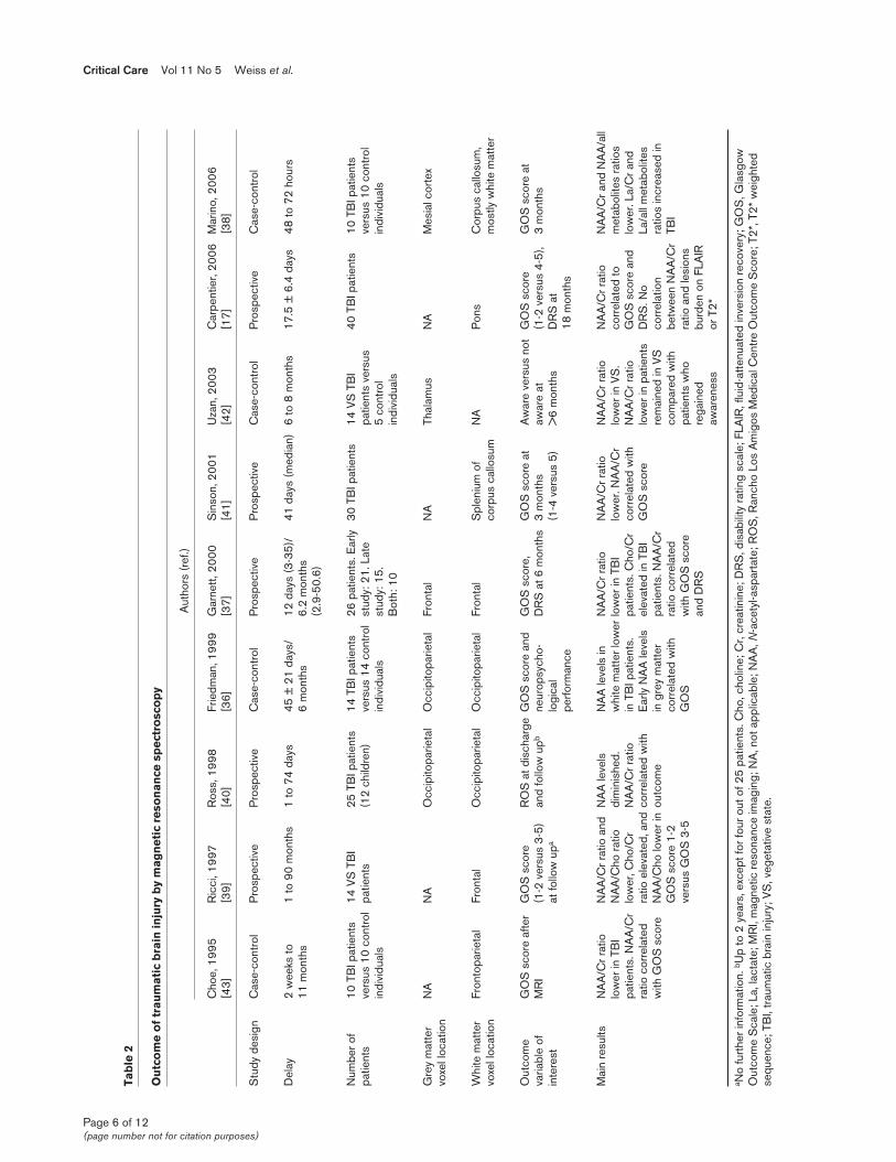
Magnetic resonance spectroscopySeveral MRS studies have been conducted in TBI patients(Table 2). Some of them were purely descriptive [33], othersassessed only the neuropsychological outcomes [34,35], andyet others focused on global outcome as evaluated using theGOS or Disability Rating Scale [17,36-42].
Compared with control individuals, TBI patients exhibiteddecreased NAA levels, decreased NAA/creatine ratios andincreased choline levels (Table 2) in all brain regionsevaluated [35-39,41,42]. Increased lactate levels wereseldom found in TBI patients, contrary to patients with otherbrain injuries [38]. The NAA/creatine ratio appeared to be thebest outcome predictor. Low NAA/creatine values correlatedwith poor outcomes when they were located in the frontal[37,39], frontoparietal [43], or occipitoparietal lobes [36,40];the splenium of the corpus callosum [41]; the thalami [42];the pons [17]; or a voxel including the corpus callosum, thewhite matter, and part of the hemispheric cortex [38].
These studies are heterogeneous (Table 2) in terms of patientselection, time from TBI to MRS, voxel location, method ofoutcome assessment and timing of outcome assessment. Forinstance, among studies of patients with TBI, one includedonly patients in a vegetative state [42], another includedpatients with severe TBI [17] and a third excluded patientswith early initial coma [36]. These differences in patientselection may be associated with differences in severity ofbrain oedema and in associated hypoxia and herniation,thereby introducing bias into the interpretation of the results.MRS findings vary greatly according to time since TBI. Fourphases may be distinguished: an acute phase, which lasts24 hours after TBI; an early subacute phase, which spansfrom the days 1 to 13; a late subacute phase, from days 14 to20; and a chronic phase, which starts on day 21. Only twostudies included patients at the acute phase [38,40], andonly one of these included all patients before 72 hours [38].Two studies were conducted from the early subacute phaseto the first month [17,37] and one began inclusion in the latesubacute phase but included patients up to 11 months afterTBI [43]. Four studies focused on the chronic phase; in twoof these studies, patients were included 3 weeks to 6 monthsafter TBI [36,39] and in the other two studies they wereincluded 2 months to 8 months after TBI [39,42].
Although NAA/creatine ratios were similar across studies, theresults should be interpreted with caution because experi-mental in vitro and in vivo data suggest differences in theunderlying pathophysiological mechanisms and in the time
course of the lesions [44-46]. To interpret these results reliably,information on NAA values over time are needed. Experimentsconducted in vitro [44] and in vivo [45,46] show an early NAAdecrease starting within a few minutes after TBI and reachingthe trough value within 48 hours. This finding explains whyspectroscopic disturbances may require 48 hours forvisualization [47]. NAA levels remain stable within the firstmonth after TBI, supporting the validity of MRS assessmentduring the second or third week [48,49]. Later on, between6 weeks and 1 year after TBI, NAA levels may decrease [9,37].Partial recovery of NAA levels has been suggested and mayindicate recovery of mitochondrial function [41].
Another important factor that varied across studies was MRSvoxel location (Table 2). Voxels were located in the hemi-sphere (the occipitoparietal, frontoparietal, or frontal lobes),corpus callosum, thalamus, or brainstem (the pons). Becausewhole brain analysis is time consuming, voxels are typicallyrestricted to the areas most affected by DAI, namely the lobarwhite matter, corpus callosum and upper brainstem [50].Estimation of NAA in the whole brain may improve theprognostic value of MRS [41]. A good compromise may be avoxel encompassing the corpus callosum, white matter andpart of the hemispheric cortex [38].
Studies also differed in their definitions of poor and goodGOS outcome groups: comparisons involved GOS score 1to 2 versus GOS score 3 to 5 [39], GOS score 1 to 4 versusGOS score 5 [41], or GOS score 1 to 2 versus GOS score4 to 5 [17]. Finally, the time from TBI to outcome assessmentvaried from 3 to 18 months (Table 2), further complicatingcomparisons because neurological status may improve for upto 1 year after TBI.
Although MRS has superseded conventional MRI, the combi-nation of these two techniques may be useful [17]. Variationsin the NAA/creatine ratio over time have not been studied in alarge TBI patient population. The above-mentioned variabilityin NAA levels constitutes the main limitation of this technique.To overcome this limitation, repeated studies at intervals of 1to 2 weeks are probably needed. In our experience, variationsin the NAA/creatine ratio are minimal in many patients. Weagree with Sinson and coworkers [41] that whole brain NAAestimation might improve the prognostic value of MRS.Absence of dysfunction by MRS is a valuable finding; in apatient with normal results by both conventional MRI andMRS, a poor outcome is unlikely. However, we have seen afew patients with normal conventional MRI and MRS findingswho had poor outcomes, probably related to white matterdamage detected as DTI abnormalities.
Diffusion tensor magnetic resonance imagingInitial reports of DTI in TBI patients suggest that thistechnique may demonstrate alterations in white matterconnections that are missed by conventional MRI [51]. DTIprovides information on the physiological status of fibre
Available online http://ccforum.com/content/11/5/230
Page 5 of 12(page number not for citation purposes)
Critical Care Vol 11 No 5 Weiss et al.
Page 6 of 12(page number not for citation purposes)
Tab
le 2
Ou
tco
me
of
trau
mat
ic b
rain
inju
ry b
y m
agn
etic
res
on
ance
sp
ectr
osc
op
y
Aut
hors
(ref
.)
Cho
e, 1
995
Ric
ci, 1
997
Ros
s, 1
998
Frie
dman
, 199
9 G
arne
tt, 2
000
Sin
son,
200
1 U
zan,
200
3 C
arpe
ntie
r, 20
06
Mar
ino,
200
6 [4
3][3
9][4
0][3
6][3
7][4
1][4
2][1
7][3
8]
Stu
dy d
esig
nC
ase-
cont
rol
Pro
spec
tive
Pro
spec
tive
Cas
e-co
ntro
lP
rosp
ectiv
eP
rosp
ectiv
eC
ase-
cont
rol
Pro
spec
tive
Cas
e-co
ntro
l
Del
ay2
wee
ks to
1
to 9
0 m
onth
s1
to 7
4 da
ys45
± 2
1 da
ys/
12 d
ays
(3-3
5)/
41 d
ays
(med
ian)
6 to
8 m
onth
s17
.5 ±
6.4
day
s48
to 7
2 ho
urs
11 m
onth
s6
mon
ths
6.2
mon
ths
(2.9
-50.
6)
Num
ber o
f 10
TB
I pat
ient
s 14
VS
TB
I 25
TB
I pat
ient
s 14
TB
I pat
ient
s 26
pat
ient
s. E
arly
30
TB
I pat
ient
s14
VS
TB
I 40
TB
I pat
ient
s10
TB
I pat
ient
s pa
tient
sve
rsus
10
cont
rol
patie
nts
(12
child
ren)
vers
us 1
4 co
ntro
l st
udy:
21.
Lat
e pa
tient
s ve
rsus
ve
rsus
10
cont
rol
indi
vidu
als
indi
vidu
als
stud
y: 1
5.
5 co
ntro
l in
divi
dual
sB
oth:
10
indi
vidu
als
Gre
y m
atte
r N
AN
AO
ccip
itopa
rieta
lO
ccip
itopa
rieta
lFr
onta
lN
ATh
alam
usN
AM
esia
l cor
tex
voxe
l loc
atio
n
Whi
te m
atte
r Fr
onto
parie
tal
Fron
tal
Occ
ipito
parie
tal
Occ
ipito
parie
tal
Fron
tal
Spl
eniu
m o
f N
AP
ons
Cor
pus
callo
sum
, vo
xel l
ocat
ion
corp
us c
allo
sum
mos
tly w
hite
mat
ter
Out
com
e G
OS
sco
re a
fter
GO
S s
core
R
OS
at d
isch
arge
GO
S s
core
and
G
OS
sco
re,
GO
S s
core
at
Aw
are
vers
us n
ot
GO
S s
core
G
OS
sco
re a
t va
riabl
e of
M
RI
(1-2
ver
sus
3-5)
an
d fo
llow
upb
neur
opsy
cho-
DR
S a
t 6 m
onth
s3
mon
ths
awar
e at
(1
-2 v
ersu
s 4-
5),
3 m
onth
sin
tere
stat
follo
w u
palo
gica
l (1
-4 v
ersu
s 5)
>6
mon
ths
DR
S a
t pe
rform
ance
18 m
onth
s
Mai
n re
sults
NA
A/C
r rat
io
NA
A/C
r rat
io a
nd
NA
A le
vels
N
AA
leve
ls in
N
AA
/Cr r
atio
N
AA
/Cr r
atio
N
AA
/Cr r
atio
N
AA
/Cr r
atio
N
AA
/Cr a
nd N
AA
/all
low
er in
TB
I N
AA
/Cho
ratio
di
min
ishe
d.
whi
te m
atte
r low
er lo
wer
in T
BI
low
er. N
AA
/Cr
low
er in
VS
. co
rrel
ated
to
met
abol
ites
ratio
s pa
tient
s. N
AA
/Cr
low
er, C
ho/C
r N
AA
/Cr r
atio
in
TB
I pat
ient
s.
patie
nts.
Cho
/Cr
corr
elat
ed w
ith
NA
A/C
r rat
io
GO
S s
core
and
lo
wer
. La/
Cr a
nd
ratio
cor
rela
ted
ratio
ele
vate
d, a
nd c
orre
late
d w
ith
Ear
ly N
AA
leve
ls
elev
ated
in T
BI
GO
S s
core
low
er in
pat
ient
s D
RS
. No
La/a
ll m
etab
olite
s w
ith G
OS
sco
reN
AA
/Cho
low
er in
out
com
ein
gre
y m
atte
r pa
tient
s. N
AA
/Cr
rem
aine
d in
VS
co
rrel
atio
n ra
tios
incr
ease
d in
G
OS
sco
re 1
-2
corr
elat
ed w
ith
ratio
cor
rela
ted
com
pare
d w
ith
betw
een
NA
A/C
r TB
Ive
rsus
GO
S 3
-5
GO
Sw
ith G
OS
sco
re
patie
nts
who
ra
tio a
nd le
sion
s an
d D
RS
rega
ined
bu
rden
on
FLA
IR
awar
enes
sor
T2*
a No
furt
her i
nfor
mat
ion.
bU
p to
2 y
ears
, exc
ept f
or fo
ur o
ut o
f 25
patie
nts.
Cho
, cho
line;
Cr,
crea
tinin
e; D
RS
, dis
abili
ty ra
ting
scal
e; F
LAIR
, flu
id-a
ttenu
ated
inve
rsio
n re
cove
ry; G
OS
, Gla
sgow
Out
com
e S
cale
; La,
lact
ate;
MR
I, m
agne
tic re
sona
nce
imag
ing;
NA
, not
app
licab
le; N
AA
, N-a
cety
l-asp
arta
te; R
OS
, Ran
cho
Los
Am
igos
Med
ical
Cen
tre
Out
com
e S
core
; T2*
, T2*
wei
ghte
dse
quen
ce; T
BI,
trau
mat
ic b
rain
inju
ry; V
S, v
eget
ativ
e st
ate.

bundles, thus complementing the metabolic and biochemicalinformation supplied by MRS. At present, little is known aboutthe prognostic value of DTI in patients with TBI. DTI findingscorrelated with clinical status in patients with multiplesclerosis or neurodegenerative disease [52,53]. In a mousemodel of TBI, DTI parameters were significantly reduced inthe injured brain, whereas conventional MRI showed nosignificant changes [54]. Furthermore, changes in relativeanisotropy correlated significantly with the density of stainedaxons on histological sections.
In a study comparing 20 TBI patients and 15 healthy controlindividuals, fractional anisotropy was reduced in the internalcapsule and splenium of the corpus callosum and correlatedwith Glasgow Coma Scale score and Rankin score atdischarge in the TBI patients [55]. Similar findings have beenreported in children [56]. Anecdotal case reports of DTIabnormalities in TBI patients have been reported [57,58]. Intwo patients who recovered partially after 6 years and19 years, respectively, in a minimally conscious state, DTIdisclosed increased anisotropy within the midline cerebellarwhite matter over an 18-month period [59]. This anisotropyincrease correlated with an increase in resting metabolism,measured using positron emission tomography, whichsuggests that axonal regrowth might underlie increases inanisotropy. Larger studies of DTI variations over time areneeded. In our institution, comatose patients have beenincluded in a prospective DTI study for the past 3 years.Patients with major connectivity abnormalities in bothhemispheres and the brainstem were at increased risk forpoor outcomes. A large multicentre prospective study isongoing in France to assess the usefulness of combining DTIwith MRS.
Magnetization transfer imagingMagnetization transfer imaging is sensitive for detecting whitematter lesions in patients with multiple sclerosis, progressivemultifocal leukoencephalopathy, or wallerian degeneration[11,12]. Preliminary results in TBI are promising [60,61]. Themagnetization transfer ratio was decreased in TBI patients[60,61]. Out of 28 TBI patients, eight had abnormalmagnetization transfer ratios, and all eight had persistentneurological deficits [62]. In another study, however, nocorrelation was found between GOS score and abnormalmagnetization transfer ratio [41].
Anoxic/hypoxic encephalopathyAnoxic/hypoxic encephalopathy is a devastating condition; itsdevelopment after prolonged cerebral hypoxia is often difficultto predict on clinical grounds. No controlled studies ofroutine MRI in large numbers of cardiac arrest patients havebeen reported. Anecdotal case reports and small series areavailable [63-67]. As with TBI, MRI findings in hypoxic/anoxicencephalopathy go through four phases [66]: an acutephase, which lasts 24 hours after anoxia or hypoxia; an earlysubacute phase, from days 1 to 13; a late subacute phase,
from days 14 to 20; and a chronic phase, starting on day 21.MRI findings in patients with hypoxic brain damage arecomplex but distinctive. Brain swelling, cortical laminarnecrosis, hypersignal of basal ganglia, delayed white matterdegeneration and atrophy occur in succession, as shown inTable 3 [63,66,67]. During the acute and early subacutephases, DWI and T2-weighted sequence show hypersignalsin the cortex, thalamus and basal ganglia. DWI may be moresensitive for detecting mild hypoxic/anoxic injury within thefirst few hours, and the hypersignal may occur first in thecerebral cortex and later in the basal ganglia. During the latesubacute phase the hypersignals previously seen by DWItend to fade, and diffuse white matter abnormalities denotingdelayed anoxic leukoencephalopathy may develop [68].During the chronic phase diffuse atrophy and dilatation of theventricles are visible, whereas DWI is normal.
The three main series published to date included ten [66],eight [67] and six [63] patients. Although the small numbersof patients is a limitation, the succession of four phases wasconfirmed in several case reports and supported by findingsof histological and animal studies [9,12,16,67], indicating fargreater vulnerability of grey matter to hypoxia as comparedwith white matter. This difference in vulnerability may explainwhy some brain regions are more susceptible than others todiffuse insults such as hypoxia or anoxia [2,11,29,66].
A few studies recorded both MRI findings and long-termoutcomes in patients with hypoxic/anoxic encephalopathy[64,67,69]. Diffuse cortical abnormalities by DWI in the acuteor early subacute phase appear to be of unfavourableprognostic significance. Of six patients with hypoxic encepha-lopathy investigated by sequential MRI, the only patient whorecovered a GOS score greater than 3 had hypersignals inwatershed zones in the parieto-occipito-temporal cortexwithout cortical hypersignal by DWI. In a study of 10 patientswho had suffered a cardiac arrest, FLAIR and DWI showedthat eight patients had diffuse abnormalities in thecerebellum, thalamus, frontal and parietal cortices, andhippocampus [69]. None of the patients with corticalstructure abnormalities recovered beyond a severely disabledstate. In another prospective study, the prognostic value ofDWI was evaluated in 12 patients within 36 hours after globalcerebral hypoxia [64]. DWI findings correlated with clinicaloutcomes after 6 months. The three patients with short resus-citation times had a good recovery and normal DWI findings.Of the remaining nine patients, all had DWI abnormalities anddeveloped a vegetative state. Thus, diffuse corticalhypersignals by DWI appear to predict a poor outcome.Conversely, several reports describe delayed anoxicencephalopathy with a good final outcome and resolution ofMRI abnormalities. Therefore, finding diffuse hypersignals inthe white matter by either DWI or T2/FLAIR weightedsequences should not lead to treatment limitation decisions.In general, whether MRI findings can be used to guidetreatment limitation decisions remains unclear. In our unit,
Available online http://ccforum.com/content/11/5/230
Page 7 of 12(page number not for citation purposes)

treatment limitation is considered in patients with diffuse corticalhypersignals by DWI or cortical laminar necrosis images afterprolonged cardiac arrest, provided the MRI findings areconsonant with the clinical examination or electrophysiologicaldata. In contrast, a patient with normal MRI findings after anoxiashould probably be re-evaluated 1 or 2 weeks later by clinicalexamination, electrophysiological testing and MRI.
Few data are available on MRS findings after anoxia [70,71].No studies were specifically designed to assess theprognostic value of DTI in patients with anoxic/hypoxicencephalopathy. The unique ability of DTI to distinguishbetween white matter and grey matter, allowing separatequantitative assessment of these two tissues, should be ofparticular interest in anoxic/hypoxic encephalopathy.
Severe hypoglycaemia has been likened to hypoxic encepha-lopathy. Imaging study data in patients with hypoglycaemiccoma are scant [63,72,73]. Interestingly, DWI abnormalitiescan mimic stroke in patients with hypoglycaemic coma[74,75]. Rapid improvements in DWI and MRI abnormalitiesafter glucose infusion were recently reported [76].
Ischaemic strokeIschaemic stroke causes coma in two main settings, namelymalignant stroke and basilar artery occlusion. We focus on
these two situations, and we do not discuss the prognosticvalue of MRI after stroke without coma.
In a study of 37 patients with acute middle cerebral arteryinfarction, early quantitative DWI findings predictedprogression to malignant stroke, which occurred in 11patients [77]. Factors that predicted malignant stroke wereas follows: size of the region with apparent diffusioncoefficient (ADC) < 80% greater than 82 ml; ADC in the coreof the stroke < 300 mm2/s; and relative ADC within the ADC< 80% of the lesion under 0.62. Another study evaluated 28patients, of whom 11 experienced malignant stroke [78]. Thebest predictor of malignant stroke within 14 hours of strokeonset was infarct volume by DWI greater than 145 cm3,which was 100% sensitive and 94% specific. Regardingbrainstem stroke, a retrospective study of 47 patientsshowed that coma, which was a feature in nine patients, wasassociated with lesions in the posterior pons and lowermidbrain [21]. The patients who died had all bilateralbrainstem lesions in this area. None of the patients withbilateral lesions survived. Although the number of patientswas small in the study, the results are consonant with clinicalexperience that brainstem stroke with coma and largebrainstem lesions has a poor outcome and that some patientswho are initially comatose with limited anterior brainsteminfarction eventually experience good outcomes.
Critical Care Vol 11 No 5 Weiss et al.
Page 8 of 12(page number not for citation purposes)
Table 3
Chronological magnetic resonance imaging findings in anoxic/hypoxic encephalopathy
Acute phase Early subacute phase Late subacute phase Chronic phase (<24 hours) (24 hours to day 13) (days 14 to 20) (>21 days)
Characteristics Brain swelling Brain swelling Absence of brain swelling Diffuse atrophy and dilatation of the ventricles
DWI Hypersignals in the cortex, Hypersignals in the cortex, Progressive disappearance Normalin the thalamus and in the in the thalamus and in the of hypersignals found basal ganglia basal ganglia previously
T2 Hypersignals in the cortex, Hypersignals in the cortex, Hypersignals of the cortex, Normal or possible in the thalamus and in the in the thalamus and in the the thalamus, the basal ganglia hypersignals of the cortex, basal ganglia basal ganglia. Possible and the pons the thalamus, the basal
subcortical hyposignals ganglia and the pons
T1 No abnormalities No abnormalities Possible spontaneous Can be normalsubcortical and basal ganglia hypersignals
T1 with No abnormalities Possible subcortical Possible subcortical No abnormalitiesgadolinium enhancement suggestive of enhancement suggestive of enhancement cortical laminar necrosis cortical laminar necrosis
Comments DWI seems more sensitive Hypersignals on both DWI and In some cases, appearance of In some cases, to mild hypoxic/anoxic injury T2 become more intense, diffuse white matter, hypersignals of the cortex in the first hours, and the particularly in the thalamus and abnormalities of delayed anoxic and hyposignals in the hypersignal in cerebral the basal ganglia leukoencephalopathy on both subcortical zone on both cortex seems more DWI and T2 T2 and T1, suggestive of precocious than in the basal cortical laminar necrosisganglia
DWI, diffusion weighted imaging; T1, T1 weighted sequence; T2, T2 weighted sequence. Adapted from [66,67].

DTI has been used to assess outcomes after stroke [79],although we are not aware of studies of MRS or DTI topredict outcomes after malignant or brainstem stroke. In astudy of 12 patients with subcortical infarcts involving theposterior limb of the internal capsule, a decrease in fractionalanisotropy was detected by DTI, indicating secondarydegeneration of the fibre tract proximal and distal to theprimary ischaemic lesion [80]. Fibre tract degenerationoccurred gradually, which might have hampered functionalrecovery. In patients with brainstem stroke or malignantstroke, DTI may be of considerable value for assessing fibretract degeneration, thus predicting chances of recovery.
Ascending reticular activating system andprognosis of brain injuriesSeveral brain areas involved in the prognosis of TBI or strokeplay a role in consciousness [17,19,21,81]. Figure 3 showsthe anatomical regions involved in arousal and conscious-ness. Brainstem lesions have been shown to influence theprognosis of patients with coma after TBI or stroke[17,19,21,81]. Bilateral brainstem lesions were associatedwith poorer outcomes [21,81], and the target area appearedto be the posterior pons and lower midbrain, where theascending reticular activating system (ARAS) nuclei arelocated. An MRI study of 88 patients in a vegetative stateafter TBI confirmed the prognostic importance of lesions inthis area [19]. The ARAS projects in part to the basal fore-brain through the hypothalamus by its ventral pathway, asshown in Figure 3. Several pathological studies showed ahigh rate of basal forebrain lesions in humans who died afterhead injuries [82], and we found that hypothalamic and basalforebrain lesions were associated with poor outcomes in TBIpatients [32]. Histological evidence of neuronal damage inthe nucleus basalis of Meynert (the main nucleus of the basalforebrain) was found in most of the patients who died afterhead injury [82]. The ARAS projects to the reticular thalamicnuclei through its dorsal pathway (Figure 3). Focal damage tothe thalami was documented in pathological studies ofpatients in vegetative state [83,84]. All three pathways leadto cortical arousal. Widespread cortical damage (asdescribed in anoxic/hypoxic encephalopathy [83,85]) andwidespread white matter damage (as described in TBIpatients [86]) may result in inability to arouse cortical areas(vegetative state). Clinical findings in patients with TBIsuggest that impairment in consciousness may correlate withdepth of the deepest lesion [20,87]. Although lesions to theARAS or its projections may correlate with severity of theinitial injury or the existence of herniation, another possibilityis that they directly contribute to the prognosis. Studiesinvolving multimodal investigations would provide valuableinsight in this area [88].
Avenues for researchData from patients with TBI, stroke, or anoxic encephalopathysuggest that specific MRI findings may hold promise foroutcome prediction. Large studies are not yet available, even
in patients with TBI. Given the major ethical, human andeconomic issues involved, there is an urgent need for largeprospective multicentre studies. Only small numbers ofpatients eligible for such studies are admitted to medical orsurgical intensive care units, and few neurosurgical orneurological intensive care units exist; therefore, a multicentredesign is essential to ensure recruitment of a sufficiently largepopulation. In our institution, which is a neurosurgicalintensive care unit in a tertiary hospital, multimodal prospec-tive imaging by conventional MRI, MRS and DTI is performedroutinely in all patients who are still comatose after 2 weeks.A multicentre study funded by the French Ministry of Health isunder way.
ConclusionPatients with severe brain injury, most notably those whoremain comatose, generate huge health care costs. Adaptingthe level of medical care to the neurological outcome is amajor challenge currently faced by neurological intensivecare. Meeting this challenge will require the development oftools that reliably predict long-term neurological outcomes.
Available online http://ccforum.com/content/11/5/230
Page 9 of 12(page number not for citation purposes)
Figure 3
Anatomical substratum of arousal and awareness. Consciousnessinvolves two main components: arousal and awareness of oneself andof the environment. Awareness is dependent on the integrity of specificanatomical regions [89]. The ascending reticular activating system(ARAS), the primary arousal structure, is located in the upper pons andlower midbrain in the posterior part of the upper two-thirds of thebrainstem [90,91]. A ventral pathway (black solid arrows) projects tothe hypothalamus (hypo) and basal forebrain (Bfb); a dorsal pathway(black dashed arrows) projects to the reticular nuclei of the thalamus(thal); and a third pathway (light grey arrows) projects directly into thecortical regions [90]. From the basal forebrain, two main bundlesproject diffusely to several cortical areas [92]. The reticular nuclei ofthe thalamus connect to other nuclei in the thalamus. They are involvedin a thalamo-cortical circuit [93] that controls cortical activity. Someregions of the cerebral cortex may also make specific contributions toconsciousness [94].

Most MRI studies to date were conducted in patients withTBI. By conventional imaging, presence of bilateral lesions inthe dorsolateral upper brainstem appears to be the factor ofgreatest adverse prognostic significance. With MRS, lowNAA/creatine ratio in the hemispheres and in the ponspredicts a poor outcome. In anoxic/hypoxic encephalopathy,the factor of greatest adverse significance appears to be thepresence of diffuse cortical abnormalities by DWI. However,data are scarcer than in the field of TBI. Finally, regardingbrainstem stroke, posterior lesions appear to be associatedwith poor outcome.
The prognostic value of imaging studies could be improvedby combining several techniques and sequences, for instanceby combining several MRI sequences or by combining MRIwith electrophysiological studies or clinical data. Completedestruction of arousal structures is consistently associatedwith poor outcome. Multimodal MRI is a promising techniquethat can be expected to provide accurate prediction ofneurological outcome in the near future.
Competing interestsThe authors declare that they have no competing interests.
References1. Oddo M, Schaller MD, Feihl F, Ribordy V, Liaudet L: From evi-
dence to clinical practice: effective implementation of thera-peutic hypothermia to improve patient outcome after cardiacarrest. Crit Care Med 2006, 34:1865-1873.
2. Celesia GG: Persistent vegetative state. Neurology 1993, 43:1457-1458.
3. Jennett B: Thirty years of the vegetative state: clinical, ethicaland legal problems. Prog Brain Res 2005, 150:537-543.
4. Payne K, Taylor RM, Stocking C, Sachs GA: Physicians’ atti-tudes about the care of patients in the persistent vegetativestate: a national survey. Ann Intern Med 1996, 125:104-110.
5. Anderson CV, Wood DM, Bigler ED, Blatter DD: Lesion volume,injury severity, and thalamic integrity following head injury. JNeurotrauma 1996, 13:35-40.
6. Brandstack N, Kurki T, Tenovuo O, Isoniemi H: MR imaging ofhead trauma: visibility of contusions and other intraparenchy-mal injuries in early and late stage. Brain Inj 2006, 20:409-416.
7. Gerber DJ, Weintraub AH, Cusick CP, Ricci PE, Whiteneck GG:Magnetic resonance imaging of traumatic brain injury: rela-tionship of T2*SE and T2GE to clinical severity and outcome.Brain Inj 2004, 18:1083-1097.
8. Scheid R, Preul C, Gruber O, Wiggins C, von Cramon DY:Diffuse axonal injury associated with chronic traumatic braininjury: evidence from T2*-weighted gradient-echo imaging at3 T. AJNR Am J Neuroradiol 2003, 24:1049-1056.
9. Brooks WM, Friedman SD, Gasparovic C: Magnetic resonancespectroscopy in traumatic brain injury. J Head Trauma Rehabil2001, 16:149-164.
10. Garnett MR, Cadoux-Hudson TA, Styles P: How useful is mag-netic resonance imaging in predicting severity and outcomein traumatic brain injury? Curr Opin Neurol 2001, 14:753-757.
11. Filippi M, Rocca MA: Magnetization transfer magnetic reso-nance imaging in the assessment of neurological diseases. JNeuroimaging 2004, 14:303-313.
12. Horsfield Ma: Magnetization transfer imaging in multiple scle-rosis. J Neuroimaging 2005, Suppl:58S-67S.
13. Pickard JD, Hutchinson PJ, Coles JP, Steiner LA, Johnston AJ,Fryer TD, Coleman MR, Smielewski P, Chatfield DA, Aigbirhio F,et al.: Imaging of cerebral blood flow and metabolism in braininjury in the ICU. Acta Neurochir Suppl 2005, 95:459-464.
14. Azouvi P: Neuroimaging correlates of cognitive and functionaloutcome after traumatic brain injury. Curr Opin Neurol 2000,13:665-669.
15. Fontaine A, Azouvi P, Remy P, Bussel B, Samson Y: Functionalanatomy of neuropsychological deficits after severe traumaticbrain injury. Neurology 1999, 53:1963-1968.
16. Jenkins A, Teasdale G, Hadley MD, Macpherson P, Rowan JO:Brain lesions detected by magnetic resonance imaging inmild and severe head injuries. Lancet 1986, 2:445-446.
17. Carpentier A, Galanaud D, Puybasset L, Muller JC, Lescot T,Boch AL, Riedl V, Cornu P, Coriat P, Dormont D, et al.: Earlymorphologic and spectroscopic magnetic resonance insevere traumatic brain injuries can detect ‘invisible brain stemdamage’ and predict ‘vegetative states’. J Neurotrauma 2006,23:674-685.
18. Firsching R, Woischneck D, Diedrich M, Klein S, Ruckert A, WittigH, Dohring W: Early magnetic resonance imaging of brainstemlesions after severe head injury. J Neurosurg 1998, 89:707-712.
19. Kampfl A, Schmutzhard E, Franz G, Pfausler B, Haring HP, UlmerH, Felber S, Golaszewski S, Aichner F: Prediction of recoveryfrom post-traumatic vegetative state with cerebral magnetic-resonance imaging. Lancet 1998, 351:1763-1767.
20. Levin HS, Mendelsohn D, Lilly MA, Yeakley J, Song J, ScheibelRS, Harward H, Fletcher JM, Kufera JA, Davidson KC, Bruce D:Magnetic resonance imaging in relation to functional outcomeof pediatric closed head injury: a test of the Ommaya-Gennarelli model. Neurosurgery 1997, 40:432-440; discussion440-441.
21. ParviziJ, Damasio AR: Neuroanatomical correlates of brainstemcoma. Brain 2003, 126:1524-1536.
22. Gentry LR: Imaging of closed head injury. Radiology 1994, 191:1-17.
23. Parizel PM, Ozsarlak, Van Goethem JW, van den Hauwe L, DillenC, Verlooy J, Cosyns P, De Schepper AM: Imaging findings indiffuse axonal injury after closed head trauma. Eur Radiol1998, 8:960-965.
24. Wilberger JE Jr, Deeb Z, Rothfus W: Magnetic resonanceimaging in cases of severe head injury. Neurosurgery 1987,20:571-576.
25. Huisman TA: Diffusion-weighted imaging: basic concepts andapplication in cerebral stroke and head trauma. Eur Radiol2003, 13:2283-2297.
26. Jennett B, Bond M: Assessment of outcome after severe braindamage. Lancet 1975, 1:480-484.
27. Paterakis K, Karantanas AH, Komnos A, Volikas Z: Outcome ofpatients with diffuse axonal injury: the significance and prog-nostic value of MRI in the acute phase. J Trauma 2000, 49:1071-1075.
28. Yanagawa Y, Tsushima Y, Tokumaru A, Un-no Y, Sakamoto T,Okada Y, Nawashiro H, Shima K: A quantitative analysis ofhead injury using T2*-weighted gradient-echo imaging. JTrauma 2000, 49:272-277.
29. Firsching R, Woischneck D, Klein S, Reissberg S, Dohring W,Peters B: Classification of severe head injury based on magneticresonance imaging. Acta Neurochir (Wien) 2001, 143:263-271.
30. Pierallini A, Pantano P, Fantozzi LM, Bonamini M, Vichi R, Zylber-man R, Pisarri F, Colonnese C, Bozzao L: Correlation betweenMRI findings and long-term outcome in patients with severebrain trauma. Neuroradiology 2000, 42:860-867.
31. Wedekind C, Hesselmann V, Lippert-Gruner M, Ebel M: Traumato the pontomesencephalic brainstem: a major clue to theprognosis of severe traumatic brain injury. Br J Neurosurg2002, 16:256-260.
32. Weiss N, Galanaud D, Carpentier A, Tezenas de Montcel S, Nac-cache L, Coriat P, Puybasset L: A combined clinical and MRIapproach for outcome assessment of traumatic head injuredcomatose patients. J Neurol 2007, in press.
33. Cecil KM, Hills EC, Sandel ME, Smith DH, McIntosh TK, MannonLJ, Sinson GP, Bagley LJ, Grossman RI, Lenkinski RE: Protonmagnetic resonance spectroscopy for detection of axonalinjury in the splenium of the corpus callosum of brain-injuredpatients. J Neurosurg 1998, 88:795-801.
34. Brooks WM, Stidley CA, Petropoulos H, Jung RE, Weers DC,Friedman SD, Barlow MA, Sibbitt WL Jr, Yeo RA: Metabolic andcognitive response to human traumatic brain injury: a quanti-tative proton magnetic resonance study. J Neurotrauma 2000,17:629-640.
35. Friedman SD, Brooks WM, Jung RE, Hart BL, Yeo RA: Proton MRspectroscopic findings correspond to neuropsychological
Critical Care Vol 11 No 5 Weiss et al.
Page 10 of 12(page number not for citation purposes)

function in traumatic brain injury. AJNR Am J Neuroradiol1998, 19:1879-1885.
36. Friedman SD, Brooks WM, Jung RE, Chiulli SJ, Sloan JH, MontoyaBT, Hart BL, Yeo RA: Quantitative proton MRS predictsoutcome after traumatic brain injury. Neurology 1999, 52:1384-1391.
37. Garnett MR, Blamire AM, Corkill RG, Cadoux-Hudson TA,Rajagopalan B, Styles P: Early proton magnetic resonancespectroscopy in normal-appearing brain correlates withoutcome in patients following traumatic brain injury. Brain2000, 123:2046-2054.
38. Marino S, Zei E, Battaglini M, Vittori C, Buscalferri A, Bramanti P,Federico A, De Stefano N: Acute metabolic brain changes fol-lowing traumatic brain injury and their relevance to clinicalseverity and outcome. J Neurol Neurosurg Psychiatry 2007, 78:501-507.
39. Ricci R, Barbarella G, Musi P, Boldrini P, Trevisan C, Basaglia N:Localised proton MR spectroscopy of brain metabolismchanges in vegetative patients. Neuroradiology 1997, 39:313-319.
40. Ross BD, Ernst T, Kreis R, Haseler LJ, Bayer S, Danielsen E,Bluml S, Shonk T, Mandigo JC, Caton W, et al.: 1H MRS in acutetraumatic brain injury. J Magn Reson Imaging 1998, 8:829-840.
41. Sinson G, Bagley LJ, Cecil KM, Torchia M, McGowan JC, Lenkin-ski RE, McIntosh TK, Grossman RI: Magnetization transferimaging and proton MR spectroscopy in the evaluation ofaxonal injury: correlation with clinical outcome after traumaticbrain injury. AJNR Am J Neuroradiol 2001, 22:143-151.
42. Uzan M, Albayram S, Dashti SG, Aydin S, Hanci M, Kuday C:Thalamic proton magnetic resonance spectroscopy in vegeta-tive state induced by traumatic brain injury. J Neurol NeurosurgPsychiatry 2003, 74:33-38.
43. Choe BY, Suh TS, Choi KH, Shinn KS, Park CK, Kang JK: Neu-ronal dysfunction in patients with closed head injury evalu-ated by in vivo 1H magnetic resonance spectroscopy. InvestRadiol 1995, 30:502-506.
44. Signoretti S, Marmarou A, Tavazzi B, Lazzarino G, Beaumont A,Vagnozzi R: N-Acetylaspartate reduction as a measure ofinjury severity and mitochondrial dysfunction following diffusetraumatic brain injury. J Neurotrauma 2001, 18:977-991.
45. Cecil KM, Lenkinski RE, Meaney DF, McIntosh TK, Smith DH:High-field proton magnetic resonance spectroscopy of aswine model for axonal injury. J Neurochem 1998, 70:2038-2044.
46. Rubin Y, Cecil K, Wehrli S, McIntosh TK, Lenkinski RE, Smith DH:High-resolution 1H NMR spectroscopy following experimentalbrain trauma. J Neurotrauma 1997, 14:441-449.
47. Alessandri B, al-Samsam R, Corwin F, Fatouros P, Young HF,Bullock RM: Acute and late changes in N-acetyl-aspartate fol-lowing diffuse axonal injury in rats: an MRI spectroscopy andmicrodialysis study. Neurol Res 2000, 22:705-712.
48. Holshouser BA, Tong KA, Ashwal S, Oyoyo U, Ghamsary M,Saunders D, Shutter L: Prospective longitudinal proton mag-netic resonance spectroscopic imaging in adult traumaticbrain injury. J Magn Reson Imaging 2006, 24:33-40.
49. Signoretti S, Marmarou A, Fatouros P, Hoyle R, Beaumont A,Sawauchi S, Bullock R, Young H: Application of chemical shiftimaging for measurement of NAA in head injured patients.Acta Neurochir Suppl 2002, 81:373-375.
50. Adams JH, Graham DI, Murray LS, Scott G: Diffuse axonal injurydue to nonmissile head injury in humans: an analysis of 45cases. Ann Neurol 1982, 12:557-563.
51. Levin HS: Neuroplasticity following non-penetrating traumaticbrain injury. Brain Inj 2003, 17:665-674.
52. Catani M: Diffusion tensor magnetic resonance imaging trac-tography in cognitive disorders. Curr Opin Neurol 2006, 19:599-606.
53. Reich DS, Smith SA, Jones CK, Zackowski KM, van Zijl PC, Cal-abresi PA, Mori S: Quantitative characterization of the corti-cospinal tract at 3T. AJNR Am J Neuroradiol 2006, 27:2168-2178.
54. Mac Donald CL, Dikranian K, Song SK, Bayly PV, Holtzman DM,Brody DL: Detection of traumatic axonal injury with diffusiontensor imaging in a mouse model of traumatic brain injury.Exp Neurol 2007, 205:116-131.
55. Huisman TA, Schwamm LH, Schaefer PW, Koroshetz WJ, Shetty-Alva N, Ozsunar Y, Wu O, Sorensen AG: Diffusion tensorimaging as potential biomarker of white matter injury in
diffuse axonal injury. AJNR Am J Neuroradiol 2004, 25:370-376.
56. Wilde EA, Chu Z, Bigler ED, Hunter JV, Fearing MA, Hanten G,Newsome MR, Scheibel RS, Li X, Levin HS: Diffusion tensorimaging in the corpus callosum in children after moderate tosevere traumatic brain injury. J Neurotrauma 2006, 23:1412-1426.
57. Ewing-Cobbs L, Hasan KM, Prasad MR, Kramer L, Bachevalier J:Corpus callosum diffusion anisotropy correlates with neu-ropsychological outcomes in twins disconcordant for trau-matic brain injury. AJNR Am J Neuroradiol 2006, 27:879-881.
58. Naganawa S, Sato C, Ishihra S, Kumada H, Ishigaki T, Miura S,Watanabe M, Maruyama K, Takizawa O: Serial evaluation of dif-fusion tensor brain fiber tracking in a patient with severediffuse axonal injury. AJNR Am J Neuroradiol 2004, 25:1553-1556.
59. Voss HU, Uluc AM, Dyke JP, Watts R, Kobylarz EJ, McCandlissBD, Heier LA, Beattie BJ, Hamacher KA, Vallabhajosula S, et al.:Possible axonal regrowth in late recovery from the minimallyconscious state. J Clin Invest 2006, 116:2005-2011.
60. Kimura H, Meaney DF, McGowan JC, Grossman RI, Lenkinski RE,Ross DT, McIntosh TK, Gennarelli TA, Smith DH: Magnetizationtransfer imaging of diffuse axonal injury following experimen-tal brain injury in the pig: characterization by magnetizationtransfer ratio with histopathologic correlation. J Comput AssistTomogr 1996, 20:540-546.
61. McGowan JC, McCormack TM, Grossman RI, Mendonca R, ChenXH, Berlin JA, Meaney DF, Xu BN, Cecil KM, McIntosh TK, et al.:Diffuse axonal pathology detected with magnetization trans-fer imaging following brain injury in the pig. Magn Reson Med1999, 41:727-733.
62. Bagley LJ, McGowan JC, Grossman RI, Sinson G, Kotapka M,Lexa FJ, Berlin JA, McIntosh TK: Magnetization transferimaging of traumatic brain injury. J Magn Reson Imaging2000, 11:1-8.
63. Fujioka M, Okuchi K, Sakaki T, Hiramatsu K, Miyamoto S, IwasakiS: Specific changes in human brain following reperfusionafter cardiac arrest. Stroke 1994, 25:2091-2095.
64. Els T, Kassubek J, Kubalek R, Klisch J: Diffusion-weighted MRIduring early global cerebral hypoxia: a predictor for clinicaloutcome? Acta Neurol Scand 2004, 110:361-367.
65. Torbey MT, Bhardwaj A: MR imaging in comatose survivors ofcardiac resuscitation. AJNR Am J Neuroradiol 2002, 23:738.
66. Arbelaez A, Castillo M, Mukherji SK: Diffusion-weighted MRimaging of global cerebral anoxia. AJNR Am J Neuroradiol1999, 20:999-1007.
67. Takahashi S, Higano S, Ishii K, Matsumoto K, Sakamoto K, IwasakiY, Suzuki M: Hypoxic brain damage: cortical laminar necrosisand delayed changes in white matter at sequential MRimaging. Radiology 1993, 189:449-456.
68. Kim HY, Kim BJ, Moon SY, Kwon JC, Shon YM, Na DG, Lee KH,Na DL: Serial diffusion-weighted MR Imaging in delayed post-anoxic encephalopathy. A case study. J Neuroradiol 2002, 29:211-215.
69. Wijdicks EF, Campeau NG, Miller GM: MR imaging in comatosesurvivors of cardiac resuscitation. AJNR Am J Neuroradiol2001, 22:1561-1565.
70. Wartenberg KE, Patsalides A, Yepes MS: Is magnetic reso-nance spectroscopy superior to conventional diagnostic toolsin hypoxic-ischemic encephalopathy? J Neuroimaging 2004,14:180-186.
71. Kucharczyk J, Moseley M, Kurhanewicz J, Norman D: MRS ofischemic/hypoxic brain disease. Invest Radiol 1989, 24:951-954.
72. Lo L, Tan AC, Umapathi T, Lim CC: Diffusion-weighted MRimaging in early diagnosis and prognosis of hypoglycemia.AJNR Am J Neuroradiol 2006, 27:1222-1224.
73. Yanagawa Y, Isoi N, Tokumaru AM, Sakamoto T, Okada Y: Diffu-sion-weighted MRI predicts prognosis in severe hypo-glycemic encephalopathy. J Clin Neurosci 2006, 13:696-699.
74. Bottcher J, Kunze A, Kurrat C, Schmidt P, Hagemann G, WitteOW, Kaiser WA: Localized reversible reduction of apparentdiffusion coefficient in transient hypoglycemia-induced hemi-paresis. Stroke 2005, 36:e20-e22.
75. Cordonnier C, Oppenheim C, Lamy C, Meder JF, Mas JL: Serialdiffusion and perfusion-weighted MR in transient hypo-glycemia. Neurology 2005, 65:175.
Available online http://ccforum.com/content/11/5/230
Page 11 of 12(page number not for citation purposes)

76. Maruya J, Endoh H, Watanabe H, Motoyama H, Abe H: Rapidimprovement of diffusion-weighted imaging abnormalitiesafter glucose infusion in hypoglycaemic coma. J Neurol Neuro-surg Psychiatry 2007, 78:102-103.
77. Thomalla GJ, Kucinski T, Schoder V, Fiehler J, Knab R, Zeumer H,Weiller C, Rother J: Prediction of malignant middle cerebralartery infarction by early perfusion- and diffusion-weightedmagnetic resonance imaging. Stroke 2003, 34:1892-1899.
78. Oppenheim C, Samson Y, Manai R, Lalam T, Vandamme X,Crozier S, Srour A, Cornu P, Dormont D, Rancurel G, et al.: Pre-diction of malignant middle cerebral artery infarction by diffu-sion-weighted imaging. Stroke 2000, 31:2175-2181.
79. Konishi J, Yamada K, Kizu O, Ito H, Sugimura K, Yoshikawa K,Nakagawa M, Nishimura T: MR tractography for the evaluationof functional recovery from lenticulostriate infarcts. Neurology2005, 64:108-113.
80. Liang Z, Zeng J, Liu S, Ling X, Xu A, Yu J, Ling L: A prospectivestudy of secondary degeneration following subcortical infarc-tion using diffusion tensor imaging. J Neurol Neurosurg Psy-chiatry 2007, 78:581-586.
81. Firsching R, Woischneck D, Klein S, Ludwig K, Dohring W: Brainstem lesions after head injury. Neurol Res 2002, 24:145-146.
82. Murdoch I, Nicoll JA, Graham DI, Dewar D: Nucleus basalis ofMeynert pathology in the human brain after fatal head injury. JNeurotrauma 2002, 19:279-284.
83. Kinney HC, Samuels MA: Neuropathology of the persistentvegetative state. A review. J Neuropathol Exp Neurol 1994, 53:548-558.
84. Graham DI, Maxwell WL, Adams JH, Jennett B: Novel aspects ofthe neuropathology of the vegetative state after blunt headinjury. Prog Brain Res 2005, 150:445-455.
85. Adams JH, Connor RC: The shocked head injury. Lancet 1966,1:263-264.
86. Adams JH, Doyle D, Ford I, Gennarelli TA, Graham DI, McLellanDR: Diffuse axonal injury in head injury: definition, diagnosisand grading. Histopathology 1989, 15:49-59.
87. Ommaya AK, Gennarelli TA: Cerebral concussion and traumaticunconsciousness. Correlation of experimental and clinicalobservations of blunt head injuries. Brain 1974, 97:633-654.
88. Laureys S, Giacino JT, Schiff ND, Schabus M, Owen AM: Howshould functional imaging of patients with disorders of con-sciousness contribute to their clinical rehabilitation needs?Curr Opin Neurol 2006, 19:520-527.
89. Laureys S, Owen AM, Schiff ND: Brain function in coma, vege-tative state, and related disorders. Lancet Neurol 2004, 3:537-546.
90. Parvizi J, Damasio A: Consciousness and the brainstem. Cogni-tion 2001, 79:135-160.
91. Plum F, Posner JB: The Diagnosis of Stupor and Coma, 3rd ed.Oxford, UK: Oxford University Press; 1980.
92. Selden NR, Gitelman DR, Salamon-Murayama N, Parrish TB,Mesulam MM: Trajectories of cholinergic pathways within thecerebral hemispheres of the human brain. Brain 1998, 121:2249-2257.
93. Steriade M: Central core modulation of spontaneous oscilla-tions and sensory transmission in thalamocortical systems.Curr Opin Neurobiol 1993, 3:619-625.
94. Laureys S, Goldman S, Phillips C, Van Bogaert P, Aerts J, LuxenA, Franck G, Maquet P: Impaired effective cortical connectivityin vegetative state: preliminary investigation using PET. Neu-roimage 1999, 9:377-382.
95. Firsching R, Woischneck D, Klein S, Ludwig K, Döhring W: Brainstem lesions after head injury. Neurol Res 2002, 24:145-146.
Critical Care Vol 11 No 5 Weiss et al.
Page 12 of 12(page number not for citation purposes)


