Embed Size (px)
Citation preview

C a na d i a n Jo u r na l of o p t o m e t ry | r e v u e C a na d i e n n e d ’o p t o m é t r i e
e S t. 1 9 3 9 vo lu m e 76 i S S u e 2
CLINICAL RESEARCH
Beyond Eye Care – Low Vision Rehabilitation of a Patient with Recent-Onset Leber’s Hereditary Optic Neuropathy: A Case Report
RECHERCHE CLINIQUE
Diverses modalités de traitement des troubles d’apprentissage scolaire par thérapies visuelles: quelles sont les évidences scientifiques?
CLINICAL RESEARCH
Overview of the Main Types of Contact Lenses for Aphakic Children Under 5

CANADiAN JOURNAL of OPTOMETRyREVUE CANADiENNE d’OPTOMéTRiEVol. 76, issue 22014iSSN 0045-5075The Canadian Journal of Optometry is the official publication of the Canadian Association of Optom-etrists (CAO) / La Revue canadienne d’optométrie est la publication officielle de l’Association canadienne des optométristes (ACO) : 234 Argyle Avenue, Ottawa, ON, K2P 1B9. Phone 613 235-7924 / 888 263-4676, fax 613 235-2025, e-mail [email protected], website www.opto.ca. Publications Mail Registration No. 558206 / Envoi de publication – Enregistrement no. 558206.The Canadian Journal of Optometry / La Revue cana-dienne d’optométrie (USPS#0009-364) is published six times per year at CDN$55, and CDN$65 for sub-sriptions outside of Canada. Address changes should be sent to CAO, 234 Argyle Avenue, Ottawa, ON K2P 1B9.
The CJO*RCO is the official publication of the CAO. However, opinions and commentaries published in the CJO*RCO are not necessarily either the official opinion or policy of CAO unless specifically identified as such. Because legislation varies from province to province, CAO advises optometrists to consult with their provincial licensing authority before following any of the practice management advice offered in CJO*RCO. The CJO*RCO welcomes new advertisers. in keeping with our goal of advancing awareness, ed-ucation and professionalism of members of the CAO, any and all advertising may be submitted, prior to its publication, for review by the National Publications Committee of the CAO. CAO reserves the right to ac-cept or reject any advertisement submitted for place-ment in the CJO*RCO.
La CJO*RCO est la publication officielle de l’ACO. Les avis et les commentaires publiés dans la CJO*RCO ne répresentent toutefois pas nécessairement la posi-tion ou la politique officielle de l’ACO, à moins qu’il en soit précisé ainsi. étant donné que les lois sont dif-férentes d’une province à l’autre, l’ACO conseille aux optométristes de vérifier avec l’organisme provincial compétent qui les habilite avant de se conformer aux conseils de la CJO*RCO sur la gestion de leurs activi-tés. La CJO*RCO est prête à accueillir de nouveaux annonceurs. Dans l’esprit de l’objectif de la CJO*RCO visant à favoriser la sensibilisation, la formation et le professionnalisme des membres de l’ACO, on pourra soumettre tout matériel publicitaire avant publication pour examen par le Comité national des publications de l’ACO. L’ACO se réserve le droit d’accepter ou de refuser toute publicité dont on a demandé l’insertion dans la CJO*RCO.
Chair, National Publications Committee / Président,Comité national des publications : Dr Paul GeneauAcademic Editors / Rédacteurs académiques :University of Waterloo, Dr B. Ralph ChouUniversité de Montréal, Dr Claude Giasson
Canadian Association of Optometrists/L’Association canadienne des optométristes
Debra yearwood, Director Marketing and Communications /Directrice du marketing et des communications
Catherine Heinmiller, Editorial/Production Assistant / Adjoint de production et réviseur
Managing Editor / Directrice de la rédactionPaula Mucci, [email protected]
Art Director / Design /Directeur artistique/DesignAndrea Mulholland, [email protected]
Group Publisher / Chef de la directionJohn Birkby, [email protected]
On the Cover
Diverses modalités de traitement des troubles d’apprentissage scolaire par thérapies visuelles: quelles sont les évidences scientifiques?
andrewjohnpublishing.com
7 CLINICAL RESEARCH
Beyond Eye Care – Low Vision Rehabilitation of a Patient with Recent-Onset Leber’s Hereditary Optic Neuropathy: A Case Report
Teresa Poon, BSc, OD | T. Labreche, BSc, OD
15 RECHERCHE CLINIQUE
Diverses modalités de traitement des troubles d’apprentissage scolaire par thérapies visuelles: quelles sont les évidences scientifiques? Amélie Ganivet, OD, M.Sc. | Isabelle Denault, OD
Rosanne Superstein, MD FRCSC | Nicole Fallaha, MD FRCSC
24 CLINICAL RESEARCH Overview of the Main Types of Contact Lenses for Aphakic Children Under 5 Marie-Eve Corbeil, OD, MSc | Amélie Ganivet, OD, MSc
Langis Michaud, OD, MSc
29 PRACTICE MANAGEMENT
Reducing Employee Turnover/ Réduire le roulement de personnel Pauline Blachford
35 CLINICAL RESEARCH
Optocase Mini AND Free Optocase for CAO Members Sanjay Sharma MD, MSc (Epid), FRCS
4 EdIToRIAL/ édIToRIAL
CLINICAL RESEARCH
Contents
C
C a na d i a n Jo u r na l of o p t o m e t ry | r e v u e C a na d i e n n e d ’o p t o m é t r i e
e S t. 1 9 3 9 vo lu m e 76 i S S u e 2
C A NA D i A N JO U R NA L o f O P T O M E T Ry | R EV U E C A NA D i E N N E d ’ O P T O M é T R i E VO L . 76 i S S U E 2 3
PRACTICE MANAGEMENTP
CLINICAL RESEARCHC

C A NA D i A N JO U R NA L o f O P T O M E T Ry | R EV U E C A NA D i E N N E d ’ O P T O M é T R i E VO L . 76 i S S U E 24
This issue presents a diverse set of papers, including a low vision case report, an article on vision therapy for learning difficulties, and the English translation of last issue’s lead article on contact lenses for pediatric aphakia patients. We also have a practice
management piece from our newest contributor, Pauline Blanchard.
The practice of optometry in Canada is characterized by its great diversity. As clinicians, we manage a wide variety of conditions for our patients, calling upon a broad knowledge base to understand how best to accomplish this. Much of this knowledge is not learned in the class-room at optometry school but acquired on the job. Even professional continuing education only serves as a foundation for the acquisition of knowledge. it is important for us to be “lifelong learners” if we are to meet our patients’ needs.
For many years, Canadian optometrists have supported research at our two Canadian Schools of Optometry through donations to the Canadian Optometric Education Trust Fund. COETF has funded research projects for my own graduate students throughout the years, and i was an early beneficiary myself. Part of the payback for COETF support is a brief technical re-port to the fund’s trustees describing the work that was done and its clinical implications, if any. There is a wealth of knowledge to be found in these reports. Beginning next spring, each issue of CJO will include at least one or two of these reports so that the profession is aware of, and can speak to the new knowledge it has made possible. The American Academy of Optometry has a slogan that today’s research is tomorrow’s practice. As a community Canadian optometry can be rightly proud of its role in fostering the next generation of optometric researchers and academics. Please read these reports and see what we can look forward to in the future practice of optometry.
B. Ralph Chou, MSc, OD, FAAO Editor-in-Chief
EdIToRIALE

Ce numéro présente un ensemble diversifié d’articles dont un rapport de cas sur la basse vision, un article sur la thérapie visuelle pour les personnes ayant des difficultés d’apprentissage et la traduction en anglais de l’article principal du dernier numéro portant
sur les lentilles cornéennes pour l’aphakie pédiatrique. Nous vous présentons également un article sur la gestion de cabinet rédigé par notre plus récente contributrice, Pauline Blanchard.
La pratique de l’optométrie au Canada se caractérise par sa grande diversité. À titre de clin-iciens, nous traitons une grande variété de problèmes pour nos patients, ce qui exige une vaste base de connaissances pour assurer l’excellence de notre travail. La plupart de ces connaissances ne sont pas acquises dans une salle de classe à l’école d’optométrie, mais plutôt avec l’expérience de travail. Même la formation professionnelle continue ne sert que de base à l’acquisition des connaissances. il est donc important pour nous d’être en apprentissage tout au long de notre vie si nous voulons répondre aux besoins de nos patients.
Depuis bon nombre d’années, les optométristes canadiens appuient la recherche dans nos deux écoles d’optométrie du Canada en versant des dons au Fonds de fiducie des optométristes canadiens pour l’éducation. Le FFOCE a financé des projets de recherche pour mes propres étudiants des cycles supérieurs au fil des ans et j’ai d’ailleurs moi-même été l’un des premiers à bénéficier de ce Fonds. En contrepartie, les bénéficiaires du FFOCE doivent, entre autres, présenter un bref rapport technique décrivant aux administrateurs du Fonds les travaux qui ont été réalisés et les conséquences cliniques de ces travaux, s’il y a lieu. Une abondance de connaissances peut être tirée de ces rapports. À partir du printemps prochain, chaque numéro de la RCO comprendra au moins un ou deux de ces rapports pour que la profession soit tenue au courant des travaux de recherche réalisés et qu’elle puisse parler des découvertes qui en découlent. Le slogan de l’American Academy of Optometry reflète bien l’idée que la recherche d’aujourd’hui est la pratique de demain. La communauté optométrique du Canada peut être fière du rôle qu’elle joue dans l’épanouissement de la prochaine génération de chercheurs et d’universitaires en optométrie. Prenez le temps de lire ces rapports et découvrez ce à quoi nous pouvons nous attendre dans la pratique de l’optométrie de demain.
B. Ralph Chou, M. Sc., O.D., F.A.A.O Éditeur en chef
édIToRIAL É
C A NA D i A N JO U R NA L o f O P T O M E T Ry | R EV U E C A NA D i E N N E d ’ O P T O M é T R i E VO L . 76 i S S U E 2 5

proven and time tested
Crowning Achievementfor comfortable, clean lenses.
© 2014 Menicon America Inc., All rights reserved. SOLOCARE AQUA®, HydroLock™ and MicroBlock® are registered trademarks of Novartis AG
www.solocareaqua.ca
For details on current promotions and to order starter kits,contact Aurium Pharma Inc. at: 877.728.7486 or [email protected]
The only multipurpose soft contact lens solution system that combines HydoLock™ comfort and
MicroBlock® lens case safety.
Fall Savings on Vision Packs!Call for Details

Abstract
Leber’s hereditary optic neuropathy (LHON) is a maternally inherited mitochondrial deoxyribonucleic acid (DNA) mutation that results in painless, sudden-onset, bilateral central vision loss and dyschromatopsia. Currently, there are no proven treatments to prevent or reverse the optic neuropathy in LHON. Accordingly, individualized rehabilitation services and assistive devices for low vision are crucial for helping people with LHON to regain independence and quality of life. This report describes the impact of multidisciplinary low vision rehabilitation on a young man with recent-onset LHON and emphasizes the importance of the provision of emotional support through counselling for low vision.
KEy words:Leber’s hereditary optic neuropathy, low vision rehabilitation, multidisciplinary, competitive enablement, counselling
Résumé
La neuropathie optique héréditaire de Lever (LHON) est une mutation de l’ADN mitochondriale transmise par la mère qui cause une perte indolore et soudaine de la vision centrale bilatérale ainsi que la dyschromatopsie. il n’existe actuellement aucun traitement éprouvé pour prévenir ou ren-verser ce type de neuropathie optique. Par conséquent, les services de réad-aptation individualisée de la basse vision et les appareils fonctionnels sont essentiels pour aider les personnes atteintes de la LHON à retrouver leur autonomie et leur qualité de vie. Le présent rapport décrit les répercus-sions de la réadaptation mutilidisciplinaire de la basse vision chez un jeune homme atteint d’un début récent de LHON et met l’accent sur l’importance de la prestation de soutien émotionnel par l’entremise de counselling relatif à la basse vision.
Mots ClÉs:Neuropathie optique héréditaire de Lever, réadaptation de la basse vision, multidisciplinaire, habilitation compétitive, counselling
Beyond Eye Care – Low Vision Rehabilitation of a Patient with Recent-Onset Leber’s Hereditary Optic Neuropathy: A Case Report
teresa poon, BSc, odt. labreche, BSc, od
University of Waterloo
CLINICAL RESEARCH C
C A NA D i A N JO U R NA L o f O P T O M E T Ry | R EV U E C A NA D i E N N E d ’ O P T O M é T R i E VO L . 76 i S S U E 2 7

C A NA D i A N JO U R NA L o f O P T O M E T Ry | R EV U E C A NA D i E N N E d ’ O P T O M é T R i E VO L . 76 i S S U E 28
INtrodUCtIoN
Leber’s hereditary optic neuropathy (LHON) is possibly the most frequently occurring mitochondrial disease, but its prevalence is still fairly rare, ranging from 1 in 30,000 to 1 in 50,000. Many individuals with the mitochondrial deoxyribonucleic acid (DNA) mutation remain asymptomatic with subclinical changes such as retinal nerve fibre layer thickening and dyschromatopsia. The penetrance in males is 45–50% and in females only 10%. As a result, approximately 85% of all individuals with LHON are male.1 LHON usually presents in young males between the ages of 15 and 30 years.2 Blood analysis showing mitochondrial DNA mutation at one of three possible nucleotide positions is diagnostic for LHON, but this may not be associated with any measurable vision loss. The severity of the vision loss is greater with mutations in positions 3460 and 11778 and milder with mutations in position 14484. These mutations affect subunits of complex i, the first site of the mitochondrial electron transport chain, which leads to decreased adenosine triphosphate (ATP) synthesis and increased oxidative stress and predisposes cells, in particular, retinal ganglion cells, to undergo apoptosis. in LHON, a tendency exists for selective damage to the papillomacular bundle and sparing of larger axons of the periphery. However, the exact mechanism of the selective death of the retinal ganglion cells is still unknown. The loss of retinal ganglion cells leads to pallor of the optic disc with subtle edema and tortuosity of vessels.1,3
Several risk factors have been identified in various retrospective studies as triggers of conversion to active LHON in unaffected carriers. These include smoking, exposure to smoke or toxins (ethanol, pesticides, cyanide, and methanol), alcoholism, deficiency of B vitamins, and intake of drugs (ethambutol, aminoglycosides, chloramphenicol, linezolid, zidovudine, and other anti-retroviral drugs) that interfere with mitochondrial respiratory function.1,4
A few case reports have shown that an absolute central scotoma in LHON can gradually shrink to a relative scotoma in the year and a half after the onset. in addition, visual acuity can improve as a result of gradual shrinkage of the central scotoma. Good visual recovery occurs even in some cases of LHON with the 11778 mutation, which has the tendency to present with a more severe visual outcome. The mechanism of visual recovery in LHON still remains unknown.5
Patients with LHON present with sudden, painless, central loss of vision in one eye, which rapidly progresses to involve the other eye within weeks to months.6 Dyschromatopsia is often present as well, with a particular decrease in the perception of red; the mechanism for this is unknown. Patients may describe their vision as being blurry, cloudy, or having a central dark spot. They may also report difficulty with activities of daily living (ADLs). Rarely, Uhthoff syndrome (decreased acuity with increased body temperature) and photopsias (colour sensations) are described.3
in the early stages of LHON, fundus examination typically reveals subtle disc edema with surrounding swelling of the retinal nerve fibre layer, circumpapillary telangiectactic microangiopathy, and tortuous vessels.7 Over time, mild optic disc pallor and retinal nerve fibre loss result.2
Differential diagnoses of LHON include infiltrative optic neuropathy and nutritional or toxic optic neuropathy. Similar to LHON, infiltrative optic neuropathy would also show a thickened optic disc; however, fluorescein angiography will show dye leakage. in addition, magnetic resonance imaging (MRi) will reveal any infiltrative lesions.1 Nutritional or toxic optic neuropathy can be confirmed with laboratory serum analysis and urinalysis for various vitamins, folic acid, and pyruvate or investigated through a detailed case history.2
CAsE rEPort
D.B., a 23-year-old Caucasian man, presented to his optometrist for a routine assessment to update his spectacle prescription. He reported some blurry vision in his left eye while watching television. The blurry vision had begun rather suddenly and continued to worsen over the course of a few days. He was unaware of any family members with notable ocular or systemic conditions. Other than being a smoker, his medical history was negative for any systemic conditions and medications.
CLINICAL RESEARCHC

Best corrected visual acuities were reduced to OD (oculus dexter, or right eye) 6/12 (20/40) and OS (oculus sinister, or left eye) 6/12 (20/40). Eye movements were unrestricted, and his pupil responses were normal and without afferent defect. intraocular pressures were within normal limits by applanation. Cover test showed no strabismus. Colour vision was unaffected when assessed using ishihara plates. Biomicroscopy results showed normal and healthy anterior segment structures. Dilated fundus examination showed normal maculae with no peripheral pathology OU (oculus uterque, or both eyes). There was edema of the left optic disc, with vascular tortuosity. Due to bilateral unexplained vision loss, D.B. was referred to an ophthalmologist for further workup and laboratory testing. Blood work results confirmed the diagnosis of LHON.
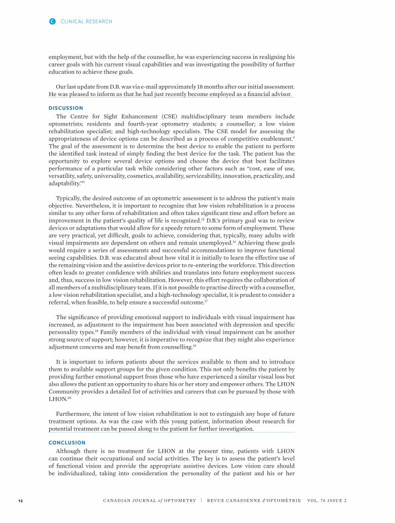
At his 3-month follow-up, best corrected visual acuities were OD 6/12 (20/40) and OS less than 6/120 (20/400). Both eyes appeared straight, and eye movements were unrestricted. Pupil responses were unremarkable and applanation intraocular pressures were within normal limits. Cover test was difficult due to poor fixation, but there was no history of strabismus. Colour vision was reduced to OS 7/9, as determined using ishihara plates, but remained normal in the right eye. Anterior segment structures were healthy. Fundus photos showed mild optic disc pallor (OS>OD) and red-free photos revealed retinal nerve fibre layer loss (Figure 1).
A month later, his vision in both eyes decreased dramatically to finger counting. Currently, there are no proven treatments to prevent or reverse the vision loss associated with LHON. D.B. was referred to the Centre for Sight Enhancement (CSE) for assessment for low vision.
management: low vision rehabilitationD.B., a carpenter by trade, is an athletic young man who played hockey and soccer. Within
6 months of being diagnosed with LHON, he was unable to participate in any of his favourite sports and he had lost his job. He could no longer see the keypad on debit machines, and he reported that grooming and crossing the street had become progressively more difficult. in addition to these concerns, he was finding each day increasingly long and was finding it difficult to occupy his time. His primary goal was to be able to return to work as soon as possible.
Presenting unaided distance visual acuity was 3/69 OD, 3/91 OS, and 3/69 OU, determined by using a Feinbloom acuity chart. Unaided near-visual acuity was 0.08/2.0M OD, 0.08/2.5M OS and 0.08/2.0M OU, determined by using a Lighthouse Single Letter near-acuity chart. Subjective refraction improved acuity to 3/61 and 3/55 for the right eye and the left eye, respectively. Contrast sensitivity measured with a Peli-Robson chart was reduced to 0.95 OU. Amsler grid testing revealed an absolute central scotoma OU.
Beyond Eye Care – Low Vision Rehabilitation
C A NA D i A N JO U R NA L o f O P T O M E T Ry | R EV U E C A NA D i E N N E d ’ O P T O M é T R i E VO L . 76 i S S U E 2 9
Figure 1. Red-free photo of the left eye, showing retinal nerve fibre layer loss after 4 months of onset of Leber’s hereditary optic neuropathy.

C A NA D i A N JO U R NA L o f O P T O M E T Ry | R EV U E C A NA D i E N N E d ’ O P T O M é T R i E VO L . 76 i S S U E 210
CLINICAL RESEARCHC
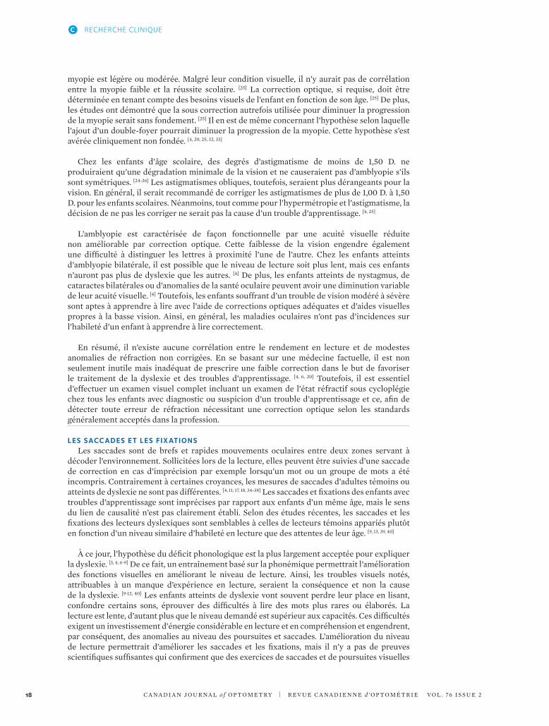
Figure 2. Right Goldmann visual field results illustrating central field loss.
Figure 3. Left Goldmann visual field results illustrating central field loss.
A Goldmann monocular visual field assessment using a V4e target confirmed an absolute central scotoma at about 5 degrees on each side. However, he still had about 130 degrees horizontally and 100 degrees vertically in each eye (Figures 2 and 3).
it was estimated that D.B. would need an equivalent viewing power of 48D in order to read 1.0M size print fluently.8 As a starting point, 24D near-devices were selected to attempt to achieve survival reading (minimal reading task, as is necessary when reading one’s mail) or 1.0M size print at threshold. Following the model of competitive enablement,9 several device types were reviewed. A 24D full-field microscope was demonstrated initially, with the microscope over OS and then over OD, which allowed him to achieve a threshold acuity of 1.2M when the microscope was placed over the right eye. Although unassisted acuity was marginally better in the left eye, this preference for the right eye may have been due to a difference in contrast sensitivity measures between the two eyes. Unfortunately, monocular contrast sensitivity testing was not performed. D.B. was counselled that this device could also be used to aid with eccentric viewing training. He found the spectacles quite heavy. A 24D illuminated hand magnifier was preferred over the full-field microscope. Performance with these devices was compared with his performance while using a portable closed-circuit television system (CCTV). With a CCTV, he readily achieved an acuity performance of 0.5M, and he greatly appreciated the enhanced visibility using enhanced contrast and reversed polarity functions. Ergonomically, he found the increased working distance with a CCTV to be much more effective for sustained viewing activities.

To address difficulties with TV viewing, the 2.1× Max TV and 4 × 20 Beecher-Mirage binocular telescopes were introduced and subsequently loaned to him to allow him to experiment and decide which one was most suitable for viewing his 46-inch television. Monocular telescopes (4 × 12 and 6 × 16) were demonstrated for distance spotting tasks and as adjuncts to aid mobility tasks such as crossing the street. Orientation and mobility training was recommended.
Although glare was a concern, D.B. did not appreciate any subjective benefits using any of the standard lens tints for those with low vision. Additional appointments were scheduled to follow up this initial assessment and for ADL consultation, a CCTV and high technology assessment, and rehabilitation counselling.
During ADL consultation, the low vision rehabilitation specialist reviewed the assistive devices that D.B. had found most useful from the initial assessment and determined the outcome of the telescopes loaned. Different types of lighting and utilization of colour and contrast were reviewed. The importance of labelling appliances, food, medications, and personal items was discussed along with demonstration of various labelling techniques and devices, including audio labellers. For communication purposes, use of bold lined paper, writing guides, and a black felt-tip marker with a white board were introduced to increase contrast. Various nonoptical devices that facilitate home safety and overall function such as liquid level indicators and oven mittens that cover the entire arm, magnifying mirrors, devices that can help identify money, and large-face and auditory-output watches and clocks were demonstrated. To provide some leisure activity, 2× super jumbo playing cards were offered.
The high-technology specialist reviewed several portable and desktop CCTV models and also demonstrated various features on the patient’s iPhone 4S. The iPad was also reviewed for comparison with D.B.’s current Kindle Touch. D.B. appreciated the higher contrast of the iPad, but no significant difference in reading was noted. The speech output function of the Kindle device was also demonstrated and was appreciated by D.B. A separate high-technology assessment to review an assortment of computer adaptations and software (including voice output and OCR software) for both PC and Mac were much appreciated.
D.B. also agreed to an appointment with the centre’s low vision counsellor, who provided support services, including career planning and vocation adjustment, information about community resources, and help with applications for financial assistance, in addition to individual and family counselling.
At the completion of the second set of assessments, D.B. indicated that he felt “hope” for the first time. He purchased the jumbo playing cards, a 24-inch CCTV, assistive computer software, and a 4× Beecher spec-mounted telescope. He had started using the appropriate accessibility features on his iPhone and Kindle reader. He was educated on the Ontario Disability Support Program (ODSP), which offers income support and employment support, and the Ontario Assistive Devices Program (ADP), which provides some financial support toward the cost of devices.10-12 Forms were completed to initiate registration for these services. The Ontario Disability Tax Credit form was completed. D.B. was also registered with the Canadian institute for the Blind (CNiB).13
D.B. was provided information about a neurologist who had been doing research on a mitochondrial cocktail consisting of creatine monohydrate, coenzyme Q10, and alpha-lipoic acid, which reduced lactate and markers of oxidative stress in patients with mitochondrial cytopathies. At the time, there was only one randomized, double-blinded clinical trial consisting of 16 patients, so further studies using a larger sample size were still required to clarify potential of this therapy.14 After discussing this option, the patient decided that he would like to be referred to this specialist.
Subsequent 6- and 9-month follow-up assessments showed some improvement in contrast sensitivity. D.B. reported continued use of the recommended assistive devices, as well as the strategies and techniques to maximize vision and facilitate ADLs. He had acquired temporary
Beyond Eye Care – Low Vision Rehabilitation
C A NA D i A N JO U R NA L o f O P T O M E T Ry | R EV U E C A NA D i E N N E d ’ O P T O M é T R i E VO L . 76 i S S U E 2 11
C A NA D i A N JO U R NA L o f O P T O M E T Ry | R EV U E C A NA D i E N N E d ’ O P T O M é T R i E VO L . 76 i S S U E 212
employment, but with the help of the counsellor, he was experiencing success in realigning his career goals with his current visual capabilities and was investigating the possibility of further education to achieve these goals.
Our last update from D.B. was via e-mail approximately 18 months after our initial assessment. He was pleased to inform us that he had just recently become employed as a financial advisor.
dIsCUssIoN
The Centre for Sight Enhancement (CSE) multidisciplinary team members include optometrists; residents and fourth-year optometry students; a counsellor; a low vision rehabilitation specialist; and high-technology specialists. The CSE model for assessing the appropriateness of device options can be described as a process of competitive enablement.9
The goal of the assessment is to determine the best device to enable the patient to perform the identified task instead of simply finding the best device for the task. The patient has the opportunity to explore several device options and choose the device that best facilitates performance of a particular task while considering other factors such as “cost, ease of use, versatility, safety, universality, cosmetics, availability, serviceability, innovation, practicality, and adaptability.”9
Typically, the desired outcome of an optometric assessment is to address the patient’s main objective. Nevertheless, it is important to recognize that low vision rehabilitation is a process similar to any other form of rehabilitation and often takes significant time and effort before an improvement in the patient’s quality of life is recognized.15 D.B.’s primary goal was to review devices or adaptations that would allow for a speedy return to some form of employment. These are very practical, yet difficult, goals to achieve, considering that, typically, many adults with visual impairments are dependent on others and remain unemployed.16 Achieving these goals would require a series of assessments and successful accommodations to improve functional seeing capabilities. D.B. was educated about how vital it is initially to learn the effective use of the remaining vision and the assistive devices prior to re-entering the workforce. This direction often leads to greater confidence with abilities and translates into future employment success and, thus, success in low vision rehabilitation. However, this effort requires the collaboration of all members of a multidisciplinary team. if it is not possible to practise directly with a counsellor, a low vision rehabilitation specialist, and a high-technology specialist, it is prudent to consider a referral, when feasible, to help ensure a successful outcome.17
The significance of providing emotional support to individuals with visual impairment has increased, as adjustment to the impairment has been associated with depression and specific personality types.18 Family members of the individual with visual impairment can be another strong source of support; however, it is imperative to recognize that they might also experience adjustment concerns and may benefit from counselling.19
it is important to inform patients about the services available to them and to introduce them to available support groups for the given condition. This not only benefits the patient by providing further emotional support from those who have experienced a similar visual loss but also allows the patient an opportunity to share his or her story and empower others. The LHON Community provides a detailed list of activities and careers that can be pursued by those with LHON.20
Furthermore, the intent of low vision rehabilitation is not to extinguish any hope of future treatment options. As was the case with this young patient, information about research for potential treatment can be passed along to the patient for further investigation.
CoNClUsIoN
Although there is no treatment for LHON at the present time, patients with LHON can continue their occupational and social activities. The key is to assess the patient’s level of functional vision and provide the appropriate assistive devices. Low vision care should be individualized, taking into consideration the personality of the patient and his or her
CLINICAL RESEARCHC

environment. Most importantly, emotional support and counselling are an essential part of the successful care of a patient with low vision.
rEFErENCEs
C A NA D i A N JO U R NA L o f O P T O M E T Ry | R EV U E C A NA D i E N N E d ’ O P T O M é T R i E VO L . 76 i S S U E 2 13
1. Sadun AA, Morgia CL, Carelli V. Leber’s hereditary optic neuropathy. Neurol Ophthalmol Otol 2011;13:109–17.
2. Chronister CL, Gurwood AS, Burns CM. Leber’s hereditary optic neuropathy: a case report. Optometry 2005;76:302–8.
3. Newman NJ. From genotype to phenotype in Leber hereditary optic neuropathy: still more questions than answers. J Neuro-Ophthalmol 2002;22(4): 257–61.
4. Man PyW, Turnbull DM, Chinnery PF. Leber’s hereditary optic neuropathy. J Med Genet 2002;39:162–9.
5. Naka M, Nitta T, Sato i. Long-term evaluation of visual field alteration in Leber’s hereditary optic neuropathy with visual recovery. J Neuro-Ophthalmol 2005;29:65–7.
6. Ehlers J, Shah C, eds. The Wills eye manual: office and emergency room diagnosis and treatment of eye disease. Philadelphia, PA: Lippincott Williams and Wilkins, 2008:260.
7. Swartz N, Savino PJ. is all nondefinable optic atrophy Leber’s hereditary optic neuropathy? Survey Ophthalmol 1994;39(2):146–50.
8. Cheong AMy, Lovie-Kitchen JE, Bowers AR. Determining magnification for reading with low vision. Clin Experiment Optometry 2002 85(4):229–37.
9. Strong JG, Jutai JW, Plotkin AD. Competitive enablement: a client-centred conceptual model for device selections in low vision rehabilitation. Everyday technology for independence and care. Assist Tech Res Series 2011;29:1033–42.
10. Ontario Disability Support Program, Ontario Ministry of Community and Social Services; 2008. income support. http://www.mcss.gov.on.ca/en/mcss/programs/social/odsp/income_support/eligibility/financial_Eligibility.asp. Accessed June 11, 2012.
11. Ontario Disability Support Program, Ontario
Ministry of Community and Social Services. (2008). Employment support. http://www.mcss.gov.on.ca/en/mcss/programs/social/odsp/employment_support/works.aspx. Accessed June 11, 2012.
12. Assistive Devices Program, Ontario Ministry of Health and Long-Term Care. (2008). Visual aids. http://www.health.gov.on.ca/en/public/programs/adp/categories.aspx. Accessed June 11, 2012.
13. CNiB, Rehabilitation and Support Services. (2012). Our range of services. http://www.cnib.ca/en/services/vision-support/range/#contentstart. Accessed June 19, 2012.
14. Tarnopolsky MA. The mitochondrial cocktail: rationale for combined nutraceutical therapy in mitochondrial cytopathies. Adv Drug Delivery Rev 2008 60:1561–7.
15. Wolffsohn JS, Cochrane AL. Design of the low vision quality-of-life questionnaire (LVQOL) and measuring the outcome of low-vision rehabilitation. Am J Ophthalmol 2000;130(6):793–802.
16. O’Day B. Employment barriers for people with visual impairments. J Visual impairment Blindness 1999;93(10):627.
17. Strong JG, Pace RJ, Plotkin AD. Low vision services: a model for sequential intervention and rehabilitation. Can J Public Health 1988;79:S50–4.
18. Tabrett DR, Latham K. Adjustment to vision loss in a mixed sample of adults with established visual impairment. invest Ophthalmol Visual Sci 2012;53(11):7227–34.
19. Bambara JK, Wadley V, Owsley C, et al. Family functioning and low vision: a systematic review. J Vision impairment Blindness 2009;103(3):137–49.
20. LHON Community. http://www.lhon.org/lhon/LHON_community.html. Accessed July 16, 2012.
Beyond Eye Care – Low Vision Rehabilitation

Richness is:
® Registered trademarks of The Bank of Nova Scotia.
Richness is:
You define richness. With the Scotia Professional® Plan, we can help with the money part. You’ve worked long and hard to build your career. It only makes sense to do everything you can to ensure your continued success, both professionally and personally. The Professional Plan is a fully customized banking package designed to help you build a strong, profitable business while ensuring your personal finances receive the attention they deserve. And that will help you focus more clearly on what you do for others.
To learn more about Scotia Professional Plan, visit your nearest Scotiabank branch or visit scotiabank.com/professional today.
File Name: SMBIZ_AD_SPP_Optomerty_1113Trim: 8.5” x 11”Bleed: 0.5" Safety: n/a Mech Res: 300dpiColours: CMYK
Publication: Canadian Journal of OptometryMaterial Deadline: November 25, 2013 Insertion Dates:
Canadian Marketing 100 Yonge Street, 16th Floor
Toronto, ON M5C 2W1
Scotia Professional Plan

Résumé
Les troubles d’apprentissage sont des désordres complexes qui entravent le développement normal des processus d’acquisition et de traitement de l’information. ils sont couramment rencontrés au sein de la population pédiatrique et peuvent se manifester sous diverses formes dont la plus fréquente est la dyslexie. Les troubles d’apprentissage ont une nature chro-nique et n’ont pas pour cause première des handicaps visuels, auditifs et/ou moteurs, la déficience intellectuelle, la perturbation affective ni même un milieu défavorisé. Cependant, il est possible que ces troubles puissent coexister avec l’un ou l’autre de ces problèmes.
Puisque les enfants qui en sont atteints rencontrent souvent des difficultés de taille lors des apprentissages fondamentaux dès leur entrée à l’école pri-maire, il est essentiel de leur offrir un soutien adapté, un diagnostic précoce et une intervention efficace. Plusieurs hypothèses impliquant divers prob-lèmes visuels ont été émises quant à la cause de la dyslexie et des troubles d’apprentissage. La présente revue de littérature traite de ces diverses hy-pothèses de traitement des troubles d’apprentissage scolaire par thérapies visuelles.
L’analyse démontre qu’il n’y a pas de preuves scientifiques suffisantes à ce jour prouvant que la thérapie visuelle, les lunettes d’entraînement, les ex-ercices de poursuites et de saccades, les exercices perceptuels, les lunettes grossissantes, les filtres ou lentilles colorées ainsi que les prismes peuvent améliorer significativement les troubles d’apprentissage.
Abstract
Learning disabilities are complex disorders that interfere with the normal acquisition and processing of knowledge. in the pediatric population, dys-lexia is the most common diagnosis. Learning disabilities are chronic in nature and are not caused by visual, auditory, motor, and intellectual dif-ficulties or environmental factors. Those conditions may, however, coexist with the disorder.
Many of these problems present when children start elementary school, and it is important to offer early diagnosis and intervention. Various theo-ries have evolved suggesting that visual troubles are the cause of dyslexia and learning disabilities. This article reviews the scientific basis of these theories and the evidence-based research.
Diverses modalités de traitement des troubles d’apprentissage scolaire par thérapies visuelles: quelles sont les évidences scientifiques?
amélie Ganivet, od, m.Sc.isabelle denault, od rosanne Superstein, md FrCSCnicole Fallaha, md FrCSC
Clinique d’optométrie Granger Bernier et Associés383 Boulevard du Séminaire Nord, suite 215Saint-Jean-sur-Richelieu, QuébecJ3B 8C5
Hôpital Sainte JustineDépartement d’Ophtalmologie3175 Chemin de la Côte-Sainte-CatherineMontréal, QuébecH3T 1C5
RECHERCHE CLINIQUE C
C A NA D i A N JO U R NA L o f O P T O M E T Ry | R EV U E C A NA D i E N N E d ’ O P T O M é T R i E VO L . 76 i S S U E 2 15

C A NA D i A N JO U R NA L o f O P T O M E T Ry | R EV U E C A NA D i E N N E d ’ O P T O M é T R i E VO L . 76 i S S U E 216
MIsE EN CoNtExtE
Les troubles d’apprentissage sont des dysfonctionnements spécifiques et fréquents qui entravent le développement normal des processus d’acquisition. En 2006, selon Statistique Canada, 3,2% des enfants Canadiens âgés entre 5 et 14 ans étaient affectés par un trouble d’apprentissage, ce qui représenterait environ un enfant par classe. [1] Plusieurs hypothèses ont été émises en regard de l’étiologie des troubles d’apprentissage dans les dernières décennies. Comme ceux-ci ne sont pas encore parfaitement bien compris, une multitude de traitements non prouvés scientifiquement en sont dérivés. De ce fait, diverses thérapies visuelles sont parfois suggérées afin de traiter ces désordres. [2, 3] Par souci d’aider ces patients à centraliser leurs ressources et efforts afin d’atteindre leurs objectifs d’apprentissage, la recommandation de ces thérapies devrait se baser sur des données factuelles et scientifiques (« evidence-based »). [3, 4]
Sous-tendant un caractère génétique, les troubles d’apprentissage sont chroniques et engendrés par une atteinte des fonctions neuropsychologiques. [5-7] Ces désordres ne sont donc pas causés par une déficience intellectuelle, un déficit sensoriel, un mauvais encadrement scolaire ou un manque d’intérêt personnel. Les enfants qui en sont affectés possèdent généralement une capacité de raisonnement moyenne ou supérieure à la moyenne même s’ils éprouvent des difficultés à acquérir, comprendre, organiser et traiter les informations. [4, 5, 7] Les principaux troubles d’apprentissage sont les suivants : la dyslexie, le trouble déficitaire de l’attention avec ou sans hyperactivité (TDA-H), la dysphasie, la dysorthographie, la dyspraxie, la dyscalculie et les troubles du spectre de l’autisme (TSA). La dyslexie est le désordre le plus fréquemment rencontré, affectant 80% des individus ayant des troubles d’apprentissage et jusqu’à 5-17% de la population générale. [4-8] Par conséquent, la dyslexie sera le trouble d’apprentissage principalement considéré dans le présent article.
Plusieurs hypothèses impliquant des atteintes visuelles ont été émises quant à la cause de la dyslexie et des troubles d’apprentissage. Bien que certaines divergences d’opinions persistent, l’hypothèse la plus largement acceptée à ce jour fait plutôt état d’un « déficit dans la composante phonologique du langage qui rend difficile l’utilisation du code alphabétique dans le décodage des mots écrits». [4] Les troubles visuels parfois rencontrés seraient en fait attribuables à un manque d’expérience en lecture. [9-12] Une étude récente démontre même qu’un entraînement en lecture basé sur la phonologie améliore non seulement les habiletés en lecture, mais également les fonctions visuelles. [9] Ainsi, les anomalies visuelles parfois évoquées seraient une conséquence du trouble d’apprentissage et non la cause. [3, 4, 6, 8-10, 13]
Puisque la dyslexie est considérée comme un trouble phonologique du langage et non une dysfonction visuelle, des publications de l’Académie Américaine de Pédiatrie ainsi que de la Société Canadienne de Pédiatrie recommandent un traitement basé sur le décodage, la fluidité, le vocabulaire et la compréhension mais aussi plus spécifiquement sur la conscience phonémique et son application. [3-5, 7, 9, 14, 15] De nature chronique, le trouble d’apprentissage ne disparaîtra pas avec l’âge. Toutefois, afin d’établir un plan d’intervention axé sur les besoins spécifiques de chaque enfant et de maximiser ses apprentissages, une prise en charge précoce s’appuyant sur la démarche scientifique est favorable. [7]
trAItEMENt dEs troUblEs d’APPrENtIssAgE sCol AIrE PAr thÉrAPIEs vIsUEllEs : ÉtAt dEs CoNNAIssANCEs
Certains problèmes de vision peuvent interférer avec le processus de lecture, créant ainsi des difficultés d’apprentissage réversibles suite à la correction du trouble visuel en cause. Par conséquent, il est primordial que tout enfant ayant un trouble ou des difficultés d’apprentissage ait un examen visuel complet dès les premières suspicions afin d’évaluer sa vision et sa santé oculaire. [3, 6, 16]
This review shows that there is not enough scientific evidence to support that vision thera-py, training glasses, pursuit and saccade exercises, magnifying glasses, coloured lenses and/or overlays and prisms significantly help in coping with learning disabilities.
RECHERCHE CLINIQUEC

Néanmoins, il a été démontré que les enfants atteints de dyslexie ou de troubles d’apprentissage n’ont pas plus d’anomalies de leur fonction visuelle et de leur santé oculaire que les enfants non affectés par ces conditions. [4, 6, 8, 14, 17-19] Leur apprentissage de matières telles que les mathématiques et le français peut être laborieux, mais ils réussissent souvent très bien dans d’autres sphères de développement nécessitant tout autant leurs aptitudes visuelles. il n’y a actuellement pas de preuves scientifiques basées sur la médecine factuelle qui démontrent que de subtiles erreurs de réfraction ou de légers problèmes visuels pourraient provoquer ou augmenter la gravité des troubles d’apprentissage, diminuer l’efficacité visuelle ou encore amenuiser la réponse aux divers traitements éducationnels. [3, 4, 17, 20] L’hypothèse selon laquelle les enfants qui participent à une thérapie visuelle seraient conséquemment plus réceptifs aux divers programmes d’apprentissage n’est pas plus fondée. [2, 4, 6, 15, 20-23]
l’ACUItÉ vIsUEllE , l A rÉFrACtIoN Et lEs lUNEttEs d’ENtrAîNEMENt
Les enfants qui débutent l’apprentissage de la lecture et de l’écriture utilisent des textes dont les lettres sont de taille assez grande. Plus le niveau scolaire augmente, plus la taille des caractères diminue et plus la demande visuelle est accrue. Bien qu’une bonne vision soit importante, il n’est pas primordial d’avoir une résolution optimale afin de discerner les caractères utilisés au tout début des apprentissages scolaires. il n’existe d’ailleurs aucune preuve démontrant que les lecteurs débutants atteints de myopie, d’hypermétropie et/ou d’astigmatisme léger à modéré ont plus de difficulté à apprendre à lire que les autres enfants. [4] De petits degrés d’hypermétropie sont considérés normaux chez les jeunes enfants et n’ont généralement pas de signification pathologique. ils constituent, au contraire, une étape du développement normal de l’œil. Les enfants ont des besoins visuels uniques basés sur leurs demandes visuelles et le développement de leur système optique. [24-28] Les besoin réfractifs des enfants ne peuvent être extrapolés en fonction des besoins des adultes. Actuellement, il n’existe pas de règles absolues précisant les montants exacts des différentes amétropies requérant une correction optique ; les recommandations sont plutôt basées sur l’expérience clinique et différents consensus de professionnels de l’optométrie et ophtalmologie pédiatriques reconnus. [4, 24-29]
Les enfants possèdent une capacité accommodative largement plus importante que les adultes. En effet, les enfants âgés entre 6 et 10 ans possèdent en moyenne 12,00 dioptries (D.) ou plus de fonction accommodative. [4, 25, 30] ils peuvent donc généralement compenser une hypermétropie modérée sans avoir de difficultés visuelles. La réfraction moyenne des enfants blancs aux états-Unis est d’environ 2,00 D. d’hypermétropie dans les 5 premières années de vie. [4] Cette hypermétropie tend à diminuer progressivement durant l’adolescence. C’est pourquoi une hypermétropie légère à modérée n’a bien souvent pas besoin d’être corrigée, car elle a peu d’impact au niveau de la fonction oculo-visuelle. [24, 25, 27]
il ne semble pas y avoir de probabilité accrue de dyslexie auprès des enfants ayant une hypermétropie non corrigée. En absence d’une diminution de l’acuité visuelle, il n’y aurait aucune corrélation entre les habiletés de lecture, les performances scolaires et le degré d’hypermétropie. [31] Des enfants de 6 ans n’ont généralement aucune réduction significative de l’acuité visuelle si l’hypermétropie n’excède pas 4,00 D. ; moins de 1% des enfants ont une hypermétropie plus forte. [25] Une erreur de réfraction élevée de nature hypermétropique peut engendrer un inconfort visuel qui peut être considérable. Ainsi, ces enfants, se désintéressant des tâches requérant un effort visuel prolongé, pourraient développer des difficultés d’apprentissage consécutives. Cependant, celles-ci seraient réversibles suite à la correction du problème visuel. À l’inverse, un enfant ayant une hypermétropie élevée non corrigée et atteint de dyslexie ou d’un réel trouble d’apprentissage pourra remarquer une amélioration de son confort, de son acuité visuelle et même de ses performances scolaires suite à la correction de son hypermétropie, son trouble d’apprentissage étant possiblement amplifié par sa mauvaise vision. Toutefois, le trouble d’apprentissage va assurément persister en raison de sa nature plutôt neurologique que sensorielle.
Les enfants atteints de myopie non corrigée observent une diminution de leur acuité visuelle en vision éloignée et, par conséquent, peuvent avoir de la difficulté à voir le tableau en classe. Toutefois, ces enfants n’ont généralement pas de difficultés avec la vision de près surtout si la
Diverses modalités de traitement des troubles d’apprentissage scolaire par thérapies visuelles: quelles sont les évidences scientifiques?
C A NA D i A N JO U R NA L o f O P T O M E T Ry | R EV U E C A NA D i E N N E d ’ O P T O M é T R i E VO L . 76 i S S U E 2 17
C A NA D i A N JO U R NA L o f O P T O M E T Ry | R EV U E C A NA D i E N N E d ’ O P T O M é T R i E VO L . 76 i S S U E 218
myopie est légère ou modérée. Malgré leur condition visuelle, il n’y aurait pas de corrélation entre la myopie faible et la réussite scolaire. [25] La correction optique, si requise, doit être déterminée en tenant compte des besoins visuels de l’enfant en fonction de son âge. [25] De plus, les études ont démontré que la sous correction autrefois utilisée pour diminuer la progression de la myopie serait sans fondement. [25] il en est de même concernant l’hypothèse selon laquelle l’ajout d’un double-foyer pourrait diminuer la progression de la myopie. Cette hypothèse s’est avérée cliniquement non fondée. [4, 20, 25, 32, 33]
Chez les enfants d’âge scolaire, des degrés d’astigmatisme de moins de 1,50 D. ne produiraient qu’une dégradation minimale de la vision et ne causeraient pas d’amblyopie s’ils sont symétriques. [24-26] Les astigmatismes obliques, toutefois, seraient plus dérangeants pour la vision. En général, il serait recommandé de corriger les astigmatismes de plus de 1,00 D. à 1,50 D. pour les enfants scolaires. Néanmoins, tout comme pour l’hypermétropie et l’astigmatisme, la décision de ne pas les corriger ne serait pas la cause d’un trouble d’apprentissage. [4, 25]
L’amblyopie est caractérisée de façon fonctionnelle par une acuité visuelle réduite non améliorable par correction optique. Cette faiblesse de la vision engendre également une difficulté à distinguer les lettres à proximité l’une de l’autre. Chez les enfants atteints d’amblyopie bilatérale, il est possible que le niveau de lecture soit plus lent, mais ces enfants n’auront pas plus de dyslexie que les autres. [4] De plus, les enfants atteints de nystagmus, de cataractes bilatérales ou d’anomalies de la santé oculaire peuvent avoir une diminution variable de leur acuité visuelle. [4] Toutefois, les enfants souffrant d’un trouble de vision modéré à sévère sont aptes à apprendre à lire avec l’aide de corrections optiques adéquates et d’aides visuelles propres à la basse vision. Ainsi, en général, les maladies oculaires n’ont pas d’incidences sur l’habileté d’un enfant à apprendre à lire correctement.
En résumé, il n’existe aucune corrélation entre le rendement en lecture et de modestes anomalies de réfraction non corrigées. En se basant sur une médecine factuelle, il est non seulement inutile mais inadéquat de prescrire une faible correction dans le but de favoriser le traitement de la dyslexie et des troubles d’apprentissage. [4, 6, 20] Toutefois, il est essentiel d’effectuer un examen visuel complet incluant un examen de l’état réfractif sous cycloplégie chez tous les enfants avec diagnostic ou suspicion d’un trouble d’apprentissage et ce, afin de détecter toute erreur de réfraction nécessitant une correction optique selon les standards généralement acceptés dans la profession.
lEs sACCAdEs Et lEs FIxAtIoNsLes saccades sont de brefs et rapides mouvements oculaires entre deux zones servant à
décoder l’environnement. Sollicitées lors de la lecture, elles peuvent être suivies d’une saccade de correction en cas d’imprécision par exemple lorsqu’un mot ou un groupe de mots a été incompris. Contrairement à certaines croyances, les mesures de saccades d’adultes témoins ou atteints de dyslexie ne sont pas différentes. [4, 11, 17, 18, 34-38] Les saccades et fixations des enfants avec troubles d’apprentissage sont imprécises par rapport aux enfants d’un même âge, mais le sens du lien de causalité n’est pas clairement établi. Selon des études récentes, les saccades et les fixations des lecteurs dyslexiques sont semblables à celles de lecteurs témoins appariés plutôt en fonction d’un niveau similaire d’habileté en lecture que des attentes de leur âge. [9, 13, 39, 40]
À ce jour, l’hypothèse du déficit phonologique est la plus largement acceptée pour expliquer la dyslexie. [3, 4, 6-9] De ce fait, un entraînement basé sur la phonémique permettrait l’amélioration des fonctions visuelles en améliorant le niveau de lecture. Ainsi, les troubles visuels notés, attribuables à un manque d’expérience en lecture, seraient la conséquence et non la cause de la dyslexie. [9-12, 40] Les enfants atteints de dyslexie vont souvent perdre leur place en lisant, confondre certains sons, éprouver des difficultés à lire des mots plus rares ou élaborés. La lecture est lente, d’autant plus que le niveau demandé est supérieur aux capacités. Ces difficultés exigent un investissement d’énergie considérable en lecture et en compréhension et engendrent, par conséquent, des anomalies au niveau des poursuites et saccades. L’amélioration du niveau de lecture permettrait d’améliorer les saccades et les fixations, mais il n’y a pas de preuves scientifiques suffisantes qui confirment que des exercices de saccades et de poursuites visuelles
RECHERCHE CLINIQUEC

pourraient aider le lecteur dyslexique à développer un meilleur niveau de lecture. [2, 4, 10, 23, 35, 40, 41]
Enfin, la majorité des individus atteints d’un trouble soit au niveau des motilités oculaires ou des mouvements oculaires ont une lecture et une compréhension normale. En effet, plusieurs enfants nés avec un strabisme important, un nystagmus ou encore avec une maladie oculaire affectant les mouvements oculaires excellent au niveau de leurs résultats académiques et de leur niveau de lecture. [3, 23, 42, 43] Ainsi, la dyslexie ne serait pas le résultat d’un déficit oculomoteur, mais plutôt le résultat d’un trouble au niveau central du traitement de l’information qui occasionne une difficulté au niveau du décodage et de la compréhension. Par conséquent, elle engendre des fixations plus longues et des saccades de corrections plus nombreuses lorsque le niveau de lecture demandé est supérieur aux capacités du lecteur.
ACCoMModAtIoNL’accommodation est la capacité à se concentrer et à faire la mise à foyer au près. Elle assure
la netteté des images selon différentes distances de vision. Les amplitudes accommodatives sont généralement maximales durant l’enfance jusqu’à l’âge de 10 ans, le pouvoir accommodatif diminuant ensuite naturellement avec l’âge. [44] il est donc rare d’observer une insuffisance accommodative chez un enfant. Un défaut accommodatif pourrait tout de même survenir chez un enfant avec une hypermétropie élevée non corrigée avec histoire d’infection virale, de trauma cérébral ou oculaire, de pathologie du tronc cérébral ou encore secondaire à la prise de médicament. [4] L’hypothèse de la faiblesse accommodative d’enfants dyslexiques ou avec trouble d’apprentissage a amené plusieurs professionnels à promouvoir l’usage d’un double-foyer afin de la compenser. Bien que certaines études aient démontré une amplitude d’accommodation légèrement plus basse des enfants dyslexiques par rapport à des enfants normaux, l’amplitude des dyslexiques se situait dans les normes attendues pour leur âge. [8, 45] Ainsi, il n’y a pas de différence significative entre la capacité accommodative de patients ayant un trouble en lecture et celle de lecteurs de niveau normal. [8, 13] De plus, il n’y a pas de preuves scientifiques qu’une augmentation du grossissement augmente l’efficacité de la lecture. Une étude a même démontré que la correction de l’insuffisance accommodative n’avait pas d’impact significatif sur les mouvements oculaires et la fluidité lors de la lecture. [46] Par conséquent, toute thérapie visant à diminuer l’effort accommodatif d’un enfant ayant des troubles d’apprentissage prétextant que son trouble en est la cause est non fondée scientifiquement. [2, 4, 6, 20]
vIsIoN bINoCUl AIrEL’équilibre oculomoteur parfait est rare dans la population pédiatrique et générale. La
plupart des individus présentent une faible ésophorie ou exophorie asymptomatique qui sont considérées dans les limites normales. [4, 17, 19] Plusieurs études ont investigué la fonction binoculaire et l’accommodation d’enfants avec troubles d’apprentissage scolaire et dyslexie. Aucune relation de causalité n’a pu être mise en évidence. [17, 19, 47]
L’insuffisance de convergence représente une difficulté à fusionner correctement et efficacement un objet situé à une distance rapprochée. Lorsqu’un effort de convergence est difficile à surmonter, il peut provoquer divers symptômes d’inconfort visuel tels que fatigue visuelle, maux de tête, vision floue en lecture, diplopie en vision rapprochée, difficulté à se concentrer pendant des périodes prolongées de travail au près. De plus, certains facteurs tels que le manque de sommeil, la maladie, une fatigue générale peuvent aggraver le problème. [4]
La prévalence de l’insuffisance de convergence serait d’environ 3% à 5% de la population. [4]
Toutefois, en raison de la différence entre les critères de diagnostic, certaines études rapportent des données qui peuvent différer. Autant une difficulté accommodative qu’une insuffisance de convergence peuvent interférer avec le confort de la lecture. [3, 8, 45] Ces troubles visuels doivent être traités s’ils sont reconnus comme problématiques selon les critères énoncés pour la population générale. De ce fait, le traitement de l’insuffisance de convergence peut aider au niveau du confort de la lecture et des travaux en vision rapprochée en permettant une lecture prolongée plus aisée. [20, 48] également, si les difficultés de lecture chez un individu sont secondaires à une anomalie de l’accommodation ou de la convergence, ces difficultés disparaîtront une fois le trouble visuel traité. [4] Toutefois, puisque ces troubles visuels ne sont pas la cause de la dyslexie, leur entraînement n’aura pas d’impact sur les capacités de décodage
Diverses modalités de traitement des troubles d’apprentissage scolaire par thérapies visuelles: quelles sont les évidences scientifiques?
C A NA D i A N JO U R NA L o f O P T O M E T Ry | R EV U E C A NA D i E N N E d ’ O P T O M é T R i E VO L . 76 i S S U E 2 19

C A NA D i A N JO U R NA L o f O P T O M E T Ry | R EV U E C A NA D i E N N E d ’ O P T O M é T R i E VO L . 76 i S S U E 220
et de compréhension en lecture. [4]
Tout comme la majorité des enfants, ceux ayant des troubles d’apprentissage aiment jouer à des jeux vidéo. L’utilisation de jeux vidéo requiert une bonne coordination œil-main, de la concentration pendant une période prolongée, une accommodation efficace, une convergence active ainsi qu’une bonne perception visuelle. Par conséquent, si ces déficits étaient la cause majeure des troubles de lecture, ces enfants rejetteraient cette tâche qui nécessite également une utilisation intensive de leurs capacités visuelles. [4, 6]
lEs lENtIllEs Et FIltrEs ColorÉs
L’utilisation de lentilles ou de filtres colorés visant à améliorer le confort et les performances en lecture chez les individus ayant des troubles d’apprentissage est très controversée. Certains sont d’avis que l’utilisation de lentilles ou filtres jaunes permettrait d’améliorer le contrôle de l’attention visuelle et des mouvements oculaires chez certains enfants par stimulation cérébrale. L’utilisation de lentilles ou filtres bleus, quant à elle, améliorerait la concentration et conséquemment, la lecture. [49, 50] D’autres avancent que le contrôle de l’accommodation et de la convergence serait influencé par une sensibilité à certaines longueurs d’ondes de la lumière, créant un stress visuel lors de la lecture. L’utilisation de certains filtres colorés correspondant à ces différentes longueurs d’onde permettrait une réduction de ce stress et une lecture plus efficace. [49, 51-53] Toutefois, plusieurs études ont statué que le choix approprié de la couleur du filtre bénéfique pour chaque individu serait inconsistant et non répétable. [6, 54-59] D’autres études ont démontré que l’utilisation de lentilles ou filtres colorés n’avait aucun effet bénéfique sur la fonction visuelle et les performances en lecture. [55, 60] Comme aucun consensus ne peut être établi à ce jour, l’utilisation de filtres colorés afin de traiter les enfants avec troubles d’apprentissage n’est pas cliniquement justifiée pour le moment. [4, 6, 14, 54, 56-59, 61-63]
lEs PrIsMEsL’utilisation de prismes chez des patients avec troubles d’apprentissage a été rapportée.
Certains affirment que des prismes base en haut pourraient être utilisés pour traiter l’exophorie ou l’insuffisance de convergence tandis que des prismes base en bas seraient employés dans le traitement de l’ésophorie ou l’excès de convergence. [20] Les prismes base en bas seraient parfois même utilisés afin de faciliter l’adaptation des individus pour qui une faible correction d’hypermétropie serait prescrite. [20]
D’autres prétendent qu’une pleine correction de toute hétérophorie permettrait de diminuer la fatigue visuelle lors de la lecture. Selon cette hypothèse, de petites hétérophories occasionneraient un effort de compensation afin de maintenir une vision binoculaire adéquate: elles devraient donc être corrigées complètement à l’aide du prisme approprié. [64, 65] Plusieurs réfutent toutefois cette hypothèse et ne recommandent pas l’utilisation de prismes car il n’améliore pas les performances en lecture. [66, 67] Les effets bénéfiques rapportés pourraient, selon eux, être attribués à un effet placebo. [66, 67] À ce jour, aucune preuve scientifique encourageant l’utilisation des prismes dans le but d’améliorer la lecture des enfants avec troubles d’apprentissage n’a été clairement établie. [4, 6, 20, 21, 57, 66, 67]
dIsCUssIoN
La dyslexie serait une difficulté d’origine neurobiologique à décoder et à comprendre un langage écrit. Elle correspond à un déficit dans le processus de la structure du son du langage écrit ou parlé (défaut phonologique). [6] il s’agit d’un désordre persistant et chronique dont les causes sont multifactorielles et sous influence génétique. [6]
Les atteintes oculo-visuelles peuvent interférer avec le processus d’apprentissage de la lecture mais elles ne sont pas la cause de la dyslexie ni des troubles d’apprentissage. En effet, bien que la vision soit fondamentale pour lire, le cerveau doit analyser les images visuelles transmises. Statistiquement, les enfants ayant de la dyslexie ou des troubles d’apprentissage associés ont le même niveau de fonction visuelle et le même niveau de santé oculaire que les enfants sans ces conditions. [3, 8, 18, 19] il n’y a pas de preuves scientifiques à ce jour démontrant que la thérapie visuelle, les lunettes d’entraînement, les exercices de poursuites et de saccades,
RECHERCHE CLINIQUEC

les exercices perceptuels, les lunettes grossissantes, les filtres ou les lentilles colorées ainsi que les prismes peuvent significativement améliorer les performances de l’enfant ayant des troubles d’apprentissage. [2, 4, 6, 7, 14, 20-22, 57, 68] Ces approches peuvent donner de faux espoirs aux parents et autres intervenants et de ce fait, possiblement retarder une intervention ayant un meilleur potentiel bénéfique pour l’enfant. De plus, les coûts engendrés par ces thérapies sont substantiels et le temps requis pour les effectuer non négligeable. Les études statuant sur l’amélioration des apprentissages via ces thérapies sont en fait des études non contrôlées scientifiquement ou encore basées sur des cas anecdotiques. [2, 4] Les bénéfices avancés seraient plutôt secondaires aux autres traitements éducationnels traditionnels souvent effectués de façon combinée avec ces thérapies visuelles et/ou à l’effet placebo de tels procédés.
CoNClUsIoN
La détection précoce des enfants ayant des troubles d’apprentissage et leur référence vers les professionnels appropriés sont essentielles afin de fournir le support nécessaire à ces enfants et leur famille. il est primordial que ces enfants aient un examen visuel complet avec cycloplégie afin de s’assurer qu’ils ne présentent pas d’atteinte visuelle entravant leur fonction visuelle et pouvant ainsi amplifier les symptômes de dyslexie/troubles d’apprentissage. Une approche multidisciplinaire est privilégiée chez ces enfants. En plus de l’évaluation de la vision, une évaluation de la santé, du développement, de l’audition et si nécessaire une intervention médicale et/ou psychologique devra être effectuée. [4, 6, 13, 14, 20, 21] En terminant, la dyslexie et les troubles d’apprentissage sont des problèmes complexes n’ayant malheureusement pas de solutions simples. Toutefois, il est recommandé que toutes les thérapies proposées soient scientifiquement justifiées afin de créer des attentes réalistes et de favoriser le développement de l’enfant.
bIblIogrAPhIE
1. Statistique_Canada. 2006; http://www.statcan.gc.ca/daily-quotidien/071203/dq071203a-fra.htm.
2. Rawstron JA, Burley CD, ad Elder MJ. A systematic review of the applicability and efficacy of eye exercises. J Pediatr Ophthalmol Strabismus 2005;42(2):82–8.
3. Granet DB. Learning disabilities, dyslexia, and vision: the role of the pediatric ophthalmologist. J AAPOS 2011;15(2):119–20.
4. Handler SM, et al. <AU: Please provide two more author names.>Learning disabilities, dyslexia, and vision. Pediatrics 2011;127(3):e818–56.
5. Gabrieli JD. Dyslexia: a new synergy between education and cognitive neuroscience. Science 2009;325(5938):280–3.
6. American Academy of Ophthalmology. Policy statement - learning disabilities, dyxlexia and vision. 2009; http://www.aao.org/about/policy/upload/Learning-Disabilities-Dyslexia-Vision-2009.pdf. Accessed May 1, 2013.
7. Siegel LS. Perspectives on dyslexia. Paediatr Child Health 2006;11(9):581–7.
8. Wahlberg-Ramsay M, et al. <AU: Please provide two more author names.>Evaluation of aspects of binocular vision in children with dyslexia. Strabismus 2012;20(4):139–44.
9. Olulade OA, Napoliello EM, Eden GF. Abnormal visual motion processing is not a cause of dyslexia. Neuron 2013;79(1):180–90.
10. Medland C, Walter H, Woodhouse JM. Eye movements and poor reading: does the developmental eye movement test measure cause or effect? Ophthalmic Physiol Opt 2010;30(6):740–7.
11. Olson RK, Kliegl R, Davidson BJ. Dyslexic and normal readers’ eye movements. J Exp Psychol Hum Percept Perform 1983;9(5):816–25.
12. Hutzler F, et al. Perhaps correlational but not causal: no effect of dyslexic readers’ magnocellular system on their eye movements during reading. Neuropsychologia 2006;44(4):637–48.
13. Olitsky SE, Nelson LB. Reading disorders in children. Pediatr Clin North Am 2003;50(1):213–24.
14. Committee on Children with Disabilities, American
Academy of Pediatrics (AAP) and American Academy of Ophthalmology (AAO), American Association for Pediatric Ophthalmology and Strabismus (AAPOS). Learning disabilities, dyslexia, and vision: a subject review. Pediatrics 1998; 102(5):1217–9.
15. A-C Bernard-Bonnin, S.c.d.p., Le recours à la médecine parallèle dans le traitement des enfants atteints de trouble de déficit de l’attention avec hyperactivité. Paediatr Child Health 2002;7(10):721–-30.
16. Koller HP. An ophthalmologist’s approach to visual processing/learning differences. J Pediatr Ophthalmol Strabismus 2002;39(3):133–42; quiz 169–70.
17. Hall PS, Wick BC. The relationship between ocular functions and reading achievement. J Pediatr Ophthalmol Strabismus 1991; 28(1):17–9.
18. Polatajko HJ. Visual-ocular control of normal and learning-disabled children. Dev Med Child Neurol 1987;29(4):477–85.
19. Palomo-Alvarez C, Puell MC. Binocular function in school children with reading difficulties. Graefes Arch Clin Exp Ophthalmol 2010;248(6):885–92.
20. Barrett BT. A critical evaluation of the evidence supporting the practice of behavioural vision therapy. Ophthalmic Physiol Opt 2009;29(1):4–25.
21. Kavale K, Mattson PD. “One jumped off the balance beam”: meta-analysis of perceptual-motor training. J Learn Disabil 1983;16(3):165–73.
22. Burke MJ. Dyslexia and vision therapy. http://www.drmilesburke.com/eye-conditions/dyslexia-and-vision-therapy.html.
23. American Academy of Ophthalmology. Complementary therapy assessment: vision therapy for learning disabilities. 2001; www.one.aao.org/complementary-therapy-assessments/vision-therapy-learning-disabilities. Accessed May 1, 2013.
24. Miller JM, Harvey EM. Spectacle prescribing recommendations of AAPOS members. J Pediatr Ophthalmol Strabismus 1998;35(1):51–2.
25. Donahue SP. Prescribing spectacles in children: a pediatric ophthalmologist’s approach. Optom Vis Sci 2007;84(2):110–4.
26. American Academy of Ophthalmology. Preferred practice pattern. 2012; http://one.aao.org/preferred-
Diverses modalités de traitement des troubles d’apprentissage scolaire par thérapies visuelles: quelles sont les évidences scientifiques?
C A NA D i A N JO U R NA L o f O P T O M E T Ry | R EV U E C A NA D i E N N E d ’ O P T O M é T R i E VO L . 76 i S S U E 2 21

C a na d i a n Jo u r na l o f o p t o m e t ry | r ev u e C a na d i e n n e d ’ o p t o m é t r i e vo l . 76 i s s u e 222
practice-pattern/amblyopia-ppp--september-2012. Accessed May 1, 2013.
27. Leat SJ, et al. Prescribing for hyperopia in childhood and teenage by academic optometrists. Optom Vis Sci 2011;88(11):1333–42.
28. Donahue SP, et al. Guidelines for automated preschool vision screening: a 10-year, evidence-based update. J AAPOS 2013;17(1):4–8.
29. Leat SJ. To prescribe or not to prescribe? Guidelines for spectacle prescribing in infants and children. Clin Exp Optom 2011;94(6):514–27.
30. Sterner B, Gellerstedt M, Sjostrom A. The amplitude of accommodation in 6-10-year-old children – not as good as expected! Ophthalmic Physiol Opt 2004;24(3):246–51.
31. Helveston EM, et al. Visual function and academic performance. Am J Ophthalmol 1985;99(3):346–55.
32. Gwiazda J, et al. A randomized clinical trial of progressive addition lenses versus single vision lenses on the progression of myopia in children. invest Ophthalmol Vis Sci 2003;44(4):1492–500.
33. Gwiazda JE, et al. Accommodation and related risk factors associated with myopia progression and their interaction with treatment in COMET children. invest Ophthalmol Vis Sci 2004;45(7):2143–51.
34. Black JL, et al. A detailed study of sequential saccadic eye movements for normal- and poor-reading children. Percept Mot Skills 1984;59(2): 423–34.
35. Brown B, et al. Tracking eye movements are normal in dyslexic children. Am J Optom Physiol Opt 1983;60(5):376–83.
36. Brown B, et al. Predictive eye movements do not discriminate between dyslexic and control children. Neuropsychologia 1983;21(2):121–8.
37. Black JL, et al. Smooth pursuit eye movements in normal and dyslexic children. Percept Mot Skills 1984;59(1):91–100.
38. Biscaldi M, Fischer B, Aiple F. Saccadic eye movements of dyslexic and normal reading children. Perception 1994;23(1):45–64.
39. Black JL, et al. Dyslexia: saccadic eye movements. Percept Mot Skills 1984;58(3): 903–10.
40. Bucci MP, et al. immaturity of the oculomotor saccade and vergence interaction in dyslexic children: evidence from a reading and visual search study. PLoS One 2012;7(3):e33458.
41. Metzger RL, Werner DB. Use of visual training for reading disabilities: a review. Pediatrics 1984;73(6):824–9.
42. MacDonald JT, et al. Reading skills in children and adults with albinism: the role of visual impairment. J Pediatr Ophthalmol Strabismus 2012;49(3):184–8.
43. Hodgetts DJ, et al. Normal reading despite limited eye movements. J AAPOS 1998;2(3):182–3.
44. Grosvenor T. Primary Care optometry, anomalies of refraction and binocular vision. 3rd ed. Oxford, UK: Butterworth-Heinemann, 1996:665.
45. Palomo-Alvarez C, Puell MC. Accommodative function in school children with reading difficulties. Graefes Arch Clin Exp Ophthalmol 2008;246(12):1769–74.
46. Abdi S, et al. The influence of accommodative insufficiency on reading. Clin Exp Optom, 2007;90(1):36–43.
47. Evans BJ, Drasdo N, Richards iL. Dyslexia: the link with visual deficits. Ophthalmic Physiol Opt 1996;16(1):3–10.
48. Rouse MW, et al. Frequency of convergence insufficiency in optometry clinic settings. Convergence insufficiency and Reading Study (CiRS)
Group. Optom Vis Sci 1998;75(2):88–96.49. Hall R, et al. A comparison of two-coloured filter
systems for treating visual reading difficulties. Disabil Rehabil 2013;35(26):2221–6.
50. Dyslexia Research Trust: Vision and coloured filters. ; www.dyslexic.org.uk/research-visual.html. Accessed July 8, 2014.
51. Harris D, MacRow-Hill SJ. Application of ChromaGen haploscopic lenses to patients with dyslexia: a double-masked, placebo-controlled trial. J Am Optom Assoc 1999;70(10):629–40.
52. Harris D. Lenses for colour vision deficiency and reading difficulties in optometric practice. 2009; http://www.optometry.co.uk/uploads/articles/April%2010%2009%20Clinical.pdf. Accessed July 8, 2014.
53. Bouldoukian J, Wilkins AJ, Evans BJ. Randomised controlled trial of the effect of coloured overlays on the rate of reading of people with specific learning difficulties. Ophthalmic Physiol Opt 2002;22(1):55–60.
54. Ritchie SJ, Della Sala S, Mcinthsoh RD. irlen colored filters in the classroom: a 1-year follow-up. Mind Brain Educ 2012;6:74–80.
55. Menacker SJ, et al. Do tinted lenses improve the reading performance of dyslexic children? A cohort study. Arch Ophthalmol 1993;111(2):213–8.
56. Woerz M, Maples WC. Test-retest reliability of colored filter testing. J Learn Disabil 1997;30(2):214–21.
57. Shainberg MJ. Vision therapy and orthoptics. Am Orthopt J 2010;60:28–32.
58. iovino i, et al. Colored overlays for visual perceptual deficits in children with reading disability and attention deficit/hyperactivity disorder: are they differentially effective? J Clin Exp Neuropsychol 1998;20(6):791–806.
59. Ritchie SJ, Della Sala S, Mcintosh RD. irlen colored overlays do not alleviate reading difficulties. Pediatrics 2011;128(4):e932–8.
60. Palomo-Alvarez C, Puell MC. Effects of wearing yellow spectacles on visual skills, reading speed, and visual symptoms in children with reading difficulties. Graefes Arch Clin Exp Ophthalmol 2013;251(3):945–51.
61. Evans BJ, Drasdo N. Tinted lenses and related therapies for learning disabilities – a review. Ophthalmic Physiol Opt 1991;11(3):206–17.
62. Wilkins AJ, Sihra N, Myers A. increasing reading speed by using colours: issues concerning reliability and specificity, and their theoretical and practical implications. Perception 2005;34(1):109–20.
63. Gole GA., et al. Tinted lenses and dyslexics--a controlled study. SPELD (S.A.) Tinted Lenses Study Group. Aust N Z J Ophthalmol 1989;17(2):137–41.
64. Pestalozzi D. Ophthalmologic aspects of dyslexia: the influence of full prismatic correction of heterophoria on dyslexic symptoms. Ann N y Acad Sci 1993;682:397–9.
65. Pestalozzi D. [Further observations of dyslexia patients with prism correction]. Klin Monbl Augenheilkd 1992;200(5):614–9.
66. Dysli M, Vogel N, Abegg M. Reading performance is not affected by a prism induced increase of horizontal and vertical vergence demand. Front Hum Neurosci 2014;8:431.
67. Kommerell G, Kromeier M. [Prism correction in heterophoria]. Ophthalmologie 2002;99(1):3–9.
68. Helveston EM. Visual training: current status in ophthalmology. Am J Ophthalmol 2005;140(5):903–10.
RECHERCHE CLINIQUEC

Quality and service from coast to coast - since 1963
TOPCON CANADA INC.
Exclusive Canadian distributor for: Topcon, Amtek, Welch Allyn, Gulden, M&S Technologies, Icare, Mortan
Eastern Canada • 1-800-361-3515
Ontario • 1-800-387-6768
Western Canada • 1-800-661-8349
www.topcon.ca [email protected]
four times the diagnostic capabilities at a touch
TRK-2PAuto Kerato-Refracto Tonometer with Pachymetry
easy to use 8.5 inch colour touch screen
rotating control panel
r/l fully automated measurement and alignment at a touch
ergonomic, compact and modern design
accurate and reliable refractive measurements
stable ioP measurement with soft air-Puff
cct measurements for auto integrated ioP compensation
4in1
The AdvAnced PRe-TesTing sTATion
CONNECTING VISIONS
VISIONS INTÉGRÉES
pub TRK-2P.indd 1 2014-11-05 10:46

C a na d i a n Jo u r na l o f o p t o m e t ry | r ev u e C a na d i e n n e d ’ o p t o m é t r i e vo l . 76 i s s u e 224
Abstract
Contact lenses are often the first choice for visual correction of aphakic children. There are several types of lens that can successfully be fitted to correct ametropia, stimulate visual selecting, and maintain ocular health. Several factors are important for choosing the type of lens. Usually, the first lens fitted is made of silicone (Elasofilcon A, Bausch & Lomb, Rochester, Ny) with an evolution to a custom silicone hydrogel lens over time. Although fitting in young aphakic children presents many challenges, contact lenses often remain the best option for the correction of refractive errors after congenital cataract surgery. An overview of the main types of contact lens available for aphakic children and their characteristics are presented.
Résumé
L’ajustement en lentille cornéenne est souvent le premier choix pour la correction visuelle des enfants aphaques. il existe plusieurs types de lentilles qui peuvent être ajustées avec succès pour corriger l’amétropie, stimuler adéquatement le développement visuel, mais également préserver la santé oculaire. Plusieurs facteurs sont déterminants pour le choix du type de lentille. Habituellement, la lentille initialement ajustée est en silicone (élastofilcon A, Bausch & Lomb, Rochester, Ny) avec une évolution vers une lentille silicone hydrogel sur mesure avec le temps. Bien que l’ajustement chez les jeunes enfants aphaques présente de nombreux défis, les lentilles cornéennes demeurent souvent l’option de choix de la correction des amétropies après une chirurgie de cataracte congénitale. Un survol des principaux types de lentilles cornéennes disponibles pour les enfants aphaques ainsi que leurs caractéristiques sera présenté.
Overview of the Main Types of Contact Lenses for Aphakic Children Under 5
marie-eve Corbeil, od, mScamélie Ganivet, od, mSclangis michaud, od, mSc école d’optométrieUniversité de MontréalCP 6128 Succursale Centre-villeMontréal, QuébecH3C 3J7
CLINICAL RESEARCHC

C a na d i a n Jo u r na l o f o p t o m e t ry | r ev u e C a na d i e n n e d ’ o p t o m é t r i e vo l . 76 i s s u e 2 25
The terms “congenital cataract” or “infantile cataract” are used to describe significant opacification of the crystalline lens in the first year of life.1 Cataract is considered as the most important avoidable cause of visual impairment in children.2,3 The prevalence of
congenital cataract is estimated at 1 to 15/10,000, depending on the criteria used in diagnosing cataract, and the population studied. if a cataract is dense, central and over 3 mm in diameter, if a back of the eye examination is not possible, or if the cataract is associated with strabismus, surgery is required.4 This should be carried out as early as possible, and certainly within the critical period for vision development, that is before 17 weeks.5
Between 2 days and 2 weeks after the surgery, refractive error secondary to aphakia must be corrected to prevent amblyopia, which can develop quickly in the very young.6 The two most common methods now used to correct vision are eyeglasses and contact lenses.3,7 insertion of an intraocular lens (iOL) is less common in children under 1 year, because of the wide variations in refractive power that develop during the early years of a child’s life as a result of axial elongation of the eye.3,8,9
The fitting of contact lenses is often the first choice in correcting the vision of aphakic children.7,10 Apart from the aesthetic improvement, image size perceived, aniseikonia, peripheral distortions, ring scotoma (known as the “Jack in the box effect”) with limited visual field, and of a pair of glasses equipped with thick lenses are all reduced.10,11 These aspects are all more important in unilateral aphakia. Moreover, changes in refractive error as a child grows can be addressed easily by changing the lens parameters as required. There are several types of lens that can be fitted successfully to young children. Whether the lens is soft, rigid gas-permeable (RGP) or scleral, the goal is to compensate for the subject’s ametropia, in order to stimulate proper vision development, and to preserve eye health by selecting a material and a design that will enable satisfactory oxygenation of the cornea. This article presents an overview of the main types of contact lens fitted to aphakic children, and their characteristics.
FIttINg oF CoNtACt lENsEs
During the first 18 months of life, the eye grows rapidly. Growth generally leads to a reduction in hypermetropia, an increase in the diameter of the cornea, and a reduction in corneal curvature. Frequent changes in the diameter, curvature and power of the lens should adjust to these developments, particularly during the first two years of life.10
Keratometer measurements of corneal power are sometimes impossible to obtain, particularly in children under 2, which means that the basic assessment may have to be done under a general anaesthetic.5,10,12 Average values from previous studies are often used as a starting point.10,12,13 Russell et al. have reported corneal power at birth between 47.00 D. and 48.50 D., as measured with the keratometer, and quicker flattening in aphakic patients. A baby has an average corneal power at initial fitting of 46.3 D. ± 2.8 D., and at 1 year of 44.6 D. ± 2.3, with an average reduction of 0.2 D. ± 0.2 D./month.12
Dioptric power also varies during the early years.5,10 The average power needed in the very young is higher (+25.5 D. ± 4 D. at 3 months) and becomes less convex over time (+17.94 ± 3.8 D. at age 3) as the eye grows. This represents a variation of 0.23 D. per month.5 Corneal diameter measures an average of 10 mm at birth, and reaches adult size with a diameter of 11.7 mm at about age 2.10
The very young lives in a near-sighted world.10 Since children who have undergone extraction of the crystalline lens lose the ability to accommodate, compensation for near vision is required. Correction of +2.50 to +3.00 D. is generally added to the distance visual correction prescription to provide appropriate near vision.8,13 The correction should be reduced to +1.00 or +1.50 at 18 to 24 months, as the child becomes more interested in distant objects. At age 3 or 4, the contact lens should be modified to correct distance vision, and eyeglasses should be provided for near vision.10
Ultraviolet (UV) light is associated with a variety of eye diseases.14,15 An aphakic eye can
Overview of the Main Types of Contact Lenses for Aphakic Children Under 5
C a na d i a n Jo u r na l o f o p t o m e t ry | r ev u e C a na d i e n n e d ’ o p t o m é t r i e vo l . 76 i s s u e 226
be more vulnerable to UV light because of the absence of the crystalline lens, which provides partial filtration. Some types of lens offer the possibility of adding UV filtration, but there are no clinical studies confirming the necessity for it.12
There are other factors leading to a succesful contact lens fitting.11 First, parents are responsible for insertion and removal. Their dexterity and motivation are therefore key factors. Numerous visits are also required, in addition to the many pre- and post-operative appointments following cataract surgery. Lenses can also be misplaced or even lost, since children tend to rub their eyes frequently. Children under 8 reportedly lose an average of one lens every 9.2 months.3 This leads to additional expense, on top of what is often a high initial cost for such lenses.12 As soon as the child is old enough to understand, it has to be explained that the contact lens is a benefit for him or her, and not a punishment.3,11 When correction is unilateral, the benefit of the lens is not always obvious to the child, accustomed as they are to using only their good eye.
FIttINg oF soFt lENsEs
Soft lenses are most often prescribed for children, because they are the easiest to handle and to fit.10 Corneal curvature in a newborn is between 48.50 D. (6.96 mm) and 47.00 D. (7.18 mm), and comes close to the adult value – 43.25 D. (7.8 mm) – at about age 3.10,12 The initial fitting is generally 0.5 mm flatter than average corneal curvature.10 For a newborn, therefore, the ideal initial curvature would be 7.4 mm. Regular changes in lens curvature is necessary, given the rapid flattening of the cornea that occurs during the first 18 months of life.10 The diameter of a soft lens is usually 2.5 to 3.0 mm larger than the horizontal diameter of the iris. Since the corneal diameter in a newborn averages 10 mm, the initial diameter selected in most cases is 12.5 to 13.0 mm, and will evolve as the child grows.10 A lens that allows too much movement will be considered too flat, and will have to be steepened. A lens fitted with too much movement, on the other hand, will have to be flattened in order to promote adequate tear flow beneath its surface. Lastly, the curvature/diameter ratio can be affected by the specific design of the lens, and its power. A larger diameter allows better centration and stability of the lens. However, it sometimes becomes difficult for parents to handle a lens if the diameter is too large.10
sIlICoNE PolyMEr soFt lENsEs
Aphakic children should use extended-wear lenses in order to reduce handling concerns and provide constant stimulation of eyesight. Only one type of lens is currently approved for 30-day extended wear in treating paediatric aphakia: the Elastofilcon A silicone lens (Silsoft; Bausch&Lomb, Rochester, Ny).16 This represents the lens of choice, since oxygen permeability (Dk) is 340 X10-11 cm2 mL O2/sec mL mm Hg with oxygen transmission (DK/t) of 58 at 0.61 mm.12 A paediatric version is available with power ranging from +23.00 D. to +32.00 D. with increments of 3 D. Diameter is 11.3 mm, and 3 base curves are available: 7.5 mm (45.00 D.), 7.7 mm. (43.75 D.) and 7.9 mm (42.75 D.).16 The 7.5 mm-base curve lens is usually fitted first, since it comes closest to the theoretical values obtained up to age 18 months.5,10 Flatter curvature is needed as the child grows: the 7.7 mm and 7.9 mm curvatures are generally used after age 2. By age 4, almost all patients require the 7.9 mm curvature.5,10,12 The Elastofilcon A has drawbacks, as well as advantages: high cost for acquisition and replacement costs can limit its use. Furthermore, lens diameter quickly becomes too small as the eye grows. it does not incorporate a UV filter. Lastly, silicone is a compound that strongly attracts tear lipids, making frequent replacement necessary: 2 to 4 times a year, depending on the patient’s tear profile.10
CoNvENtIoNAl hydrogEl soFt lENsEs
Conventional hydrogel soft lenses for daily wear are available with a range of parameters, which makes fitting easier. The material is much more resistant to lipid deposits, although it has a strong affinity for proteins. These lenses can be replaced annually, and a UV filter can be added. The initial and replacement costs of hydrogel lenses offer substantial savings for parents. On the other hand, oxygen transmission is not adequate at high convex power, to maintain ocular health, and this very much limits their appeal.10,17,18 They can in fact cause neovascularization, stromal edema and even chronic endothelial dysfunction.19
CLINICAL RESEARCHC

C a na d i a n Jo u r na l o f o p t o m e t ry | r ev u e C a na d i e n n e d ’ o p t o m é t r i e vo l . 76 i s s u e 2 27
dIsPosAblE hydrogEl soFt lENsEsFew disposable hydrogel soft lenses are available in high convex powers corrections. The
Benz-G lens offers custom specifications for this category of patient.20 The omafilcon A lens offers up to +20 D, with a curvature of 8.6 mm and a diameter of 14.2 mm, and requires monthly replacement.21 Regular lens replacement minimizes deposit adsorption and the risk of infection. The oxygen permeability problem remains, but the ease of movement optimizes circulation tear flow under the lens surface. Movement partially offsets the inherent risk of infection from debris accumulated under the surface of the lens.22 in many cases, the flatter base curvature and greater diameter limit use of such lenses to older patients: from age 3 and over.
hydrogEl sIlICoNE soFt lENsEs
Disposable lenses are also available in silicone hydrogel. Basically, this is a hydrophilic material offering greater permeability through the addition of silicone to the matrix. The modulus – or relative rigidity – of the lens is increased, as is its wetting angle, which can cause discomfort. These lenses sometimes lead to irritation and the development of giant papillary conjunctivitis resulting from mechanical stress on the ocular surface. They are available in various parameters, and can even compensate for refractive astigmatism. However, because of the thickness of the lens associated with a high refractive power, correction remains easier and more accurate with the use of a spherical lens combined with eyeglasses to correct residual astigmatism.10 This type of lens is not available with a UV filter.20
gAs-PErMEAblE lENsEs
Gas-permeable lenses can also be prescribed for children of any age, provided parents are careful about insertion, removal and care. They offer many advantages. They have far superior oxygen permeability and can be manufactured to almost any specification. A UV filter can be added. Even irregular corneal astigmatism can be corrected, which is most helpful in the case of traumatic cataract involving the cornea.10 infections and corneal neovascularization are much less frequent compared with soft lenses.5,6,10,19 On the other hand, a comfortable initial fit can be harder to achieve, and if a child rubs his or her eyes frequently, this can cause irritation, corneal abrasion, and even ejection of the lens.10
sClErAl lENsEs
Mini-scleral and scleral lenses can now be considered at any age. They are more complicated to fit, but this is largely offset by the superior quality of vision obtained, particularly in the presence of corneal astigmatism or irregular cornea. They do not touch the cornea, but they are supported partly by the tear fluid under its surface and the conjunctiva, where they land. They offer comfort comparable to that provided by soft lenses, with all of the benefits of semi-gas permeable lenses. They have higher oxygen permeability than hydrogels, and are comparable to silicone hydrogels if fluid clearance is optimal. The material offers a full UV protection. Deposits rarely form in sufficient quantity to impair vision or patient comfort. Scleral lenses offer an excellent alternative.23 Paediatric use is not common at the present time, however, because of the initial cost – about 10 times that of smaller gas-permeable lenses – and their larger diameter, which can make them more difficult for parents to handle initially.10
Follow-UPRegular changes in the type of contact lens are required as a child grows. The Elastofilcon A
lens generally used in an initial trial will be replaced in time by a custom silicone hydrogel. The transition is usually prompted by the limitations associated with the diameter of the silicone lens and the presence of deposits that make very frequent replacement necessary; both can cause patient discomfort. Product replacement cost is also a factor in lens selection.5,10,12
young patients wearing contact lenses must be monitored regularly in order to avoid secondary effects. Studies have shown that in school-age children, contact lenses can be used with a safety factor comparable to that for older patients.11 The psychological impact of wearing contact lenses is not insignificant: better social acceptance, improved self-esteem, more involvement in sports and physical activity, better overall vision and an improved quality of life.9,24,25 Even children who love their eyeglasses see benefits in wearing contacts, particularly
Overview of the Main Types of Contact Lenses for Aphakic Children Under 5

C a na d i a n Jo u r na l o f o p t o m e t ry | r ev u e C a na d i e n n e d ’ o p t o m é t r i e vo l . 76 i s s u e 228
when refraction issues are of the kind seen in aphakic children.
CoNClUsIoN
While the fitting of contact lenses in young aphakic children presents numerous challenges, they remain in many cases the option of choice for the correction of refractive error after congenital cataract surgery. The choice of soft contact lenses for aphakic children is limited by the availability of some parameters, and by the question of oxygen permeability. Developments in the availability of materials are making it easier to select a lens that meets patient needs. it is now possible to provide appropriate stimulation of vision development, while preserving ocular health.
bIblIogrAPhy
1. Francis PJ, Berry V, Bhattacharya SS, Moore AT. The genetics of childhood cataract. J Med Genet 2000;37:481–8.
2. Foster A, Gilbert C, Rahi J. Epidemiology of cataract in childhood: a global perspective. J Cataract Refract Surg 1997;23:601–4.
3. Ma JJ, Morad y, Mau E, et al. Contact lenses for the treatment of pediatric cataracts. Ophthalmology 2003;110:299–305. doi:10.1016/s0161-6420(02)01557-9.
4. Vasavada AR, N. B. Pediatric cataract surgery. Curr Opin Ophthalmol 2006;17:54–61.
5. de Brabander J, Kok JH, Nuijts RM, Wenniger-Prick LJ. A practical approach to and long-term results of fitting silicone contact lenses in aphakic children after congenital cataract. CLAO J 2002;28:31–5.
6. Saltarelli DP. Hyper oxygen-permeable rigid contact lenses as an alternative for the treatment of pediatric aphakia. Eye Contact Lens 2008;34 :84–93. doi:10.1097/iCL.0b013e31811eadaa.
7. Ozbek Z, Durek i, Berk TA. Contact lenses in the correction of childhood aphakia. CLAO J 2002;28:28–30.
8. Collins MJ, Franklin R, Davis BA. Optical considerations in the contact lens correction of infant aphakia. Optom Vis Sci 2002;79:234–40.
9. Walline JJ. Children: an untapped population of contact lens wearers. Contact Lens Spectrum 2002;February.
10. Lindsay RG, Chi JT. Contact lens management of infantile aphakia. Clin Exp Optom 2010;93:3–14.
11. Chia A, J. K, Martin F. Use of contact lenses to correct aphakia in children. Clin Exp Optom 2002;30:252–4.
12. Russell, B, Ward MA, Lynn M, et al. The infant aphakia treatment study contact lens experience: one-year outcomes. Eye Contact Lens 2012;38:234–9. doi:10.1097/iCL.0b013e3182562dc0.
13. Moore BD. Optometric management of congenital cataracts. J Am Optom Assoc 61994;5:719–24.
14. Ooi JL, Sharma NS, Papalkar D, et al. Ultraviolet fluorescence photography to detect early sun damage
in the eyes of school-aged children. Am J Ophthalmol 2006;141:294–8. doi:10.1016/j.ajo.2005.09.006.
15. Roh S, Weiter JJ. Light damage to the eye. J Fla Med Assoc 1994;81:248–51.
16. Vaughan (ON): Bausch & Lomb. http://www.bausch.ca/-/m/BL/Canada/Files/Package%20inserts/Vision%20Care/patient-guides/Silsoft-PiFG.pdf. <AU: Please provide author names and article title.>
17. Brennan NA, Efron N, Weissman BA, Harris MG. Clinical application of the oxygen transmissibility of powered contact lenses. CLAO J 1991;17:169–72.
18. Holden BA, Mertz GW. Critical oxygen levels to avoid corneal edema for daily and extended wear contact lenses. invest Ophthalmol Vis Sci 1984;25:1161–7.
19. Bruce AS, Brennan NA. Corneal pathophysiology with contact lens wear. Surv Ophthalmol 1990;35:25–58.
20. Lapierre M, Quesnel NM. Répertoire: Les Lentilles cornéennes et les solutions. 2014.
21. CooperVision Canada Corp. Lentilles cornéennes souples (hydrophiles) en Omafilcon A. Remplacement planifié ou Jetables à port quotidien. Richmond Hill (ON): CooperVision Canada Corp. https://coopervision.ca/sites/coopervision.ca/files/Omafilcon_A_Daily_Wear_Pi_LF0174A_Rev_3_Sept_2012_(French).pdf.
22. Paugh JR, Stapleton F, Keay L, Ho A. Tear exchange under hydrogel contact lenses: methodological considerations. invest Ophthalmol Vis Sci 2001;42:2813–20.
23. Pullum KW, Whiting MA, Buckley RJ. Scleral contact lenses: the expanding role. Cornea 2005;24:269–277.
24. Walline JJ, Gaume A, Jones LA, et al. Benefits of contact lens wear for children and teens. Eye Contact Lens 2007;33 :317–21. doi:10.1097/iCL.0b013e31804f80fb.
25. Walline JJ, Jones LA, Sinnott L, et al. Randomized trial of the effect of contact lens wear on self-perception in children. Optom Vis Sci 2009;86:222–32. doi:10.1097/OPX.0b013e3181971985.
CLINICAL RESEARCHC

C a na d i a n Jo u r na l o f o p t o m e t ry | r ev u e C a na d i e n n e d ’ o p t o m é t r i e vo l . 76 i s s u e 2 29
Pauline Blachford consults optometrists on how to reduce unbooked appointments, increase eyewear sales, and improve employee productivity. She has abundant experience in the eye health industry, including 17 years at White Rock Optometry in B.C. Pauline frequently presents at optometry conferences and is a regular columnist for the CJO. For more information, visit paulineblachford.com.
“Darn it! Another employee resignation, just when we were hitting our stride…”
i hear this all the time. Employee turnover is one of the most frustrating aspects of running a practice.
When an employee resigns, an optometrist has to sink scarce time and energy into finding a replacement. Meanwhile, the rest of the staff has to shoulder a heavier workload, and the departure decreases office morale.
it all costs you. Research has found that replacing an employee costs a business 150% of that employee’s annual salary.1 This is, in part, due to the resources required to find and train a replacement.
To mitigate this impact, many employers scale back on the time, energy, and financial resources that they invest in their staff.
This approach is predicated on the assumptions that an employer cannot do anything to reduce employee turnover; that employees are bound to leave; and that investing in them through training or time will only increase what is already a costly process. Approaching the problem from this perspective considers employees as costs to be reduced and liabilities that must be hedged.
Research, however, shows that the opposite approach yields better results. Treating your staff as assets – ones that warrant significant and continual investment – reduces employee turnover.2 Moreover, this approach has shown to dramatically increase employee productivity, subsequently boosting the business’s financial returns over the long run.3
in 2003, Bassi and McMurrer conducted a study in which they only invested in companies that aggressively invested in employee development. Two years later, their portfolio had outperformed the S&P 500 index by 17–35%.4
As indicated by ample empirical evidence, as well as my two decades of experience working on the business side of optometry, here are three ways that i recommend you invest in your employees to reduce turnover while increasing productivity.
rAMP UP CoMMUNICAtIoN
in an adapted article from his book Leading with Trust is like Sailing Downwind, Robert T. Whipple states that in nearly every employee satisfaction survey he conducts, communication surfaces as a top issue. To address it efficiently and effectively, employers need to establish a constant, two-way flow of information with their employees.1
To open these channels of communication, try conducting an employee satisfaction survey.5
Also, hold weekly team meetings,6 and ask each employee to create a personal development plan.7
Reducing Employee Turnoverpauline Blachford
PRACTICE MANAGEMENT P

C A NA D i A N JO U R NA L o f O P T O M E T Ry | R EV U E C A NA D i E N N E d ’ O P T O M é T R i E VO L . 76 i S S U E 230
Admittedly, communicating with your employees and implementing their suggestions will take a lot of your time, but it is a small investment compared with the risks and costs involved in constantly having to hire and train new staff.
INvEst IN trAININg yoUr EMPloyEEs
investing in training your employees indicates that you view your team members as assets that you want to grow with you over the long term. it also provides your employees with career stimulation without their having to seek out new opportunities with a different company.
Optometry conferences are a good start and the Optometric Assistant Course offered by the Canadian Association of Optometrists (CAO) has four modules that will help your staff develop practical skills and knowledge. Bringing in the right optometry consultant will provide your employees with coaching that is tailored to their individual strengths and weaknesses and to your practice’s unique goals.
you can also look outside the optometry sector. For example, offer to enroll one of your budding young employees in a social or digital media course, and then task her with website and social media responsibilities.
A common concern associated with paying for employees to receive training is that it may indirectly cause them to leave. While training will increase an employee’s value within your practice, it will also increase their value in the job market.
Despite this, research supports training employees. A study that surveyed 2833 Dutch pharmacy assistants who were sent for training by their employers found that employees view skills training as a gift and that this increases their loyalty to their employer. Furthermore, the study found no evidence to support the theory that an employee’s intention to quit is positively related to their participation in general training.2
rEwArd good PErForMANCE
Rewarding good performance will give your most productive employees reason to stay with your practice.
you may choose to reward your employees in many ways. i believe that the most underrated form of recognition is a thank-you note or a thoughtful email that expresses appreciation for a particular act.
Another effective form of recognition is an incentive program. Whether they are trips, prizes, or cash, incentive programs keep employees engaged and excited about their work.1
A research report by the international Society of Performance improvement found that reward programs can increase performance by an average of 22% and that team incentives can increase performance by up to 44%.3
A further benefit that is unique to incentive programs is that unlike most investments, you do not have to pay into an incentive program unless you receive the desired returns.
Design, however, is key. individualized incentive programs can introduce counterproductive competitiveness among your team, and group incentive programs can cause resentment if the team perceives certain employees to be freeloading.
To avoid these pitfalls, have your employees assist you in designing the program from the outset, and check in regularly to ensure that the program is having its desired effects.
inevitably, employee turnover will happen. But you can significantly reduce the rate at which it occurs by treating your employees as assets and investing in them accordingly. Doing so will improve the morale at your practice as well as your bottom line.
PRACTICE MANAGEMENTP

C a na d i a n Jo u r na l o f o p t o m e t ry | r ev u e C a na d i e n n e d ’ o p t o m é t r i e vo l . 76 i s s u e 2 31
I am currently conducting research for an article on how to grow your client base, which will be published in an upcoming issue of the CJO. Do you have any questions or recommendations related to this topic? If so, please contact me at [email protected].
rEFErENCEs
1. Whipple RT. 10 Keys for reducing turnover. Leadergrow inc. 2010; www.leadergrow.com.
2. Koster F, et al. Does perceived support in employee development affect personnel turnover? int J Hum Resource Manage 2011;22(11): 2403–18.
3. Smith MM. Building the business case for investing in employees. Compensation Benefits Rev 2013;44(6):327–331.
4. Bassi L, McMurrer D. How’s your return on people? Harvard Business Rev 2009;18.
5. Check out some of the demos on this website: http://www.hr-survey.com/EmployeeOpinion.htm.
6. 100 ideas to engage & retain employees. HR insights blog. http://www.yourerc.com/blog/post/100-ideas-to-Engage-Retain-Employees.aspx. (See recommendations 5, 16, 60 and 98).
7. Heathfield SM. Steps to Create a Career Development Plan. About Money Blog. http://humanresources.about.com/od/careersuccess/qt/steps-to-create-a-career-development-plan.htm.
Proptix Medical Inc. www.poptix.com (416)848-4577 (866)379-6560
Optimize your investment!
Visit us at (www.proptix.com) for the full range of instrument we cover.
Quick turn-around, warranty, 100% satis-faction, competitive rate, Free loaner.
GET EXTRA 10% OFF BY USING CODE: CJO2014
Reducing Employee Turnover

C a na d i a n Jo u r na l o f o p t o m e t ry | r ev u e C a na d i e n n e d ’ o p t o m é t r i e vo l . 76 i s s u e 232
Pauline Blachford consulte les optométristes sur la façon de réduire les rendez-vous non pris, d’accroître les ventes d’articles de lunetterie et d’augmenter la productivité des employés. Elle a acquis une vaste expérience dans l’industrie de la santé oculaire, dont 17 ans pour White Rock Optometry en Colombie-Britannique. Pauline donne fréquemment des conférences sur l’optométrie et elle est une chroniqueuse régulière de la RCO. Pour de plus amples renseignements, consultez le paulineblachford.com.
« Bon sang! Une autre démission d’employé! Au moment même où nous trouvions notre rythme. »
J’entends cela constamment. Le roulement de personnel est l’un des aspects les plus frustrants d’une pratique.
Quand un employé démissionne, un optométriste doit consacrer le peu de temps et d’énergie qui lui reste encore pour trouver un remplaçant. Pendant ce temps, la charge de travail des autres employés s’alourdit, et le départ mine leur moral au bureau.
Tous en paient le prix. La recherche a établi que le remplacement d’un employé coûte à une entreprise 150 % du salaire annuel de cet employé1. Cela s’explique, en partie, par les ressources nécessaires à la recherche et à la formation d’un remplaçant.
Dans le but d’atténuer cet effet, de nombreux employeurs réduisent le temps, l’énergie et les ressources financières qu’ils consacrent à leurs employés.
Cette approche est fondée sur les hypothèses suivantes : un employeur ne peut rien faire pour réduire le roulement de personnel, des employés partiront inévitablement, et la formation et le temps consacrés aux employés ne font qu’accroître le coût d’un processus déjà ruineux. En considérant le problème sous cet angle, on perçoit les employés comme des coûts et des éléments de passif à réduire.
La recherche démontre toutefois que l’approche contraire produit de meilleurs résultats. En traitant ses employés comme des éléments d’actif, des éléments qui exigent un investissement considérable et constant, on parvient à réduire le roulement de personnel2. En outre, cette approche a montré qu’elle permet d’accroître de façon spectaculaire la productivité des employés, ce qui fait augmenter par la suite le rendement financier de l’entreprise à long terme3.
En 2003, Bassi et McMurrer ont mené une étude durant laquelle ils ont investi exclusivement dans des entreprises qui investissaient activement dans le perfectionnement de leurs employés. Deux ans plus tard, le rendement de leur portefeuille avait surpassé de 17 % à 35 % l’indice du Standard and Poor’s 5004.
Comme l’ont démontré une multitude de preuves empiriques et mes 20 ans d’expérience liés à l’aspect commercial de l’optométrie, je vous recommande les trois moyens suivants pour investir dans vos employés et ainsi réduire le roulement tout en augmentant la productivité.
INtENsIFIEr l A CoMMUNICAtIoN
Dans un article adapté de son ouvrage Leading with Trust is like Sailing Downwind, Robert T. Whipple mentionne que la communication figure parmi les enjeux de premier plan dans presque tous les sondages qu’il réalise sur la satisfaction des employés. Pour s’en occuper avec efficience et efficacité, les employeurs doivent établir une communication bidirectionnelle et
Réduire le roulement de personnelpauline Blachford
GESTIoN dE LA PRATIQUEP

C A NA D i A N JO U R NA L o f O P T O M E T Ry | R EV U E C A NA D i E N N E d ’ O P T O M é T R i E VO L . 76 i S S U E 2 33
constante avec leurs employés1.
Pour ouvrir les voies de communication, tentez de réaliser un sondage sur la satisfaction des employés5. Tenez également des réunions d’équipe toutes les semaines6 et demandez à chaque employé de préparer un plan de perfectionnement professionnel7.
il faut reconnaître que la communication avec vos employés et la mise en application de leurs suggestions peuvent prendre beaucoup de votre temps, mais il s’agit d’un petit investissement comparativement aux risques et aux coûts liés à l’embauche et à la formation constantes de nouveaux employés.
INvEstIr dANs l A ForMAtIoN dE vos EMPloyÉs
Un investissement dans la formation de vos employés signifie que vous considérez les membres de votre équipe comme des éléments d’actif que vous souhaitez voir se développer avec vous à long terme. Cela motive également vos employés sur le plan professionnel et les incite à ne pas se mettre à la recherche de nouvelles possibilités dans une autre entreprise.
Les conférences sur l’optométrie constituent un bon départ, et le cours pour les assistants optométriques de l’Association canadienne des optométristes (ACO) compte quatre modules qui aideront votre personnel à perfectionner leurs compétences pratiques et leurs connaissances. Faire appel au bon consultant en optométrie vous permettra d’offrir un accompagnement adapté aux forces et aux faiblesses de chacun de vos employés ainsi qu’aux objectifs uniques de votre pratique.
Vous pouvez tourner votre regard à l’extérieur du secteur de l’optométrie. Par exemple, proposez à l’une de vos jeunes employées débutantes de l’inscrire à un cours sur les médias sociaux ou numériques, puis confiez-lui des responsabilités se rapportant au site Web et aux médias sociaux.
L’une des craintes couramment liées au paiement de formation pour les employés est qu’une telle formation pourrait causer indirectement leur départ. La formation augmente la valeur d’un employé pour votre pratique, mais elle accroît également sa valeur sur le marché du travail.
Malgré cela, la recherche appuie la formation des employés. Une étude menée auprès de 2,833 assistants en pharmacie inscrits à des formations par leurs employeurs a révélé que ces employés considéraient la formation professionnelle comme un cadeau et que cela accentuait leur loyauté envers leur employeur. En outre, l’étude a conclu qu’aucun élément probant n’étaye la théorie selon laquelle l’intention d’un employé à quitter son travail est reliée positivement à sa participation à des formations générales2.
rÉCoMPENsEr lE rENdEMENt sAtIsFAIsANt
Récompenser le rendement satisfaisant offrira à vos employés les plus productifs des raisons de demeurer au sein de votre pratique.
Vous pouvez décider de récompenser vos employés de nombreuses façons. Je crois que la forme de reconnaissance la plus sous-estimée est une note de remerciement ou un courriel attentionné pour exprimer votre reconnaissance à la suite d’une action précise.
Un programme incitatif est une autre forme de reconnaissance efficace. Qu’ils offrent des voyages, des prix ou de l’argent, les programmes incitatifs maintiennent l’engagement et le dynamisme des employés à l’égard de leur travail1.
Un rapport de recherche de l’international Society of Performance improvement a révélé que les programmes de récompenses peuvent faire augmenter le rendement de 22 % en moyenne et que les primes d’équipe peuvent accroître le rendement jusqu’à 44 %3.
Un autre avantage propre aux programmes incitatifs est que, contrairement à la plupart des
Réduire le roulement de personnel

C a na d i a n Jo u r na l o f o p t o m e t ry | r ev u e C a na d i e n n e d ’ o p t o m é t r i e vo l . 76 i s s u e 234
investissements, vous n’avez pas à payer pour un programme incitatif à moins d’en obtenir les rendements voulus.
Toutefois, le secret du succès réside dans le concept. Des programmes incitatifs personnalisés peuvent introduire une concurrence contre-productive au sein de votre équipe, et les programmes incitatifs collectifs peuvent causer de l’animosité si l’équipe perçoit certains employés comme des gens qui en profitent à leurs dépens.
Pour éviter ces pièges, demandez à vos employés de vous aider à concevoir le programme dès le départ et à vérifier régulièrement si le programme produit les effets escomptés.
inévitablement, il y aura un roulement de personnel. Mais vous pouvez réduire considérablement le rythme auquel il survient en traitant vos employés comme des éléments d’actif et en investissant en eux en conséquence. Ainsi, vous gonflerez le moral au sein de votre pratique ainsi que vos résultats financiers.
Je suis présentement en train de mener une recherche sur le sujet suivant « Comment augmenter votre clientèle », qui sera publiée dans une prochaine édition de RCO. Si vous avez des questions où des recommandations sur ce sujet, veuillez me les faire parvenir au courriel suivant [email protected]
rEFErENCEs1. Whipple RT. 10 Keys for reducing turnover. Leadergrow
inc. 2010; www.leadergrow.com. 2. Koster F, et al. Does perceived support in employee
development affect personnel turnover? int J Hum Resource Manage 2011;22(11): 2403–18.
3. Smith MM. Building the business case for investing in employees. Compensation Benefits Rev 2013;44(6):327–331.
4. Bassi L, McMurrer D. How’s your return on people? Harvard Business Rev 2009;18.
5. Check out some of the demos on this website: http://www.hr-survey.com/EmployeeOpinion.htm.
6. 100 ideas to engage & retain employees. HR insights blog. http://www.yourerc.com/blog/post/100-ideas-to-Engage-Retain-Employees.aspx. (See recommendations 5, 16, 60 and 98).
7. Heathfield SM. Steps to Create a Career Development Plan. About Money Blog. http://humanresources.about.com/od/careersuccess/qt/steps-to-create-a-career-development-plan.htm.
Photo courtesy Fudem and CooperVision
Your donation will allow Optometry Giving Sight to support more programs like the Windows of Light (Ventanitas de Luz) program in El Salvador.
Implemented by local NGO FUDEM, the program screens tens of thousands
of children from under served communities each year and provides eye care and eye wear to those in need.
These children can now make the best use of their education to break the cycle of poverty and transform their lives.
Donate now via mobile!
Give the gift of vision
this holiday season.
Please donate today at www.givingsight.org
or call 1-800-585-8265 ext 4
A donation of $100 this holiday season could help fund a school screening in an under
served community.
GESTIoN dE LA PRATIQUEP

C a na d i a n Jo u r na l o f o p t o m e t ry | r ev u e C a na d i e n n e d ’ o p t o m é t r i e vo l . 76 i s s u e 2 35
A 62-year-old man presented with sudden loss of vision in conjunction with distorted vision in his right eye. He denied seeing flashing lights or new floaters or having any previous trauma. His visual acuity was 6/24 in his right eye and 6/6 in his left eye.
The fundus photograph is compatible with the patient having which of the following?
1. Branch retinal vein occlusion 2. Branch retinal artery occlusion 3. Diabetic retinopathy 4. Cytomegalovirus retinitis
Please find the answer on page 36.
Optocase Mini AND Free Optocase for CAO Members
Sanjay Sharma md, mSc (epid), FrCS
Professor of Ophthalmology & Epidemiology, Queen’s University and Editor-in-Chief, optocase.com
CLINICAL RESEARCH C
C A NA D i A N JO U R NA L o f O P T O M E T Ry | R EV U E C A NA D i E N N E d ’ O P T O M é T R i E VO L . 76 i S S U E 2 35

C A NA D i A N JO U R NA L o f O P T O M E T Ry | R EV U E C A NA D i E N N E d ’ O P T O M é T R i E VO L . 76 i S S U E 236
ANswEr to oPtoCAsE MINI
1. Branch retinal vein occlusion
Sudden painless loss of vision is typically associated with retinal conditions. Common causes of painless loss of vision include retinal vein occlusion, retinal artery occlusion, retinal detachment, and vitreous hemorrhage.
Retinal vein occlusion can be clinically classified as branch hemi-, or central, depending on where the retinal hemorrhage is located. in central retinal vein occlusion, the hemorrhage is located in all four quadrants. in hemi-retinal vein occlusion, the hemorrhage is located in only half of the retina. in branch retinal vein occlusion, the hemorrhage is located in a specific area drained by a specific retinal venule.
Retinal vein occlusion is thought to be related to the common adventitial sheath that is shared by a retinal vein and artery.1 in almost all (99%) patients in this condition, the artery is located anterior to the vein.2 The high differential pressure that exists between the artery and vein is though to cause the vein to collapse as it is compressed by the artery on one side and the retina on the other. This collapse causes venous return turbulence eddies that in turn lead to formation of an intravenous thrombosis at the arterial venous crossing.1 Although this is the typical pathogenic mechanism for formation of retinal vein occlusion, other factors, such as hypercoagulable states, diabetes mellitus, glaucoma, and prothrombotic conditions, could be implicated also.1,3
Patients with retinal vein occlusion have a variable prognosis for improvement in vision.4,5 Poor vision can be associated with macular hemorrhage, macular edema that might complicate branch retinal vein occlusion, or vitreous hemorrhage secondary to posterior segment neovascularization.1
The Branch Retinal Vein Study, a multicentre, randomized clinical trial sponsored by the United States National institutes of Health, demonstrated that laser photocoagulation was beneficial in two situations: when the occlusion was complicated by macular edema and a patient’s visual acuity was less than 6/12,4 and when the occlusion was complicated by formation of new blood vessels in the retina.5 More recently intravitreal injection with anti-VEGF compounds and steroids have been proven to increase the risk of significant visual improvement.6,7,8
MANAgEMENt
The patient was referred to an ophthalmologist on an urgent basis. Dilated funduscopic examination demonstrated mild hypertensive retinopathy in the contralateral eye and a branch retinal vein occlusion in the affected eye. The patient was also noted to have substantial macular edema. He was referred to a retinal specialist, and the diagnosis of branch retinal vein occlusion with macular edema was confirmed. The patient also had a systemic workup that included blood pressure measurement; a fasting blood sugar test; prothrombin time and partial thromboplastin time test; and blood test for abnormalities in proteins C and S, antithrombin iii, and a screen for a mutation in factor V.
The patient was offered intravitreal injection with an anti-VEGF compound. After a series of injections the vision had improved to 6/9.
rECoMMENdAtIoN
Patients with sudden loss of vision should be evaluated by an ophthalmologist on an urgent basis. Patients with branch retinal vein occlusion should have a thorough systemic evaluation because many of these patients have some systemic abnormality. Patients with branch retinal vein occlusion can develop macular edema and retinal neovascularization. These complications can be treated with intravitreal injection with an anti-VEGF compound, intraocular steroids or laser photocoagulation.
CLINICAL RESEARCHC

C a na d i a n Jo u r na l o f o p t o m e t ry | r ev u e C a na d i e n n e d ’ o p t o m é t r i e vo l . 76 i s s u e 2 37
FrEE oPtoCAsE For CAo MEMbErs
Learn more about hypertension and the eye, online at Optocase.com
To learn more about how hypertension can present in the back of the eye, please consider taking the COPE-approved case study on Hypertensive Retinopathy online at www.optocase.com (worth two hours of COPE credit). The CAO has arranged that members can take the case for free until January 31, 2015 - use coupon code HRFREE on the Cases page (you will need to register for free first).
rEFErENCEs
1. Sharma S, Vander J. Retinal venous obstruction. in: Sharma S, Vander J. Current ocular therapy. Philadelphia, Pa: WB Saunders; 2000.
2. Zhao J, Sastry SM, Sperduto RD, Chew Ey, Remaley NA. Arteriovenous crossing patterns in branch retinal vein occlusion. The Eye Disease Case-Control Study Group. Ophthalmology 1993;100:423-8.
3. Sperduto RD, Hiller R, Chew E, Seigel D, Blair N, Burton TC, et al. Risk factors for hemiretinal vein occlusion: comparison with risk factors for central and branch retinal vein occlusion. The Eye Disease Case-Control Study. Ophthalmology 1998;105:765-71.
4. Branch Retinal Vein Occlusion Study Group. Aragon laser photocoagulation for macular edema and branch vein occlusion. Am J Ophthalmol 1984;98:271-82.
5. Branch Retinal Vein Occlusion Study Group. Aragon scatter laser photocoagulation for prevention of neovascular and vitreous hemorrhage in branch vein occlusion. A randomized clinical trial. Arch Ophthalmol 1986;104:34-41.
6. Campochiaro PA, Heier JS, Feiner L, Gray S, Saroj N, Rundle AC, Murahashi Wy, Rubio RG; BRAVO
investigators. Ranibizumab for macular edema following branch retinal vein occlusion: six-month primary end point results of a phase iii study. Ophthalmology 2010 Jun;117(6):1102-1112.e1. doi: 10.1016/j.ophtha.2010.02.021. Epub 2010 Apr 15.
7. Campochiaro PA, Clark WL, Boyer DS, Heier JS, Brown DM, Vitti R, Kazmi H, Berliner AJ, Erickson K, Chu KW, Soo y, Cheng y, Haller JA. intravitreal Aflibercept for Macular Edema Following Branch Retinal Vein Occlusion: The 24-Week Results of the ViBRANT Study. Ophthalmology 2014 Oct 11. pii: S0161-6420(14)00790-8. doi: 10.1016/j.ophtha.2014.08.031. [Epub ahead of print]
8. Haller JA, Bandello F, Belfort R Jr, Blumenkranz MS, Gillies M, Heier J, Loewenstein A, yoon yH, Jiao J, Li Xy, Whitcup SM; Ozurdex GENEVA Study Group, Li J. Randomized, sham-controlled trial of dexamethasone intravitreal implant in patients with macular edema due to retinal vein occlusion. Ophthalmology 2011 Dec;118(12):2453-60. doi: 10.1016/j.ophtha.2011.05.014. Epub 2011 Jul 20.
Optocase Mini AND Free Optocase for CAO Members

RIVERFRONT RENDEZVOUS
SUR LA RIVIÈRE34th Biennial CAO Congress
July 15-18, 2015 Fredericton, NB, Canada

Donate
Ph
oto
co
urt
esy
Fu
de
m a
nd
Co
op
erV
isio
n
this holiday season.Give the gift of vision
A donation of just $100 this holiday season could help fund a school screening in an under served community