Embed Size (px)
Citation preview

1589
Int J Ophthalmol, Vol. 12, No. 10, Oct.18, 2019 www.ijo.cnTel: 8629-82245172 8629-82210956 Email: [email protected]
·Clinical Research·
Anatomical and functional changes after dexamethasone implant and ranibizumab in diabetic macular edema: a retrospective cohort studyLeonardo Mastropasqua1, Silvio Di Staso2, Rossella D’Aloisio3, Alessandra Mastropasqua1, Luca Di Antonio1, Alfonso Senatore1, Marco Ciancaglini2, Marta Di Nicola4, Giuseppe Di Martino5, Daniele Tognetto3, Lisa Toto1
1Department of Medicine and Science of Ageing, Ophthalmology Clinic, University “G. d’Annunzio” Chieti-Pescara, Chieti 66100, Italy2Department of Life, Health and Environmental Sciences, Ophthalmology Clinic, University of L’Aquila, L’Aquila 67100, Italy3Department of Medicine, Surgery and Health Sciences, Eye Clinic, University of Trieste, Trieste 34129, Italy4Department of Medical, Oral and Biotechnological Sciences, Laboratory of Biostatistics, University “G. d’Annunzio” Chieti-Pescara, Chieti 66100, Italy5Department of Medicine and Science of Ageing, School of Hygiene and Preventive Medicine, University “G. d’Annunzio” Chieti-Pescara, Chieti 66100, ItalyCo-first authors: Leonardo Mastropasqua and Silvio Di StasoCorrespondence to: Rossella D’Aloisio. Department of Medicine, Surgery and Health Sciences, Eye Clinic, University of Trieste, Trieste, Piazza Ospedale 1 34129, Italy. [email protected]: 2018-08-12 Accepted: 2019-04-16
Abstract● AIM: To investigate the efficacy and safety of ranibizumab (RZB group) and dexamethasone implant (DEX group) intravitreal treatments in patients with treatment-naïve center involved diabetic macular edema (DME) by means of functional and morphological assessments.● METHODS: This retrospective cohort study included 50 eyes of 50 patients with DME treated either with RBZ or DEX. Best-corrected visual acuity (BCVA) and microperimetry were evaluated at baseline and during a 6-month follow-up. In addition, central macular thickness (CMT) by means of structural optical coherence tomography (OCT) and retinal capillary plexus density and choriocapillary density by means of OCT angiography were assessed in all cases.● RESULTS: Functional and morphological parameters significantly improved during the study period in both groups. BCVA improved significantly in both groups with
a greater increase in the DEX group compared to the RBZ group (P=0.030). Microperimetry significantly differed during follow-up between the two treatments (P=0.031). In both groups CMT significantly decreased (P<0.001) without statistically significant differences between the two groups. A statistically significant increase of deep capillary plexus density was detected in both groups at 30d after therapy. The retreatment rate was 0.70±0.10 and 0.65±0.10 in the RBZ group and 0.65±0.10 and 0.50±0.11 in DEX group at 120 and 180d respectively. Two out of 25 patients in DEX group showed intraocular pressure increase requiring hypotonic eye drops.● CONCLUSION: Both treatments are very effective for DME treatment during 6mo of follow-up with a lower retreatment rate in DEX group.● KEYWORDS: optical coherence tomography angiography; diabetic macular edema; intravitreal dexamethasone implant; intravitreal ranibizumab injectionsDOI:10.18240/ijo.2019.10.11
Citation: Mastropasqua L, Di Staso S, D’Aloisio R, Mastropasqua A, Di Antonio L, Senatore A, Ciancaglini M, Di Nicola M, Di Martino G, Tognetto D, Toto L. Anatomical and functional changes after dexamethasone implant and ranibizumab in diabetic macular edema: a retrospective cohort study. Int J Ophthalmol 2019;12(10): 1589-1597
INTRODUCTION
D iabetic macular edema (DME) is a leading cause of visual impairment in diabetic retinopathy (DR) and may
occur at any stage of the disease[1-2].In the past laser photocoagulation demonstrated its efficacy in prevention of vision loss but did not always consistently improve visual acuity[3]. Nowadays, intravitreal treatment either with anti-vascular endothelial growth factor (VEGF) or steroids agents has become among the most used and effective therapy for DME condition due to their effect on the retinal vascular permeability and anti-inflammatory action[4-8].
1590
Anti-VEGF intravitreal treatment is considered a first-line therapy for center-involved DME improving visual acuity in a large percentage of patients with best visual results in monthly fixed regimen compared to pro re nata (PRN) regimen clinical trials[9-12]. Dexamethasone intravitreal implant (DEX) has demonstrated its efficacy in DME and has been proposed as a second line therapy in DME refractory to anti-VEGF treatments. Recently some reports reported DEX use and efficacy in treatment naïve DME[13-17].The aim of the study was to investigate the efficacy and safety of the intravitreal ranibizumab (RZB) treatment and the DEX in treatment-naïve DME patients by means of a functional and morphological retrospective study. A comparison between both groups of treatments, in terms of qualitative and quantitative parameters was performed.SUBJECTS AND METHODSEthical Approval This retrospective cohort study included fifty eyes of 50 patients with center involved DME treated at the Ophthalmologic Clinic of University “G. d’Annunzio”, Chieti-Pescara, Italy between December 2016 and October 2017. This retrospective observational study adhered to the tenets of the Declaration of Helsinki and our Institutional Review Board approved the retrospective consecutive chart review. Written informed consent was obtained from all subjects enrolled.The inclusion criteria were: 1) treatment naïve patients with no proliferative moderate DR stage (simplified version of the ETDRS classification)[18] and center-involved DME type without subretinal fluid component; 2) central macular thickness (CMT) >300 µm as measured using the spectral-domain optical coherence tomography (SD-OCT) at the baseline examination; 3) age >18y; 4) best corrected visual acuity (BCVA) greater than 0.5 logMAR in the study eye at baseline examination; 5) treatment with RBZ or DEX implant. If both eyes of a patient met the inclusion/exclusion criteria, the eye with higher CMT was selected as the study eye.The patients treated with RZB (Lucentis, Genentech, Inc., South San Francisco, California, and Novartis Pharma AG, Basel, Switzerland), were included if three consecutive monthly intravitreal injections of 0.5 mg ranibizumab followed by PRN regimen had been administered.The patients treated with DEX were included if an intravitreal implant of 0.7 mg sustained-release dexamethasone (DEX implant; Ozurdex, Allergan, Irvine, CA, USA) followed by PRN treatment, administered not before 4mo from the first implant had been administered during a 6-month follow-up. PRN regimen consisted of a new injection starting from month 3 in RBZ group and from month 4 in DEX group, in patients with recurrence/persistence of DME (CMT>300 µm) associated or not with loss of BCVA.
The exclusion criteria were: 1) any previous ocular surgery in the last 6mo; 2) laser treatments; 3) retinal vascular diseases; 4) medium lens opacities according to Lens Opacities Classification System (LOCS)[19]. All patients were diagnosed with DR and DME using fundoscopy examination, fluorescein angiography (FA), SD-OCT and were evaluated with a comprehensive ophthalmologic examination. CMT using SD-OCT (XR Avanti®; Optovue, Inc., Fremont, CA, USA), foveal and parafoveal vessel density using optical coherence tomography angiography (OCTA; XR Avanti®
AngioVue, Optovue Inc., Fremont, CA, USA, SSADA software version 2017.1.0.144)[20-22], BCVA and microperimetry (MP; MP-1 Microperimeter, Nidek Technologies, Padova, Italy) were assessed at baseline, 30, 60, 90, 120, 150 and 180d after the first intravitreal injection of ranibizumab and DEX implant.SD-OCT Angiography with XR Avanti The XR Avanti AngioVue OCTA is a device with a high speed of 70 000 axial scans per second that uses a light source of 840 nm and an axial resolution of 5 μm. This system is based on the SSADA algorithm (version 2017.1.0.144), which uses blood flow as intrinsic contrast. Flow is detected as a variation over time in a speckle pattern formed by the interference of light scattered by red blood cells and adjacent tissue structures. OCTA scans were acquired following a standardized protocol as previously described[23].Vascular Layer Segmentation Vascular retinal layers were visualized and segmented as previously described in the superficial capillary plexus (SCP), the deep capillary plexus (DCP) and the choriocapillaris (CC)[24]. The projection-resolved algorithm was used to remove projection artifacts from the inner vascular plexus in the deep vascular plexus. This algorithm retains flow signals from blood vessels while suppressing projected flow signals in deeper layers. Images were reviewed by two investigators (Toto L and D’Aloisio R) for segmentation accuracy; if segmentation errors were observed, then they were corrected using the segmentation and propagation tool from AngioVue. (Angiovue, Optovue, Freemont CA, USA). Final images were reviewed again to confirm segmentation placement in all B-Scans.Quantitative Vessel Analysis Objective quantification of vessel density was carried out for each eye using SSADA software. A quantitative analysis was performed on the OCTA en-face images for each eye using AngioVue software as previously described[23].Vessel densities of the SCP, DCP and CC were automatically calculated by software on OCTA 3×3-mm volume scans in the whole foveal and parafoveal area, foveal area, parafoveal area and in the superior and inferior hemi-macular areas. Vessel
Intravitreal treatment in diabetic macular edema

1591
Int J Ophthalmol, Vol. 12, No. 10, Oct.18, 2019 www.ijo.cnTel: 8629-82245172 8629-82210956 Email: [email protected]
density was defined as the percentage of the area occupied by vessels in a circular region of interest (ROI) of 3 mm in diameter positioned on the center of the foveal avascular zone and including the foveal area (1 mm of diameter) and the parafoveal area, which constitute the remaining part inside the ROI. Foveal and Parafoveal Retinal Thickness Analysis Foveal and parafoveal macular thickness from the internal limiting membrane (ILM) to the retinal pigment epithelium (ILM-RPE) were automatically calculated by software on OCTA 3×3-mm volume scans (XR Avanti1; Optovue, Inc., Fremont, CA, USA). A circular ROI centred on the foveal avascular zone with a diameter of 3.0 mm was used for retinal thickness analysis: the central foveal area (1 mm in diameter) and the parafoveal area constituted the remaining part inside the ROI (full parafoveal area or parafoveal area in the temporal, superior, nasal and inferior quadrants). Sample Size Determination and Statistical Analysis The estimation of the number of eyes was based on the main endpoint criteria. A planned sample size of 40 patients was expected to provide 80% power for a two-sided test with significance level of 0.05, assuming an effect size of 17% in difference of BCVA after seven days of implantation with between subjects’ pooled standard deviation of 0.3 logMAR.A Shapiro-Wilk’s test was performed to evaluate the departures from normality distribution for each variable. Student’s test was performed to compare quantitative parameters between DEX and RZB group. Analysis of variance (ANOVA) for repeated-measures with linear trend analysis was performed to evaluate the effect of time (within factor), type of therapy (between factor) and interaction separately for each quantitative parameter. The Kaplan-Meier method was applied to estimate the re-treatment rates stratified respect to treatment group (DEX vs RBZ). The false discovery rate (FDR) correction was used to control the family-wise type I error rate and an FDR adjusted P value less than 0.05 was determined to be statistically significant. Statistical analysis was performed using IBM®
SPSS Statistics version 20.0 software (SPSS Inc., Chicago, Illinois, USA).RESULTSDemographic Data A total of 50 patients were enrolled in this study from December 2016 throughout October 2017. Totally 25 eyes of 25 type 2 diabetic patients (RZB group, 13 males; 12 females; mean age of 61.4±7.3y) with DME treated with 3 monthly ranibizumab injections followed by a PRN regimen and 25 eyes of 25 type 2 diabetic patients (DEX group, 10 males; 15 females; mean age of 62.1±6.8y) with DME treated with one DEX followed by a PRN treatment, were evaluated for the analysis (P=0.752 and P=0.755 for gender and age, respectively).
No treatment-related complications were observed during the follow-up, except for two patients of DEX group that showed intraocular pressure increase requiring hypotonic eye drops.Thirteen out of 25 eyes in the RZB group and fourteen out of 25 eyes, in the DEX group were pseudophakic. Functional Parameters at Baseline The mean BCVA and 4° MP values of the two groups of patients at the baseline are reported in Table 1. No statistically significant difference was found between the two groups of patients in terms of BCVA (P=0.120) and microperimetry sensitivity (P=0.948). The mean BCVA at the baseline was 0.4±0.3 logMAR in the RZB group and 0.5±0.1 logMAR in the DEX group (Table 1). The mean microperimetry sensitivity at the baseline was 5.7±5.5 dB in the RZB group and 5.8±5.3 dB in the DEX group (Table 1).Morphological Parameters at Baseline At the baseline no statistically significant difference was found between the RZB group and DEX group in terms of morphological parameters
Table 1 Baseline parameters of patients
VariableRZB group
(n=25)DEX group
(n=25)Pa
CMT (µm)
Fovea 460.3±125.2 479.1±100.6 0.561
Parafovea 412.8±73.1 447.7±76.0 0.104
SCPD (µm)
Whole 39.8±4.4 40.1±4.2 0.806
Fovea 26.8±5.8 29.3±5.2 0.115
Parafovea 41.5±4.9 41.3±4.3 0.878
Parasuperior 41.3±5.1 40.0±5.8 0.404
Parainferior 40.8±5.0 41.0±6.1 0.899
DCPD (µm)
Whole 45.9±5.1 45.4±5.1 0.730
Fovea 20.7±7.7 19.8±7.4 0.675
Parafovea 47.7±4.9 48.3±3.9 0.634
Parasuperior 48.8±4.9 46.5±6.0 0.144
Parainferior 47.7±3.9 47.5±5.0 0.875
CCD (µm)
Whole 61.3±7.0 63.0±1.8 0.254
Fovea 61.4±6.2 60.8±5.7 0.723
Parafovea 61.5±6.0 63.0±3.3 0.279
Parasuperior 59.9±8.8 62.0±2.1 0.252
Parainferior 61.5±6.3 62.0±3.3 0.727
4° MP (dB) 5.7±5.5 5.8±5.3 0.948
BCVA (logMAR) 0.4±0.3 0.5±0.1 0.120aStudent’s t-test DEX group vs RZB group; CMT: Central macular thickness; SCPD: Superior capillary plexus density; DCPD: Deep capillary plexus density; CCD: Choriocapillaris density; MP: Microperimetry; BCVA: Best corrected visual acuity. Data are expressed as mean and standard deviation.
1592
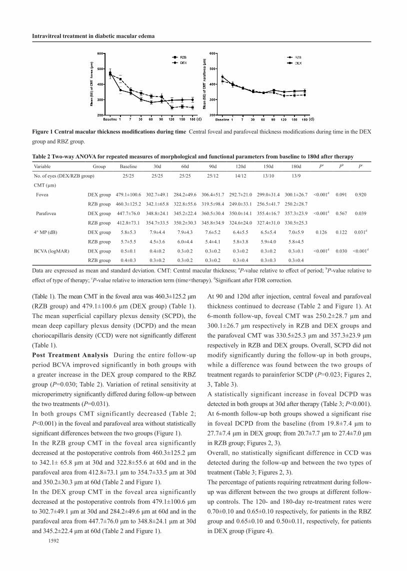
(Table 1). The mean CMT in the foveal area was 460.3±125.2 μm (RZB group) and 479.1±100.6 μm (DEX group) (Table 1). The mean superficial capillary plexus density (SCPD), the mean deep capillary plexus density (DCPD) and the mean choriocapillaris density (CCD) were not significantly different (Table 1).Post Treatment Analysis During the entire follow-up period BCVA improved significantly in both groups with a greater increase in the DEX group compared to the RBZ group (P=0.030; Table 2). Variation of retinal sensitivity at microperimetry significantly differed during follow-up between the two treatments (P=0.031).In both groups CMT significantly decreased (Table 2; P<0.001) in the foveal and parafoveal area without statistically significant differences between the two groups (Figure 1). In the RZB group CMT in the foveal area significantly decreased at the postoperative controls from 460.3±125.2 μm to 342.1± 65.8 μm at 30d and 322.8±55.6 at 60d and in the parafoveal area from 412.8±73.1 μm to 354.7±33.5 μm at 30d and 350.2±30.3 μm at 60d (Table 2 and Figure 1).In the DEX group CMT in the foveal area significantly decreased at the postoperative controls from 479.1±100.6 μm to 302.7±49.1 μm at 30d and 284.2±49.6 μm at 60d and in the parafoveal area from 447.7±76.0 μm to 348.8±24.1 μm at 30d and 345.2±22.4 μm at 60d (Table 2 and Figure 1).
At 90 and 120d after injection, central foveal and parafoveal thickness continued to decrease (Table 2 and Figure 1). At 6-month follow-up, foveal CMT was 250.2±28.7 μm and 300.1±26.7 μm respectively in RZB and DEX groups and the parafoveal CMT was 330.5±25.3 μm and 357.3±23.9 μm respectively in RZB and DEX groups. Overall, SCPD did not modify significantly during the follow-up in both groups, while a difference was found between the two groups of treatment regards to parainferior SCDP (P=0.023; Figures 2, 3, Table 3).A statistically significant increase in foveal DCPD was detected in both groups at 30d after therapy (Table 3; P<0.001). At 6-month follow-up both groups showed a significant rise in foveal DCPD from the baseline (from 19.8±7.4 μm to 27.7±7.4 μm in DEX group; from 20.7±7.7 μm to 27.4±7.0 μm in RZB group; Figures 2, 3).Overall, no statistically significant difference in CCD was detected during the follow-up and between the two types of treatment (Table 3; Figures 2, 3).The percentage of patients requiring retreatment during follow-up was different between the two groups at different follow-up controls. The 120- and 180-day re-treatment rates were 0.70±0.10 and 0.65±0.10 respectively, for patients in the RBZ group and 0.65±0.10 and 0.50±0.11, respectively, for patients in DEX group (Figure 4).
Table 2 Two-way ANOVA for repeated measures of morphological and functional parameters from baseline to 180d after therapyVariable Group Baseline 30d 60d 90d 120d 150d 180d Pa Pb Pc
No. of eyes (DEX/RZB group) 25/25 25/25 25/25 25/12 14/12 13/10 13/9
CMT (µm)
Fovea DEX group 479.1±100.6 302.7±49.1 284.2±49.6 306.4±51.7 292.7±21.0 299.0±31.4 300.1±26.7 <0.001d 0.091 0.920
RZB group 460.3±125.2 342.1±65.8 322.8±55.6 319.5±98.4 249.0±33.1 256.5±41.7 250.2±28.7
Parafovea DEX group 447.7±76.0 348.8±24.1 345.2±22.4 360.5±30.4 350.0±14.1 355.4±16.7 357.3±23.9 <0.001d 0.567 0.039
RZB group 412.8±73.1 354.7±33.5 350.2±30.3 345.8±34.9 324.6±24.0 327.4±31.0 330.5±25.3
4° MP (dB) DEX group 5.8±5.3 7.9±4.4 7.9±4.3 7.6±5.2 6.4±5.5 6.5±5.4 7.0±5.9 0.126 0.122 0.031d
RZB group 5.7±5.5 4.5±3.6 6.0±4.4 5.4±4.1 5.8±3.8 5.9±4.0 5.8±4.5
BCVA (logMAR) DEX group 0.5±0.1 0.4±0.2 0.3±0.2 0.3±0.2 0.3±0.2 0.3±0.2 0.3±0.1 <0.001d 0.030 <0.001d
RZB group 0.4±0.3 0.3±0.2 0.3±0.2 0.3±0.2 0.3±0.4 0.3±0.3 0.3±0.4
Data are expressed as mean and standard deviation. CMT: Central macular thickness; aP-value relative to effect of period; bP-value relative to effect of type of therapy; cP-value relative to interaction term (time×therapy). dSignificant after FDR correction.
Figure 1 Central macular thickness modifications during time Central foveal and parafoveal thickness modifications during time in the DEX group and RBZ group.
Intravitreal treatment in diabetic macular edema

1593
Int J Ophthalmol, Vol. 12, No. 10, Oct.18, 2019 www.ijo.cnTel: 8629-82245172 8629-82210956 Email: [email protected]
DISCUSSIONManagement of DR and its most common complications, such as DME, have improved with the development of different intravitreal drugs[4-8]. Intravitreal treatment of anti-VEGF, specifically targeting the VEGF and corticosteroids with their action of blockage the inflammatory mediators’ production, are largely widespread in the treatment of the DME condition[25]. Ranibizumab, a monoclonal anti-VEGF antibody fragment, is a safe treatment for DME with the early effects detectable as early as 7d after the first injection[9-11,26].Similarly, dexamethasone, an anti-inflammatory agent approved by Food and Drug Administration in 2014 for intravitreal treatment, represents an efficacy DME treatment[27]. In our retrospective 6-month follow-up study no significant difference in terms of morphological and functional parameters
was found between the two groups that underwent DEX and RBZ for DME treatment.CMT showed a significant decrease in both groups compared to preoperative values. Patients treated with DEX showed a tendency to a higher decrease in comparison with RZB in the short-term period. In DEX group, the greatest reduction of foveal CMT was observed at 2mo. Conversely, in RZB group the greatest reduction of foveal CMT was detected at 120d. The effect peak of the dexamethasone implant has been already previously reported to be at 30d with a mean duration of the treatment being at 4mo[28]. Several studies have already demonstrated DEX efficacy in DME improvement[6,29]. It has been described 34% of CMT reduction at 30d after DEX implantation[16]. Similarly, in literature, the ranibizumab efficacy in CMT decrease of DME patients has been reported[30]. Callanan et al have compared dexamethasone
Figure 2 OCTA images of the SCP (A and B, left panel), DCP (A and B, middle panel) and CC (A and B, right panel) At baseline (A) vessel rarefaction surrounding the foveal avascular zone in the SCP and DCP, microaneurysms and diffuse vessel rarefaction in the DCP and focal areas with no apparent flow in the CC can be observed; corresponding structural SD-OCT images centred on the fovea (A and B, left, middle and right panel with overlying segmentation bands at the level of the SCP, DCP and CC, respectively) show increased retinal thickness due to cystoid macular edema and presence of subretinal fluid. After a loading dose of ranibizumab injection (B) a restoration of vessel density mainly in the DCP can be observed with corresponding resolution of macular edema and partial resolution of subretinal fluid.

1594
Figure 3 OCTA images of the SCP (A and B, left panel), DCP (A and B, middle panel) and CC (A and B, right panel) At baseline (A) vessel rarefaction surrounding the foveal avascular zone in the SCP and DCP, microaneurysms and diffuse vessel rarefaction in the DCP and focal areas with no apparent flow in the CC can be observed; corresponding structural SD-OCT images centred on the fovea (A and B, left, middle and right with overlying segmentation bands at the level of the SCP, DCP and CC, respectively) show increased retinal thickness due to cystoid macular edema and presence of subretinal fluid. After dexamethasone implant (B) a restoration of vessel density mainly in the DCP can be observed with corresponding resolution of macular edema and subretinal fluid.
with ranibizumab for the treatment of DME and demonstrated that the mean decrease in CMT from baseline was greater with the corticosteroids than the anti-VEGF at 1 and 2mo after injections[30]. In our study the re-treatment rate in patients treated with anti-VEGF injections was higher than those
treated with DEX implantation. A three-year randomized sham-controlled trial reported a mean of 4-5 injections over 3y in DME patients treated with DEX[6]. Similarly to our findings, the Bevordex study[31] reported a comparison between anatomic and functional outcomes using DEX and bevacizumab during over 12-month follow-up. Anatomic findings were significantly better in patients treated with DEX with fewer injections (mean of 2.7 injections) compared to patients treated with the anti-VEGF (8.6 injections).However, the final CMT was 300.1±26.7 µm in DEX group and 250.2±28.7 µm in the RZB group at 180d in our series thus probably leading to earlier retreatment in the DEX group.Using OCTA analysis, we also investigated retinal superficial and deep vessel densities and CC density in both treatment groups. Nowadays, in clinical practice routinely use of OCTA allows for a better and precise evaluation of the microvascular retinal changes in DR and DME patients[16,32]. Some studies have already reported retinal capillary network and CC
Figure 4 Kaplan-Meier curve of re-treatment according to group Continuous line is relative to patients in the RBZ group while dotter line is relative to patients in DEX group.
Intravitreal treatment in diabetic macular edema

1595
Int J Ophthalmol, Vol. 12, No. 10, Oct.18, 2019 www.ijo.cnTel: 8629-82245172 8629-82210956 Email: [email protected]
Table 3 Two-way ANOVA for repeated measures of morphological parameters from baseline to 180d after therapyVariable Group Baseline 30d 60d 90d 120d 150d 180d Pa Pb Pc
No. of eyes (DEX/RZB group) 25/25 25/25 25/25 25/12 14/12 13/10 13/9
SCPD (µm)
Whole DEX group 40.1±4.2 40.8±3.0 41.4±3.5 41.5±2.9 41.1±3.2 39.7±0.1 39.8±1.1 0.077 0.069 0.035
RZB group 39.8±4.4 40.7±6.2 42.1±3.4 43.2±4.4 45.2±3.6 45.5±3.1 45.7±3.1
Fovea DEX group 29.3±5.2 25.2±7.2 23.4±8.9 28.0±8.6 28.1±2.9 28.1±1.8 28.0±1.9 0.188 0.582 0.701
RZB group 26.8±5.8 27.2±7.1 25.3±5.2 28.6±5.0 26.3±7.1 25.6±6.0 25.0±7.5
Parafovea DEX group 41.3±4.3 41.0±5.3 41.7±5.0 40.8±3.3 41.2±3.4 40.7±1.3 41.3±1.1 0.059 0.088 0.040
RZB group 41.5±4.9 43.0±5.0 44.8±2.9 46.0±3.1 46.1±2.7 46.5±2.4 47.1±3.1
Parasuperior DEX group 40.0±5.8 42.2±5.1 42.3±5.0 41.1±3.3 41.9±3.3 40.7±.2.1 40.5±1.8 0.478 0.074 0.285
RZB group 41.3±5.1 43.4±5.4 44.5±3.9 46.0±3.0 45.1±3.7 46.0±2.9 46.1±3.9
Parainferior DEX group 41.0±6.1 42.8±4.3 40.4±4.1 41.2±3.9 41.5±3.8 40.7±0.2 41.1±0.5 0.585 0.028 0.023
RZB group 40.8±5.0 43.1±5.0 45.2±2.9 46.5±2.7 46.9±3.0 47.2±3.1 46.7±3.1
DCPD (µm)
Whole DEX group 45.4±5.1 48.3±3.8 47.2±4.4 43.0±6.2 46.5±5.7 46.5±3.1 46.4±2.7 0.482 0.788 0.344
RZB group 45.9±5.1 47.5±5.1 47.7±4.9 50.0±4.5 49.1±3.5 49.3±2.9 49.0±3.2
Fovea DEX group 19.8±7.4 26.1±8.0 24.8±6.0 27.1±6.1 27.5±8.2 27.6±8.0 27.7±7.4 0.001d 0.821 0.199
RZB group 20.7±7.7 26.2±8.0 24.4±7.2 28.8±7.0 27.8±10.4 27.2±6.4 27.4±7.0
Parafovea DEX group 48.3±3.9 50.8±4.0 48.4±4.0 47.4±5.1 48.4±6.0 47.8±3.9 48.1±3.8 0.622 0.654 0.199
RZB group 47.7±4.9 49.1±6.4 49.4±4.8 51.3±4.5 50.2±3.1 50.3±4.0 50.7±4.0
Parasuperior DEX group 46.5±6.0 50.8±4.6 49.4±4.0 47.3±6.3 50.1±4.7 47.8±3.1 47.4±2.2 0.301 0.901 0.284
RZB group 48.8±4.9 50.4±6.8 49.7±6.0 51.0±4.4 51.4±4.4 50.8±4.0 49.9±4.3
Parainferior DEX group 47.5±5.0 50.3±4.9 47.7±5.0 45.4±5.4 47.1±7.2 47.7±4.4 47.3±5.0 0.411 0.561 0.310
RZB group 47.7±3.9 48.7±5.0 49.1±4.4 51.3±4.2 50.1±3.8 51.2±3.0 51.2±4.0
CCD (µm)
Whole DEX group 63.0±1.8 65.0±1.8 65.5±1.5 64.4±1.8 60.8±6.8 57.7±8.4 58.0±9.0 0.511 0.399 0.301
RZB group 61.3±7.0 64.5±2.2 64.0±2.0 65.3±2.2 65.5±1.7 65.5±1.4 65.5±1.3
Fovea DEX group 60.8±5.7 65.9±2.8 65.4±5.4 64.5±1.8 59.3±8.0 54.8±6.0 55.4±7.1 0.522 0.822 0.598
RZB group 61.4±6.2 63.7±2.5 62.7±5.0 64.7±4.0 63.3±3.4 64.9±1.9 65.2±2.8
Parafovea DEX group 63.0±3.3 64.8±1.9 65.1±1.8 64.4±1.4 58.7±7.1 57.8±12.7 57.3±11.0 0.488 0.374 0.362
RZB group 61.5±6.0 63.4±3.1 63.9±2.0 65.2±2.7 65.8±1.9 66.8±1.5 66.9±1.7
Parasuperior DEX group 62.0±2.1 64.7±1.9 65.9±1.4 63.8±1.9 61.9±6.4 56.7±12.4 56.8±9.7 0.188 0.878 0.154
RZB group 59.9±8.8 61.8±2.2 63.8±2.7 65.1±3.8 65.3±2.7 64.5±4.3 64.7±2.3
Parainferior DEX group 62.0±3.3 65.0±1.7 65.4±1.9 65.0±2.0 58.9±7.7 57.2±10.0 58.0±8.9 0.502 0.368 0.448
RZB group 61.5±6.3 63.2±2.5 63.6±2.4 65.5±2.9 65.6±1.8 65.9±1.6 66.2±1.7
Data are expressed as mean and standard deviation. CMT: Central macular thickness; SCPD: Superior capillary plexus density; DCPD: Deep capillary plexus density; CCD: Choriocapillaris density. aP-value relative to effect of period; bP-value relative to effect of type of therapy; cP-value relative to interaction term (time×therapy). dSignificant after FDR correction.
modifications in DR patients, such as a decrease of vessel density and a significant decrease of capillary perfusion density values as retinopathy progresses[32-33]. It has been described that the reduction of vessel density was more evident in the DCP compared to the superficial plexus[32-33].In our study, deep vessel density increased significantly after both RZB and DEX injections; on the contrary, we did not find significant modifications of foveal and parafoveal retinal superficial vascular density after the two treatments.In several studies it has been observed that in DME patients DCP is severely damaged showing reduced density, ectatic vessels, no flow areas corresponding to cysts[32,34-35]. In
particular, sites of macular edema are mainly localized in the deep plexus in regions of reduced or absent flow.It has been speculated that DCP could be a potential predictor of the effectiveness of the DME treatment. Lee et al[32] found a significant correlation between the status of DCP and the therapy response. We hypothesize that the modification of vessel density after treatment could be related to two factors: disappearance of macular edema and steroid and anti-VEGF effect on vessel diameter.The modification of vessel density in DR complicated by DME could be in part related to vessel displacement by intraretinal fluid particularly when retinal cysts are present, thus edema

1596
reduction or resolution could modify the vessel distribution. In addition, vessel caliber could change due to the drug effect thus influencing vessel density assessment. The blockage of VEGF due to intravitreal steroid such as dexamethasone or anti-VEGF injections can lead to a reduction of arteriolar or venular vessel diameter with a resolution or improvement of macular edema[36-37]. The CCD after treatment did not show any significant increase in both groups.Capillary perfusion density has been found reduced in patients suffering from DR with greater reduction at increasing disease severity[33]. As previously reported in retinal vein occlusion complicated by macular edema it can be hypothesized that overlying retinal edema could attenuate the OCT signal of the CC[38]. A role of anti-VEGF and dexamethasone in influencing directly CCD could be considered and investigated. Regarding functional parameters, overall retinal sensitivity detected with microperimetry increased significantly after therapy. It is probably related to a rearrangement of foveal architecture and to the status of photoreceptors, also after the improvement of DME[39]. On the contrary, the BCVA showed a statistically significant increase in both groups of treatment, with the highest gain at 60d post implant in the patients treated with DEX. This study has some limitations such as the relatively small sample of eyes examined presenting only no proliferative moderate DR stage, the short follow-up and the retrospective nature.In conclusion, RBZ and DEX appeared both safe and effective therapies for DME. The corticosteroid medication showed an earlier short-term effect with a lower retreatment rate compared to ranibizumab. Nevertheless, the two different intravitreal treatments both allowed a fast improvement of the pathology in terms of anatomical and functional outcomes.ACKNOWLEDGEMENTSConflicts of Interest: Mastropasqua L, None; Di Staso S, None; D’Aloisio R, None; Mastropasqua A, None; Di Antonio L, None; Senatore A, None; Ciancaglini M, None; Di Nicola M, None; Di Martino G, None; Tognetto D, None; Toto L, None.REFERENCES
1 Habib SL, Rojna M. Diabetes and risk of cancer. ISRN Oncol
2013;2013:583786.
2 Varma R, Bressler NM, Doan QV, Gleeson M, Danese M, Bower
JK, Selvin E, Dolan C, Fine J, Colman S, Turpcu A. Prevalence of and
risk factors for diabetic macular edema in the United States. JAMA
Ophthalmol 2014;132(11):1334-1340.
3 Photocoagulation for diabetic macular edema. Early Treatment Diabetic
Retinopathy Study report number 1. Early Treatment Diabetic Retinopathy
Study research group. Arch Ophthalmol 1985;103(12):1796-1806.
4 Funatsu H, Yamashita H, Sakata K, Noma H, Mimura T, Suzuki M,
Eguchi S, Hori S. Vitreous levels of vascular endothelial growth factor
and intercellular adhesion molecule 1 are related to diabetic macular
edema. Ophthalmology 2005;112(5):806-816.
5 Martidis A, Duker JS, Greenberg PB, Rogers AH, Puliafito CA, Reichel
E, Baumal C. Intravitreal triamcinolone for refractory diabetic macular
edema. Ophthalmology 2002;109(5):920-927.
6 Boyer DS, Yoon YH, Belfort R Jr, Bandello F, Maturi RK, Augustin
AJ, Li XY, Cui H, Hashad Y, Whitcup SM, Ozurdex MEAD Study
Group. Three-year, randomized, sham-controlled trial of dexamethasone
intravitreal implant in patients with diabetic macular edema. Ophthalmology
2014;121(10):1904-1914.
7 Zhioua I, Semoun O, Lalloum F, Souied EH. Intravitreal dexamethasone
implant in patients with ranibizumab persistent diabetic macular edema.
Retina 2015;35(7):1429-1435.
8 Totan Y, Güler E, Gürağaç FB. Dexamethasone intravitreal implant
for chronic diabetic macular edema resistant to intravitreal bevacizumab
treatment. Curr Eye Res 2016;41(1):107-113.
9 Nguyen QD, Brown DM, Marcus DM, et al. Ranibizumab for diabetic
macular edema: results from 2 phase III randomized trials: RISE and
RIDE. Ophthalmology 2012;119(4):789-801.
10 Mitchell P, Bandello F, Schmidt-Erfurth U, et al. The RESTORE
study: ranibizumab monotherapy or combined with laser versus
laser monotherapy for diabetic macular edema. Ophthalmology
2011;118(4):615-625.
11 Massin P, Bandello F, Garweg JG, et al. Safety and efficacy of
ranibizumab in diabetic macular edema (RESOLVE Study): a 12-month,
randomized, controlled, double-masked, multicenter phase II study.
Diabetes Care 2010;33(11):2399-2405.
12 Egan C, Zhu HG, Lee A, et al. The United Kingdom Diabetic
Retinopathy Electronic Medical Record Users Group, Report 1: baseline
characteristics and visual acuity outcomes in eyes treated with intravitreal
injections of ranibizumab for diabetic macular oedema. Br J Ophthalmol
2017;101(1):75-80.
13 Guigou S, Pommier S, Meyer F, Hajjar C, Merite PY, Parrat E,
Rouhette H, Rebollo O, Matonti F. Efficacy and safety of intravitreal
dexamethasone implant in patients with diabetic macular edema.
Ophthalmologica 2015;233(3-4):169-175.
14 Dutra Medeiros M, Postorino M, Navarro R, Garcia-Arumí J, Mateo
C, Corcóstegui B. Dexamethasone intravitreal implant for treatment
of patients with persistent diabetic macular edema. Ophthalmologica
2014;231(3):141-146.
15 Matonti F, Guigou S, Pommier S, Meyer F, Hajjar C, Merite PY, Parrat
E, Rouhette H, Rebollo O, Soler V. Dexamethasone implants in patients
with naive diabetic macular edema. Ophthalmologica 2016;235(4):244.
16 Toto L, D’Aloisio R, Di Nicola M, Di Martino G, Di Staso S,
Ciancaglini M, Tognetto D, Mastropasqua L. Qualitative and quantitative
assessment of vascular changes in diabetic macular edema after
dexamethasone implant using optical coherence tomography angiography.
Int J Mol Sci 2017;18(6):E1181.
Intravitreal treatment in diabetic macular edema

1597
Int J Ophthalmol, Vol. 12, No. 10, Oct.18, 2019 www.ijo.cnTel: 8629-82245172 8629-82210956 Email: [email protected]
17 Iglicki M, Busch C, Zur D, et al. Dexamethasone implant for diabetic
macular edema in naive compared with refractory eyes: the international
retina group real-life 24-month multicenter study. the IRGREL-DEX
study. Retina 2019;39(1):44-51.
18 Wilkinson CP, Ferris FL 3rd, Klein RE, Lee PP, Agardh CD, Davis
M, Dills D, Kampik A, Pararajasegaram R, Verdaguer JT, Global
Diabetic Retinopathy Project Group. Proposed international clinical
diabetic retinopathy and diabetic macular edema disease severity scales.
Ophthalmology 2003;110(9):1677-1682.
19 Karbassi M, Khu PM, Singer DM, Chylack LT Jr. Evaluation of lens
opacities classification system III applied at the slitlamp. Optom Vis Sci
1993;70(11):923-928.
20 Jia YL, Tan O, Tokayer J, Potsaid B, Wang YM, Liu JJ, Kraus
MF, Subhash H, Fujimoto JG, Hornegger J, Huang D. Split-spectrum
amplitude-decorrelation angiography with optical coherence tomography.
Opt Express 2012;20(4):4710.
21 Jia YL, Morrison JC, Tokayer J, Tan O, Lombardi L, Baumann B, Lu
CD, Choi W, Fujimoto JG, Huang D. Quantitative OCT angiography of
optic nerve head blood flow. Biomed Opt Express 2012;3(12):3127-3137.
22 Mastropasqua R, Toto L, Di Antonio L, Borrelli E, Senatore A, Di
Nicola M, Di Martino G, Ciancaglini M, Carpineto P. Corrigendum:
Optical coherence tomography angiography microvascular findings in
macular edema due to central and branch retinal vein occlusions. Sci Rep
2017;7:42570.
23 Samara WA, Shahlaee A, Sridhar J, Khan MA, Ho AC, Hsu J.
Quantitative optical coherence tomography angiography features
and visual function in eyes with branch retinal vein occlusion. Am J
Ophthalmol 2016;166:76-83.
24 Toto L, Borrelli E, Mastropasqua R, Senatore A, Di Antonio L, Di
Nicola M, Carpineto P, Mastropasqua L. Macular features in retinitis
pigmentosa: correlations among ganglion cell complex thickness,
capillary density, and macular function. Invest Ophthalmol Vis Sci
2016;57(14):6360-6366.
25 Miyamoto K, Khosrof S, Bursell SE, Rohan R, Murata T, Clermont
AC, Aiello LP, Ogura Y, Adamis AP. Prevention of leukostasis and
vascular leakage in streptozotocin-induced diabetic retinopathy via
intercellular adhesion molecule-1 inhibition. Proc Natl Acad Sci USA
1999;96(19):10836-10841.
26 Nguyen QD, Shah SM, Khwaja AA, et al. Two-year outcomes of
the ranibizumab for edema of the mAcula in diabetes (READ-2) study.
Ophthalmology 2010;117(11):2146-2151.
27 Shah AR, Xi MQ, Abbey AM, Yonekawa Y, Faia LJ, Hassan TS, Ruby
AJ, Wolfe JD. Short-term efficacy of intravitreal dexamethasone implant
in vitrectomized eyes with recalcitrant diabetic macular edema and prior
anti-VEGF therapy. J Ophthalmic Vis Res 2016;11(2):183-187.
28 Panozzo G, Gusson E, Panozzo G, Dalla Mura G. Dexamethasone
intravitreal implant for diabetic macular edema: indications for a PRN
regimen of treatment. Eur J Ophthalmol 2015;25(4):347-351.
29 Mastropasqua R, Toto L, Borrelli E, Di Antonio L, De Nicola C,
Mastrocola A, Di Nicola M, Carpineto P. Morphology and function
over a one-year follow up period after intravitreal dexamethasone
implant (ozurdex) in patients with diabetic macular edema. PLoS One
2015;10(12):e0145663.
30 Callanan DG, Loewenstein A, Patel SS, Massin P, Corcóstegui B, Li
XY, Jiao J, Hashad Y, Whitcup SM. A multicenter, 12-month randomized
study comparing dexamethasone intravitreal implant with ranibizumab in
patients with diabetic macular edema. Graefes Arch Clin Exp Ophthalmol
2017;255(3):463-473.
31 Gillies MC, Lim LL, Campain A, Quin GJ, Salem W, Li J, Goodwin
S, Aroney C, McAllister IL, Fraser-Bell S. A randomized clinical
trial of intravitreal bevacizumab versus intravitreal dexamethasone
for diabetic macular edema: the BEVORDEX study. Ophthalmology
2014;121(12):2473-2481.
32 Lee J, Moon BG, Cho AR, Yoon YH. Optical coherence tomography
angiography of DME and its association with anti-VEGF treatment
response. Ophthalmology 2016;123(11):2368-2375.
33 Agemy SA, Scripsema NK, Shah CM, Chui T, Garcia PM, Lee JG,
Gentile RC, Hsiao YS, Zhou Q, Ko T, Rosen RB. Retinal vascular
perfusion density mapping using optical coherence tomography
angiography in normals and diabetic retinopathy patients. Retina
2015;35(11):2353-2363.
34 Mastropasqua R, Di Antonio L, Di Staso S, Agnifili L, Di Gregorio
A, Ciancaglini M, Mastropasqua L. Optical coherence tomography
angiography in retinal vascular diseases and choroidal neovascularization.
J Ophthalmol 2015;2015:343515.
35 Spaide RF. Retinal vascular cystoid macular edema: Review and New
Theory. Retina 2016;36(10):1823-1842.
36 Semeraro F, Russo A, Rizzoni D, Danzi P, Morescalchi F, Costagliola
C. Diameters and wall-to-lumen ratio of retinal arterioles in patients with
retinal vein occlusion before and after treatment with dexamethasone
intravitreal implants. J Ocul Pharmacol Ther 2014;30(7):573-579.
37 Wickremasinghe SS, Rogers SL, Gillies MC, Zhu MD, Wong TY. Retinal
vascular caliber changes after intravitreal triamcinolone treatment for diabetic
macular edema. Invest Ophthalmol Vis Sci 2008;49(11):4707-4711.
38 Kim AY, Chu ZD, Shahidzadeh A, Wang RK, Puliafito CA, Kashani
AH. Quantifying microvascular density and morphology in diabetic
retinopathy using spectral-domain optical coherence tomography
angiography. Invest Ophthalmol Vis Sci 2016;57(9):OCT362-OCT370.
39 Querques G, Lattanzio R, Querques L, Triolo G, Cascavilla ML,
Cavallero E, Del Turco C, Casalino G, Bandello F. Impact of intravitreal
dexamethasone implant (Ozurdex) on macular morphology and function.
Retina 2014;34(2):330-341.