Embed Size (px)
Citation preview

1
Clinical Practice Guideline for Diagnosis and Management of Acute Otitis Media
(AOM) in Children in Japan
Subcommittee of Clinical Practice Guideline for Diagnosis and Management of Acute
Otitis Media in Children
(Japan Otological Society, Japan Society for Pediatric Otorhinolaryngology, Japan
Society for Infectious Diseases in Otolaryngology)
January 27, 2015
1. Summary
Objective: To 1) indicate methods of diagnosis and testing for childhood (15 years)
acute otitis media (AOM); and 2) recommend methods of treatment in accordance with
the evidence-based consensus reached by the Subcommittee of Clinical Practice
Guideline for Diagnosis and Management of AOM in Children (Subcommittee of
Clinical Practice Guideline), in light of the causative bacteria and their drug sensitivity
of AOM in Japan. Methods: We investigated the most recently detected bacteria
causing childhood AOM in Japan as well as antibacterial sensitivity and the worldwide
progress of vaccination, produced Clinical Questions concerning the diagnosis, testing
methods, and treatment of AOM, searched literature published during 2000–2004, and
issued the 2006 Guidelines. In the 2009 and 2013 Guidelines we performed the same
investigation with the addition of literature, which were not included in the 2006
Guidelines and published during 2005–2008 and during 2009–2012, respectively.
Results: We categorized AOM as mild, moderate, or severe on the basis of tympanic
membrane findings and clinical symptoms, and presented recommended treatment for
each degree of severity. Conclusion: Accurate assessment of tympanic membrane
findings is important for judging the degree of severity and selecting a method of
treatment. Some of the new antimicrobial agents and pneumococcal vaccination are
recommended as new treatment options.
2. Authors

2
The membership of the Subcommittee of Clinical Practice Guideline for
Diagnosis and Management of AOM in Children is shown in Table 1. This committee
is composed of three organizations: the Japan Otological Society (JOS) , the Japan
Society for Infectious Diseases in Otolaryngology (JSIDO), and the Japan Society for
Pediatric Otorhinolaryngology (JSPO). The first committee meeting was held on
January 8, 2003, and the 2006 Guidelines were published in March that year on the
web site of the JSIDO , in the journals of the JOS and the JSPO, on the web site of the
Japan Council for Quality Healthcare, and in printed form (Otol Jpn 2006; Pediatr
Otorhinolaryngol Jpn 2006; and Japan Council for Quality Healthcare, Kanehara
Shuppan, 2006). The 2006 Guidelines underwent evaluation, and work on the
production of a revised edition began at the 13th committee meeting on January 7,
2007. The 2009 Guidelines were published in January, 2009 by Kanehara Shuppan Co.,
and they first appeared on the website of the Medical Information Network
Distribution Service (MINDS) on October 19, 2010. The production of the revised
edition (i.e., the 2013 edition) started in May 2010 (Table 1).
Table 1. The Members of the Subcommittee of Clinical Practice Guideline
Name Affiliation
Fumiyo Kudo (chairman) Dept. of Dietetics, Chiba Prefectural University of Health Sciences
Haruo Takahashi Dept. of Otolaryngology Head and Neck Surgery, Nagasaki University
Yukio Iino Dept. of Otolaryngology, Jichi Medical University Saitama Medical Center
Yoshifumi Uno UNO ENT Clinic
Yousuke Kamide Kamide ENT Clinic
Hidenobu Taiji Dept. of Otolaryngology, Saiseikai Central Hospital
Noboru Yamanaka Dept. of Otolaryngology Head and Neck Surgery, Wakayama Medical University
Kenji Suzuki Dept. of Otolaryngology, Banbuntane Houtokukai Hospital, Fujita Health University School of Medicine
Takeo Nakayama Dept. of Health Informatics, Kyoto University School of Public Health
Ken Kitamura (advisor) Dept. of Otolaryngology, Chigasaki Central Hospital

3
References
1. Clinical Practice Guideline for Diagnosis and Management of Acute Otitis Media
in Children (2006). Otol Jpn 2006; 16; Suppl 1 (in Japanese).
2. Clinical Practice Guideline for Diagnosis and Management of Acute Otitis Media
in Children. Pediatric Otorhinolaryngology Japan 2006; 27:71-107 (in Japanese).
3. The Japan Council for Quality Healthcare - Minds (http://minds.jcqhc.or.jp/).
4. Shounikyuuseichuujien shinryougaidorain 2006 nenban. Ed by the Japan
Otological Society, the Japan Society for Infectious Diseases in Otolaryngology,
and the Japan Society for Pediatric Otorhinolaryngology, Kaneharashuppan,
Tokyo, 2006.
5. Shounikyuuseichuujien shinryougaidorain 2009 nenban. Ed by the Japan
Otological Society, the Japan Society for Infectious Diseases in Otolaryngology,
and the Japan Society for Pediatric Otorhinolaryngology, Kaneharashuppan,
Tokyo, 2009.
3. Financial Backers and Sponsors
Production of these Guidelines was funded by JOS operating expenses. The
JOS does not receive support from any specific organizations or companies. A list of
organizations and companies that posed non-personal financial conflicts of interest to
members of the Subcommittee of Clinical Practice Guideline during the production of
these Guidelines is provided (attachment).
Attachment. List of pharmaceutical companies those posed non-personal financial conflicts of
interest to members of the Subcommittee of Clinical Practice Guideline (alphabetical)
Astellas Pharma Inc. AstraZeneca K.K.
Bayer Yakuhin, Ltd. Chugai Pharmaceutical Co., Ltd.
Daiichi Sankyo Company, Limited Eisai Co., Ltd.
GlaxoSmithKline K.K. KISSEI PHARMACEUTICAL CO., LTD.
KYORIN Pharmaceutical Co., Ltd. Kowa Pharmaceutical Co. Ltd.
Kowa Shinyaku Co., Ltd. Kyowa Hakko Kirin., Co. Ltd.
Lumenis Japan Co., Ltd. MSD K.K.
Meiji Seika Pharma Co. Ltd. Mitsubishi Tanabe Pharma Corporation
Nippon Boehringer Ingelheim Co., Ltd. NIPPON SHINYAKU CO., LTD.
ONO PHARMACEUTICAL CO., LTD. Otsuka Pharmaceutical Co. Ltd.
Pfizer Japan Inc. SHIONOGI & CO., LTD.
Sanofi K.K. Senju Pharmaceutical Co., Ltd.

4
Sumitomo Dainippon Pharma Co., Ltd. TAIHO PHARMACEUTICAL CO., LTD.
Taisho Toyama Pharmaceutical Co., Ltd. Takeda Pharmaceutical Company Limited
4. Introduction
AOM is a typical upper respiratory inflammation commonly affecting
children and is mainly treated by otolaryngologists. Its exact frequency of occurrence
in Japan is unknown, however. According to reports from Europe and the USA, 62%
of children aged less than one year and 83% of those up to the age of three suffer from
at least one bout of AOM (Teele et al. 1989). Faden et al (1998) have reported that it
affects 75% of children up to the age of one.
Some authors in Europe and the USA do not recommend the use of
antimicrobial agents for AOM. In the Netherlands, it has been proposed that
antimicrobial agents are unnecessary in at least 90% of cases, and that patients should
be observed for 3-4 days without antimicrobial agent administration (van Buchem et
al. 1985, Damoiseaux et al. 2000). Rosenfeld et al. have also reported observation as
management option (Rosenfeld et al. 2003a, b, c), and more recent studies have also
found no significant difference in clinical outcome if antimicrobial agents are not given
immediately but are prescribed if there is no improvement in symptoms after 48 or 72
hours (Spiro et al. 2006, Little et al. 2006). A Cochrane Review, which examined
randomized controlled trials of antimicrobial agent administration versus placebo also
found that antimicrobial agents had little effect on childhood AOM (Glaziou et al.
2004). In addition, a double-blind randomized controlled trial of amoxicillin (AMPC)
and a placebo found no significant difference in therapeutic efficacy between the two
(Le Saux et al. 2005, McCormick et al. 2005). Dagan et al. (2000, 2001) and Toltzis
et al. 2005), in a review and case-control study, advised that antimicrobial agent use
would be reduced because the use of a wide variety of drugs increases the survival of
resistant Streptcoccus pneumonia (S. pneumonia) in the epipharynx, which can cause
additional infections in middle-ear (ME) fluid.
In Japan, regular nationwide surveys are performed of the causative bacteria
for AOM, acute sinusitis, acute tonsillitis, and peritonsillar abscess. These surveys
have reported that antimicrobial agent -resistant bacteria are now being detected more
frequently (Suzuki et al. 2000, Nishimura et al. 2004), which means that
recommendations for no administration of antimicrobial agents proposed in Europe

5
and the USA is not applied. In addition, the criteria and assessment levels used in
conventional clinical assessment are not necessarily uniform even within Europe and
the USA (Chan et al. 2001). Investigation and unified evaluation of the diagnosis and
treatment of childhood AOM are therefore required, based on the actual situation in
Japan. Based on this perspective, the JOS, the JSIDO, and the JSPO produced 2006
Clinical Practice Guidelines consistent with evidence-based medicine (EBM) with the
aim of supporting the diagnosis and treatment of childhood AOM (Nakayama, 2004).
A survey of otolaryngologists and pediatricians in Ishikawa Prefecture
showed that 85% of otolaryngologists and 52% of pediatricians were aware of the 2006
edition of the Guidelines, with 56% of those otolaryngologists and 49% of those
pediatricians reporting that they used them in practice (Ito et al. 2008). Therapeutic
outcomes of clinical practice that adhered to the guidelines were also generally good
(Hayashi et al. 2007, Sugawara et al. 2008). In light of these results, JOS, JSIDO, and
JSPO decided to revise the 2006 Guidelines and issue a new edition in 2009.
Thereafter, AOM guidelines from Canada (Forgie et al. 2009) and Italy
(Marchisio et al. 2010) were published. In Italy’s guidelines, it is noteworthy that, as
in Japan’s guidelines, the identification and description of detailed tympanic
membrane findings are highlighted, and one of the options for a pediatrician when s/he
cannot identify or describe the tympanic membrane findings is to transfer the patient
to an otolaryngologist who can examine the tympanic membrane precisely by
microscopy and/or endoscopy. That principle seems to agree well with our Guidelines,
which propose the management of AOM based on the detailed observations of
tympanic membrane findings. In the 2013 AOM Guidelines published by the U.S. as
a revision of their 2004 Guidelines, the necessity of detailed observation of the
tympanic membrane findings was emphasized (Lieberthal et al. 2013).
In our present 2013 Guidelines, the changes of pathogens and their drug
sensitivity and the grading system of AOM including signs and symptoms determining
the grade were revised. Descriptions were expanded based on new data obtained by
rapid tests for the detection of pneumococcal antigen, vaccinations for Streptococcus
pneumoniae, new antibiotics, Japanese herbal medicine, and more. Although no
remarkable change has been made to the other parts of the 2009 guidelines, items
described in the 2006 and 2009 guidelines were included in the 2013 Guidelines.
These Clinical Practice Guidelines are issued only to assist clinical practice,

6
and have no binding authority on treatment (Note 1). How they should actually be used
for patients in the clinical setting is a matter to be decided in light of the patient’s
wishes and values, and based on the medical practitioner’s specialist knowledge and
experience. The fact that there is no high level of evidence demonstrating a treatment
method’s efficacy does not directly mean that it is ineffective or that it should not be
carried out. When using such methods of treatment, however, an extra degree of
consideration is required concerning evaluations of its clinical efficacy and
communication with the patient. It must be re-emphasized that recommendations in
clinical practice guidelines are not legal grounds for the content of medical treatment
that should be practiced in individual clinical situations (Hurwitz 1999). These
Guidelines will be periodically revised to reflect the opinions of users and patients and
as a result of external evaluation, in the same way as the 2006 and 2009 Guidelines
were revised after their publication.
Note 1: Guidelines are ranked as follows:
Regulations directives recommendations guidelines (From A Dictionary of
Epidemiology, trans. Japan Epidemiological Association ed. J. Last, (with additions)
References
1. Teele DW, Klein JO, Rosner B, The Greater Boston Otitis Media Study Group.
Epidemiology of otitis media during the first seven years of life in children in
Greater Boston: a prospective cohort study. J Infect Dis 1989; 160:83-94.
2. Faden H, Duffy L, Boeve M. Otitis media: back to basics. Pediatr Infect Dis J
1998; 17:1105-13.
3. van Buchem FL, Peeters NIF, van't Hof MA. Acute otitis media: a new treatment
strategy. BMJ 1985; 290:1033-7.
4. Damoiseaux R, van Balen FAM, Hoes A, Verheij T, deMelker R. Primary care
based randomised, double blind trial of amoxicillin versus placebo for acute otitis
media in children aged under 2 years. BMJ 2000; 320:350-4.
5. Rosenfeld RM, Bluestone CD eds. Evidence-Based Otitis Media 2nd ed.
2003(a):199-226.
6. Rosenfeld RM, Kay D. Natural history of untreated otitis media. In: Rosenfeld RM,
Bluestone CD eds. Evidence-Based Otitis Media 2nd ed. 2003(b):180-98.

7
7. Rosenfeld RM. Kay D. Natural history of untreated otitis media. Laryngoscope
2003(c); 113:1645-57.
8. Spiro DM, Tay KY, Arnold DH, Dziura JD, Baker MD, Shapiro ED. Wait-and-see
prescription for the treatment of acute otitis media: a randomized controlled trial.
JAMA 2006;296:1235-41.
9. Little P, Moore M, Warner G, Dunleavy J, Williamson I. Longer term outcomes
from a randomised trial of prescribing strategies in otitis media. Br J Gen Pract
2006; 56:176-82.
10. Glasziou PP, Del Mar CB, Sanders SL, Hayem M. Antibiotics for acute otitis media
in children. Cochrane Database of Systematic Reviews: 2004, Issue 1.
11. Le Saux N, Gaboury I, Baird M, Klassen TP, MacCormick J, Blanchard C, Pitters
C, Sampson M, Moher D. A randomized, double-blind, placebo-controlled
noninferiority trial of amoxicillin for clinically diagnosed acute otitis media in
children 6 months to 5 years of age. CMAJ 2005; 172:335-41.
12. McCormick DP, Chonmaitree T, Pittman C, Saeed K, Friedman NR, Uchida T,
Baldwin CD. Nonsevere acute otitis media: a clinical trial comparing outcomes of
watchful waiting versus immediate antibiotic treatment. Pediatrics 2005; 115:
1455-65.
13. Dagan R. Treatment of acute otitis media - challenges in the era of antibiotic
resistance. Vaccine 2000; 19 Suppl 1:S9-S16.
14. Dagan R, Leibovitz E, Cheletz G, Leiberman A, Porat N. Antibiotic treatment in
acute otitis media promotes superinfection with resistant Streptococcus
pneumoniae carried before initiation of treatment. J Infect Dis 2001; 183:880-6.
15. Toltzis P, Dul M, O'Riordan MA, Toltzis H, Blumer JL. Impact of amoxicillin on
pneumococcal colonization compared with other therapies for acute otitis media.
Pediatr Infect Dis J 2005; 24:24-8.
16. Suzuki K. The status quo of drug-resistant bacteria in pediatric otolaryngological
infectious diseases. Pediatr Otorhinolaryngol Jpn 2000; 21:26-31 (in Japanese).
17. Nishimura T, Suzuki K, Oda M, Kobayashi T, Yajin K, Yamanaka N, et al. The
third nationwide survey of clinical isolates from patients with otolaryngological
field infections. J Jpn Soc Infect Dis Otolaryngol 2004; 22:12−23 (in Japanese).
18. Chan LS, Takata GS, Shekelle P, Morton SC, Mason W, Marcy SM. Evidence
assessment of management of acute otitis media: II. Research gaps and priorities

8
for future research. Pediatrics 2001; 108:248-54.
19. Nakayama T. Clinical Practice Guideline Consistent with Evidence-based
Medicine Manual for Development and Application. Kanehara Shuppan, Tokyo、
2004 (in Japanese).
20. Ito M, Furukawa M. Usefulness of the Guideline of Diagnosis and Management of
Acute Otitis Media. Pediatr Otorhinolaryngol Jpn 2008; 29:20-4 (in Japanese).
21. Hayashi T, Abe Y, Ueda S, Otaka T, bando N, Katada A, et al. Treatment outcome
of acute otitis media in children treated with the newly released guideline in Japan.
Otol Jpn 2007; 17:118-23 (in Japanese).
22. Sugahara K, Yamashita Y. Treatment based on the guidelines for pediatric acute
otitis media. Pediatr Otorhinolaryngol Jpn 2007; 28:206-10 (in Japanese).
23. Forgie S, Zhanel G, J Robinson J. Management of acute otitis media. Paediatr
Child Health 2009; 14: 457–60.
24. Marchisio P, Bellussi L, Di Mauro G, Doria M, Felisati G, Longhi R, Novelli A,
Speciale A, Mansi N, Principi N. Acute otitis media: From diagnosis to prevention.
Summary of the Italian guideline. Int J Pediatr Otorhinolaryngol. 2010; 74:1209-
16.
25. Lieberthal AS, Carroll AE, Chonmaitree T, Ganiats TG, Hoberman A, Jackson MA,
Joffe MD, Miller DT, Rosenfeld RM, Sevilla XD, Schwartz RH, Thomas PA,
Tunkel DE. The diagnosis and management of acute otitis media. Pediatrics 2013;
131:e964-e99
26. Hurwitz B. Legal and political considerations of clinical practice guidelines. BMJ
1999; 318:661-4.
5. Objective and Aim of Production
These Guidelines were produced to describe diagnostic and testing methods
for childhood AOM (below the age of 15* [see note]), and represent the evidence-
based consensus of the members of the Subcommittee of Clinical Practice Guideline
on recommended treatment methods in light of the causative bacteria and their
antimicrobial agents sensitivity in cases of AOM in Japan. The aim is for these
Guidelines to be used to assist clinical decision-making in the care of childhood AOM,
and for them to prove beneficial in the diagnosis and treatment of patients with AOM.
9
*Note: In the Ministry of Health, Labor and Welfare’s Pharmaceutical Affairs Bureau
Notification No. 1334, Guidance Concerning Clinical Trials of Drugs in Pediatric
Populations, released on December 15, 2000, the following have been proposed as age
categories for the design of clinical trials of drugs on pediatric patients: preterm
neonates, full-term neonates (0–27 days), infants (28 days to 23 months), children (2–
11 years), and adolescents (12–16 or 12–18 years). In these Guidelines, we have used
the general criterion for children of 15 years.
References
1. Guidance Concerning Clinical Trials of Drugs in Pediatric Populations. Ministry
of Health, Labor and Welfare’s Pharmaceutical Affairs Bureau Notification No.
1334, released on December 15, 2000.
6. Users
The main users of these Guidelines will be otolaryngologists who perform
otological procedures including the accurate evaluation of tympanic membrane
findings and myringotomy.
7. Subjects
The subjects of these Guidelines are AOM patients aged 15 years who were
free from AOM or otitis media with effusion (OME) within one month prior to onset,
who do not have a ventilation tube inserted, have no cranial or facial deformity, and
do not suffer from immunodeficiency. Patients with the following conditions are
excluded as subjects: AOM with complications including facial palsy and inner ear
disorder, elevated pinna with acute mastoiditis, and AOM with Gradenigo’s syndrome
or similar findings. It can be difficult to distinguish between AOM and bullous
myringitis, but the latter is not covered by these Guidelines.
The consensus reached by the Subcommittee of Clinical Practice Guideline
on recurrent otitis media (ROM) (with a proposed definition explained below) has been
included as an additional statement.
Treatment algorithms are included at the end of the Guidelines, in which
cases that have not improved after tertiary treatment according to each treatment
algorithm are classed as intractable. The care of intractable cases is not covered in
10
these Guidelines.
[Addendum] Proposals for the management of ROM
(a) Definition of ROM
The definition of ROM has yet to be standardized either in Japan or
internationally, but in these Guidelines it has been defined as three or more occurrences
of AOM within the previous six months, or four or more within the previous 12 months,
as generally used in comparatively recent studies(Sher et al. 2005,Ables et al. 2004,
Arrieta et al. 2004).
(b) Pathophysiology of and risk factors for ROM
The pathophysiology of ROM can be categorized into two types: recurrent
simple AOM, and recurrent AOM occurring as an acute exacerbation in patients
suffering from OME.
Proposed risk factors for ROM include young age, resistant causative
bacteria, immunity of the affected individual, and lifestyle and environmental factors.
Genetic make-up has also been reported as a risk factor in young children aged 2
years(Wiertsema et al. 2006). In terms of causative bacteria, multidrug-resistant
pneumococci are reportedly responsible in many cases(van Kempen et al. 2004),
with incomplete elimination from the nasopharynx owing to reduced antimicrobial
agent efficacy regarded as one cause of recurrence. The involvement of decreased
immune response by the host to the causative bacteria is also important(Yamanaka et
al. 2008). A link between immunity received from the mother via breast milk and the
onset of ROM has also been conjectured, with the absence of breastfeeding
constituting a strong risk factor for ROM(Lubianca Neto et al. 2006). Lifestyle and
environmental risk factors include having siblings, attending daycare, and pacifier use
(Lubianca Neto et al. 2006).
Regarding other risk factors for ROM, the involvement of gastroesophageal
reflux (GERD) has been reported based on the results of monitoring. Lelepic et al.
(2000) presented results obtained by continual 24-h esophageal pH monitoring of
GERD, and other reports provided data concerning the amount of pepsin/pepsinogen
protein content of MEE (Taker et al. 2002, Rozmanic et al. 2002). It was reported that
two randomized trials found no benefit of antireflux treatment with proton pump
inhibitors (Miura et al. 2011)
11
(c) Treatment of ROM
With the factors described above assumed to constitute risk factors for ROM,
bacterial sensitivity tests must always be carried out prior to antimicrobial agent
administration to counteract resistant causative bacteria, and an appropriate dose of
antimicrobial agents must be selected. Recommended antimicrobial agents are listed
in these Guidelines.
Pneumococcal conjugate vaccine is used in Europe and the USA to prevent
ROM. In a double-blind randomized controlled trial of a 7-valent pneumococcal
conjugate vaccine and pneumococcal polysaccharide vaccine in Holland, there was no
significant reduction in the frequency of occurrence of ROM (Brouwer et al. 2005).
Although a Cochrane Review accepts the utility of pneumococcal polysaccharide
vaccine, it does not recommend the conjugate vaccine (Straetemans et al. 2004). In a
double-blind randomized controlled trial in the Czech Republic, however, 11-valent
pneumococcal capsular polysaccharide vaccine conjugated to protein D had a
significant protective effect against AOM caused by pneumococci or influenza viruses
(Prymula et al. 2006). In Japan, 7-valent pneumococcal conjugate vaccine was
approved for use in 2010. This vaccine covers 60.6% of pneumococcal serotypes
isolated from the middle ears of childhood AOM patients in Japan and 87% of
antimicrobial agent-resistant bacteria, and is anticipated to provide up to about 17%
protection against all forms of AOM.
One form of treatment unique to Japan that has been proposed is the use of
Chinese herbal medicines for their protective effect in boosting immunity, and Juzen-
taiho-to has been reported as effective (Maruyama et al. 2008, Yoshizaki et al. 2012).
Adenoidectomy has not been shown to reduce the frequency of ROM as a surgical
treatment in double-blind randomized controlled trials, nor is it regarded as having any
preventive effect (Oomen et al. 2005,Hammaren-Malmi et al. 2005,Koivunen et al.
2004). Myringotomy has not been shown to have any significant effect in reducing the
frequency of occurrence of ROM in research on patients in Japan (Nomura et al. 2005),
but insertion of a ventilation tube for one year and short-term insertion for one month
significantly reduce the frequency of occurrence (Uno 2007a, b). As measures to deal
with lifestyle and environmental factors, group daycare should be discontinued and
breastfeeding is desirable.
12
[Addendum] Proposals for the management of refractory otitis media (OM) and
prolonged OM
Among patients with AOM diagnosed as having a moderate or severe degree
of AOM at the first visit, some show no improvement or even worsening in the
symptoms or tympanic membrane findings after treatments based on the Guidelines;
for example, otorrhea continues or the bulging tympanic membrane recurs after the
closure of a myringotomy.
However, the tympanic membrane in some patients with a special condition
called semi-hot ear (Sade 1979) shows findings similar to AOM without significant
symptoms of AOM. Due to the lack of symptoms of AOM, we clinicians sometimes
encounter such cases.
Cases showing no improvement in the signs even after a third-line treatment
from the Guidelines should be regarded as refractory OM, and it is necessary to
consider the pathophysiology and background of such cases. We herewith propose a
definition of refractory OM, and we describe its background and pathophysiology.
1) Definition of refractory OM in the treatment of AOM
The definition of refractory OM has not yet been standardized in Japan or
internationally. In the field of infectious disease medicine, a condition tends to be
regarded as refractory when it does not improve despite continuous treatment.
According to this concept, refractory OM may be defined as incurable OM resistant to
treatments based on the Guidelines, including no improvement of tympanic membrane
findings and/or the symptoms persist or even become worse.
Refractory OM can thus be defined as a state in which symptoms and/or
findings of tympanic membrane persist or become worse despite treatments for
infection.
2) Background and pathophysiology of refractory OM
When severe tympanic membrane findings continue despite treatment
including a sufficient amount of antibiotics and drainage by myringotomy, it is
necessary to recheck aspects of the affected individual’s condition such as immunity,
neutrophil dysfunction, vulnerability to infection, GERD (Velepic et al. 2000, Tasker

13
et al. 2002, Rozmanic et al. 2002, Miura et al. 2012) and causative bacteria, and to
reconsider administering more intensive treatments including intravenous
antimicrobial agents and the insertion of a tympanostomy tube.
3) Definition of prolonged OM in the treatment of AOM
The definition of prolonged OM includes acute onset, abnormal findings of tympanic
membrane similar to AOM persisting for more than 3 weeks, and no appearance of
signs of acute symptoms such as otalgia and fever. This state is called semi-hot ear
(Sade 1979) or the state of subacute phase, as reported in the report of a research
conference on recent advances in OME (Senturia et al. 1980).
4) Definitions and classifications of terms associated with AOM
The definitions and classifications of terms associated with AOM at this stage are as
follows.
ROM: The state in which AOM occurs more than three times within the last 6
months, or more than four times within the last 12 months (Sher et al. 2005,
Ables et al. 2004, Arrieta et al. 2004, Clinical Practice Guideline for the
Diagnosis and Management of AOM in Children 2009 in Japan).
Refractory OM: The state in which symptoms and/or findings of the tympanic
membrane persist or even become worse despite continuing infection treatment.
Prolonged OM: The state in which abnormal findings of the tympanic membrane
similar to AOM persist for more than 3 weeks without the appearance of acute
symptoms such as otalgia and fever.
The definitions of relapse and recurrence are as follows.
Relapse: The tympanic membrane findings become worse and symptoms of
AOM reappear after the findings and symptoms improved.
Recurrence: AOM occurs within 3 weeks after the tympanic membrane findings
became normalized.
References
1. Sher L, Arguedas A, Husseman M, Pichichero M, Hamed KA, Biswas D, Pierce P,
Echols R. Randomized, investigator-blinded, multicenter, comparative study of
14
gatifloxacin versus amoxicillin/clavulanate in recurrent otitis media and acute
otitis media treatment failure in children. Pediatr Infect Dis J 2005; 24:301-8.
2. Ables AZ, Warren PK. High-dose azithromycin or amoxicillin-clavulanate for
recurrent otitis media? J Fam Pract 2004; 53:186-8.
3. Arrieta A, Singh J. Management of recurrent and persistent acute otitis media: new
options with familiar antibiotics. Pediatr Infect Dis J 2004; 23:S115-24.
4. Wiertsema SP, Herpers BL, Veenhoven RH, Salimans MM, Ruven HJ, Sanders
EA, Rijkers GT. Functional polymorphisms in the mannan-binding lectin 2 gene:
effect on MBL levels and otitis media. J Allergy Clin Immunol 2006; 117:1344-50.
5. van Kempen MJ, Vaneechoutte M, Claeys G, Verschraegen GL, Vermeiren J,
Dhooge IJ. Antibiotic susceptibility of acute otitis media pathogens in otitis-prone
Belgian children. Eur. J Pediatr 2004; 163:524-9.
6. Yamanaka N, Hotomi M, Billal DS. Clinical bacteriology and immunology in acute
otitis media in children. J Infect Chemother 2008; 14:180-7.
7. Lubianca Neto JF, Hemb L, Silva DB. Systematic literature review of modifiable
risk factors for recurrent acute otitis media in childhood. J Pediatr (Rio J). 2006;
82:87-96.
8. Velepic M, Rozmanic V, Velepic M, Bonifacic M. Gastroesophageal reflux, allergy
and chronic tubotympanal disorders in children. Int J Pedatr Otorhinolaryngol
2000; 55:187-190.
9. Tasker A, Dettmar PW, Panetti M, Koufman JA, P Birchall J, Pearson JP. Is gastric
reflux a cause of otitis media with effusion in children? Laryngoscope 2002;
112:1930–1934.
10. Rozmanic V, Velepic M, Ahel V, Bonifacic D, Velepic M. Prolonged esophageal
pH monitoring in the evaluation of gastroesophageal reflux in children with
chronic tubotympanal disorders. J Pediatr Gastroentrol Nutr 2002; 34: 278–80.
11. Miura MS, Mascaro M, Rosenfeld RM. Association between otitis media and
gastroesophageal reflux: a systematic review. Otolaryngol Head Neck Surg
2012;146: 345-52. Epub 2011 Dec 9.
12. Brouwer CN, Maille,AR, Rovers MM, Veenhoven RH, Grobbee DE, Sanders EA,
Schilder AG. Effect of pneumococcal vaccination on quality of life in children with
recurrent acute otitis media: a randomized, controlled trial. Pediatrics 2005; 115:
273-9.

15
13. Straetemans M, Sanders EAM, Veenhoven RH, Schilder AGM, Damoiseaux
RAMJ, Zielhuis GA. Pneumococcal vaccines for preventing otitis media.
Cochrane Database of Systematic Reviews. 2004 issue 1.
14. Prymula R, Peeters P, Chrobok V, Kriz P, Novakova E, Kaliskova E, Kohl I,
Lommel P, Poolman J, Prieels JP, Schuerman L. Pneumococcal capsular
polysaccharides conjugated to protein D for prevention of acute otitis media caused
by both Streptococcus pneumoniae and non-typable Haemophilus influenzae: a
randomised double-blind efficacy study. Lancet 2006; 367:740-8.
15. Maruyama Y, Hoshida S, Furukawa M, Ito M. Effects of Japanese herval medicine,
Juzen-taiho-to, in otitis-prone children -a preliminary study. Acta Otolaryngol
2008; 12:1-5 (Epub ahead of print).
16. Yoshizaki T: Multicenter, randomized controlled study on the efficacy of Juzen-
taiho-to for children with recurrent otitis media (H21- clinical research 007).
Report for Japan Health and Labor Sciences Research Grant, 2012.
17. Oomen KP, Rovers MM, van den Akker EH, van Staaij BK, Hoes AW, Schilder
AG. Effect of adenotonsillectomy on middle ear status in children. Laryngoscope
2005; 115:731-4.
18. Hammaren-Malmi S, Saxen H, Tarkkanen J, Mattila PS. Adenoidectomy does not
significantly reduce the incidence of otitis media in conjunction with the insertion
of tympanostomy tubes in children who are younger than 4 years: a randomized
trial. Pediatrics 2005; 116(1):185-9.
19. Koivunen P, Uhari M, Luotonen J, Kristo A, Raski R, Pokka T, Alho OP.
Adenoidectomy versus chemoprophylaxis and placebo for recurrent acute otitis
media in children aged under 2 years: randomised controlled trial. BMJ (Clinical
research ed.) 2004; 328:487.
20. Nomura Y, Ishibashi T, Yano J, Ichikawa T, Shinogami M, Monobe H, Hirai R,
Kaga K. Effect of myringotomy on prognosis in pediatric acute otitis media. Int J
Pediatr Otorhinolaryngol 2005; 69:61-4.
21. Uno Y. Effects of long-term tympanostomy tubes in children with intractable otitis
media. Otol Jpn 2007(a); 17:16-25 (in Japanese).
22. Uno Y. Effects of short-term tympanostomy tube on intractable recurrent otitis
media in children. Otol Jpn 2007(b); 17:194-202 (in Japanese).
23. Sade J: 1.Clinical picture: Secretory otitis media and its sequelae (ed by Jacob

16
Sade). New York: Churchill Livingston, 1979 (Monographs in Clinical
Otolaryngology:v.1) :p1-11
24. Senturia BH, Paparella MM, Lowery HW, Klein JO, Arnold WJ, Lim DJ, Axelsson
GA, Paradise J, Bluestone CD, Sade J, Howie VM, Woods Raymond, Hussl B,
Wullstein HL, Ingelstedt S, Wullstein SR. Panel I-A Definition and
Classification. Ann Otol Rhinol Laryngol 1980;89 (Suppl 68):4-8.
8. Definition of AOM
In this clinical practice guideline, AOM is defined as “an acute occurrence
of middle ear infection that may be associated with otalgia, fever, or otorrhea.” The
following notes are further added.
Notes:
(i) Acute occurrence is defined as a case in which a person complains of an
acute symptom or such is observed by his/her parent/guardian, and the
person is seen in a clinic within 48 hours (Harabuchi et al. 2001). The
duration of acute inflammation is commonly defined as not longer than
three weeks, though no clear evidence serves as the basis for this
definition. This guideline also adopts these common definitions (Senturia
et al. 1980). It should be noted that acute aggravation of chronic OM is
excluded from these definitions because its pathological condition differs.
(ii) The clinical practice guideline for diagnosis and management of AOM
reported by the American Academy of Pediatrics (Subcommittee on
Management of Acute Otitis Media 2004) provides that a diagnosis of AOM
requires the following signs and symptoms:
(1) Recent, usually abrupt, onset of signs and symptoms of middle-ear
inflammation and MEE;
(2) The presence of MEE, indicated by any bulging of the tympanic
membrane, limited or absent mobility of the tympanic membrane, air-
fluid level behind the tympanic membrane, and otorrhea; and
(3) Signs or symptoms of middle-ear inflammation as indicated by either
distinct erythema of the tympanic membrane or distinct otalgia.
In the AOM Clinical Practice Guidelines issued by the U.S. in 2004,
the following three items were required for the diagnosis of AOM: acute onset

17
of symptoms, presence of ME effusion, and the presence of signs manifesting
the acute inflammation of the ME. As stated in Clinical Question 19-1, the
2013 U.S. AOM Guidelines recommended the following three features for the
definition of AOM (Lieberthal et al. 2013).
(1) The presence of moderate to severe bulging of the tympanic membrane or
new onset of otorrhea not due to acute otitis externa.
(2) The presence of mild bulging of the tympanic membrane and an acute
(within 48 hours) onset of otalgia (holding, tugging, rubbing of the ear in a
nonverbal child) or intense erythema of the tympanic membrane.
(3) Excluding patients who do not have MEE (based on pneumatic otoscopy
and/or tympanometry).
References
1. Harabuchi Y, Kodama H, Faden H. Outcome of acute otitis media and its relation
to clinical features and nasopharyngeal colonization at the time of diagnosis. Acta
Oto-Laryngologica 2001; 121: 908-14.
2. Senturia BH, Paparella MM, Lowery HW, KleinJO, Arnold WJ, Lim DJ, Axelsson
GA, Paradise J, Bluestone CD, Sade J, Howie VM, Woods Raymond, Hussl B,
Wullstein HL, Ingelstedt S, Wullstein SR. Panel I-A Definition and
Classification. Ann Otol Rhinol Laryngol 1980; 89 (Suppl 68):4-8.
3. American Academy of Pediatrics Subcommittee on Management of Acute Otitis
Media 2004. Diagnosis and management of acute otitis media. Subcommittee on
Management of Acute Otitis Media: Pediatrics. 2004; 113: 1451-65.
4. Lieberthal AS, Carroll AE, Chonmaitree T, Ganiats TG, Hoberman A, Jackson MA,
Joffe MD, Miller DT, Rosenfeld RM, Sevilla XD, Schwartz RH, Thomas PA,
Tunkel DE: The diagnosis and management of acute otitis media. Pediatrics 2013;
131:e964-e99
9. Bacteria isolated from children with AOM in Japan and antibacterial activity
(1) Bacteria isolated from children with AOM
The report of the Fourth Nationwide Surveillance of Clinical Isolates from
Patients with Otorhinolaryngological Infections in 2007 (conducted from January
through June 2007, Suzuki et al. 2008) showed yearly changes in frequencies of
18
bacteria isolated from patients of all ages with AOM in the four previous surveillances
conduced in 1994, 1998, 2003, 2007 and 2012 (Figure 1 and Table 2). S. pneumoniae
tended to decrease from 34.1% of isolates in the surveillance of 2007 to 29.2% in 2012.
Haemophilus influenzae (H. influenzae) tended to increase during the surveillance of
2003, and remained at approx. 25% until 2012. Staphylococcus aureus (S. aureus),
which decreased to 4.4% in 2007, increased again to 14.4% in 2012. Moraxella
catarrhalis (M. catarrhalis) was detected in 7.1% in 2003 and in 4.4% in 2007, but
increased to 11.3% in 2012.
H. influenzae, S. pneumoniae, M. catarrhalis, and Streptococcus pyogenes
are thought to be significant as bacteria causing AOM. However, S. aureus appears to
enter mainly from the external ear canal and is unlikely to be a causative bacterium.
Reports from the U.S. and Europe also show that H. influenzae, S. pneumoniae, and
M. catarrhalis are the three predominant causative bacteria. Turner et al. (2002)
reported that H. influenzae had been detected in 34%, was S. pneumoniae in 46%, and
M. catarrhalis in 2% of isolates from 109 infants who experienced 122 episodes of
AOM within two months after birth. Commisso et al. (2000) also reported from
Argentina that S. pneumoniae and H. influenzae had been detected in the majority of
isolates (39.4% and 32.7%, respectively).
The cases reported in the 2007 Nationwide Surveillance of Clinical Isolates
from Patients with Otorhinolaryngological Infections including adult patients were
AOM (94 patients), acute sinusitis (95 patients), acute tonsillitis (91 patients),
peritonsillar abscess (69 patients), chronic OM (95 patients), and chronic sinusitis (90
patients). Of 63 H. influenzae strains isolated from these patients, 26 strains (41.3%)
were β-lactamase-non-producing ampicillin-susceptible H. influenzae (BLNAS), 33
strains (52.4%) were β-lactamase-non-producing ampicillin-resistant H. influenzae
(BLNAR), and four strains (6.3%) were β-lactamase-producing ampicillin-resistant H.
influenzae (BLPAR). BLNAR strains, which are significant as drug-resistant bacteria,
were recovered from 52.4% of the patients, showing an increasing trend each year. Of
the S. pneumoniae strains, 42 strains (53.8%) were penicillin-susceptible S.
pneumoniae (PSSP), 26 strains (33.3%) were penicillin-intermediately resistant S.
pneumoniae (PISP), and 10 strains (12.8%) were penicillin-resistant S. pneumoniae
(PRSP). Drug-resistant bacteria, PISP and PRSP combined, accounted for
approximately 50%, showing a decreasing trend from 60% in 2004.
19
According to the results of a nationwide surveillance of bacterial pathogens
from patients with the six main diseases encountered in the otorhinolaryngological
field conducted by the Surveillance Committee of the Japanese Society of
Chemotherapy, the Japanese Association for Infectious Diseases, and the Japanese
Society for Clinical Microbiology in 2012 (2012 Nationwide surveillance by 3
organizations, Suzuku et al, 2015 in press), the incidences of pathogens harvested from
ears with AOM were nearly the same as those examined in 2007, but the proportion of
H. influenzae was found to increase in children under the age of 15 (Fig. 1).
A multicenter clinical study was conducted in 701 patients in Japan from
February 2005 to February 2008. Among isolates in nasopharyngeal swab specimens
of 684 patients, S. pneumoniae was detected in 486 patients, H. influenzae in 427
patients, and M. catarrhalis in 333 patients. Among isolates in MEE of 592 patients,
S. pneumoniae was detected in 183 patients, H. influenzae in 208 patients, and M.
catarrhalis in 38 patients. Combined, among the 701 patients, S. pneumoniae was
detected in 490 patients (69.9%), H. influenzae in 438 patients (62.4%), and M.
catarrhalis in 340 patients (48.5%). Of the 183 S. pneumoniae strains detected in MEE,
65 strains (35.5%) were PSSP, 68 strains (37.2%) were PISP, and 50 strains (27.3%)
were PRSP. Drug-resistant bacteria, PISP and PRSP combined, accounted for a large
proportion (approximately 65%) of the isolates (Figure 2). This analysis also showed
that, of the 208 H. influenzae strains derived from MEE, 62 strains (29.8%) were
BLNAS, 144 strains (69.3%) were BLNAR, and two strains (0.9%) were BLPAR.
BLNAR strains, which are significant as drug-resistant bacteria, accounted for a large
proportion (approximately 70%) of the isolates (Figure 3).
According to the 2012 nationwide surveillance by the three organizations
noted above, there was a decrease in the ratio of PRSP in S. pneumoniae; in 113 strains
of S. pneumoniae harvested, 57 strains (50.4%) were PSSP, 42 strains (37.2%) were
PISP, and the remaining 14 strains (12.4%) were PRSP (Fig. 4). The same study
revealed almost the same ratio or a slight increase in the ratio of BLNAR in H.
influenzae; among 106 strains of H. influenzae harvested from MEE, BLNAS was
observed in 36 strains (34.0%), BLNAR in 54 strains (50.9%), and BLPAR in 16
strains (15.1%, Fig. 5).
Uno (2009a, b) collected isolates from the upper pharynx of patients with
AOM or acute sinusitis younger than 15 years who visited his clinic from 2003 to 2007
20
and analyzed antibacterial activity against 5,720 S. pneumoniae strains and 5,297 H.
influenzae strains. PRSP was detected in 51.2%, PISP in 40.1%, and PSSP in 8.7% in
2003. PRSP was detected in 37.1%, PISP in 36.8%, and PSSP in 26.1% in 2007. The
proportion of S. pneumoniae strains that are resistant to antibacterial agents tended to
decrease (Figure 6). BLNAS strains were detected in 55.1%, low BLNAR strains in
18.1%, BLNAR strains in 21.1% and BLPAR strains in 5.7% in 2003. BLNAS strains
were detected in 76.7%, low BLNAR strains in 9.6%, BLNAR strains in 2% and
BLPAR strains in 11.7% in 2007. The proportion of BLPAR strains of H. influenzae
tended to increase but that of BLNAR strains tended to decrease (Figure 7).
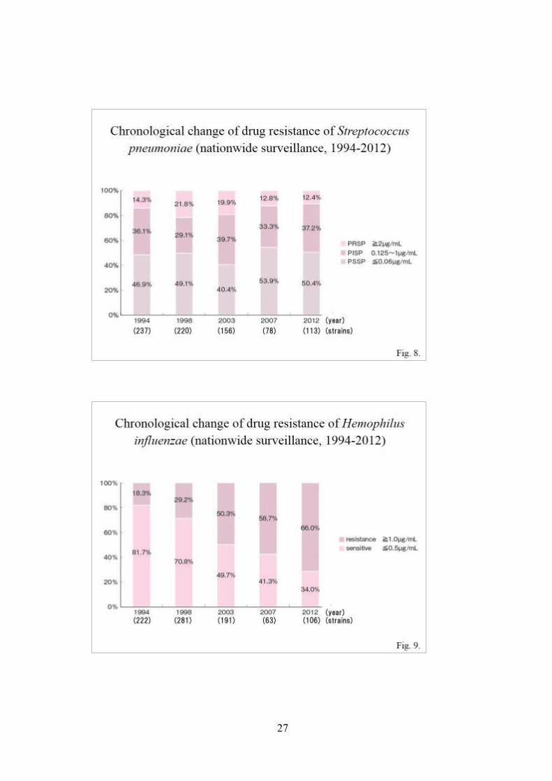
According to periodical surveillance (including the 2012 nationwide
surveillance by three organizations), the ratio of drug-resistant strains in S. pneumoniae
peaked in 2003, then showed a decrease in 2007, and remained the same in 2012 (Fig.
8). The ratio of H. influenzae has shown an increasing tendency since 2003, and
demonstrated a rapid increase in 2012 (Fig. 9).
(2) Antibacterial activity of various agents against predominantly detected bacteria
a. Antibacterial activity against S. pneumoniae
The antibacterial activity results of oral β-lactam antimicrobial agents
against S. pneumoniae reported in the 2007 Nationwide Surveillance show that
amoxicillin (AMPC) and clavulanate/amoxicillin (CVA/AMPC [1:14] formulation)
are superior to ampicillin/sulbactam (ABPC/SBT) by one tube. Cefditoren pivoxil
(CDTR-PI) and faropenem (FRPM) are also effective. Macrolide antimicrobial agents
are ineffective. New quinolone antimicrobial agents are relatively effective. In
particular, sitafloxacin (STFX), tosfloxacin (TFLX), and moxifloxacin (MFLX) are
effective, but now only TFLX is approved for use in children in Japan. Telithromycin
(TEL) is also effective. However, none of these antimicrobial agents are indicated in
children at this point. Among injections, cephems, such as cefpirom (CPR) and
ceftriaxone (CTRX), and carbapenems, such as panipenem (PAPM), meropenem
(MEPM), and doripenem (DRPM), are very useful. Cefmenoxime (CMX), approved
as an eardrop and the only approved nebulizer agent, also has relatively high
antibacterial activity (Table 3).
When we compare the results of two surveys done in 2007 and 2012,
although the surveys were done by two different facilities, we observed that macrolides

21
showed deterioration in MIC although there was no remarkable change in β-lactam
agents. Tebipenem pivoxil (TBPM-PI) and garenoxacin (GRNX) showed extremely
good minimum inhibitory concentrations (MICs, Table 4).
The analysis results of the multicenter clinical study show that S.
pneumoniae has relatively high susceptibility to AMPC and CVA/AMPC (1:14
formulation) (Table 5). CDTR-PL and CFPN-PI also have high antibacterial activity.
TFLX, a new quinolone antimicrobial agent indicated in children, has high
antibacterial activity. Injections, such as CTRX and a carbapenem antimicrobial agent
PAPM/BP also exhibit high antibacterial activity.
Yamanaka et al. (2012a) reported that S. pneumonia showed extremely good
sensitivity to BPM-PI, and the antimicrobial activities of CDTR-PI and cefcapene
pivoxil (CFPN-PI) were also good. The new quinolone agent TFLX, which is indicated
for children, had good antimicrobial activities too (Fig. 10).
b. Antibacterial activity against H. influenzae
According to the 2007 Nationwide Surveillance, ABPC is superior to AMPC
against H. influenzae by one tube among oral penicillin antimicrobial agents, but a
high dose is required based on the MIC. Among cephem antimicrobial agents, CDTR-
PI and cefteram-pivoxil (CFTM-PI) have favorable MIC values, but susceptibility of
H. influenzae to other antimicrobial agents is low. All new quinolones have very high
antibacterial activity, but cannot be used in children at this point. Among injections,
CTRX and CMX, cephem antimicrobial agents, and MEPM, a carbapenem
antimicrobial agent, are very useful (Table 6).
According to the analysis in the multicenter clinical study, AMPC does not
necessarily have high antibacterial activity, with MIC ≥8 μg/mL against more than a
half of H. influenzae strains. Approximately 40% of H. influenzae strains are
susceptible to CVA/AMPC (1:14 formulation). The antibacterial activity of CDTR-PI
is favorable. Approximately 96% of H. influenzae strains are susceptible to
azithromycin (AZM). According to Yamanaka, et al, TFLX, a new quinolone
antimicrobial agent, has extremely high antibacterial activity. Injections, such as
CTRX and MEPM, a carbapenem antimicrobial agent, also have high antibacterial
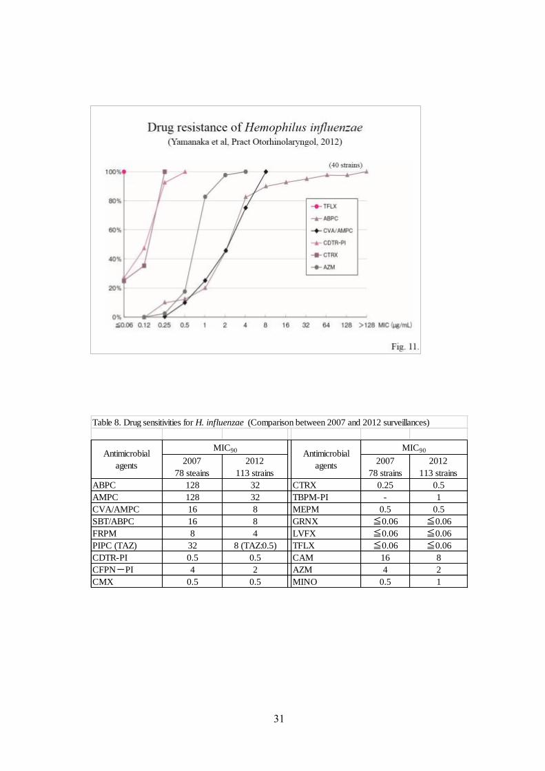
activity (Table 7). Yamanaka et al. (2012b) reported that TFLX, the only quinolone
22
agent approved for use in children, showed extremely high antimicrobial activity
against H. influenzae (Fig. 11).
When we compared the results of the 2007 and 2012 surveys, we noted that
although the surveys were done by two different facilities, the sensitivities of
penicillins and macrolides showed slight improvements in 2012 (Table 8).
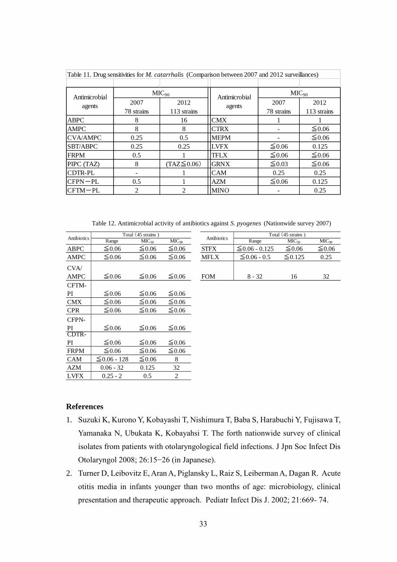
c. Antibacterial activity against M. catarrhalis
While M. catarrhalis is of low pathogenicity, 94% of M. catarrhalis strains
produce β-lactamase, as shown in the Third Nationwide Surveillance. When they are
present with pathogens, they inactivate β-lactam antimicrobial agents. They are
therefore significant as so-called indirect causative bacteria. As shown in Table 8, the
results of the 2007 Nationwide Surveillance show that there are a total of only 20
strains, and all antimicrobial agents except ABPC, AMPC, PIPC, CPR, and
fosfomycin (FOM) can be used against M. catarrhalis. All antibacterial agents are
effective if they are stable with β-lactamase (Table 9). In the multicenter clinical study,
CVA/AMPC (1:14 formulation), CDTR-PI, CFPN-PI, CTRX, and LVFX showed
effective antibacterial activity (Table 10).
Our comparison of the 2007 and 2012 surveys by two different facilities
revealed that the sensitivities of some of the β-lactam agents deteriorated slightly.
Those of LVFX and AZM also deteriorated, while TFLX, which can be used for
children, showed good antimicrobial activity (Table 11).
d. Antibacterial activity against S. pyogenes
S. pyogenes is not detected at high frequency, but is a significant causative
bacterium with strong pathogenicity (Figure 1). As all antibacterial agents except
macrolides and FOM are expected to be effective, safe agents can be selected for use
(Table 12).

23
Table 2. Transition of isolates from acute otitis media (Nationwide survey, Suzuki et al. 2013)
1994 1998 2003 2007 2012
S.aureus 25.1% 27.7% 17.0% 4.4% 14.4%
S.epidermidis 5.7% 3.3% 6.6%
other CNS 9.9% 7.5% 15.4%
CNS 24.6% 15.6% 10.8%
S.pneumoniae 15.5% 18.3% 24.1% 34.1% 29.2%
S.pyogenes 2.9% 3.5% 4.1% 2.2% 2.1%
S.agalactiae 1.0%
other Streptococcus spp. 1.0% 2.5% 4.4%
Enterococcus spp. 1.6% 1.0%
M.(B.) catarrhalis 2.9% 4.0% 7.1% 4.4% 11.3%
H.influenzae 15.3% 17.5% 27.4% 24.2% 26.7%
other Haemophilus spp. 0.2% 0.8%
Enterobacteriaceae 0.8% 2.0% 1.2% 1.1%
P.aeruginosa 2.9% 4.7% 2.1% 1.1% 0.5%
other NFGNR 5.5% 2.5% 2.9%
other G(-) rod 2.9%
Candida spp. 1.2% 1.1%
others 1.1% 15.8%
No. of strains Total 386 405 241 91 195
Year & Percentages of strains of each organism

24

25

26
27

28
Table 3. Antimicrobial activity of antibiotics against S. pneumoniae (Nationwide survey 2007)
Range MIC50 MIC90 Range MIC50 MIC90 Range MIC50 MIC90
PCG ≦0.06 ≦0.06 ≦0.06 0.125 - 1 0.5 1 2 2 2
AMPC ≦0.06 ≦0.06 ≦0.06 ≦0.06 - 2 0.25 1 0.5 - 2 1 2
PIPC ≦0.06 - 0.25 ≦0.06 0.125 ≦0.06 - 2 1 2 1 - 4 2 4
SBT/ABPC ≦0.06 - 0.125 ≦0.06 ≦0.06 ≦0.06 - 2 0.5 2 1 - 4 2 4
CVA/AMPC ≦0.06 ≦0.06 ≦0.06 ≦0.06 - 2 0.25 1 0.5 - 2 1 2
CFTM-PI ≦0.06 - 0.5 0.125 0.5 ≦0.06 - 4 0.5 1 0.5 - 4 1 2
FMOX 0.125 - 0.25 0.125 0.25 0.25 - 4 1 4 2 - 8 4 8
CMX ≦0.06 - 0.5 0.125 0.25 ≦0.06 - 2 0.5 1 0.5 - 1 0.5 1
CTRX ≦0.06 - 0.5 0.125 0.5 ≦0.06 - 2 0.5 1 0.5 - 1 0.5 1
CPR ≦0.06 - 0.5 0.125 0.25 ≦0.06 - 1 0.5 0.5 0.25 - 1 0.5 0.5
CFPN-PI ≦0.06 - 0.5 0.25 0.5 ≦0.06 - 4 0.5 1 0.5 - 2 1 1
PAPM/BP ≦0.06 ≦0.06 ≦0.06 ≦0.06 - 0.125 ≦0.06 0.125 ≦0.06 - 0.25 0.125 0.125
CDTR-PI ≦0.06 - 0.25 0.125 0.25 ≦0.06 - 2 0.25 1 0.25 - 1 0.5 1
FRPM ≦0.06 ≦0.06 ≦0.06 ≦0.06 - 0.5 ≦0.06 0.25 0.125 - 1 0.25 0.5
DRPM ≦0.06 ≦0.06 ≦0.06 ≦0.06 - 0.25 ≦0.06 0.25 0.125 - 0.5 0.25 0.5
CAM ≦0.06 - 128 128 128 ≦0.06 - 128 4 128 ≦0.06 - 128 2 128
AZM ≦0.06 - 32 32 32 ≦0.06 - 32 32 32 0.125 - 32 8 32
LVFX 1 - 2 1 2 0.5 - 1 1 1 0.25 - 1 1 1
TFLX 0.125 - 0.25 0.25 0.25 0.125 - 0.25 0.125 0.25 ≦0.06 - 0.25 0.125 0.25
STFX ≦0.06 - 0.125 ≦0.06 ≦0.06 ≦0.06 ≦0.06 ≦0.06 ≦0.06 ≦0.06 ≦0.06
MFLX 0.125 - 0.25 0.25 0.25 0.125 - 0.25 0.125 0.25 ≦0.06 - 0.25 0.125 0.25
MEPM ≦0.06 ≦0.06 ≦0.06 ≦0.06 - 0.5 0.125 0.5 0.25 - 0.5 0.5 0.5
TEL ≦0.06 - 0.5 ≦0.06 0.25 ≦0.06 - 0.5 ≦0.06 0.25 ≦0.06 - 0.5 ≦0.06 0.125
PISP( 26 strains )Antibiotics
PSSP( 42 strains ) PRSP( 10 strains )
Table 4. Drug sensitivities for S. pneumoniae (Comparison between 2007 and 2012 surveillances)
2007
78 strains
2012
113 strains
2007
78 strains
2012
113 strains
PCG 2 2 TBPM-PL - ≦0.06
AMPC 2 2 PAPM/BP 0.125 0.25
CVA/AMPC 2 2 MEPM 0.5 0.5
SBT/ABPC 4 4 GRNX 0.06 ≦0.06
FRPM 0.5 0.5 LVFX 2 2
PIPC 4 2 TFLX 0.25 0.25
CDTR-PL 1 0.5 CAM 128 ≦128
CMX 1 0.5 AZM 32 ≦128
CTRX 1 1 VCM - 0.25
Antimicrobial agents
MIC90
Antimicrobial agents
MIC90

29
Antimicrobials <0.031 0.0625 0.125 0.25 0.5 1 2 4 8 16 32 64 128 256 MIC50 MIC90 Susceptible(%) Total
PCG 70 25 25 18 28 53 103 7 0.5 2 22.8 329
Amoxicillin 71 21 26 26 38 104 39 4 0.5 2 98.78 329
CVA/AMPC 75 30 20 28 40 102 30 4 0.5 1 98.78 329
Cefcapene 1 76 6 59 129 38 9 7 2 2 0.5 1 NA 329
Cefditoren 20 22 58 47 131 38 7 3 3 0.5 1 NA 329
Cetriaxone 17 4 33 57 72 125 16 3 2 0.5 1 93.62 329
Levofloxacin 1 2 41 200 85 0.5 1 100 329
Paipenem 81 40 48 90 64 2 2 2 0.25 0.5 NA 329
Clarithromycin 24 3 6 34 32 20 20 6 3 2 1 178 256 256 8.21 329
Azithromycin 4 13 10 2 13 16 38 27 11 1 194 256 256 5.17 329
NA:not available
Table 5. Antimicrobial activity of antibiotics against S. pneumoniae (Multicenter clinical study)
MIC (µg/ml)

30
Range MIC50 MIC90 Range MIC50 MIC90 Range MIC50 MIC90
ABPC 0.125 - 0.5 0.25 0.5 1 - 8 2 8 1 - 128 32 128
AMPC 0.125 - 1 0.5 0.5 2 - 32 8 16 2 - 128 128 128
PIPC ≦0.06 ≦0.06 ≦0.06 ≦0.06 - 0.5 0.125 0.25 0.125 - 32 16 32
SBT/ABPC 0.125 - 1 0.25 0.5 1 - 16 4 8 0.5 - 16 4 16
CVA/AMPC 0.125 - 1 0.5 0.5 2 - 32 8 16 0.5 - 16 4 16
CFTM-PI ≦0.06 - 0.125 ≦0.06 ≦0.06 ≦0.06 - 1 1 1 ≦0.06 - 1 0.5 1
FMOX 0.25 - 2 0.5 1 2 - 16 8 16 0.5 - 16 8 16
CMX ≦0.06 ≦0.06 ≦0.06 ≦0.06 - 0.5 0.25 0.5 ≦0.06 - 0.25 0.25 0.25
CTRX ≦0.06 ≦0.06 ≦0.06 ≦0.06 - 0.25 0.25 0.25 ≦0.06 - 0.25 0.125 0.25
CFPN-PI ≦0.06 - 0.25 ≦0.06 ≦0.06 ≦0.06 - 8 2 4 ≦0.06 - 4 2 4
PAPM-BP ≦0.06 - 2 0.25 1 0.25 - 8 1 4 0.25 - 4 0.5 4
CDTR-PI ≦0.06 ≦0.06 ≦0.06 ≦0.06 - 1 0.25 0.5 ≦0.06 - 0.25 0.25 0.25
FRPM 0.25 - 2 0.5 1 0.5 - 8 4 8 0.25 - 4 4 4
DRPM ≦0.06 - 0.25 ≦0.06 0.125 0.125 - 4 0.5 2 ≦0.06 - 2 0.5 2CAM 0.125 - 16 8 16 1 - 8 8 8 4 - 16 4 16AZM ≦0.06 - 4 2 4 0.5 - 4 1 2 0.5 - 4 1 4MINO 0.125 - 2 0.25 1 0.125 - 2 0.25 0.5 0.125 - 0.5 0.125 0.5LVFX ≦0.06 - 2 ≦0.06 ≦0.06 ≦0.06 - 0.125 ≦0.06 ≦0.06 ≦0.06 ≦0.06 ≦0.06TFLX ≦0.06 - 8 ≦0.06 ≦0.06 ≦0.06 - 0.125 ≦0.06 ≦0.06 ≦0.06 ≦0.06 ≦0.06STFX ≦0.06 - 0.5 ≦0.06 ≦0.06 ≦0.06 ≦0.06 ≦0.06 ≦0.06 ≦0.06 ≦0.06MFLX ≦0.06 - 8 ≦0.06 0.125 ≦0.06 - 0.25 ≦0.06 ≦0.06 ≦0.06 ≦0.06 ≦0.06MEPM ≦0.06 - 0.125 ≦0.06 ≦0.06 ≦0.06 - 2 0.25 0.5 ≦0.06 - 0.25 0.125 0.25TEL ≦0.06 - 4 4 4 1 - 4 2 2 1 - 2 2 2
Table 6. Antimicrobial activity of antibiotics against H. influenzae (Nationwide survey 2007)
AntibioticsBLNAS( 26 strains ) BLNAR( 33 strains ) BLPAR( 4 strains )
Table 7. Antimicrobial activity of antibiotics against H. influenzae (Multicenter clinical study)
Antimicrobials <0.031 0.0625 0.125 0.25 0.5 1 2 4 8 16 32 64 128 256 MIC50 MIC90 Susceptible(%) Total
Ampicillin 21 6 60 21 15 22 35 68 45 16 7 2 18 4 16 36.6 336
Amoxicillin 1 22 72 13 11 22 43 68 50 13 3 18 8 32 NA 336
CVA/AMPC 1 20 73 14 12 24 42 68 59 18 2 3 8 16 42.85 336
Cefcapene 110 2 4 21 18 45 78 37 8 4 2 6 1 1 4 NA 336
Cefditoren 118 25 33 90 41 9 4 3 3 3 4 3 0.125 0.5 NA 336
Cetriaxone 120 24 31 99 30 11 6 3 4 1 4 3 0.125 0.5 95.54 336
Levofloxacin 314 22 0.031 0.031 100 336
Meropenem 16 56 37 37 65 53 39 16 3 8 3 3 0.5 2 NA 336
Clarithromycin 1 1 15 104 165 47 2 1 16 32 36.01 336
Azithromycin 9 2 38 90 99 51 36 9 2 1 4 96.73 336
MIC (µg/ml)
31
Table 8. Drug sensitivities for H. influenzae (Comparison between 2007 and 2012 surveillances)
2007
78 steains
2012
113 strains
2007
78 strains
2012
113 strains
ABPC 128 32 CTRX 0.25 0.5
AMPC 128 32 TBPM-PI - 1
CVA/AMPC 16 8 MEPM 0.5 0.5
SBT/ABPC 16 8 GRNX ≦0.06 ≦0.06
FRPM 8 4 LVFX ≦0.06 ≦0.06
PIPC (TAZ) 32 8 (TAZ:0.5) TFLX ≦0.06 ≦0.06
CDTR-PI 0.5 0.5 CAM 16 8
CFPN-PI 4 2 AZM 4 2
CMX 0.5 0.5 MINO 0.5 1
Antimicrobial
agents
MIC90 Antimicrobial
agents
MIC90

32
Table 9. Antimicrobial activity of antibiotics against M. catarrhalis (Nationwide survey 2007)
Range MIC50 MIC90 Range MIC50 MIC90
ABPC ≦0.06 - 8 2 8 TFLX ≦0.06 ≦0.06 ≦0.06
AMPC ≦0.06 - 8 4 8 STFX ≦0.06 ≦0.06 ≦0.06
PIPC ≦0.06 - 8 0.25 8 FOM 8 - 16 8 16
SBT/
ABPC ≦0.06 - 0.25 0.125 0.25CVA/
AMPC ≦0.06 - 0.25 0.125 0.25CFTM-
PI ≦0.06 - 4 1 2
CMX ≦0.06 - 1 0.5 1
CPR ≦0.06 - 4 1 4CFPN-
PI ≦0.06 - 1 0.5 1
FRPM ≦0.06 - 0.5 0.25 0.5
CAM ≦0.06 - 0.5 0.125 0.25
AZM ≦0.06 - 0.125 ≦0.06 ≦0.06
LVFX ≦0.06 ≦0.06 ≦0.06
AntibioticsTotal (20 strains )
AntibioticsTotal (20 strains )
Table 10. Antimicrobial activity of antibiotics against M. catarrhalis (Multicenter clinical study)
Antimicrobials <0.031 0.0625 0.125 0.25 0.5 1 2 4 8 16 32 64 128 256 MIC50 MIC90 Susceptible(%) Total
Amoxicillin 2 1 4 6 7 17 33 28 9 2 2 16 64 NA 111
CVA/AMPC 4 3 13 73 11 4 1 2 0.25 0.5 NA 111
Cefcapene 8 4 14 51 26 4 3 1 0.5 1 NA 111
Cefditoren 3 2 4 6 29 33 21 8 4 1 1 4 NA 111
Cetriaxone 3 3 2 9 39 33 12 5 5 0.5 2 NA 111
Levofloxacin 81 28 1 1 <0.031 0.0625 NA 111
Panipenem 15 11 9 11 15 10 11 6 7 4 5 7 0.5 32 NA 111
Clarithromycin 68 5 23 10 1 1 3 0.0625 0.5 NA 111
Azithromycin 5 10 35 55 2 1 3 0.5 0.5 NA 111
NA:not available
MIC (µg/ml)
33
References
1. Suzuki K, Kurono Y, Kobayashi T, Nishimura T, Baba S, Harabuchi Y, Fujisawa T,
Yamanaka N, Ubukata K, Kobayahsi T. The forth nationwide survey of clinical
isolates from patients with otolaryngological field infections. J Jpn Soc Infect Dis
Otolaryngol 2008; 26:15−26 (in Japanese).
2. Turner D, Leibovitz E, Aran A, Piglansky L, Raiz S, Leiberman A, Dagan R. Acute
otitis media in infants younger than two months of age: microbiology, clinical
presentation and therapeutic approach. Pediatr Infect Dis J. 2002; 21:669- 74.
Table 11. Drug sensitivities for M. catarrhalis (Comparison between 2007 and 2012 surveillances)
2007
78 strains
2012
113 strains
2007
78 strains
2012
113 strains
ABPC 8 16 CMX 1 1
AMPC 8 8 CTRX - ≦0.06
CVA/AMPC 0.25 0.5 MEPM - ≦0.06
SBT/ABPC 0.25 0.25 LVFX ≦0.06 0.125
FRPM 0.5 1 TFLX ≦0.06 ≦0.06
PIPC (TAZ) 8 (TAZ≦0.06) GRNX ≦0.03 ≦0.06
CDTR-PL - 1 CAM 0.25 0.25
CFPN-PL 0.5 1 AZM ≦0.06 0.125
CFTM-PL 2 2 MINO - 0.25
Antimicrobial
agents
MIC90 Antimicrobial
agents
MIC90
Table 12. Antimicrobial activity of antibiotics against S. pyogenes (Nationwide survey 2007)
Range MIC50 MIC90 Range MIC50 MIC90
ABPC ≦0.06 ≦0.06 ≦0.06 STFX ≦0.06 - 0.125 ≦0.06 ≦0.06
AMPC ≦0.06 ≦0.06 ≦0.06 MFLX ≦0.06 - 0.5 ≦0.125 0.25
CVA/
AMPC ≦0.06 ≦0.06 ≦0.06 FOM 8 - 32 16 32
CFTM-
PI ≦0.06 ≦0.06 ≦0.06
CMX ≦0.06 ≦0.06 ≦0.06
CPR ≦0.06 ≦0.06 ≦0.06
CFPN-
PI ≦0.06 ≦0.06 ≦0.06CDTR-
PI ≦0.06 ≦0.06 ≦0.06
FRPM ≦0.06 ≦0.06 ≦0.06
CAM ≦0.06 - 128 ≦0.06 8
AZM 0.06 - 32 0.125 32
LVFX 0.25 - 2 0.5 2
AntibioticsTotal (45 strains )
AntibioticsTotal (45 strains )

34
3. Commisso R, Romero-Orellano F, Montanaro PB, Romero-Moroni F, Romero-
Diaz R. Acute otitis media: bacteriology and bacterial resistance in 205 pediatric
patients. Int J Pediatr Otorhinolaryngol. 2000; 56:23-31.
4. Suzuki K, Kurono Y, Ikeda K, et al. The fifth nationwide survey of clinical isolates
from patients with otolaryngological field infections. JSIAO 2015 (in press).
5. Uno Y. Changes in antimicrobial susceptibilities of Streptococcus pneumoniae
detected in the nasopharynx of children with upper airway infection. Pediatr
Otorhinolaryngol Jpn 2009; 30:232-247 (in Japanese).
6. Uno Y. Changes in antimicrobial susceptibilities of Haemophilus influenzae
detected in nasopharynx of children with upper airway infection. Pediatr
Otorhinolaryngol Jpn 2009; 30:308-320 (in Japanese).
7. Yamanaka N, Sugita R, Uno Y, Matsubara S, Hayashi Y、Sawada S. Clinical
Efficacy of Tosfloxacin Tosilate Hydrate for the Treatment of Acute Otitis Media
in Children. Pract Otol (Kyoto) 105;381-392, 2012 (in Japanese).
8. Yamanaka N, Suetake M, Tomiyama M, Sugita R, Matsubara S, Sawada S, Uno Y,
Kanesada K, Uchizono A. Efficacy evaluation of TBPM-PI, an oral carbapenem
antibacterial agent, in children with acute otitis media including recurrent/
persistent cases. Pract Otol (Kyoto) 105; 687-698, 2012 (in Japanese).
10. Gathering evidence
During the preparation of these Guidelines, existing evidence (literature)
was gathered with respect to the following clinical questions by means of the
procedures described below:52)
(a) Diagnosis
(b) Testing methods
(c) Treatment
(i) Databases used
For the 2006 and 2009 Guidelines, PubMed and Japan Centra Revuo
Medicina Web version 3 and 4 were used, and for the 2013 Guidelines, PubMed, the
Cochrane Library, and Japan Centra Revuo Medicina Web version 5 were used.
(ii) Search period

35
For the 2006 Guidelines, searches were performed in the databases of
literature published during 2000–2004. For the 2009 Guidelines articles published in
2004 but not included in the 2006 Guidelines, were added, and we also added articles
which were published after 2005 and searchable on April 10, 2008. For the 2013
Guidelines, articles published after 2008 and searchable on January 26, 2012 were
added.
(iii) Criteria for use
Priority was given to articles comprising systematic reviews of randomized
controlled trials or describing individual randomized controlled trials, and if these were
not available then articles describing observational studies such as cohort studies and
case controlled studies were used. If these were insufficient, the scope was widened to
include articles describing case series. Articles concerning animal experiments and
basic science were excluded.
(iv) Method of use
For the 2006 Guidelines, the keyword中耳炎 (chuujien, “otitis media”) was
used to search the Japan Centra Revuo Medicina Web version 3 database with the
“meta-analysis,” “randomized controlled trial,” “controlled clinical trial,” and
“comparative research” research design tags checked, but no articles suitable for use
in these Clinical Practice Guidelines were found. In PubMed, searches were performed
with the following keywords: (1) otitis media, treatment; (2) otitis media, antimicrobial
agents; (3) acute otitis media, treatment; and (4) acute otitis media, antimicrobial
agents. For meta-analyses and systematic reviews using the Cochrane Collaboration,
the search format “English [la] AND otitis media [ti] AND (Cochrane Database Syst
Rev [jour] OR meta-analysis [pt]) AND 2000:2004 [dp]”was used. Articles cited in the
American Academy of Pediatrics Guidelines (2004) were also analyzed. In addition to
the literature searches described above, articles published before 2000, those published
during 2003–2005 while the Guidelines were in preparation, and those published in
Japanese and international journals that were considered to be required for the
preparation of the Guidelines were also identified, resulting in a total of 82 articles for
investigation.

36
For the 2009 and 2013 Guidelines, the search format (otitis media/TH or
otitis media/AL) and (PT = NOT conference report and RD = meta-analysis,
randomized controlled trial, semi-randomized controlled trial, controlled study,
clinical practice guidelines) was used to search Japan Centra Revuo Medicina Web
version 4 and 5, yielding hits for 104 articles (2003–2008) and 233 articles (2008–
2012), respectively. The abstracts or main texts of these articles were studied, and
seven and 50 articles were selected for inclusion, respectively.
In PubMed, searches were performed using the following keywords: Search
(English[la] OR Japanese[la]) AND (otitis media) AND (treatment OR antimicrobial
agents) AND (randomized controlled trial [pt]) AND 2004:2007 [dp]; and Search
(English [la] OR Japanese [la]) AND (otitis media) AND (treatment OR antimicrobial
agents) AND (meta-analysis[pt] OR Cochrane Database Syst Rev [ta]) AND 2004:
2007 [dp], yielding 118 articles. A further 268 articles published between 2004 and
April 2007 and containing “otitis media” in their title, abstract, or keywords were also
identified from the Cochrane Reviews, Clinical Trials, Other Reviews, Technology
Assessments, and Economic Evaluations included in the Cochrane Library. A total of
386 articles found by the above searches were studied and 60 of 386 articles were
added to the 2009 Guidelines, excluding those already used in the 2006 Guidelines. In
addition, with the cooperation of the Japan Council for Quality Healthcare Medical
Information Network Distribution System EBM Medical Information Department, a
search of PubMed for articles published after April 1, 2007 was performed on April 10,
2008 using the search format ((“otitis media” [MeSH] AND “therapy”[Subheading])
OR (“otitis media” [MeSH] AND antimicrobial agents) OR (“acute otitis media” AND
“therapy”[Subheading]) OR (“acute otitis media” AND antimicrobial agents)) AND
((“meta-analysis”[pt] OR “randomized controlled trial”[pt]) NOT ”Cochrane database
of systematic reviews (Online)”[Jour] AND “humans” [MeSH] AND (English [la] OR
Japanese [la]) AND 2007/4/1 [edat]: 2008/3/31 [edat]. This identified 11 articles, of
which five were selected for study.
In addition to the literature searches described above, three other articles
were added that were considered required for preparation of the Guidelines, resulting
in 75 articles being added to those used in the 2006 Guidelines giving a final total of
157 articles used in the Guidelines. In the 2013 Guidelines, a further literature search
was conducted using the same method, picking up 650 articles, and finally 208 articles

37
were added. In the 2013 guidelines, the abstract table was inserted on the homepage
of the Japan Otological Society (http://www.otology.gr.jp).
11. Criteria for deciding recommendation grades
The method proposed by the Japan Stroke Society to indicate the level of
evidence was used in the preparation of these Guidelines, as shown below.
Level of evidence
Ia Meta-analysis (with homogeneity) of randomized controlled trials
Ib At least one randomized controlled trial
IIa At least one well-designed, controlled study but without randomization
IIb At least one well-designed, quasi-experimental study
III At least one well-designed, non-experimental descriptive study
(e.g., comparative studies, correlation studies, case studies)
IV Expert committee reports, opinions and/or experience of respected authorities
Recommendation grades were determined based on the evidence obtained
by the search policies described above and the anticipated degree of benefit or harm.
During this process, reference was made to items according to the proposed grades
outlined below. Five levels of recommendation grades were established, based on the
U.S. Preventive Services Task Force report
(http://www.ahrq.gov/clinic/ajpmsuppl/harris3.htm#tb6fn5). In the 2013 Guidelines,
considering the consistency with the previous two editions, the same five levels
described below were used as well.
A: (strongly recommended: strong evidence is available, benefits substantially
outweigh harms)
B: (recommended: sufficient evidence is available, benefits outweigh harms)
C: (no recommendation made: fair evidence is available, but the balance of benefits
and harms is close)
D: (recommended against: harms outweigh benefits)
I: (insufficient evidence to determine the balance of benefits and harms)

38
The specification of recommendation grades is one of the most important
roles expected of clinical practice guidelines, but there is great debate concerning the
sort of factors that should be taken into account when determining recommendation
grades. The Subcommittee of Clinical Practice Guideline made overall judgments
taking into consideration the factors below, with reference to the proposals of Fukui
and Tango (Shinryou gaidorain sakusei no tebiki dai 4-pan, “Guide to the Preparation
of Clinical Practice Guidelines, 4th edition”) and those of the MINDS Guide to the
Preparation of Clinical Practice Guidelines (MINDS Shinryou gaidorain sakusei no
tebiki, Igakushoin, 2007).
Level of evidence
Quality of evidence
Consistency of evidence (supported by multiple studies)
Directness (magnitude of clinical efficacy, external validity, indirect evidence,
evaluation by surrogate outcomes)
Clinical applicability
Evidence concerning harm or costs
No Level I study reports on AOM in Japan were found. Accordingly, Grade
A recommendations were determined based on the existence of at least one piece of
level I evidence from Europe or the USA that was judged by the committee to be
applicable to Japanese circumstances. The condition for determination of Grade B
recommendations was the existence of at least one piece of Level II evidence
demonstrating efficacy that was judged by the committee to be applicable to Japanese
circumstances.
Opinions on these recommendations were solicited from the directors and
executive committee members of the JOS, the JSIDO, and the JSPO before the final
decision was made by the Subcommittee of Clinical Practice Guideline. The
committee endeavored to maintain objectivity and transparency when deciding on
recommendation grades, but it was not possible to guarantee this in every case.
A system will be put in place in future for accepting comments and
suggestions from users concerning the content of recommendations and
recommendation grades, with a view to the future revision of these Guidelines.

39
References
1. Atkins D, Best D, Briss PA, Eccles M, Falck-Ytter Y, Flottorp S, Guyatt GH,
Harbour RT, Haugh MC, Henry D, Hill S, Jaeschke R, Leng G, Liberati A, Magrini
N, Mason J, Middleton P, Mrukowicz J, O'Connell D, Oxman AD, Phillips B,
Schunemann HJ, Edejer TT, Varonen H, Vist GE, Williams JW Jr, Zaza S; GRADE
Working Group. Grading quality of evidence and strength of recommendations.
BMJ. 2004;328(7454):1490.
2. Fukui T, Tango T. Guide to the Preparation of Clinical Practice Guidelines, 4th edition EBM
Journal 2003;4:284-92 (in Japanese).
3. MINDS Shinryou gaidorain sakusei no tebiki (MINDS Guide to the Preparation of
Clinical Practice Guidelines). Igakushoin, Tokyo, 2007. (in Japanese)
12. Procedures for consolidating evidence
To consolidate the evidence, the main findings from each article were
identified and an evidence table was prepared. The features of each finding were
compared and evaluated. When meta-analyses were found during the literature search,
their results were used as reference. No new meta-analyses or decision analyses were
conducted in the preparation of these Guidelines.
13. Pre-release review
Before these Guidelines were released for general use, they were reviewed
with reference to the Conference on Guideline Standardization (COGS) proposals
concerning publication format54) and the Appraisal of Guidelines for Research &
Evaluation (AGREE) appraisal instrument for assessing content.55)
Before publication of the 2006 edition of the Guidelines, opinions were
solicited from JOS, JSIDO, and JSPO, and pediatricians, and corrections were made
where necessary. Otolaryngologists, regarded as the general users of the Guidelines,
were also surveyed regarding their utility in the clinical setting, and the results were
reflected where appropriate. The 2013 Guidelines were assessed before publication by
the Subcommittee of the Clinical Practice Guidelines for the Diagnosis and
Management of Acute Otitis Media in Children with reference to the appraisal
instruments of AGREE II (http://www.agreetrust.org/resource-centre/agree-ii).

40
References
1. Shiffman RN, Shekelle P, Overhage JM, Slutsky J, Grimshaw J, Deshpande AM.
Standardized reporting of clinical practice guidelines: a proposal from the
Conference on Guideline Standardization. Ann Intern Med 2003;139: 493-8.
2. Appraisal of Guidelines, Research, and Evaluation in Europe (AGREE) Collaborative Group.
Guideline development in Europe. An international comparison. Int J Technol Assess Health
Care 2000; 16:1039-49.
14. Planned Updates
These Guidelines are scheduled to be updated in around 3–5 years. After
their publication, work will begin toward the organization of a new Subcommittee of
Clinical Practice Guideline. Newly published evidence will be systematically assessed
and reviews carried out, with a Working Group established to contribute resources for
the updated Guidelines. Should partial updates to the Guidelines be required, these will
be published on the societies’ web sites as appropriate.
15. Recommendations and explanation of reasons
These Guidelines were formulated for otolaryngologists as users, but they
are also expected to be used as reference in all situations in which clinical judgments
are made concerning the diagnosis and treatment of childhood AOM, by all medical
professionals involved in the treatment of this condition, in a wide variety of clinical
settings. The specific relationships between recommendations and the literature on
which they are based are described in each section of the Guidelines. It must again be
emphasized that the recommendation grades indicated by these Guidelines do not
constitute an alternative to the judgment of an experienced medical practitioner, but
are only provided to assist his or her decision-making.
16. Patients’ wishes
In the process of deciding the recommendations in the 2006 edition of the
Guidelines, the wishes of patients or their parents or guardians were listened to but not
actively taken into account. When dealing with individual patients and clinical
situations, however, to apply the recommendations in the Guidelines without exception

41
in every case is to mistake what is important, in light of the spirit of the Guidelines as
an aid to decision-making in actual clinical situations. It must again be emphasized
that decision-making in actual clinical situations must always be carried out by taking
into account the evidence and recommendations contained in the Guidelines and
elsewhere, the experience and specialist knowledge of the medical practitioner, and
the wishes and values of the patient and his or her parents or guardians. Future
revisions of the Guidelines will consider efforts to reflect the wishes of patients and
their parents and guardians to a greater extent.
17. Algorithms
The generally recommended algorithms according to the degree of severity
of AOM are included at the end of the Guidelines.
18. Practical consideration
In principle, in these Guidelines medications are referred to by their generic
names rather than brand names. The reasons for this include concerns that it would be
unfair to refer only to selected products by name in the Guidelines as well as the strong
influence of expert opinion. In addition to which all generic products are fully included,
and updating this information to include brand names would pose too great a burden
on the Subcommittee of Clinical Practice Guideline. For this reason, we advise the
preparation of clinical paths or manuals that take account of the status of medications
used and other specific attributes of individual facilities, to enable the smooth
acceptance of the recommendations in these Guidelines in actual clinical settings.
19. Diagnosis, examinations
Clinical question 19-1: Under what condition is AOM diagnosed?
Recommendation
AOM is diagnosed when the following eardrum findings are recognized, and
thus, detailed inspection of the eardrum is indispensable for its diagnosis (Typical
eardrum findings are in Figure 8, by Kamide Y56)). (Grade of Recommendation: B)
Hyperemia, protrusion, diminishment of the light reflex, thickening, bullar formation,
cloudiness (turbidity), perforation, MEE, otorrhea, edema of middle-ear mucosa

42
Reference used to assess the recommendation level: Rosenfeld et al., 200157) (Level
IIb)
[Addendum] Otomicroscopic or otoendoscopic observation of the eardrum is most
desirable, but recent model of the pneumatic otoscope is also acceptable.
Background
As AOM is acute inflammation of the middle-ear mucosa, confirmation by
inspection of the eardrum findings manifesting middle-ear inflammatory effusion
and/or inflammatory change is indispensable for the diagnosis of AOM.
Comments
As for the eardrum findings suitable for the diagnosis of AOM, various
descriptions such as hyperemia, cloudiness (turbidity), protrusion, thickening, bullar
formation, perforation, and change of the light reflex, have been observed, and there
has not been a uniform standard that was used uniformly in the literature issued to date.
Among those findings, eardrum protrusion is a finding that is frequently observed and
is also suspected of the existence of MEE (Shaikh et al. 2012). Eardrum protrusion is,
therefore, in combination with the eardrum color and mobility, the finding that is most
suspected of AOM (Karma et al. 1989, Pelton 1998, Pichichero 2000, Shaikh et al.
2012). Eardrum turbidity frequently represents en edema of the eardrum except those
due to scar tissue. Although hyperemia of the eardrum by acute inflammation is also
frequently observed with AOM, those due to crying or systemic fever should be
discriminated, and also differential diagnosis includes viral OM (Weiss et al. 1996). It
is sometimes the case that eardrum hyperemia is not distinct in spite of apparent
protrusion in AOM of infant under the age of one.
AOM is diagnosed based on subjective symptoms (acute onset of symptoms
of inflammation in the ME) and objective signs (tympanic membrane findings
including hyperemia, bulging, otorrhea, MEE, etc.) (Lee et al. 2012). The 2013
American AOM Guidelines recommended the following three features for the
definition of AOM (Lieberthal et al. 2013).
(1) the presence of moderate to severe bulging of the tympanic membrane or new
onset of otorrhea not due to acute otitis externa
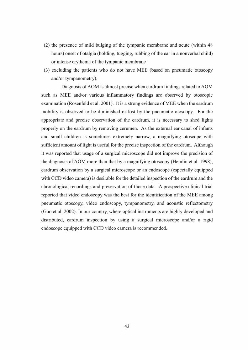
43
(2) the presence of mild bulging of the tympanic membrane and acute (within 48
hours) onset of otalgia (holding, tugging, rubbing of the ear in a nonverbal child)
or intense erythema of the tympanic membrane
(3) excluding the patients who do not have MEE (based on pneumatic otoscopy
and/or tympanometry).
Diagnosis of AOM is almost precise when eardrum findings related to AOM
such as MEE and/or various inflammatory findings are observed by otoscopic
examination (Rosenfeld et al. 2001). It is a strong evidence of MEE when the eardrum
mobility is observed to be diminished or lost by the pneumatic otoscopy. For the
appropriate and precise observation of the eardrum, it is necessary to shed lights
properly on the eardrum by removing cerumen. As the external ear canal of infants
and small children is sometimes extremely narrow, a magnifying otoscope with
sufficient amount of light is useful for the precise inspection of the eardrum. Although
it was reported that usage of a surgical microscope did not improve the precision of
the diagnosis of AOM more than that by a magnifying otoscopy (Hemlin et al. 1998),
eardrum observation by a surgical microscope or an endoscope (especially equipped
with CCD video camera) is desirable for the detailed inspection of the eardrum and the
chronological recordings and preservation of those data. A prospective clinical trial
reported that video endoscopy was the best for the identification of the MEE among
pneumatic otoscopy, video endoscopy, tympanometry, and acoustic reflectometry
(Guo et al. 2002). In our country, where optical instruments are highly developed and
distributed, eardrum inspection by using a surgical microscope and/or a rigid
endoscope equipped with CCD video camera is recommended.

44
References
1. Kamide Y. Classification of tympanic membrane finding son acute otitis media
in children and their verification. Oto-Rhino-Laryngology Tokyo 2003; 46:17-30
(in Japanese).
2. Rosenfeld RM, Casselbrant ML, Hannley MT. Implications of the AHRQ
evidence report on acute otitis media. Otolaryngol Head Neck Surg 2001;
125:440-8.
3. Shaikh N, Hoberman A, Rockette HE, Kurs-Lasky M: Development of an
algorithm for the diagnosis of otitis media. Acad Pediatr 2012; 12:214-8.
4. Karma PH, Penttila MA, Sipila MM, Kataja MJ. Otoscopic diagnosis of middle
ear effusion in acute and non-acute otitis media. I. The value of different
otoscopic findings. Int J Pediatr Otolaryngol 1989; 17:37–49.
5. Pelton SI. Otoscopy for the diagnosis of otitis media. Pediatr Infect Dis J 1998;
17:540–3.
6. Pichichero ME. Acute otitis media: Part I. Improving diagnostic accuracy. Am
Fam Physician 2000; 61: 2051-6.

45
7. Weiss JC, Yates GR, Quinn LD. Acute otitis media: making an accurate diagnosis.
Am Fam Physician 1996; 53:1200-6.
8. Lee HJ, Park SK, Choi KY, Park SE, Chun YM, Kim KS, Park SN, Cho YS, Kim
YJ, Kim HJ, Korean Otologic Society: Korean clinical practice guidelines: otitis
media in children. J Korean Med Sci 2012; 27: 835-48.
9. Lieberthal AS, Carroll AE, Chonmaitree T, Ganiats TG, Hoberman A, Jackson
MA, Joffe MD, Miller DT, Rosenfeld RM, Sevilla XD, Schwartz RH, Thomas PA,
Tunkel DE: The diagnosis and management of acute otitis media. Pediatrics 2013;
131:e964-e99
10. Hemlin C, Hassler E, Hultcrantz M, Papatziamos G, Krakau I. Aspects of
diagnosis of acute otitis media. Fam Practice 1998; 15:133-7.
11. Guo YC, Shiao AS. Diagnostic methods for otitis media with effusion in children.
Zhonghua Yi Xue Za Zhi (Taipei). 2002; 65:372-7.
Clinical question 19-2: How is the severity of AOM assessed?
Recommendation
Severity of AOM is classified as mild, moderate and severe according to
eardrum findings and clinical manifestations. (Grade of Recommendation: A)
Reference used to assess the recommendation level: Hotomi et al., 2004, 2005 (Level
IIa), Friedman et al., 2006 (Level Ib), Biner et al., 2007 (Level Ib)
Manifestations and findings and their scores used for the classification of the
severity of AOM (proposal from the AOM Guideline Committee)
3 points are automatically given for the age younger than 24 months
Otalgia is scored as 0, 1, 2, according to the severity
0: absent, 1: present, 2: present - continuous severe pain
Fever is scored as 0, 1, 2, according to the severity
0: lower than 37.4 degree centigrade (oC), 1: higher than 37.5 but lower
than 38.4 oC, 2: higher than 38.5 oC
Crying and/or bad temper is scored as 0, 1, according to the severity
0: absent, 1: present
Hyperemia of the eardrum is scored as 0, 2, 4, according to the severity

46
0: absent, 2: present – 1st degree at manubrium of malleus, or at a part of
eardrum, 4: present at whole eardrum
Protrusion of the eardrum is scored as 0, 4, 8, according to the severity
0: absent, 4: present at a part of eardrum, 8: present at whole eardrum
Otorrhea is scored as 0, 4, 8, according to the severity
0: absent, 4: present eardrum is visible, 8: present and eardrum is invisible
Classification of severity of AOM according to the total score
Mild - < 5
Moderate – 6 - 11
Severe - > 12
A sample of a score chart used for assessing the severity of AOM in the clinic
is shown in Table 13.
Background
For AOM, appropriate treatments according to its severity are required. In
younger age, discrepancy between general condition and the eardrum finding is often
seen in the convalescent stage of AOM; where the eardrum findings are not improved
even general condition is improved well (Hotomi et al., 2004, 2005). Thus, precise
assessment of the eardrum findings and therefore severity of AOM lead to the
appropriate choice its treatment (Friedman et al., 2006).
Comments
In this guideline, severity of eardrum findings and clinical manifestations
was scored, and the severity of AOM was assessed by summing those scores.
Friedman et al. (2006) reported a similar study, in which they assessed the severity of
AOM by summing the scores of both total impressions of the child by the guardian
and eardrum findings, and concluded that such an assessment is important for the
appropriate choice of its treatment. Casey et al. (2011) also discussed the grading of
the severity of AOM by a scoring system for the diagnosis and evaluation of treatments
of AOM, and they did so on the basis of symptoms (otalgia, bad temper, etc.), findings
of eardrum, and systemic signs (fever: presence or absence, and degree). In our

47
previous guideline issued in 2006, 3 factors of the eardrum findings including
hyperemia (yellowish change), protrusion, otorrhea were chosen as an assessing tool
of the severity of AOM, while eardrum turbidity and diminishment of the light reflex
were reported to be important for assessing the cure of AOM (Hotomi et al. 2004,
2005). For these reasons, in the present guideline, we reconsidered the factors of
eardrum findings used for assessing the severity of AOM by way of normal group
technique (NGT, see the note for detail), and agreed to pick up ‘diminishment of the
light reflex’ in addition to hyperemia, protrusion and otorrhea. Then, in stratifying
those factors, we assigned the score 0 (normal) and 4 (diminished or absent) on the
newly-added factor of ‘diminishment of the light reflex’. Analysis of the influence of
the factor (addition of the light reflex) on the score of severity of AOM using 721
children revealed that it did not affect significantly the distribution of the severity.
Until this 2013 issue was edited, the light reflex was reevaluated within the
subcommittee to determine whether it is an appropriate and significant factor to assess
the severity of AOM, and the conclusion was that it did not contribute sufficiently
because of the difficulty of judgment due to the narrow and/or curved ear canal in
newborns and infants and due to the lack of an objective or digital index, and also
because of possibility of misjudgment due to differences in the observation apparatus
or light source, for example.
A multicenter clinical study assessing the factors determining the severity of
AOM revealed that the light reflex showed the smallest change in score after treatment,
and that the light reflex score did not change even in patients showing successful
treatment outcomes. In light of these results, it has been pointed out that the light reflex
may not be suitable as a factor determining the severity of AOM, which is directly and
closely related to the treatment algorithm (Yamanaka et al. 2012). Thus, in the 2013
Guidelines, the factor ‘light reflex’ was deleted from the factors, and consequently the
lineup of factors has become the same as in the 2006 Guidelines.
In the previous issue of our guideline in 2006, we recommended that both
eardrum protrusion and otorrhea should not be scored simultaneously, because both
cannot exist at the same time, in other words, protrusion must disappear when otorrhea
occurs. But in reality, there were not few children showing both of them, and many
clinicians were found to score both of them. This is the reason why we recommended
to score both of them in the present guideline. Analysis of the influence of this change

48
on the distribution of the severity of AOM using clinical data of two groups consisting
of 1196 and 721 children with AOM revealed that this change did not affect much the
distribution of the severity of AOM.
In the present guideline, as important clinical manifestations for assessing
the severity of AOM, we chose three factors including otalgia, fever, and crying/bad
temper with the same score, which were chosen in the previous guideline in 2006 by
NGT, as well.
As for the body temperature, Kaleida et al. (1991) classified mild and severe
AOM at 39.0 oC and 39.5 oC. Considering that the body temperature is measured at
axilla in our country, the grades of fever were classified into three groups according to
<37.4 oC, 37.5 – 38.4 oC, and >38.5 oC from discussion within the committee using
NGT. It was defined that the score of the fever was determined by the body
temperature measured at the first visit to a clinic. Although it is possible that fever is
not necessarily related with the severity of AOM, fever was adopted as one of the
factors determining the severity of AOM, because it is one of the basic signs and
symptoms for diagnosing acute pediatric febrile diseases including AOM. In their
report discussing the scoring of AOM, Casey et al. (2011) noted that the presence or
absence of fever and/or the degree of body temperature were considered as two of the
factors determining the score.
Since younger age is apparently one of the risk factors aggravating and/or
prolonging AOM (Hotomi et al. 2004, Rovers et al. 2004, Ovetchkine et al. 2003,
Block et al. 2000), ‘younger than 3 years of age’ was adopted as one of the factors
determining the severity of AOM in our previous guideline in 2006. In this edition,
considering the results of several other reports (Rovers et al. 2004, 2007, Ovetchkine
et al. 2003, Block et al. 2000), we changed it into ‘younger than 24 months’. Analysis
of the clinical data of 681 children with AOM revealed that this change caused only a
slight shift of the distribution of the severity of AOM.
From all the results of reviews, we defined the scores for each factors as
follows; otalgia: 0, 1, 2, fever: 0, 1, 2, crying and/or bad temper: 0, 1, eardrum
hyperemia: 0, 2, 4, eardrum protrusion: 0. 4, 8, otorrhea: 0, 4, 8, and added 3 points
for younger than 24 months of age. As the total score, smaller than 5, from 6 to 11,
and greater than 12 were defined as mild, moderate, and severe AOM, respectively.

49
Table 13. AOM Clinical Score Sheet
Patient ID:
Name:
Age: year months
Date: Body weight: Body temperature: oC
Sex: male female Other features:
[Scores chart]
Age (<24 mo.) 0 3
Otalgia 0 1 (present) 2 (continuous & severe)
Fever 0 (<37.4 oC), 1 (37.5-38.4 oC) 2 (>38.5 oC)

50
Crying bad temper 0 1
Eardrum hyperemia 0 2 (1st degree or partial) 4 (whole eardrum)
Eardrum protrusion 0 4 (partial) 8 (whole eardrum)
Otorrhea 0 4 (visible eardrum) 8(invisible eardrum)
Light reflex 0 4 (diminished or absent due to turbidity)
Total score
[Assessment]
Mild (0-9) Moderate (10-15) Severe (>15)
[Addendum] normal group technique (NGT)
A method to make a decision on an issue, on which general agreement
(consensus) is not obtained due to the lack of positive scientific evidence, or to
existence of adverse evidence, is called consensus method. In consensus method, on
the basis of individual opinions on an issue, final consensus is attained by intensively
discussing and assessing those opinions. As those in the field of medicine or health
care, Delphi’s method and Normal group technique (NGT) are often used. Both are
characterized by collection and quantification of opinions and its feedback to the
participants. In NGT, specialists of the field of the issue get together, directly give
opinions, feed them back, and again give opinions with reference to the other’s
opinions. A consensus is obtained by repeating those processes. Although this method
has a disadvantage that some relationship between the participants may affect the final
conclusion, it has a greater advantage that all the participants can directly exchange
opinions. In this guideline, by using this method, an agreement was able to be attained
on the selection of necessary items of eardrum findings and clinical manifestations for
assessing severity of AOM, stratification of each item, scores allotted to the three levels
of severity (mild, moderate, severe), as well as the selection of treatment for AOM
with each three level of severity. Although the present consensus is not an objective
results based on definite scientific evidence, it is expected to be a useful tool for the
appropriate judgement in the clinical practice, as it is considered an extract of precious
opinions of experienced specialists.
51
Raferences
1. Hotomi M, Yamanaka N, Shimada J, Ikeda Y, Faden H. Factors associated with
clinical outcome in acute otitis media. Ann Otol Rhinol Laryngol 2004; 113: 846-
52.
2. Hotomi M, Yamanaka N, Samukawa T, Suzumot M, Sakai A, Shimada J, Ikeda Y,
Faden H. Treatment and outcome of severe and non-severe acute otitis media. Eur
J Pediatr 2005; 164: 3-8.
3. Friedman NR, McCormick DP, Pittman C, Chonmaitree T, Teichgraeber DC,
Uchida T, Baldwin CD, Saeed KA.Development of a practical tool for assessing
the severity of acute otitis media. Pediatr Infect Dis J 2006; 25:101-7.
4. Biner B, Celtik C, Oner N, Küçükuğurluoğlu Y, Güzel A, Yildirim C, Adali MK.
The comparison of single-dose ceftriaxone, five-day azithromycin, and ten-day
amoxicillin/clavulanate for the treatment of children with acute otitis media. Turk
J Pediatr 2007; 49:390-6.
5. Casey JR, Block S, Puthoor P, Hedrick J, Almudevar A, Pichichero ME:A simple
scoring system to improve clinical assessment of acute otitis media. Clinical
pediatrics 2011; 50: 623-9.
6. Kamide Y. Classification of tympanic membrane finding son acute otitis media in
children and their verification. Oto-Rhino-Laryngology Tokyo 2003; 46:17-30 (in
Japanese).
7. Yamanaka N, Sugita R, Uno Y, Matsubara S, Hayashi Y、Sawada S. Clinical
Efficacy of Tosfloxacin Tosilate Hydrate for the Treatment of Acute Otitis Media
in Children. Pract Otol (Kyoto) 105; 381-392, 2012 (in Japanese).
8. Kaleida PH, Casselbrant ML, Rockette HE, Paradise JL, Bluestone CD, Blatter
MM, Reisinger KS, Wald ER, Supance JS. Amoxicillin or myringotomy or both
for acute otitis media: Results of a randomized clinical trial. Pediatrics 1991; 87:
466-74.
9. Rovers MM, Schilder AGM, Zielhuis GA, Rosenfeld RM. Otitis Media. Lancet
2004; 363:465-73.
10. Ovetchkine P, Cohen R. Shortened course of antibacterial therapy for acute otitis
media. Paediatr Drugs 2003; 5:133-40.
11. Block SL, Kratzer J, Nemeth MA, Tack KJ. Five-day cefdinir course vs. ten-day

52
cefprozil course for treatment of acute otitis media. Pediatr Infect Dis J 2000 (c);
19(12 Suppl):S147-52.
12. Rovers MM, Glasziou P, Appelman CL, Burke P, McCormick DP, Damoiseaux
RA, Little P, Le Saux N, Hoes AW. Predictors of pain and/or fever at 3 to 7 days
for children with acute otitis media not treated initially with antibiotics: a meta-
analysis of individual patient data. Pediatrics 2007; 119:579-85.
CQ19-3: Is tympanometry useful to diagnose AOM?
Recommendation
Tympanometry is recommended to identify the presence of MEE after the
diagnosis of AOM is confirmed by a precise otoscopic finding. (Grade of
Recommendation: B, Saeed et al. 200473); Level IIa)
Background
Tympanometry is reliable test to identify the presence of MEE in the
tympanic cavity. An acoustic reflectometry, recommend to identify the effusion in
European countries and the US (Laine et al. 2012), is not recommended in Japan
because it has not been available since 1994.
Comments
Tympanometry is a tool to measure the compliance change of middle ear
conduction system consisting of the eardrum, ossicles and tympanic cavity by forcing
the positive and negative pressures in the sealed external ear canal. The type of
tympanogram is roughly divided into three; type A, C, and B. Tympanometry is very
reliable to detect the presence of MEE and the negative pressure in the middle ear
(Subcommittee on Management of Acute Otitis Media 2004, Saeed et al. 2004,
Sakaguchi et al. 1994). Although MEE can be detected using tympanometry, the stage
of AOM, i.e. acute stage or resolution stage, cannot be identified (Rosenkranz et al.
2012). Therefore, it is necessary to observe the eardrum precisely using an
otomicroscopy or an otoendoscopy. As children with AOM are usually younger than
those with OME, we have to take care the following conditions when doing

53
tympanometry; presence of pain, impact cerumen, crying, insufficiency of an ear probe
insertion and lack of a patient’s compliance. In addition, there has been a report that
the incidence of antimicrobial agent use did not alter if MEE was detected by
tympanometry (Spiro et al. 2004). From these reasons, the reliability of tympanometry
for the diagnosis of AOM must be limited.
References
1. Saeed K, Coglianese CL, McCormick DP, Chonmaitree T. Otoscopic and
tympanometric findings in acute otitis media yielding dry tap at tympanocentesis.
Pediatr Infect Dis J 2004; 23:1030-4.
2. Laine MK, Tähtinen PA, Helenius KK, Luoto R, Ruohola A: Acoustic
reflectometry in discrimination of otoscopic diagnoses in young ambulatory
children. Pediatr Infect Dis J 2012; 31:1007-11.
3. American Academy of Pediatrics Subcommittee on Management of Acute Otitis
Media 2004. Diagnosis and management of acute otitis media. Subcommittee on
Management of Acute Otitis Media: Pediatrics 2004; 113: 1451-65.
4. Sakaguchi M, Taguchi K, Ishiyama T, Netsu K, Katsuno S. Tympanometric
changes following acute otitis media in Japanese children. Eur Arch
Otorhinolaryngol 1994; 251:113-6.
5. Rosenkranz S, Abbott P, Reath J, Gunasekera H, Hu W : Promoting diagnostic
accuracy in general practitioner management of otitis media in children: findings
from a multimodal, interactive workshop on tympanometry and pneumatic
otoscopy. Qual Prim Care 2012; 20:275-85.
6. Spiro DM, King WD, Arnold DH, Johnston C, Baldwin S. A randomized clinical
trial to assess the effects of tympanometry on the diagnosis and treatment of acute
otitis media. Pediatrics 2004; 114:177-81.
CQ 19-4 Is it necessary to ask patient’s history to diagnose AOM?
Recommendation
It is very important to ask background, past history and family history of a
patient to predict the carriage of drug resistant bacteria and intractability of AOM.
(Grade of Recommendation: B, Hotomi et al 200464); level IIa, Damiseaux et al. 20068);
level IIa)

54
Background
AOM is mostly seen in infants. Young aged children and children in day-
care attendance frequently associated with drug resistant bacterial infection and tend
to be invasive (Ito et al. 1999, Hotomi et al. 2004). It is also important to know the
presence of siblings because bacterial infection also transfers at home (Lubianca Neto
et al. 2006).
Comments
Drug-resistant bacteria are frequently detected as pathogens of children in
day-care attendance (Ito et al. 1999). A home-based child also has a chance to be
exposed to drug-resistant bacterial infection if its siblings are taken care in a nursery
school. There have been reports from Japan and European countries that AOM in
young aged children tends to be severe or intractable (Hotomi et al. 2004, Rovers et al.
2004). The history of recurrent AOM suggests the carriage of drug-resistant bacteria
and the presence of immunological weakness in each child. These data can be provided
from detailed questionnaire, which is very valuable to predict the severity of AOM and
the presence of immunological condition of a child. However, AOM cannot be
diagnosed only with the history and/or symptoms (Laine et al. 2012). A sample of a
questionnaire for a child with AOM is shown in Table 14.
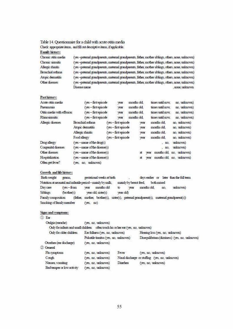
55

56
References
1. Hotomi M, Yamanaka N, Shimada J, Ikeda Y, Faden H. Factors associated with
clinical outcome in acute otitis media. Ann Otol Rhinol Laryngol 2004; 113:846-
52.
2. Damoiseaux RA, Rovers MM, Van Balen FA, Hoes AW, de Melker RA. Long-
term prognosis of acute otitis media in infancy: determinants of recurrent acute
otitis media and persistent middle ear effusion. Fam Pract 2006; 23:40-5.
3. Ito M, Shirai A, Tatsumi A, Yoshizaki T, Nishimura T, Furukawa M. Incidence of
penicillin resistant Streptococus pneumoniae in a nursery school. Pract Otol
(Kyoto) 1999; 92:1071-19.
4. Lubianca Neto JF, Hemb L, Silva DB. Systematic literature review of modifiable
risk factors for recurrent acute otitis media in childhood. J Pediatr (Rio J) 2006;
82:87-96.
5. Rovers MM, Schilder AGM, Zielhuis GA, Rosenfeld RM. Otitis Media. Lancet
2004; 363:465-73.
6. Laine MK, Tähtinen PA, Ruuskanen O, Huovinen P, Ruohola A. Symptoms or
symptom-based scores cannot predict acute otitis media at otitis-prone age.
Pediatrics 2010; 125, e1154-61.
[Addendum]
It is desirable to determine the presence of sensorineural hearing loss by pure
tone audiometry in patients with AOM.
Sensorineural hearing loss is well-known complication of AOM. The association of
sensorineural hearing loss suggests the extension and severity of the acute
inflammation in the temporal bone.
The recommended method how to obtain a specimen for microbial test is
shown in Table 15.
AOM is caused by the invasion of nasopharyngeal pathogens to the middle
ear via the Eustachian tube. Therefore, it is valuable to take a specimen of the
nasopharynx not orally but nasally. The pathogens detected from otorrhea and the
nasopharynx were the same in 90% of S.pneumoniae and in 80% of H.influenzae
(Konno et al. 1999).

57
Table 15. The method of sampling for microbial test
To detect pathogens of AOM, it is desirable to take specimens not only from middle ear effusion
or otorreha but also from nasopharynx. The main pathogens of AOM such as Streptococcus
pneumoniae and Heamophilus influenzae are very weak under dry condition. A swab should be
immediately inoculated into a porter for bacteriological examination.
1) A case without eardrum perforation
After sterilization of the external ear canal with alcohol or popvidone-iodine, the myringotomy
is made. The middle ear effusion is taken for a microbial test using Seed-Swab No. 2.
2) A case with eardrum perforation
After removal of otorrhea in the external ear canal followed by the sterilization of the external
ear canal with alcohol or popvidone-iodine, the middle ear effusion is taken for a microbial test
using Seed-Swab No. 2.
3) Nasopharynx
After removal of nasal discharge followed by the sterilization of a nostril, nasopharyngeal smear
is obtained endonasally.
Reference
1. Konno M, Ubukata K. eds., Penishirintaiseihaiennkyuukin-Revised.
Kyouwakikakutuushin, 1999, pp53-63.
[Addendum] Clinical usefulness of the Pneumococcal Antigen Rapid Detection Kit
(Rapiran-HS®)
The kit has been on the Japan health insurance list since November 2011.
The kit is a detection tool for pneumococci in middle ear effusion, ear discharge or
nasopharyngeal secretion, and it is not used for other body samples including serum
and urine.
The efficiency of the kit was evaluated in a clinical experiment based on
microbiological culture results (Hotomi et al. 2012). In middle ear effusion or ear
discharge, true positivity was found to be 81.4% (48/59), true negativity was 80.5%
(165/205) and the rate of concordance was 80.7% (213/264). In nasopharyngeal
secretion, true positivity was 75.2% (121/161), true negativity was 88.8% (95/107) and
the rate of concordance was 80.6% (216/268). Moreover, when the results of whole
58
samples were combined, the true positivity was 76.8% (169/220), true negativity was
83.3% (260/312) and the rate of concordance was 80.6% (429/532). The concordance
rates with microbiological culture results in both types of samples were favorable.
Based on those results, the kit is considered to be efficient for the diagnosis of
pneumococcal infections of the upper respiratory tract including OM and rhinosinusitis.
This kit is effective in the diagnosis of pneumococcus; however, it does not
supply information about bacterial resistance or other types of pathogenic bacteria.
However, the use of the kit may be efficient when considering the risk factors related
to drug resistance in pediatric patients, including children under two years old
(Yamanaka 2008), those enrolled in daycare and those administered an antibiotic
within the prior month. However, the identification of pathogenic bacteria and the
evaluation of drug resistance by bacterial culture methods should have priority. A
recent multicenter study on the clinical usefulness of the kit for the management of
AOM revealed that the kit was useful for identifying the infecting organism, selecting
appropriate antibacterial agents, and giving the information to explain to patients’
guardians about the management of OM (Uchizono et al. 2012, Yamanaka et al. 2014).
The kit will be useful to select appropriate antimicrobial selections in the
following cases of AOM.
1. In mild cases showing no improvement after observation, after 3 days’
administration of AMPC, the result of the kit will be useful for the subsequent
treatment selection.
2. In moderate cases showing no improvement after the first antimicrobial
therapy, the result of the kit will be useful for the subsequent treatment selection.
3. In severe cases at the first visit or cases showing no improvement after the
first therapy, the result of the kit will be useful for the subsequent treatment selection.
H. influenzae Antigen Detection: ELISA
For OM or acute sinusitis, the detection of H. influenzae antigen in middle
ear effusion or ear discharge and nasopharyngeal cavity or nasal secretion has been
covered by the Japan health insurance since November 2012.
The test specifically detects the P6 protein localized in the outer membrane
of all serotypes and nontypeable H. influenzae by an enzyme-linked immunosorbent
assay (ELISA) and makes the diagnosis of influenzae infection. The remaining fluid
59
from the pneumococcus rapid detection kit can be used as a test sample, and the result
is available within 3 hours (hospital laboratory or private laboratory). For middle ear
effusion or ear discharge, the sensitivity was 83.3% (75/90), the specificity was 85.6%
(143/164) and the rate of concordance was 84.8% (218/257), and for nasopharyngeal
samples, the sensitivity was 71.5% (113/158), the specificity was 92.5% (99/107) and
the rate of concordance was 80.0% (212/265) based on the microbiological culture
standards.
References
1. The supplement for Rapiran Pneumococcus HS (Otitis media and sinusitis).
(Otsuka) (in Japanese).
2. Hotomi M, Togawa A, Takei S et al. Evaluation of a rapid immune-
chromatographic ODK-0901 test for detection of pneumococcal antigen in middle
ear fluids and nasopharyngeal secretions. PLoS One, March 2012, vol 7, issue 3,
e33620: 1-7.
3. Yamanaka N. Infections in the field of otolaryngology, diagnosis and treatment 96:
81-86, 2008 (in Japanese).
4. Uchizono A, Yamanaka N. Efficiency of rapiran pneumococcus HS (otitis media
and sinusitis) and H. influenzae antigen detection kit. J Jpn Soc Infect Dis
Otolaryngol 30; 31-35, 2012 (in Japanese).
5. Yamanaka N, Sugita R, Fujimaki Y et al. Usefulness of Streptococcus pneumoniae
antigen detection kit for the management of acute otitis media. Pract Otol (Kyoto)
107; 7-14, 2014 (in Japanese).
6. The supplement in the H. influenzae ELISA kit (Otsuka) (in Japanese).
20. Prophylaxis
CQ 20-1: Is PCV-7 effective for preventing AOM in children?
Recommendation
PCV-7 is effective for the prevention of infant AOM. (Grade of
Recommendation: A, Benninger 2008 (levelⅠa), Dinleyici 2010 (levelⅠa),
Boonacker et al. 2011 (levelⅠa), Gisselsson-Solen et al. 2011 (levelⅠb), and van Gils

60
et al. 2011(levelⅠb)).
Background
Concerning vaccines against pneumococcus in Japan, the 23-valent
pneumococcal polysaccharide vaccine Pneumovax® was released in the 1980s.
However, this vaccine cannot be used on infants under 2 years of age, and since re-
vaccination within five years involves risk, the usefulness and safety of the vaccine are
low. On February 24, 2010, a 7-valent pneumococcal conjugate vaccine (PCV-7,
Prevenar®), which can be used for infants from 2 months of age was released. In Japan,
this vaccine covers 62.9% of serotypes and 78.0% of drug-resistant bacteria of
pneumococcus isolated from middle ear fluid of childhood AOM. Preventive effects
of 34.4%–62.5% against pneumococcus and 39.8%–49.1% against drug-resistant
pneumococcus are expected, respectively.
Comments
As of May 2013, pneumococcal vaccines that can be used for children in
Japan are the above-mentioned two types. Pneumovax® is 23-valent and the coverage
is good at 93.7%; however, this vaccine is primarily intended for the elderly. PCV-7
is a 7-valent (4, 6B, 9V, 14, 18C, 19F, 23F) vaccine and the coverage is 62.9%. A near-
lifelong immunity can be achieved by inoculating the individual four times between
the age of 2 months and 15 months. PCV-7 has been used as a routine childhood routine
vaccination in the U.S. since 2000, mainly for preventing ROM. Although the
usability of a pneumococcal polysaccharide vaccine against ROM has been confirmed,
conjugate vaccines including PCV-7 are not recommended (Straetemans et al. 2004:
Ia). In a double-blind randomized controlled trial of the 7-valent pneumococcal
conjugate vaccine and pneumococcal polysaccharide vaccine, there was no significant
reduction in the incidence frequency of ROM (Brouwer et al. 2005: Ib) and no effects
of PncCRM (PCV-7) were indicated against AOM when evaluated by Ply-PCR (Palmu
et al. 2009: Ib).
However, the usability of the vaccines has been indicated in many Western
research studies of cost-effectiveness with a high level of evidence (Jansen et al. 2008:
Ib; Dinleyici et al. 2010: Ia; Gisselsson-Solen et al. 2011: Ib; Tyo et al. 2011: III).
Inoculation of this vaccine reduces the contained pneumococcus serotype (Boonacker

61
et al. 2011: Ia; Van Gils et al. 2011: Ib), but on the other hand, it is also reported that it
increases serotypes of 19A and 16A and H. influenzae that are not included (Benninger
2008: Ia; Sox 2011: Ia). In Japan, the preventive effect of PCV-7 against OM is
reported to be 7% to 9%; however, for AOM, when the cost-effectiveness of the
vaccination is calculated, the usability is sufficiently high, contributing to an annual
reduction of 31.4 billion yen in medical expenditures even after the total cost of
vaccination is subtracted (Kamiya et al. 2008: III).
PCV-13 (a 13-valent vaccine) and PHiD-CV (a 10-valent vaccine including
H. influenzae) have already been adopted in Western countries, and their usability has
been demonstrated (Ib). A comparison study of the three types of vaccines is in
progress, showing the effectiveness of all three: PCV-13, PHiD-CV, and PCV-7. PCV
inoculation is expected to reduce the incidence and symptoms of OM (Shea et al. 2011:
Ia; Grijalva et al. 2011: Ia). The cost-effectiveness of all three PCV inoculations was
high (Dinleyici et al. 2010: Ia); however, there are reports pointing out the necessity
of considering various factors in order to evaluate the true cost-effectiveness (Tyo et
al. 2011: III). There are also reports indicating that the economic effect of PCV-13
(Strutton et al. 2012: III) or PHiD-CV (Robberstad et al. 2011: III) is better than those
of the other two vaccines. In Japan, PCV-13 was approved in May 2013, and it was
approved for production and sales in June of the same year. If PCV-13 and PHiD-CV
become available in the near future, the coverage is expected to expand further and the
usability is expected to rise. In their guidelines for the treatment for AOM revised in
2013, the U.S. strongly recommended a pneumococcal vaccine for all children
(Lieberthal et al. 2013).
[Addendum – June 2014]
In Japan, from November 1st, 2013, PCV-13 [Prevenar13®: a 13-valent
pneumococcus conjugate vaccine with a 6-valent serotype (1, 3, 5, 6A, 7F, 19A) newly
added to PCV-7] has been used in place of PCV-7 in routine vaccinations. According
to the Japanese epidemiological data, the 7-valent serotype accounts for 37% and the
newly added 6-valent serotype accounts for 30% of invasive infections by
pneumococcus.
As for the use of PCV-13, in clinical trials, the incidence of side effects such
as erythema reaction at the injection site was observed to be more than 70% (single

62
inoculation) and systemic reactions such as fevers (of 37.5°C or more) were observed
in more than half of the cases, and therefore, sufficient care should be taken to monitor
patients for adverse reactions (Ministry of Health, Labor and Welfare, 9/10/2013).
References
1. Benninger MS. Acute bacterial rhinosinusitis and otitis media: changes in
pathogenicity following widespread use of pneumococcal conjugate vaccine.
Otolaryngol Head Neck Surg 2008; 138:274-8.
2. Dinleyici EC. Current status of pneumococcal vaccines: lessons to be learned and
new insights. Expert Rev Vaccines 2010; 9:1017-22.
3. Boonacker CW, Broos PH, Sanders EA, Schilder AG, Rovers MM. Cost
effectiveness of pneumococcal conjugate vaccination against acute otitis media in
hildren: a review. Pharmacoeconomics 2011; 29:199-211.
4. Gisselsson-Solén M, Melhus A, Hermansson AGisselsson-Solén M, Melhus A,
Hermansson A. Pneumococcal vaccination in children at risk of developing
recurrent acute otitis media - a randomized study. Acta Paediatr 2011; 100:1354-8.
5. van Gils EJ, Veenhoven RH, Rodenburg GD, Hak E, Sanders EA. Effect of 7-
valent pneumococcal conjugate vaccine on nasopharyngeal carriage with
Haemophilus influenzae and Moraxella catarrhalis in a randomized controlled
trial.Vaccine 2011; 29:7595-8.
6. Straetemans M, Sanders EA, Veenhoven RH, Schilder AG, Damoiseaux RA,
Zielhuis GA.:Pneumococcal vaccines for preventing otitis media. Cochrane
Database Syst Rev 2004;(1):CD001480.
7. Brouwer CN, Maillé AR, Rovers MM, Veenhoven RH, Grobbee DE, Sanders EA,
Schilder AG. Effect of pneumococcal vaccination on quality of life in children with
recurrent acute otitis media: a randomized, controlled trial. Pediatrics 2005;
115:273-9.
8. Palmu AA, Saukkoriipi A, Jokinen J, Leinonen M, Kilpi TM. Efficacy of
pneumococcal conjugate vaccine against PCR-positive acute otitis media. Vaccine
2009; 27:1490-1
9. Jansen AG, Sanders EA, Hoes AW, van Loon AM, Hak E. Effects of influenza plus
pneumococcal conjugate vaccination versus influenza vaccination alone in
preventing respiratory tract infections in children: a randomized, double-blind,
63
placebo-controlled trial. J Pediatr 2008; 153:764-7
10. Tyo KR, Rosen MM, Zeng W, Yap M, Pwee KH, Ang LW, Shepard DS. Cost-
effectiveness of conjugate pneumococcal vaccination in Singapore: comparing
estimates for 7-valent, 10-valent, and 13-valent vaccines. Vaccine 2011; 29:6686-
94.
11. Sox C. Acute otitis media: antibiotics are moderately effective and mildly increase
the risk of adverse effects; prevalence of different causative bacteria changed after
introduction of the heptavalent pneumococcal conjugate vaccine. Evid Based Med
2011; 16:181-2.
12. Kamiya H, et al. Economic analysis of pneumococcal conjugate vaccination in
Japan. Jpn J Pediatr 2008; 61:2233-2241. (In Japanese).
13. Shea KM, Weycker D, Stevenson AE, Strutton DR, Pelton SI. Modeling the decline
in pneumococcal acute otitis media following the introduction of pneumococcal
conjugate vaccines in the US. Vaccine 2011; 29:8042-8.
14. Grijalva CG, Pelton SI. A second-generation pneumococcal conjugate vaccine for
prevention of pneumococcal diseases in children. Curr Opin Pediatr 2011; 23: 98-
104.
15. Strutton DR, Farkouh RA, Earnshaw SR, Hwang S, Theidel U, Kontodimas S,
Klok R, Papanicolaou S. Cost-effectiveness of 13-valent pneumococcal conjugate
vaccine: Germany, Greece, and The Netherlands. J Infect 2012; 64:54-67.
16. Robberstad B, Frostad CR, Akselsen PE, Kværner KJ, Berstad AK. Economic
evaluation of second generation pneumococcal conjugate vaccines in Norway.
Vaccine 2011; 29:8564-74.
17. Lieberthal AS, Carroll AE, Chonmaitree T, Ganiats TG, Hoberman A, Jackson MA,
Joffe MD, Miller DT, Rosenfeld RM, Sevilla XD, Schwartz RH, Thomas PA,
Tunkel DE: The diagnosis and management of acute otitis media. Pediatrics 2013;
131:e964-e99
21. Treatment
The outcome of the treatment recommended by the present guideline is
defined by improvement of otoscopic findings such as “injection, swelling, thickness,
blister formation, dullness and perforation of the eardrum, the presence of MEE,
otorrhea, and edema of the middle ear mucosa”, at the point of 3 weeks after the onset.

64
Otoscopic score 0 of each item except for age factor (under 24 months) is judged as
cure of AOM.
A patient receiving antimicrobial agents before the onset of AOM is also
classified into three stages of AOM such as mild, moderate or severe, and is adapted
to the algorithm of the present guideline. However, we should consider an
antimicrobial agent which a patient has been given before, and should take otoscopic
findings at the examination carefully.
CQ 21-1 Is it reasonable not to administer antimicrobial agents for mild AOM?
Recommendation
Watchful waiting for 3 days without use of antimicrobial agents is
recommended for a mild AOM. (Grade of Recommendation: A, Damoiseaaux et al.
2000, Glasziou et al. 2000 (level Ia), Little et al. 2006 (level IIa))
Background
It has been reported that most of cases of AOM improve without use of
antimicrobial agents (van Buchem et al. 1985, Damoiseaux et al. 2000, Rosenfeld et
al. 2003 a, b, Jacobs et al. 2001, Glasziou et al. 2000). However, as the incidence of
AOM caused by drug resistant bacteria is popular in Japan, it is important for us to
diagnose mild AOM precisely by the finding of the eardrum, and follow a child strictly
when we do not use antibiotics.
Comments
The use of antibiotics is closely related with the increase of drug resistant
bacteria in the treatment of infectious diseases. It has been reported that most of cases
of AOM improve without use of antimicrobial agents (van Buchem et al. 1985,
Damoiseaux et al. 2000, Rosenfeld et al. 2003 a, b, Jacobs et al. 2001, Glasziou et al.
2000, Wald et al. 2001). Takata et al. (2001) reported the metaanalysis consisted with
74 comparative studies, and concluded that the cases with simple AOM treated without
antimicrobial agents rarely showed complications and the effect of AMPC was
minimum for the cases. In the randomized clinical trial (RCT) between AMPC and
placebo for children with AOM, in whom 80% of the cases were classified as moderate
65
AOM, the placebo group did not show poorer recovery compared with the AMPC
group (Le Saux et al. 2005). McCormick et al. (2005) reported the comparative study
between the groups with and without administration of AMPC for cases with non-
severe AOM, the eradication rate was high but the carriage rate of drug resistant S.
pneumoniae increased in cases treated by AMPC. They concluded that watchful
waiting was recommended when the following items were resolved; evaluation of
severity of AOM, education for parents, the treatment for relief of the symptom, easy
access to the hospital for follow-up, and antimicrobial agent use if necessary.
Little et al. (2001) reported the multicenter-comparative trial between two
groups, one with immediate use of antimicrobial agents and the other with use of
antimicrobial agents only when the symptom relief was not obtained for 72 hours, and
the results were as follows; there was no significant differences between the two
groups if the patients did not show high fever, restlessness and vomiting. However,
immediate use of antimicrobial agents significantly decreased the incidence of
restlessness and sleep disturbance if the patients showed high fever, restlessness and
vomiting. In addition, Little et al. (2006) reported the long-term outcome i.e. three
months and one year after the randomized trail regarding antimicrobial agent use in
the two groups described above. As the results, there was no statistically different in
the long-term outcome between the two groups. In the other report, wait-and-see for
48 hours without prescription of antimicrobial agents is useful if the symptom does not
become worse (Spiro et al. 2006).
Rover et al. (2007) reported the meta-analysis of randomized trial, in which
the prognosis of the two groups, i.e., wait-and-see group and immediate antimicrobial
agent use group, was compared regarding pain and /or fever at 3 and 7 days. They
concluded that the symptoms in children younger than 2 years persisted twice longer
than those in children at the age of 2 years and older. In the RCT between the
immediate use of antimicrobial agents and wait-and-see for 72 hours if a child does
not complain otalgia, fever and some other symptoms, the immediate use of
antimicrobial agent group showed significantly less otalgia than the wait-and-see
group 3 months after the trial particularly in cases of recurrent AOM. However, one
year after the trial, there was no difference in the outcome between the two groups
(Little et al. 2006). From the results of these reports, it is possible to do watchful
waiting for children with good general condition, but it is necessary to evaluate the

66
clinical signs and symptoms in children with risk factors if they are not administered
antimicrobial agents.
In Japan, AOM caused by drug resistant bacteria has been increasing.
Hotomi et al. 2005) classified children with AOM into two categories, mild and severe
cases, and children with mild AOM were not given antimicrobial agents . As the results,
it is possible not to use antimicrobial agents for mild AOM for 5 days. Although they
showed that the clinical symptoms improved in 94 % of the children with both mild
and server AOM, otoscopic findings improved in 55 % of mild AOM and in only 10%
of severe AOM at day five. Therefore, when a child is followed without antimicrobial
agent use, it is necessary to follow the child watchfully and to prepare the
circumstances to give antimicrobial agents to a child anytime if he/she has failed to
show improvement. There has been another trial that parents could give a child
antimicrobial agents, which had been prescribed beforehand, when needed. As the
results, only 31 % (Siegel et al. 2003) and 34% (McCormick et al. 2005) of children
were administered antimicrobial agents. Rover et al. (2004) also reported that some
children improved without the use of antimicrobial agents, but it is important to follow
them for 2~3 days strictly.
References
1. Damoiseaux R, van Balen FAM, Hoes A, Verheij T, deMelker R. Primary care
based randomised, double blind trial of amoxicillin versus placebo for acute otitis
media in children aged under 2 years. BMJ 2000; 320:350-4.
2. Glasziou PP, Del Mar CB, Hayem M, Sanders SL. Update of: Cochrane Database
Syst Rev. 2000;(2):CD000219. Antibiotics for acute otitis media in children.
Cochrane Database Syst. Rev 2000;(4):CD000219.6.
3. Little P, Moore M, Warner G, Dunleavy J, Williamson I. Longer term outcomes
from a randomised trial of prescribing strategies in otitis media. Br J Gen Pract
2006; 56:176-82.
4. van Buchem FL, Peeters NIF, van't Hof MA. Acute otitis media: a new treatment
strategy. BMJ 1985; 290:1033-7.
5. Rosenfeld RM, Kay D. Natural history of untreated otitis media. In: Rosenfeld
RM, Bluestone CD eds. Evidence-Based Otitis Media 2nd ed. 2003(a):180-98.
6. Rosenfeld RM, Kay D. Natural history of untreated otitis media. Laryngoscope
67
2003 (b); 113:1645-57.
7. Jacobs J, Springer DA, Crothers D. Homeopathic treatment of acute otitis media
in children: a preliminary randomized placebo-controlled trial. Pediatr Infect Dis
J 2001; 20:177-83.
8. Wald ER, Mason EO Jr, Bradley JS, Barson WJ, Kaplan SL, US Pediatric
Multicenter Pneumococcal Surveillance Group. Acute otitis media caused by
Streptococcus pneumoniae in children’s hospitals between 1994 and 1997. Pediatr
Infect Dis J 2001; 20:34-9.
9. Takata GS, Chan LS, Shekelle P, Morton SC, Mason W, Marcy SM. Evidence
assessment of management of acute otitis media: I. The role of antibiotics in
treatment of uncomplicated acute otitis media. Pediatrics 2001; 108:239-47.
10. Le Saux N, Gaboury I, Baird M, Klassen TP, MacCormick J, Blanchard C, Pitters
C, Sampson M, Moher D. A randomized, double-blind, placebo-controlled
noninferiority trial of amoxicillin for clinically diagnosed acute otitis media in
children 6 months to 5 years of age. CMAJ 2005; 172:335-41.
11. McCormick DP, Chonmaitree T, Pittman C, Saeed K, Friedman NR, Uchida T,
Baldwin CD. Nonsevere acute otitis media: a clinical trial comparing outcomes of
watchful waiting versus immediate antibiotic treatment. Pediatrics 2005; 115:
1455-65.
12. Little P, Gould C, Williamson I, Moore M, Warner G, Dunleavey J. Pragmatic
randomized controlled trial of two prescribing strategies for childhood acute otitis
media. BMJ 2001; 322(7282):336-42.
13. Spiro DM, Tay KY, Arnold DH, Dziura JD, Baker MD, Shapiro ED. Wait-and-see
prescription for the treatment of acute otitis media: a randomized controlled trial.
JAMA 2006; 296:1235-41.
14. Rovers MM, Glasziou P, Appelman CL, Burke P, McCormick DP, Damoiseaux
RA, Little P, Le Saux N, Hoes AW. Predictors of pain and/or fever at 3 to 7 days
for children with acute otitis media not treated initially with antibiotics: a meta-
analysis of individual patient data. Pediatrics 2007; 119:579-85.
15. Hotomi M, Yamanaka N, Samukawa T, Suzumot M, Sakai A, Shimada J, Ikeda Y,
Faden H. Treatment and outcome of severe and non-severe acute otitis media. Eur
J Pediatr 2005; 164: 3-8.
16. Siegel RM, Kiely M, Bien JP, Joseph EC, Davis JB, Mendel SG, Pestian JP,
68
DeWitt TG. Treatment of otitis media with observation and a safety-net antibiotic
prescription. Pediatrics 2003; 112:527-31.
17. Rovers MM, Schilder AGM, Zielhuis GA, Rosenfeld RM. Otitis Media. Lancet
2004; 363:465-73.
CQ 21-2 Are antimicrobial agents useful for the analgesic treatment of AOM?
Recommendation
The efficacy of antimicrobial agents specifically for otalgia is unknown.
(Grade of Recommendation: I)
Background
Otalgia is the main clinical symptom of AOM to be treated, but
contradictory results concerning the analgesic effect of antimicrobial agents have
been reported.
Comments
Glasziou et al.(2004) reported that antimicrobial agents did not
significantly improve otalgia compared to the natural course. In contrast, Bascelli et
al. (2001) reported that the duration of subjective otalgia was significantly shorter
and the consumption of analgesics was significantly reduced in an antimicrobial
agents-treated group in a randomized controlled study in which antimicrobial agents
were administered immediately after onset and 3 days after onset in cases showing
no remission tendency. Therefore, the effect of antimicrobial agents on otalgia is
unclear. The effect of analgesics on otalgia has also not been fully investigated. In a
multicenter randomized double-blind controlled study performed by Bertin et al.
(1996), the effect of ibuprofen was significant compared to a placebo, but no
69
significant analgesic effect of acetaminophen was observed.
Note: In the current situation in Japan, acetaminophen is selected for analgesic
treatment for infants aged 3 years or younger.
[Addendum]
It was reported in 2008 that the local administration of 2% lignocaine into
the external acoustic meatus significantly reduced pain to 50% of the pretreatment
level at 10 and 30 minutes after ear drop treatment in a double-blind randomized
controlled study (Bolt et al. 2008), for which additional study results are anticipated.
References
1. Glasziou PP, Hayem M, Del Mar CB. Update in: Cochrane Database Syst Rev.
2000;(4):CD000219. Antibiotics for acute otitis media in children. Cochrane
Database Syst Rev 2004; (1):CD000219.
2. Bascelli LM, Losh DP. How does a "wait and see" approach to prescribing
antibiotics for acute otitis media (AOM) compare with immediate antibiotic
treatment? J Fam Pract 2001; 50:469.
3. Bertin L, Pons G, d’Athis P, Duhamel JF, Maudelonde C, Lasfargues G, Guillot
M, Marsac A, Debregeas B, Olive G. A randomized, double-blind, multicentre
controlled trial of ibuprofen versus acetaminophen and placebo for symptoms of
acute otitis media in children. Fundam Clin Pharmacol 1996; 10:387-92.
4. Bolt P, Barnett P, Babl FE, Sharwood LN.Topical lignocaine for pain relief in
acute otitis media: results of a double-blind placebo-controlled randomized trial.
Arch Dis Child 2008;93:40-4.
CQ 21-3 Which antimicrobial agents should be used for AOM?
70
Recommendation
Recommended antimicrobial agents depend on bacterial resistance and the
severity of AOM, but the following are often recommended. (Grade of
Recommendation: A)
P.O.: Amoxicillin (AMPC), amoxicillin/clavulanate (AMPC/CVA), cefditoren pivoxil
(CDTR-PI), and DIV: Ampicillin (ABPC), ceftriaxone (CTRX)
Studies used for the recommendation: Ghaffar et al. 2002, 2000 (Level Ib), Piglansky
et al. 2003 (Level Ib), Haiman et al. 2002 (Level Ib)
Background
Based on the current situation in Japan, about 50-65% of S.pneumoniae
and about 50-70% of H. influenzae is drug-resistant, therefore, it is recommended to
select the above antimicrobial agents corresponding to the severity of AOM based on
the sensitivity of pathogens. This does not mean that other antimicrobial agents are
not recommendable, but the above antimicrobial agents are recommended in
consideration of the current drug sensitivity in Japan.
Comments
Based on bacteria detected in infants with AOM in Japan and antimicrobial
agent activities on these, AMPC, CVA/AMPC (1:14), CDTR-PI, TFLX and TBPM-
PI as peroral agents, and CTRX and ABPC are selected as injection corresponding to
the severity. Reports on AMPC treatment in Japan demonstrated their usefulness
(Hotomi et al. 2005, Harabuchi et al. 2001). In the U.S. guidelines revised in 2013,
AMPC is still recommended as the first-line antimicrobial agent (Lieberthal et al.
2013).
Regarding studies on AMPC and AMPC/CVA in Western countries, a

71
prospective observational study reported significant therapeutic results of AMPC
(Brook et al. 2002). Lund et al. (2001) investigated changes in bacterial flora in the
oropharynx and epipharyngeal space in 12 patients treated with AMPC/CVA and 17
patients treated with cefuroxime axetil (CMX-AX) in a randomized controlled study,
and observed similar effects on S.pneumoniae, H.influenzae, and M. catarrhalis in
the epipharyngeal space in both groups. The administration of AMPC/CVA at an
increased dose (90/6.4 mg/kg/day, 10 days) was effective with regard to the
eradication rates of S.pneumoniae, PRSP, and H.influenzae in a multicenter
randomized controlled study (Dagan et al. 2001). Piglansky et al. (2003) also
reported that high-dose AMPC (80 mg/kg/day, 10 days) was effective as an early
treatment in a prospective observational study. In contrast, high-dose AMPC was
neither beneficial nor non-beneficial for antimicrobial agent-resistant low-risk AOM
in a double-blind randomized controlled study (Garrison et al. 2004). Casellas et al.
(2005) observed no difference in efficacy between AMPC/SBT and AMPC/CVA in a
multicenter single-blind randomized controlled study. Regarding non-AMPC
responders, Block et al. (2001) reported that many patients in whom the disease was
not remitted by AMPC were infected by S.pneumoniae in a retrospective
observational study. Regarding the dosing frequency, Damrikarnlert et al. (2000)
observed no significant difference between twice— and 3 times-per-day
administrations of AMPC/CVA in a multicenter randomized controlled study.
Haiman et al. (2002) investigated changes in S.pneumoniae in the
epipharyngeal space (before, in the middle of, and after treatment) in a randomized
controlled study in which ceftriaxone (CTRX) was injected 3 times, and observed a
significant reduction of S.pneumoniae. Heikkinen et al. (2000) also reported that the
intramuscular injection of CTRX significantly decreased H.influenzae in the
epipharyngeal space. Toltzis et al. (2007) reported that the potentiation of fecal gram-
negative bacterial growth by a single intramuscular injection of CTRX was similar to

72
that by cefprozil (CFPZ), AMPC, and azithromycin (AZM) in a randomized
controlled study. Wang et al. (2004) also reported that a single administration of
CTRX was as effective as the 10—day administration of AMPC/CVA (40 mg/kg).
The intramuscular injection of CTRX is not indicated in Japan, but once-a-day
administration to infants was approved on November 13, 2007, and intravenous
antimicrobial agent injection at outpatient clinics became available for infants, as in
adults.
Ioannidis et al. (2001) performed a meta-analysis of multicenter
randomized controlled studies, and identified no significant differences in the
efficacy or safety of AZM for upper airway infection compared to other antimicrobial
agents. Hoberman et al. (2005) compared high-dose CVA/AMPC (6.4/90 mg/day)
and AZM, and observed that CVA/AMPC was more useful than AZM, but Guven et
al. (2006) noted no significant difference between AZM and AMPC/CVA (45/6.4
mg/day) in a single-blind randomized controlled study. Arguedas et al. (2005)
reported that AZM exhibited an effect equivalent to that of high-dose AMPC (90
mg/kg) and was superior in compliance in a multicenter double-blind randomized
controlled study. Regarding the current state of pathogens in Japan, AZM may be
selected for H.influenzae.
In light of the present status of pathogens of AOM in Japan, cefaclor
(CCL), cefpodoxime proxetil (CPDX-PR) cannot be treatment options, and CFPZ
and gatifloxacin (GFLX) are also not allowed; they are not approved in Japan.
TFLX is a rare new quinolone agent, the safety of which for children is
recognized. TFLX shows excellent antimicrobial activities to gram-positive and -
negative bacteria, anaerobic bacteria, several β-lactamase producing bacteriae, and
methicillin-resistant Staphylococcus aureus (MRSA). TFLX was also clinically
demonstrated to have an excellent effect on OM (Suzuki et al. 2010, Yamanaka et al.
2012a), as well as in basic and clinical studies in the field of otorhinolaryngology

73
(Kobayashi et al. 1988).
TBPM-PI, which is the only peroral carbapenem agent, has a wide range of
antimicrobial spectrum and antimicrobial activities that are stronger than those of
intravenous carbapenem agents. A randomized controlled trial (RCT) of TBPM-PI on
AOM in children showed an effect equivalent to that of a high dose of CDTR-PI
(Suzuki et al. 2009). Its good transition to ME tissue was also reported (Baba et al.
2009). A recent multicenter clinical trial revealed its high efficacy on AOM in
children, including those with ROM and prolonged OM (Yamanaka et al. 2012b).
The present AOM Guideline Subcommittee recommends that TFLX and TBPM-PI
should be used only in cases in which the effect of other antimicrobial agents cannot
be expected.
References
1. Ghaffar F, Muniz LS, Katz K, Smith JL, Shouse T, Davis P, McCracken GH Jr.
Effects of large dosages of amoxicillin/clavulanate or azithromycin on
nasopharyngeal carriage of Streptococcus pneumoniae, Haemophilus influenzae,
nonpneumococcal alpha-hemolytic streptococci, and Staphylococcus aureus in
children with acute otitis media. Clin Infect Dis 2002; 34:1301-9.
2. Ghaffar F, Muniz LS, Katz K, Reynolds J, Smith JL, Davis P, Friedland IR,
McCracken GH Jr. Effects of amoxicillin/clavulanate or azithromycin on
nasopharyngeal carriage of Streptococcus pneumoniae and Haemophilus
influenzae in children with acute otitis media. Clin Infect Dis 2000; 31:875-80.
3. Piglansky L, Leibovitz E, Raiz S, Greenberg D, Press J, Leiberman A, Dagan R.
Bacteriologic and clinical efficacy of high dose amoxicillin for therapy of acute
otitis media in children. Pediatr Infect Dis J 2003; 22:405-13.
4. Haiman T, Leibovitz E, Piglansky L, Press J, Yagupsky P, Leiberman A, Dagan
R. Dynamics of pneumococcal nasopharyngeal carriage in children with

74
nonresponsive acute otitis media treated with two regimens of intramuscular
ceftriaxone. Pediatr Infect Dis J 2002; 21:642-7.
5. Hotomi M, Yamanaka N, Samukawa T, Suzumot M, Sakai A, Shimada J, Ikeda
Y, Faden H. Treatment and outcome of severe and non-severe acute otitis media.
Eur J Pediatr 2005; 164: 3-8.
6. Harabuchi Y, Kodama H, Faden H. Outcome of acute otitis media and its
relation to clinical features and nasopharyngeal colonization at the time of
diagnosis. Acta Oto-Laryngol 2001; 121:908-14.
7. Lieberthal AS, Carroll AE, Chonmaitree T, Ganiats TG, Hoberman A, Jackson
MA, Joffe MD, Miller DT, Rosenfeld RM, Sevilla XD, Schwartz RH, Thomas
PA, Tunkel DE: The diagnosis and management of acute otitis media. Pediatrics
2013; 131:e964-e99
8. Brook I, Gober AE. Effect of amoxicillin and co-amoxiclav on the aerobic and
anaerobic nasopharyngeal flora. J Antimicrob Chemother 2002; 49:689-92.
9. Lund B, Edlund C, Rynnel-Dagoo B, Lundgren Y, Sterner J, Nord CE.
Ecological effects on the oro- and nasopharyngeal microflora in children after
treatment of acute otitis media with cefuroxime axetil or amoxycillin-
clavulanate as suspensions. Clin Microbiol Infect 2001; 7:230-7.
10. Dagan R, Hoberman A, Johnson C, Leibovitz EL, Arguedas A, Rose FV, Wynne
BR, Jacobs MR. Bacteriologic and clinical efficacy of high dose
amoxicillin/clavulanate in children with acute otitis media. Pediatr Infect Dis
J. 2001; 20:829-37.
11. Garrison GD, Sorum PC, Hioe W, Miller MM. High-dose versus standard-dose
amoxicillin for acute otitis media. Ann Pharmacother 2004; 38:15-9.
12. Casellas JM, Israele V, Marin M, Ishida MT, Heguilen R, Soutric J, Arenoso H,
Sibbald A, Stamboulian D. Amoxicillin-sulbactam versus amoxicillin-clavulanic
acid for the treatment of non-recurrent-acute otitis media in Argentinean
75
children. Int J Pediatr Otorhinolaryngol 2005; 69:1225-33.
13. Block SL, Hedrick JA, Tyler RD, Smith RA, Harrison CJ. Microbiology of
acute otitis media recently treated with aminopenicillins. Pediatr Infect Dis J
2001; 20:1017-21.
14. Damrikarnlert L, Jauregui AC, Kzadri M. Efficacy and safety of amoxycillin/
clavulanate (Augmentin) twice daily versus three times daily in the treatment of
acute otitis media in children. The Augmentin 454 Study Group. J Chemother.
2000; 12:79-87.
15. Heikkinen T, Saeed KA, McCormick DP, Baldwin C, Reisner BS, Chonmaitree
T. A single intramuscular dose of ceftriaxone changes nasopharyngeal bacterial
flora in children with acute otitis media. Acta Paediatr 2000; 89:1316-21.
16. Toltzis P, Dul M, O'Riordan MA, Toltzis H, Blumer JL. Comparative effects of
single-dose ceftriaxone versus three oral antibiotic regimens on stool
colonization by resistant bacilli in children. Pediatr Infect Dis J 2007; 26:25-30.
17. Wang CY, Lu CY, Hsieh YC, Lee CY, Huang LM. Intramuscular ceftriaxone in
comparison with oral amoxicillin-clavulanate for the treatment of acute otitis
media in infants and children. J Microbiol Immunol Infect 2004; 37:57-62.
18. Oguz F, Unuvar E, Suoglu Y, Erdamar B, Dundar G, Katircioglu S, Sidal M.
Etiology of acute otitis media in childhood and evaluation of two different
protocols of antibiotic therapy: 10 days cefaclor vs. 3 days azithromycin. Int J
Pediatr Otorhinolaryngol 2003; 67:43-51.
19. Catania S, Gallo A. Clinical efficacy and tolerability of short course therapy with
cefaclor compared with long-term therapy for treatment of acute otitis media in
children. Infez Med 2004; 12:259-65.
20. Ioannidis JP, Contopoulos-Ioannidis DG, Chew P, Lau J. Meta-analysis of
randomized controlled trials on the comparative efficacy and safety of
azithromycin against other antibiotics for upper respiratory tract infections. J

76
Antimicrob Chemother 2001; 48:677-89.
21. Hoberman A, Dagan R, Leibovitz E, Rosenblut A, Johnson CE, Huff A,
Bandekar R, Wynne B. Large dosage amoxicillin/clavulanate, compared with
azithromycin, for the treatment of bacterial acute otitis media in children.
Pediatric Infect Dis J.2005;24:525-32.
22. Guven M, Bulut Y, Sezer T, Aladag I, Eyibilen A, Etikan I. Bacterial etiology of
acute otitis media and clinical efficacy of amoxicillin-clavulanate versus
azithromycin. Int J Pediatr Otolaryngol 2006; 70:915-23.
23. Arguedas A, Emparanza P, Schwartz RH, Soley C, Guevara S, de Caprariis. A
randomized, multicenter, double blind, double dummy trial of single dose
azithromycin versus high dose amoxicillin for treatment of uncomplicated acute
otitis media. Pediatric Infect Dis J 2005; 24:153-61.
24. Suzuki K, Iino Y, Kudo F, Taiji H, Sunakawa K. An uncontrolled, open label
study of tosfloxacin granules 10% in pediatric acute otitis media (AOM). Jpn J
Chemotherapy 2010; 58(S-2):50-68 (in Japanese).
25. Yamanaka N, Sugita R, Uno Y, Matsubara S, Hayashi Y, Sawada S. Clinical
efficacy of tosufloxacin tosilate hydrate for the treatment of acute otitis media in
children. Pract Otol (Kyoto) 105; 381-392,2012 (in Japanese).
26. Kobayashi T, Baba S, Suzuki K, Shimada J, Soyano K, Maaruo T, Ito H.
Laboratory and clinical studies on T-3262 in otorhinolaryngological infections.
Chemotherapy 36: 1296-1302,1988 (in Japanese).
27. Baba S, Kasahara H, Morita J, Aizawa K, Sunakawa K. Tissue and aural
discharge distribution of tebipenem pivoxil. Jpn J Antibiotics 2009;62:127-135
(in Japanese).
28. Suzuki K, Baba S, Totsuka K, Hori S, Ubukata K, Nakashima M, Sunakawa K.
Double-blind comparative study of tebipenem pivoxil and high-dose cefditoren
pivoxil in children with acute otitis media (Phase III).Jpn J Chemotherapy

77
57(Suppl.1): 167-185,2009 (in Japanese).
29. Baba S,Suzuki K,Totsuka K,Hori S, Ubukata K, Sunakawa K. General
study of tebipenem pivoxil in children with acute otitis media and acute
rhinosinusitis (Phase III). Jpn J Chemotherapy 57(S-1): 151-166,2009 (in
Japanese).
30. Yamanaka N, Suetake M, Tomiyama M, Sugita R, Matsubara S, Sawada S, Uno
Y, Kanesada K, Uchizono A. Efficacy evaluation of TBPM-PI, an oral
carbapenem antibacterial agent, in children with acute otitis media including
recurrent/persistent cases Pract Otol (Kyoto) 105; 687-698, 2012 (in Japanese).
CQ 21-4 How long is the period for administration of antibacterial agent?
Recommendation:
In moderate and severe cases, the patient should be treated with 5-day
administration of antibacterial agent and disease status should be evaluated at the third
or fourth day of the treatment. (Grade of Recommendation: A)
Studies used for the recommendation: Ovetchkine et al. 2003 (Level Ia), Kozyrskyj
et al. 2000 (Level Ia), Bezakova et al. 2009 (Level Ib), Gulani et al. 2010 (Level Ia),
Hoberman et al. 2011 (Level Ia), Tahtinen et al. 2011 (Level Ib)
Background
Although the period of antibacterial therapy is generally 5-day, 7-day or 10-
day, the period is recommended according to the pathogenicity of bacteria and the
78
efficacy of antibacterial therapy.
Comments
In a prospective study, Pichichero et al. (2001) evaluated 5-day, 7-day and
10-day period of antibacterial therapy for AOM. Although 10-day treatment was found
to have better cure and improvement in cases with an episode of AOM in the preceding
month or in cases with more than one AOM episodes, no significant difference was
observed in the outcome of 5-day, 7-day and 10-day treatment groups between
antibacterial types and ages.
On the other hand, a meta-analysis based on 7 randomized controlled trials
revealed that longer period of antibacterial therapy was better in the outcome than
shorter course in children younger than 2 years of age, attending daycare centers and
those with perforated tympanic membrane, while shorter course was recommended in
children elder than 2 years even with previous antibacterial treatment or recurrent
AOM history (Ovetchkine et al. 2003, Kozyrskyj et al. 2000) compared the clinical
efficacy of antibacterial therapies less than seven days and longer course (8 - 19 days)
in another meta-analysis of 32 randomized controlled studies. The five-day
antibacterial therapy was found to be effective for uncomplicated AOM. Leibovitz et
al. (2000) compared 1-day 50mg/kg/day vs. 3-day 50-mg/kg/day of intramuscular
CTRX treatment in non-responsive AOM and found 3-day regime to be significantly
effective especially for PISP and PRSP. However, intramuscular administration of
CTRX is not approved in Japan. Pichicero et al. (2000) found no significant difference

79
for clinical efficacy between 5 days vs. 10 days treatment of cefuroxime axetil (CXM-
AX) and concluded that short course treatment is effective because of better drug
compliance. However, CXM-AX is not a treatment alternative in Japan when
considering the etiologic bacterial agents. In a multicenter single-blind (doctor
blinded) randomized study, Roos et al. (2000) compared 5 days versus 10 days
treatment with ceftibuten (CETB) 9mg/kg/day for recurrent AOM and reported that 10
days treatment showed lower recurrence rate in short term follow-up. Another
prospective study on duration of antibacterial therapy for cases not previously received
antibacterial agents suggested 5 days treatment for AOM in children over 2 years of
age (Manarey et al. 2002).
An RCT comparing the effect by questionnaire of 3-day AMPC (40 mg/kg)
and a placebo revealed no significant difference in the incidence of recurrence
(Bezakova et al. 2009). A systematic review analyzing 35 RCTs comparing the effects
of short-term (< 4 days) and long-term (> 5 days) administrations of peroral
antimicrobial agents on AOM in children revealed that the risk of treatment failure
increased in the short-term group when short-acting antimicrobial agents were used
(Gulani et al. 2010). These results suggested that at least more than 4 days’
administration of antimicrobial agents is recommended whenever necessary.
In another RCT comparing the efficacy of 7-day CVA/AMPC and placebo
on AOM in children, a significant difference between the two groups was shown in the
incidence of treatment failure from the third treatment day (Tahtinen et al. 2011).
Hoberman et al. (2011) also conducted an RCT comparing the effects of 10-day

80
CVA/AMPC and placebo on AOM in children, and they reported a significant
difference in the incidence of the clinical failure from the 4th or 5th administration
days. These results may indicate that the clinical efficacy of antimicrobial agents
appears on the third administration day at the earliest, and thus, the assessment of
efficacy on the third administration day is recommended.
In the U.S. AOM Guidelines revised in 2013, a 10-day administration of
antimicrobial agents is recommended for infants under the age of 2 or severe cases, a
7-day administration is recommended for moderate or mild grades in children 2 to 5
years of age, and for moderate or mild grades in children older than 6 years, the
Guidelines state that 5–7 days’ administration is sufficient. Also, the U.S. Guidelines
recommend the assessment of the effect of the agent on the 2nd or 3rd day of
administration in cases of initial use (Lieberthal et al. 2013).
References
1. Ovetchkine P, Cohen R. Shortened course of antibacterial therapy for acute otitis
media. Paediatr Drugs 2003; 5:133-40.
2. Kozyrskyj AL, Hildes-Ripstein GE, Longstaffe SE, Wincott JL, Sitar DS,
Klassen TP, Moffatt ME. Short course antibiotics for acute otitis media.
Cochrane Database Syst Rev 2000;CD001095.
3. Bezakova N,Damoiseaux RA、Hoes AW, Schilder AG, Rovers MM. Recurrence
up to 3.5 years antibiotic treatment of acute otitis media in very young Dutch
children: survey of trial participants. BMJ 2009; 339:b2525
4. Gulani, A,Sachdev HP, Qazi SA. Efficacy of short course (<4 days) of

81
antibiotics for treatment of acute otitis media in children: a systematic review of
randomized controlled trials. Indian pediatr 2010; 47,74-87.
5. Hoberman A, Paradise JL, Rockette HE, Shaikh N, Wald ER,Kearney DH,
Colborn DK, Kurs-Lasky M, BhatnagarS, Haralam MA, ZoffelLM, Jenkins C,
Pope MA, Balentine TL, Barbadora KA. Treatment of Acute Otitis Media in
Children under 2 Years of Age. New Engl J Med 2011; Jan 13,105-115.
6. Tahtinen PA, Laine MK, Huovinen P, Ruuskanen O, RuoholaA. A Placebo-
Controlled Trial of Antimicrobial Treatment for Acute Otitis Media. The New
England Journal of Medicine 2011; 13,116-126
7. Pichichero ME, Marsocci SM, Murphy ML, Hoeger W, Francis AB, Green JL.
A prospective observational study of 5-, 7-, and 10-day antibiotic treatment for
acute otitis media. Otolaryngol Head Neck Surg 2001; 124:381-7.
8. Leibovitz E, Piglansky L, Raiz S, Press J, Leiberman A, Dagan R.
Bacteriologic and clinical efficacy of one day vs. three day intramuscular
ceftriaxone for treatment of nonresponsive acute otitis media in children.
Pediatr Infect Dis J 2000; 19:1040-5.
9. Pichicero M. Short courses of antibiotic in acute otitis media and sinusitis
infections. J Int Med Res 2000; 28 Suppl 1:25A-36A.
10. Roos K, Larsson P. Efficacy of ceftibuten in 5 versus 10 days treatment of
recurrent acute otitis media in children. Int J Pediatr Otorhinolaryngol 2000;
55:109-15.
11. Manarey CR, Westerberg BD, Marion SA. Clinical decision analysis in the
treatment of acute otitis media in a child over 2 years of age. J Otolaryngol
2002; 31:23-30.
12. Lieberthal AS, Carroll AE, Chonmaitree T, Ganiats TG, Hoberman A, Jackson
MA, Joffe MD, Miller DT, Rosenfeld RM, Sevilla XD, Schwartz RH, Thomas
PA, Tunkel DE: The diagnosis and management of acute otitis media. Pediatrics
82
2013; 131:e964-e99
CQ 21-5 What are appropriate indications for myringotomy?
Recommendation
The indication should be considered depending on severity of AOM. (Grade
of Recommendation: I)
Background
In AOM, there is fluid accumulation due to inflammatory pathology in
middle ear, therefore drainage of inflammatory fluid by myringotomy would be
efficient for early cure of the disease. However, currently there are only a limited
number of studies about the clinical efficacy of myringotomy on early cure of the
disease.
Comments
All of the reports showing the clinical efficacy of myringotomy have been of the
retrospective study. Myringotomy was performed for cases with infection signs after
48 hours of antibacterial therapy and all cases showed improvement 48 hours after
the procedure (Babin et al. 2003). Hotomi et al. (2002) concluded that myringotomy
was necessary depending on severity of the disease. In a case-control study, Nomura
et al. (2005) reported that myringotomy significantly decreased the deterioration rate
of AOM into OME, while it was not effective for prevention of early relapse or
recurrence of AOM. A randomized controlled trial comparing the three groups
including myringotomy only, antibacterial therapy only, and myringotomy with

83
antibacterial therapy in severe cases revealed that there was no clinically significant
difference among them, even myringotomy added to antibacterial therapy (van
Buchem et al. 1985). In another randomized trial of severe cases, Kalaida et al.
(1991) compared three regimens including amoxicillin only, amoxicillin and
myringotomy, and placebo and myringotomy (2 years and older) and found that the
treatment failure was higher in placebo and myringotomy group compared with
others in severe cases. Based on this trial, it can be inferred that myringotomy only
may not be of an effective treatment; nevertheless myringotomy would be effective
in combination with antibacterial therapy.
The position statement released in 2009 by the Canadian Paediatric Society
recommended a referral to an otolaryngologist for tympanocentesis when a patient
with β-lactam allergy does not improve after first-line treatment of antimicrobials and
when a patient does not improve after a second-line treatment with antimicrobials
(Forgie et al. 2009).
One of the concerns is that most previous studies concerning the effect of
myringotomy were based on data obtained under the circumstances of no significant
antimicrobial-resistant bacteria. It seems that patients with refractory OM caused by
PRSP and/or BLNAR were not included in these studies. Yamanaka and Hotomi
(2006) compared the otoscopic findings of the tympanic membrane in patients with
severe AOM between those who received antimicrobial treatment with myringotomy
and those who received antimicrobial treatment, and they found that the otoscopic
findings 2 weeks after the treatment were improved significantly in the myringotomy
group compared to the only-antimicrobial treatment group.
Uno (2008) evaluated the effect of myringotomy in patients with AOM, and
he concluded that in severe cases, as defined by the severity evaluation, the disease
was ameliorated significantly earlier after myringotomy than without it in patients with
an ear pain score of 2 and fever score of 1 or 2 on a clinical symptom evaluation, a
flare score of 2 and a bulge score of 4 or 8 on otoscopic evaluation, and an otorrhea
score of 0. He also found that early in the 10-day follow-up, the overall improvement
rate was significantly higher after myringotomy than in the non-myringotomy cases,
but the difference was not significant in the final outcome. The recurrence rate in
severe cases was also significantly lower in the myringotomy group than in the non-
myringotomy group.

84
References
1) Babin E, Lemarchand V, Moreau S, Goullet de Rugy M, Valdazo A, Bequignon
A. Failure of antibiotic therapy in acute otitis media. J Laryngol Otol 2003;
117:173-6.
2) Hotomi M, Yamanaka N. Antimicrobial therapy for intractable otitis media
caused by drug-resistant bacteria. Monthly Book ENTONI 2002; 15:15-22 (in
Japanese).
3) Nomura Y, Ishibashi T, Yano J, Ichikawa T, Shinogami M, Monobe H, Hirai R,
Kaga K. Effect of myringotomy on prognosis in pediatric acute otitis media. Int J
Pediatr Otorhinolaryngol 2005; 69:61-4.
4) van Buchem FL, Peeters NIF, van't Hof MA. Acute otitis media: a new treatment
strategy. BMJ 1985; 290:1033-7.
5) Kaleida PH, Casselbrant ML, Rockette HE, Paradise JL, Bluestone CD, Blatter
MM, Reisinger KS, Wald ER, Supance JS. Amoxicillin or myringotomy or both
for acute otitis media: Results of a randomized clinical trial. Pediatrics 1991; 87:
466-74.
6) Forgie S, Zhanel G, J Robinson J. Management of acute otitis media. Paediatr
Child Health 2009; 14: 457–60.
7) Yamanaka N, Hotomi M. TOF-27 Is there no different clinical effect between the
myringotomy and antimicrobial treatment? Management in pediatric acute otitis
media (Yamanaka N & Hotomi M eds). Iyaku Journal, Osaka 2006, pp73-6 (In
Japanese).
8) Uno Y. Current frequency and efficacy of myringotomy for acute otitis media in
children. Pediatr Otorhinolaryngol Jpn 2008; 29:20-4 (in Japanese).
CQ 21-6 What are appropriate indications for topical otic treatment?
85
Recommendation
In cases who underwent ventilation tube insertion, antibacterial eardrops is
recommended only for the condition securing full access of antibacterial solution with
high antimicrobial activity into middle ear through the tube. (Grade of
Recommendation: A)
Studies used for the recommendation: Dohar J et al. 2006 (level Ib), Schmelzle et al.
2008 (level Ia)
Background
Topical otic treatment can achieve high concentration of antibacterial agent
in middle ear and have an indication in selected cases.
Comments
In a prospective study, 4 drops of topical ciprofloxacin (CPFX 0.3%) /
dexamethasone (0.1%) twice daily for 7 days and oral suspension of AMPC/CVA
(600mg/42.9mg) every 12 hours for 10 days was compared in AOM cases with
otorrhea through tympanostomy tubes, otic drops obtained significant earlier cure and
the otic treatment was found to be effective in earlier cessation of otorrhea through
tympanostomy tubes (Dohar et al. 2006). Concerning the type of otic treatment, a
randomized clinical trial comparing CPFX/dexamethasone and ofloxacin (OFLX)
revealed CPFX/ dexamethasone to be significantly superior (Roland et al. 2004).

86
Schmelzle et al. (2008) reviewed the evidence regarding the antibiotic
treatment of AOM in children with tympanostomy tubes, and they discussed antibiotic
resistance and ototoxicity, using articles providing level I evidence (randomized
controlled trials) for treatment. They reported that the current evidence suggests that a
topical fluoroquinolone, with or without a corticosteroid, results in a faster resolution
of otorrhea, and that this is the treatment of choice for AOM with tympanostomy tubes.
[Addendum]
In a double-blind, randomized trial in 2008 evaluating eardrops for pain relief,
topical 2% lignocaine was found to reduce significantly pain scores at 10, 20 and 30
minutes by 50% from the baseline (Bolt et al. 2008).
References
1) Dohar J, Giles W, Roland P, Bikhazi N, Carroll S, Moe R, Reese B, Dupre S, Wall
M, Stroman D, McLean C, Crenshaw K. Topical ciprofloxacin/dexamethasone
superior to oral amoxicillin/clavulanic acid in acute otitis media with otorrhea
through tympanostomy tubes. Pediatrics 2006; 118:e561-9.
2) Roland PS, Dohar JE, Lanier BJ, Hekkenburg R, Lane EM, Conroy PJ, Wall.
Topical ciprofloxacin/dexamethasone otic suspension is superior to ofloxacin otic
solution in the treatment of granulation tissue in children with acute otitis media
with otorrhea through tympanostomy tubes. Otolaryngol Head Neck Surg 2004;
130:736-41.
3) Schmelzle J, Birtwhistle RV, Tan AKW. Acute otitis media in children with

87
tympanostomy tubes. Can Fam Physician 2008; 54:1123-7
4) Bolt P, Barnett P, Babl FE, Sharwood LN. Topical lignocaine for pain relief in acute
otitis media: results of a double-blind placebo-controlled randomised trial. Arch
Dis Child 2008; 93:40-4.
CQ 21-7 Risk factors deteriorating AOM and medications other than
antibacterial agents
Recommendation
Since younger age and daycare attendance have an important role on
deterioration of the disease, the attention should be paid during the treatment. (Grade
of Recommendation: A)
In cases of AOM complicated with nasal disease, nasal treatments should be
considered as complementary to the treatment of AOM (Recommendation level I).
The report used for the recommendation: Ovetchkine et al. 2003 (Level Ia),
Montanari 2010 (level IIa)
Comments
Flynn et al. (2002) compared anti-inflammatory drug, antihistamine agents
and combined treatment of both agents with placebo in a meta-analysis including 13
randomized controlled trials and they did not find any benefit both in anti-
inflammatory and antihistamine agents receiving groups. Furthermore, in a more

88
recent meta-analysis of Flynn et al. (2007) including 15 randomized controlled trials,
combination of decongestants and antihistamine agents was evaluated and no benefit
was found in the improvement of AOM. Therefore, decongestants are not
recommended and standard administration of antihistamine agents is also not
recommended. Young age under 2 years is a risk factor for recurrent AOM and
persistent MEE after AOM (Kobayashi et al. 2006,Damoiseaux et al. 2006). Hotomi
et al. (2004, 2005) found that severe AOM cases were more common in children with
younger age and male gender while there was no relation between daycare attendance
and severity of AOM. However, Ovetchkine et al. (2003) showed that in addition to
tympanic membrane perforation and young age below 2 years, daycare attendance
showed significant role on the severity of AOM.
In meta-analysis of Glasziou et al (2000) consisting of 7 randomized
controlled trials, antimicrobial agent use was reported to be unnecessary in mild AOM
cases, while antimicrobial agent administration was effective in risk group for
mastoiditis.
Pacifier use was reported to be a risk factor for both AOM and respiratory
infections (Niemela et al. 2000), and it was also found as a risk factor for recurrent
AOM (Lubianca Neto et al. 2006).
The role of nasal intervention in treatment of AOM is not clearly defined.
Ito et al. (2002) evaluated effects of nasal intervention on bacterial flora of
nasopharynx in AOM cases by a prospective study. They found that bacterial
population of nasopharynx contained lower detection rates of PRSP (57%) and
BLNAR (60%) when the nasal intervention was performed. In a prospective study,
89
Irimada et al. (1999) evaluated the change of bacterial population in nasal discharge in
a patient group performing nasal wash without antimicrobial agent treatment. They
reported that amount of nasal discharge and postnasal drips decreased and were
normalized in 55% and 71% of cases, respectively. Moreover, bacteria quantity
decreased or disappeared to 80% of S. pneumoniae and 60% of H. influenzae.
Although the evidence level is not high enough, normalization of bacterial flora in
nasopharyngeal cavity by nasal intervention is very likely to bring a benefit of S.
pneumoniae and 60% of H. influenzae. Although the evidence level is not high enough,
the normalization of bacterial flora in the nasopharyngeal cavity by nasal intervention
is very likely to provide a benefit for the treatment of AOM by improving the
eustachian tube function. Only the Japan guidelines had recommended nasal
intervention for the management of AOM. However, recently, the removal of nasal
discharge was also recommended by the Italian AOM guidelines (Marchisio et al.
2010). Nowadays, it has been accepted worldwide that nasal intervention is important
for the management of AOM. In a prospective study, the use of a nasal aspirator and
physiological saline solution (the Narhinel method) was shown to be more effective
compared to the use of physiological saline alone in the prevention of acute
rhinosinusitis and AOM in children (Montanari 2010).
References
1. Ovetchkine P, Cohen R. Shortened course of antibacterial therapy for acute otitis
media. Paediatr Drugs 2003; 5:133-40.
2. Montanari G. Observation study on the performance of the Narhinel method
(nasal aspirator and physiological saline solution) versus physiological saline

90
solution in the prevention of recurrences of viral rhinititis and associated
complications of the upper respiratory tract infections(URTI), with a special focus
on acute rhinosinusitis and acute otitis of the middle ear. Minerva pediatrica 2010;
62:9-16,17-21.
3. Flynn CA, Griffin G, Tudiver F. Decongestants and antihistamines for acute otitis
media in children. Cochrane Database of Systematic Reviews 2002; (1):
CD001727.
4. Flynn CA, Griffin GH, Schultz JK. Decongestants and antihistamines for acute
otitis media in children. Cochrane Database of Systematic Reviews. 2007,Issue 2.
5. Kibayashi N, Fukushima K, Kariya S, Kataoka Y, Nishizaki K. The trend of otitis
media in the municipal hospital. J Jpn Soc Infect Dis Otolaryngol 2006; 24:83-86
(in Japanese).
6. Damoiseaux RA, Rovers MM, Van Balen FA, Hoes AW, de Melker RA. Long-
term prognosis of acute otitis media in infancy: determinants of recurrent acute
otitis media and persistent middle ear effusion. Fam Pract 2006; 23:40-5.
7. Hotomi M, Yamanaka N, Shimada J, Ikeda Y, Faden H. Factors associated with
clinical outcome in acute otitis media. Ann Otol Rhinol Laryngol 2004; 113:846-
52.
8. Hotomi M, Yamanaka N, Samukawa T, Suzumot M, Sakai A, Shimada J, Ikeda Y,
Faden H. Treatment and outcome of severe and non-severe acute otitis media. Eur
J Pediatr 2005; 164:3-8.
9. Glasziou PP, Del Mar CB, Hayem M, Sanders SL. Update of: Cochrane Database
Syst Rev. 2000;(2):CD000219. Antibiotics for acute otitis media in children.
Cochrane Database Syst Rev. 2000;(4):CD000219.
10. Niemela M, Pihakari O, Pokka T, Uhari M. Pacifier as a risk factor for acute otitis
media: A randomized, controlled trial of parental counseling. Pediatrics 2000;
106: 483-8.
11. Lubianca Neto JF, Hemb L,Silva DB. Systematic literature review of modifiable
risk factors for recurrent acute otitis media in childhood. J Pediatr (Rio J). 2006;
82:87-96.
12. Ito M, Shirai A, Yoshizaki T, Nishimura T, Miwa T, Furukawa M. The Efficacy of
Nasopharyngeal Topical Treatment. Pract Otol (Kyoto) 2002; 95:145-151 (in
Japanese).

91
13. Irimada M, Suetake M, Takayanagi R, Endo H. The effectiveness of nasal
irrigation in the treatment of infect nasal problems. Jpn J Rhinol 1999; 8:230-234
(in Japanese).
14. Marchisio P, Bellussi L, Di Mauro G, Doria M, Felisati G, Longhi R, Novelli A,
Speciale A, Mansi N, Principi N. Acute otitis media: From diagnosis to prevention.
Summary of the Italian guideline. Int J Pediatr Otorhinolaryngol 2010; 74:1209-
16.
CQ 21-8 Is a revitalizing stimulant in Japanese herbal medicine effective for ROM?
Recommendation
As a revitalizing stimulant in Japanese herbal medicine, Juzen-taiho-to has the ability
to stimulate an immune response and improves the nutritional status, and thus we
recommend it for the treatment of ROM in children (Grade of Recommendation: B,
references used to assess the recommendation level: Maruyama et al. 2008 [level IIb],
Yoshizaki et al. 2012 [level IIa]).
Background
The prevalence of ROM is quite high in children under 2 years, who show
an immature immune system against microorganisms. It has been reported that a
revitalizing stimulant in Japanese herbal medicine, Juzen-taiho-to, showed significant
efficacy for the improvement of ROM in children.
Comments
In Japanese herbal medicine, revitalizing stimulants are administered for
patients with poor physical strength to improve their weakness and homeostasis. There
are two revitalizing stimulants in Japanese herbal medicine: Juzen-taiho-to and
Hochu-ekki-to. There have been many clinical and basic reports concerning the effects
of Juzen-taiho-to on the immune-defense system. Clinically, they are: inhibitory
effects on rhinovirus infection, weight gain and decrease of the episodes of common
cold in patients with COPD (chronic obstructive pulmonary disease), and inhibitory
effects on MRSA and Candida infections. In addition, Juzen-taiho-to showed efficacy
for pediatric patients with perianal abscess, and its administration has become a gold
92
standard for the treatment of perianal abscess. The effects of Juzen-taiho-to include
the improvement of the immune-defense system and nutritional status by the
stimulation of phagocytotic activity, modulation of cytokine production, and
stimulation of NK cell activity.
Maruyama et al. (2009) reported that otitis-prone children who received
Juzen-taiho-to for 3 months showed a reduction in the number of the episodes of AOM,
improved febrile duration, shortened duration of the antibiotic administration, and
fewer visits to an emergency room. They concluded that the efficacy rate of Juzen-
taiho-to was 95.2% in otitis-prone-children. In a randomized study, the episodes of
AOM and acute rhinitis associated with the common cold and the use of antimicrobial
agents were significantly reduced in otitis-prone children treated with Juzen-taiho-to
compared with those without it (Yoshizaki et al. 2012). In particular, those authors
concluded that Juzen-taiho-to was significantly more effective for otitis-prone children
with the following characteristics:
1) severe cases with frequent episodes of AOM
2) under 2 years of age
3) day-nursery attendance
4) children exposed to tobacco smoking
Of note, in the Japanese insurance heath program, Juzen-taiho-to is not
supported for OM, but it is supported for conditions such as general fatigue after
surgery, tired feeling, loss of appetite, night sweats, cold feeling of extremities, and
anemia.
References
1. Maruyama Y, Hoshida S, Furukawa M, Ito M. Effects of Japanese herbal medicine,
Juzen-taiho-to, in otitis-prone children - a preliminary study. Acta Otolaryngol.
2009;129: 14-8.
2. Yoshizaki T. Multicenter, randomized controlled study on the efficacy of Juzen-
taiho-to for children with recurrent otitis media (H21 - clinical research 007).
Report for Japan Health and Labor Sciences Research Grant, 2012.
CQ 21-9 Treatment algorithms for AOM in children
The following figures show algorithms for treating AOM, which were made

93
by combining the results of our review of evidence and the opinions of expert-members
of the Guideline subcommittee, and the algorithms are recommended for
uncomplicated cases of AOM. In clinical practice, therapeutic strategies should be
chosen based on the situation of each individual patient. Cases showing no
improvement in the signs even after the third-line treatment in these Guidelines should
be regarded as having refractory OM, and they are not considered the subjects of these
Guidelines.

94
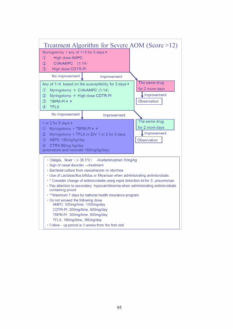
95