Embed Size (px)
Citation preview

OBSTETRICS AND GYNECOLOGY/CLINICAL POLICY
Clinical Policy: Critical Issues in the Initial Evaluation andManagement of Patients Presenting to the Emergency
Department in Early Pregnancy
From the American College of Emergency Physicians Clinical Policies Subcommittee (Writing Committee) on Early Pregnancy:
Sigrid A. Hahn, MD, (Subcommittee Chair)Eric J. Lavonas, MDSharon E. Mace, MDAnthony M. Napoli, MDFrancis M. Fesmire, MD, (Committee Chair)
Members of the American College of Emergency Physicians Clinical Policies Committee (Oversight Committee):
DSRSSS
R
R
AS
Francis M. Fesmire, MD (Chair 2011-2012)Douglas Bernstein, MD (EMRA Representative 2011-
2012)Michael D. Brown, MD, MScJohn H. Burton, MDDeborah B. Diercks, MD, MScSteven A. Godwin, MDSigrid A. Hahn, MDJason S. Haukoos, MD, MSc (Methodologist)J. Stephen Huff, MDEric J. Lavonas, MDGail Lenehan, EdD, RN, FAEN, FAAN (ENA
Representative 2011-2012)Sharon E. Mace, MD
Edward Melnick, MDfollowing critical questions: (1) Should the emergency physician
owacttfrpE
Volume , . : September
evorah J. Nazarian, MDusan B. Promes, MDichard D. Shih, MDcott M. Silvers, MDtephen J. Wolf, MDtephen V. Cantrill, MD (Liaison with Quality andPerformance Committee)
obert E. O’Connor, MD, MPH (Board Liaison 2010-2012)
honda R. Whitson, RHIA, Staff Liaison, Clinical PoliciesCommittee and Subcommittees
pproved by the ACEP Board of Directors April 10, 2012upported by the Emergency Nurses Association, June
21, 2012Policy statements and clinical policies are the official policies of the American College of EmergencyPhysicians and, as such, are not subject to the same peer review process as articles appearing in the printjournal. Policy statements and clinical policies of ACEP do not necessarily reflect the policies and beliefsof Annals of Emergency Medicine and its editors.
0196-0644/$-see front matterCopyright © 2012 by the American College of Emergency Physicians.http://dx.doi.org/10.1016/j.annemergmed.2012.04.021
[Ann Emerg Med. 2012;60:381-390.]
ABSTRACTThis clinical policy from the American College of Emergency
Physicians is the revision of the 2003 Clinical Policy: CriticalIssues in the Initial Evaluation and Management of PatientsPresenting to the Emergency Department in Early Pregnancy.1
A writing subcommittee reviewed the literature to deriveevidence-based recommendations to help clinicians answer the
btain a pelvic ultrasound in a clinically stable pregnant patientho presents to the emergency department (ED) with
bdominal pain and/or vaginal bleeding and a beta humanhorionic gonadotropin (�-hCG) level below a discriminatoryhreshold? (2) In patients who have an indeterminateransvaginal ultrasound, what is the diagnostic utility of �-hCGor predicting possible ectopic pregnancy? (3) In patientseceiving methotrexate for confirmed or suspected ectopicregnancy, what are the implications for ED management?
vidence was graded and recommendations were developedAnnals of Emergency Medicine 381

�ahwpiacirpdErrac
m�tppscspSbolcudd
mcsil
mAakta
at
*
Clinical Policy
based on the strength of the available data in the medicalliterature.
A literature search was also performed for a critical questionfrom the 2003 clinical policy.1 Is the administration of anti-Dimmunoglobulin indicated among Rh-negative women during thefirst trimester of pregnancy with threatened abortion, completeabortion, ectopic pregnancy, or minor abdominal trauma? Becauseno new, high-quality articles were found, the managementrecommendations from the previous policy are discussed in theintroduction.
INTRODUCTIONEmergency physicians frequently evaluate and manage patients
with abdominal pain and/or vaginal bleeding in the first trimesterof pregnancy (also referred to here as “early pregnancy”). Theirprimary concern in this group of patients is to identify ectopicpregnancy. The prevalence of ectopic pregnancy in symptomaticemergency department (ED) patients is as high as 13% in someseries, which is much higher than the prevalence in the generalpopulation.2,3 With wide availability of bedside ultrasound inacademic EDs and increasing access in community settings, moreproviders are now routinely using ultrasound in their evaluation ofthese patients.4
The term bedside ultrasound is used here to refer to pelvicultrasounds that are performed in the ED by the emergencyclinician, rather than in the radiology department. With the termpelvic ultrasound, the use of a transvaginal approach is impliedunless transabdominal images have identified an intrauterinepregnancy. According to the 2006 ACEP policy statementEmergency Ultrasound Imaging Criteria Compendium, the primaryindication for bedside ultrasound of the pelvis is to evaluate for thepresence of intrauterine pregnancy, minimizing the likelihood of anectopic pregnancy when modifying factors such as infertilitytreatment (putting patients at risk of heterotopic pregnancy)are not present.5 A bedside ultrasonographer may or may notvisualize the adnexa. A recent meta-analysis found thatbedside ultrasound performed by emergency physicians canbe used as a screening tool for ectopic pregnancy.6 Pooledanalysis included 10 studies and a total of 2,052 patientswith 152 ectopic pregnancies; of those with ectopicpregnancy, 99.3% (95% confidence interval [CI] 96.6% to100%) had no intrauterine pregnancy identified on bedsideultrasound.6 A comprehensive ultrasound, in contrast, isusually performed in a radiology department and is expectedto include views of the uterus, adnexa, and cul-de-sac.Studies using either or both categories of ultrasound werereviewed and this distinction was highlighted in the text andEvidentiary Table. This policy is not intended to review theevidence supporting the use of bedside ultrasound byemergency physicians.
Ultrasound has facilitated the evaluation of complications ofearly pregnancy; however, diagnostic algorithms still varyconsiderably among providers and institutions. Algorithms guidingthe evaluation of abdominal pain or vaginal bleeding in early
pregnancy generally incorporate the results of quantitative serum z382 Annals of Emergency Medicine
-hCG measurements and pelvic ultrasonography. Manylgorithms apply the principle of the discriminatory threshold,*istorically defined as the level at which the sensitivity of ultrasoundas thought to approach 100% for the detection of intrauterineregnancy; the presumptive diagnosis of ectopic pregnancy is madef an intrauterine pregnancy is not visualized when the �-hCG isbove a defined cutoff. This threshold depends on what ultrasoundriteria are used to define an intrauterine pregnancy and isnstitution, operator, and patient dependent, but is commonlyeported as ranging from 1,000 to 2,000 mIU/mL for radiologist-erformed transvaginal sonography.7,8 Although the traditionallyefined discriminatory threshold is widely used, its applicability toD practice is not as well established, and the concept itself has
ecently been called into question.9,10 For these reasons, this policyefers to the general concept of a discriminatory threshold, whereppropriate, but the discussion is not limited to any specific �-hCGutoff.
The first critical question deals with the diagnostic andanagement variability that occurs when the clinician obtains a-hCG result, and it is below a commonly defined discriminatory
hreshold. Some clinicians may not perform an ultrasound in theseatients based on the incorrect assumption that an ectopicregnancy is unlikely because the �-hCG level is low. In someettings, the emergency physician may be unable to obtain aomprehensive ultrasound in the radiology department for theame reason. However, it is well documented that ectopicregnancies can present at almost any �-hCG level, high or low.7
ome clinicians may defer an ultrasound when the �-hCG level iselow the discriminatory threshold because they think that the riskf rupture is low. However, rupture has been documented at veryow �-hCG levels.7,11 Other clinicians may defer imaging in theseases because they believe that the diagnostic utility of pelvicltrasound is low when the �-hCG level is below theiscriminatory threshold or assume that there is little harm inelaying the diagnostic ultrasound.
The emergency physician is faced with another diagnostic andanagement question when an ultrasound is indeterminate, also
alled “nondiagnostic” or a “pregnancy of unknown location.” Theecond critical question examines this subgroup of patients withndeterminate ultrasounds and addresses whether the initial �-hCGevel can help risk-stratify these patients.
The third critical question explores the implications ofethotrexate therapy for emergency medicine practice.dministration of methotrexate is an accepted and widely usedlternative to laparoscopic surgery for the management ofnown or suspected ectopic pregnancy.12-14 Methotrexateherapy is a complex intervention, and complications of therapyre frequently evaluated in the ED.
In the previous policy,1 one of the critical questions alsoddressed the issue of which Rh-negative patients in the firstrimester of pregnancy with threatened abortion, complete
The discriminatory threshold is also referred to as the discriminatory
one or level.Volume , . : September

ccomGeesacc
geiwrptfpcmsbaogpqaaql(w
pc
pcos
mmbtc
ma
Clinical Policy
abortion, ectopic pregnancy, or minor abdominal traumarequired the administration of anti-D immunoglobulin. Thelevel B recommendation was to administer 50 �g of anti-Dimmunoglobulin to Rh-negative women in all cases ofdocumented first-trimester loss of established pregnancy toprevent Rh-D alloimmunization. There was insufficientevidence to recommend for or against its use in treatingthreatened abortion or ectopic pregnancy. There was also a levelC recommendation to consider anti-D immunoglobulin use incases of minor abdominal trauma. These recommendations werebased on theoretic construct, multiple limited observationalstudies, and 1 limited randomized controlled trial. An updatedliterature search was performed on the topic, excludingabdominal trauma, and no high-quality studies were foundaddressing this issue. As a result, the patient managementrecommendations for this question remain unchanged and arenot discussed in further detail in this policy update.
METHODOLOGYThis clinical policy was created after careful review and
critical analysis of the medical literature. Multiple searches ofMEDLINE and Google Scholar were performed. All searcheswere limited to English-language sources and human studies.Specific key words/phrases and years used in the searches areidentified under each critical question. In addition, relevantarticles from the bibliographies of included studies and morerecent articles identified by committee members and reviewerswere included.
The reasons for developing clinical policies in emergencymedicine and the approaches used in their development havebeen described.15 This policy is a product of the AmericanCollege of Emergency Physicians (ACEP) clinical policydevelopment process, including expert review, and is based on
the existing literature; when literature was not available, cVolume , . : September
onsensus of emergency physicians was used. Expert reviewomments were received from emergency physicians, membersf ACEP’s Emergency Ultrasound Section, and individualembers of the American College of Obstetricians andynecologists. Their responses were used to further refine and
nhance this policy; however, their responses do not implyndorsement of this clinical policy. Clinical policies arecheduled for revision every 3 years; however, interim reviewsre conducted when technology or the practice environmenthanges significantly. ACEP was the funding source for thislinical policy.
All articles used in the formulation of this clinical policy wereraded by at least 2 subcommittee members for strength ofvidence. The articles were classified by the subcommittee membersnto 3 classes of evidence on the basis of the design of the study,ith design 1 representing the strongest design and design 3
epresenting the weakest design for therapeutic, diagnostic, andrognostic clinical reports, respectively (Appendix A). Articles werehen graded on dimensions related to the study’s methodologicaleatures, including but not necessarily limited to randomizationrocesses, blinding, allocation concealment, methods of dataollection, outcome measures and their assessment, selection andisclassification biases, external validity, generalizability, and
ample size. Articles received a final grade (Class I, II, III) on theasis of a predetermined formula, taking into account the designnd study quality (Appendix B). Articles identified with fatal flawsr that were not relevant to the critical question received an “X”rade and were not used in formulating recommendations for thisolicy. Grading was done with respect to the specific criticaluestions; thus, the level of evidence for any one study may varyccording to the question. As such, it was possible for a singlerticle to receive different levels of grading as different criticaluestions were answered from the same study. Question-specificevel of evidence grading may be found in the Evidentiary Tableavailable online at http://www.annemergmed.com and at http://ww.acep.org/clinicalpolicies/).
Clinical findings and strength of recommendations regardingatient management were then made according to the followingriteria:
Level A recommendations. Generally accepted principles foratient management that reflect a high degree of clinicalertainty (ie, based on strength of evidence Class I orverwhelming evidence from strength of evidence Class IItudies that directly address all of the issues).
Level B recommendations. Recommendations for patientanagement that may identify a particular strategy or range ofanagement strategies that reflect moderate clinical certainty (ie,
ased on strength of evidence Class II studies that directly addresshe issue, decision analysis that directly addresses the issue, or strongonsensus of strength of evidence Class III studies).
Level C recommendations. Other strategies for patientanagement that are based on Class III studies, or in the
bsence of any adequate published literature, based on panel
onsensus.Annals of Emergency Medicine 383

paS
buitturmemrawcfba
Dw
tmpc(“rpbso
ssowibd6sws
pipu
Clinical Policy
There are certain circumstances in which the recommendationsstemming from a body of evidence should not be rated as highly asthe individual studies on which they are based. Factors such asheterogeneity of results, uncertainty about effect magnitude andconsequences, and publication bias, among others, might lead tosuch a downgrading of recommendations.
When possible, clinically oriented statistics (eg, likelihood ratios[LRs], number needed to treat) were presented to help the reader betterunderstand how the results may be applied to the individual patient.For a definition of these statistical concepts, see Appendix C.
This policy is not intended to be a complete manual on theevaluation and management of patients with abdominal pain orvaginal bleeding in early pregnancy but rather a focusedexamination of critical issues that have particular relevance tothe current practice of emergency medicine.
It is the goal of the Clinical Policies Committee to provide anevidence-based recommendation when the medical literatureprovides enough quality information to answer a critical question.When the medical literature does not contain enough qualityinformation to answer a critical question, the members of theClinical Policies Committee believe that it is equally important toalert emergency physicians to this fact.
Recommendations offered in this policy are not intended torepresent the only diagnostic and management options that theemergency physician should consider. ACEP clearly recognizes theimportance of the individual physician’s judgment. Rather, thisguideline defines for the physician those strategies for whichmedical literature exists to provide support for answers to thecrucial questions addressed in this policy.
Scope of Application. This guideline is intended forphysicians working in hospital-based EDs.
Inclusion Criteria. This guideline is intended for stablepatients (with normal blood pressure and pulse rate) in the firsttrimester of pregnancy with abdominal pain or vaginal bleeding,without a previously confirmed intrauterine pregnancy.
Exclusion Criteria. This guideline is not intended toaddress the care of patients who are clinically unstable, have hadabdominal trauma, or are at higher risk for heterotopicpregnancy such as those who are undergoing fertility treatments.
CRITICAL QUESTIONS1. Should the emergency physician obtain a pelvicultrasound in a clinically stable pregnant patient whopresents to the ED with abdominal pain and/or vaginalbleeding and a �-hCG level below a discriminatorythreshold?
Patient Management RecommendationsLevel A recommendations. None specified.Level B recommendations. None specified.Level C recommendations. Perform or obtain a pelvic
ultrasound for symptomatic pregnant patients with a �-hCG levelbelow any discriminatory threshold.
Key words/phrases for literature searches: ultrasound, �-
hCG, transvaginal, ectopic pregnancy, pelvic pain, abdominal u384 Annals of Emergency Medicine
ain, vaginal bleeding, emergency department, and variationsnd combinations of the key words/phrases, years 1980 througheptember 2009.
Articles were reviewed for evidence of (1) the potential diagnosticenefit of performing an emergent bedside or comprehensive pelvicltrasound in patients with abdominal pain and/or vaginal bleeding
n early pregnancy and a �-hCG level below a discriminatoryhreshold, or (2) documented harm in deferring the ultrasound inhis same group of patients. Assessing the safety of deferring a pelvicltrasound, however, requires large numbers to detect the relativelyare event of a patient experiencing significant morbidity orortality because of an ectopic pregnancy, and no study was large
nough to confidently assess this risk. Furthermore, ED patientsay have difficulty arranging appropriate follow-up; this fact is not
eflected in the studies presented below but must be taken intoccount when deciding on the appropriate plan for any patientith a possible ectopic pregnancy. There was no attempt to
ompare the benefit of bedside versus comprehensive ultrasoundor diagnostic performance, ED wait times, length of stay, or costecause this was beyond the scope of the question. However, whenvailable, the use of bedside ultrasound may expedite the diagnosis.
iagnostic benefit of performing a pelvic ultrasound in patientsith a �-hCG level below a discriminatory thresholdThe previous policy provided level C recommendations to consider
ransvaginal ultrasound in patients with a �-hCG level below 1,000IU/mL because it may detect intrauterine pregnancy or an ectopic
regnancy.1 This was based on the moderate sensitivity of aomprehensive ultrasound for detecting intrauterine pregnancyranging from 40% to 67% across the studies), using presence of agestational sac” as the diagnostic criterion for intrauterine pregnancy,ather than a yolk sac or fetal pole.8,16-18 Modest diagnosticerformance of ultrasound in this group of patients with a �-hCG levelelow 1,000 mIU/mL was also observed for ectopic pregnancy, with aensitivity of 19% and specificity of 100% in one series and a sensitivityf 39% in another study.3,19
In addition to the previously reviewed articles, 4 additionaltudies directly or indirectly address this question. A Class IItudy by Barnhart et al20 examined the diagnostic performancef a comprehensive ultrasound in patients presenting to the EDith symptomatic early pregnancy and stratified the results by
nitial �-hCG level. For patients presenting with a �-hCG levelelow 1,500 mIU/mL, the sensitivity of ultrasound for theiagnosis of intrauterine pregnancy was 33% (95% CI 10% to5%), and specificity was 98% (95% CI 90% to 100%). Theensitivity of ultrasound for the diagnosis of ectopic pregnancyas similar, at 25% (95% CI 5% to 57%), as was the
pecificity, at 96% (95% CI 87% to 99%).Two Class III studies evaluated the diagnostic
erformance of a comprehensive ultrasound at presentationn patients who had the final diagnosis of ectopicregnancy.21,22 Cacciatore21 conducted a review of theltrasounds that he had performed. He found that
ltrasound had 92% sensitivity for an ectopic pregnancyVolume , . : September

ea
ai
atucS
trppirTsauddwisapvcshc
frqipLtmrepiowecd
Clinical Policy
with �-hCG level below 1,000 mIU/mL (95% CI 79% to97%).21 Counselman et al22 found that among patients witha �-hCG below 1,000 mIU/mL, a comprehensive ultrasoundwas suggestive of an ectopic pregnancy in 86% (95% CI60% to 96%) of cases that had the diagnosis confirmed.
One Class III study examined 74 patients with a bedsideultrasound result suggestive or diagnostic of an ectopic pregnancy,in which emergency physicians performed pelvic ultrasounds thatincluded views of the uterus, adnexa, and cul-de-sac.23 Of the 47patients with a suggestive or diagnostic initial ultrasound result anda final diagnosis of an ectopic pregnancy, 36% had a presenting�-hCG level below 1,000 mIU/mL.
Potential harm of deferring pelvic ultrasound in patients with a�-hCG level below a discriminatory threshold
Algorithms that defer ultrasounds in stable patients with a�-hCG level below the discriminatory threshold may resultin diagnostic delays. Unfortunately, the published studies donot allow us to estimate the risk of rupture or death amongthese patients. One Class III study reviewed the safety of astrategy of discharging symptomatic but stable, low-riskpatients for urgent outpatient ultrasound withinapproximately 12 to 24 hours.24 The authors retrospectivelyidentified all patients who ultimately received a diagnosis ofectopic pregnancy. They found no adverse events, defined asdeath or need for fluid bolus because of hemodynamicinstability, in 37 patients despite a median delay toultrasound of 14 hours (range 0 to 126 hours), with 62% ofpatients waiting 12 hours or longer. The mean �-hCG levelin this group was 2,887 mIU/mL (range 85 to 26,000 mIU/mL), but the number of patients with a �-hCG level lessthan the discriminatory threshold was not provided. Thesmall number of patients in this study does not allow us todraw conclusions about the safety of delaying ultrasounds.
Another Class III study observed the performance of analgorithm that deferred ultrasounds in patients with an initial �-hCG level below 1,500 mIU/mL (until their � level plateaued orincreased above this threshold).7 For these 69 patients with a finaldiagnosis of an ectopic pregnancy, the authors found that meantime to diagnosis was 5.2 days.7 There was no comparison groupwhen ultrasound was performed immediately for patients with a�-hCG level below 1,500 mIU/mL. There were a small number ofpatients in this study with evidence of rupture at the time ofdiagnosis, but their initial �-hCG level was not provided, makingthe true risk of increased morbidity or mortality associated with thisapproach impossible to determine. However, some patients orclinicians might consider a delay in diagnosis unacceptable.
2. In patients who have an indeterminate transvaginalultrasound, what is the diagnostic utility of �-hCG forpredicting possible ectopic pregnancy?
Patient Management Recommendations
Level A recommendations. None specified. aVolume , . : September
Level B recommendations. Do not use the �-hCG value toxclude the diagnosis of ectopic pregnancy in patients who haven indeterminate ultrasound.
Level C recommendations. Obtain specialty consultation orrrange close outpatient follow-up for all patients with anndeterminate pelvic ultrasound.
Key words/phrases for literature searches: pelvic pain,bdominal pain, vaginal bleeding, ectopic pregnancy, �-hCG,ransvaginal, ultrasound, emergency department, indeterminateltrasound, pregnancy of unknown location, and variations andombinations of the key words/phrases, years 1980 througheptember 2009.
A majority of patients who have a pelvic ultrasound duringheir ED evaluation for symptomatic early pregnancy willeceive a diagnosis of an intrauterine pregnancy or an abnormalregnancy (eg, ectopic pregnancy, fetal demise, or molarregnancy). A significant minority, however, will have anndeterminate, or nondiagnostic, ultrasound; most ED literatureeports an indeterminate study rate of 20% to 30%.3,9,25-28
his rate depends on a number of factors, including the clinicaletting, patient population, ultrasound machine and operator,nd criteria used for each diagnostic category. ED studiessually require the presence of a yolk sac or fetal pole toiagnose an intrauterine pregnancy. This is in contrast toiagnostic criteria more frequently used by radiologists, inhich a “gestational sac” is diagnostic of intrauterine pregnancy
f a “double decidual” sign is seen, even in the absence of a yolkac or fetal pole. Diagnostic criteria for ectopic pregnancy varys well, and some studies stratify findings into possible,robable, or definite ectopic pregnancy according to what isisualized in the adnexa or cul-de-sac. This can complicateomparisons among studies, and the definitions used in eachtudy are noted in the Evidentiary Table (available online atttp://www.annemergmed.com and at http://www.acep.org/linicalpolicies/).
Indeterminate ultrasounds pose a management dilemmaor the clinician. Authors for the ACEP 2003 clinical policyeviewed literature through 2000 to answer the relateduestion, “Above what �-hCG level is the absence ofntrauterine pregnancy by transvaginal ultrasoundresumptive evidence of ectopic pregnancy?” and provided aevel B recommendation that patients with a indeterminateransvaginal ultrasound and a �-hCG level above 2,000IU/mL have follow-up arranged because they have a higher
isk of ectopic pregnancy.1 For this revision, the authorsxamined the broader question of whether the risk of ectopicregnancy can be predicted in patients who have anndeterminate ultrasound with any �-hCG level and reportedr calculated LRs from the available data to determinehether these could be applied to estimate a posttest risk of
ctopic pregnancy that would be high or low enough tohange management (Table). A positive test result wasefined as an indeterminate ultrasound with a �-hCG level
bove a discriminatory threshold, and a negative test result asAnnals of Emergency Medicine 385

t1c
pppcidmapdisroct
ccwmanmny
TulL
Clinical Policy
an indeterminate ultrasound with a �-hCG level below adiscriminatory threshold. Therefore, a positive LR estimatesthe risk of an ectopic pregnancy when the �-hCG level isabove a discriminatory threshold, and a negative LRestimates the risk of an ectopic pregnancy when the �-hCGlevel is below a discriminatory threshold. When LRs werenot available or could not be calculated, other statisticalresults were reported. Although not described in detail in thetext, relative risk for ectopic pregnancy below a given �-hCGcutoff was also calculated (Table). The issue of serial �-hCGmeasurements is not addressed because this is not relevant todecisionmaking at the time of initial evaluation in the ED.
Nine Class II studies examined the initial �-hCG level inpatients with an indeterminate ultrasound and found that itcould not be used to predict final diagnosis.3,9,26,27,29-33 Tworeported on the performance of bedside ultrasounds.9,27 Thefirst study aimed to test the traditional concept of thediscriminatory threshold in ED patients and found thatusing a �-hCG cutoff of 3,000 mIU/mL to try to predictwhich patients had an ectopic pregnancy had virtually nodiagnostic utility (positive LR 0.8; negative LR 1.1).9 Theauthors of the study also attempted to identify a more usefuldiscriminatory threshold for bedside ultrasonography.Although there was no cutoff at which 100% of theintrauterine pregnancies were identified, a �-hCG level ofmore than 25,000 mIU/mL identified 88% (87 of 99intrauterine pregnancies). The other study examiningindeterminate bedside ultrasounds found that at the initialED visit, median �-hCG level was not significantly differentwhether the final diagnosis was intrauterine pregnancy(1,304 mIU/mL), embryonic demise (1,572 mIU/mL), orectopic pregnancy (1,147 mIU/mL) (P�NS).27
Six of the Class II studies examined indeterminatecomprehensive ultrasounds.3,26,29-32 Two studies ofsymptomatic ED patients from the same institution found
�-hCG Threshold,mIU/mL
Study
Author Year Class
1,000 Condous29 2005 IIDart26 2002 IIKaplan3 1996 IIMol33 1998 IIDart36 1998 III
1,500 Condous29 2005 II
2,000 Condous29 2005 IIMol33 1998 IIMateer34 1996 III
3,000 Wang9 2011 IIDart35 1997 III
*Relative risk was calculated with the online calculator http://ktclearinghouse.ca†Negative LRs were determined based on having a �-hCG level below the stated
‡Positive LRs were determined based on having a �-hCG level above the stated thresho386 Annals of Emergency Medicine
hat the negative LRs with a discriminatory threshold of,000 mIU/mL were not large or small enough to help withlinical decisionmaking.3,26
Four other Class II studies took place in an earlyregnancy unit, which is a specialized evaluation center foratients with symptomatic or asymptomatic earlyregnancy.29-32 The first study examined several differentommon discriminatory thresholds for patients withndeterminate ultrasounds and found LRs close to 1 foriscriminatory thresholds of 1,000 mIU/mL, 1,500 mIU/L, and 2,000 mIU/mL.29 Two studies by Condous et
l30,31 found that the mean initial �-hCG level for ectopicregnancies was not significantly different than for the finaliagnostic categories of intrauterine pregnancy or failing
ntrauterine pregnancy. The fourth study also found noignificant difference in median initial �-hCG levelegardless of the final diagnosis and reported that the receiverperating characteristic curve for �-hCG level is close tohance for predicting the need for intervention (area underhe curve [AUC]�0.47; P�NS).32
The last Class II study examined indeterminateomprehensive ultrasounds performed by obstetricians andalculated LRs for different strata of �-hCG levels.33 Dataere extracted only for those patients without an ectopicass or fluid in the pouch of Douglas. For �-hCG level
bove 1,000 mIU/mL, the positive LR was 3.1 and theegative LR was 0.7. When a �-hCG level above 2,000IU/mL was used as a cutoff, the positive LR was 25 and
egative LR was 0.6. This is the single instance of a studyielding a strongly predictive positive LR.
Five Class III studies addressed this topic as well.28,34-37
wo examined bedside ultrasound and 3, comprehensiveltrasounds. Four of these studies also found that �-hCG
evel was poorly predictive of ectopic pregnancy, based onRs (Table).28,34-36 A study examining expectant
Relative Risk ofEctopic Below
Threshold* (95%CI)
Likelihood Ratios (95%CI)
Negative†
Positive‡
0.6 (0.3–1.1) 0.9 (0.8–1.0) 1.7 (0.9–3.1)7.1 (3.4–14.9) 2.3 (1.9–2.7) 0.3 (0.2–0.5)3.8 (1.4–9.8) 2.5 (1.4–4.5) 0.5 (0.2–0.9)0.4 (0.2–0.5) 0.7 (0.5–0.8) 3.1 (2.0–4.8)2.2 (1.0–4.5) 1.8 (1.1–2.9) 0.7 (0.5–1.0)
0.4 (0.2–0.9) 0.9 (0.8–1.0) 2.3 (1.1–4.9)
0.5 (0.2–1.1) 0.9 (0.8–1.0) 2.3 (0.9–5.7)0.2 (0.1–0.3) 0.6 (0.5–0.8) 25 (7.9–81)0.5 (0.3–0.8) 0.7 (0.5–0.9) 2.3 (1.2–4.3)
1.3 (0.6–2.6) 1.1 (0.8–1.5) 0.8 (0.5–1.4)2.1 (0.9–4.8) 1.4 (1.0–1.8) 0.6 (0.3–1.1)
/practise/ca/calculators/statscalc.old.
N
52763572
262220
527
52726295
141194
/cebmthresh
ld.
Volume , . : September

mau
2mrosilmt
ls2
fa1lmfroppl
ptwNpr
fpmehrat
r
ct
Clinical Policy
management of pregnancies of uncertain location found nosignificant difference in mean �-hCG level between ectopicpregnancy requiring treatment and other final outcomes.37
3. In patients receiving methotrexate for confirmed or,suspected ectopic pregnancy, what are the implications forED management?
Patient Management RecommendationsLevel A recommendations. None specified.Level B recommendations. (1) Arrange outpatient follow-
up for patients who receive methotrexate therapy in the ED fora confirmed or suspected ectopic pregnancy.
(2) Strongly consider ruptured ectopic pregnancy in thedifferential diagnosis of patients who have received methotrexateand present with concerning signs or symptoms.
Level C recommendations. None specified.
Key words/phrases for literature searches: methotrexate,ectopic pregnancy, side effects, drug interactions, emergencyservice, and variations and combinations of the keywords/phrases, years 2000 to May 2008.
Methotrexate administration in the ED is an alternative tosurgical treatment for known or suspected early ectopicpregnancy.12,13 The decision to administer methotrexate or useanother treatment approach is complex. Methotrexate is givento hemodynamically stable patients with an unruptured ectopicpregnancy as a single intravenous or intramuscular dose of 50mg/m2, after which they are discharged to outpatientmanagement.14 Because treatment with a single dose ofmethotrexate is often ineffective, patients may require repeateddoses of methotrexate until their �-hCG levels are clearlydecreasing. Laboratory testing, including a CBC count withdifferential and platelet counts, hepatic enzyme level, and renalfunction tests, is recommended before initiation of methotrexatetherapy.14 Methotrexate therapy is contraindicated in patientswith alcoholism, immunodeficiency, peptic ulcer, or activedisease of the lungs, liver, kidneys, or hematopoietic system andrelatively contraindicated in patients with an ectopic gestationalsac larger than 3.5 cm or with embryonic cardiac motionobserved on ultrasound.14 Treatment success rates are also lowerin patients who have a �-hCG level of 5,000 mIU/L or more.13
Treatment failure, with rupture of the ectopic pregnancy, isone of the most serious complications of methotrexate therapy.In several cohort studies, more than 20% of patients receivingmethotrexate required surgery.38-42 Therefore, ruptured ectopicpregnancy must be considered in the differential diagnosis ofpatients who present to the ED with concerning symptoms orsigns after methotrexate therapy.
This section is an update of the 2003 policy1 questionexamining the failure rate of methotrexate treatment and itsimplications for ED management. Nineteen Class I, II, and IIIstudies are included for discussion below.13,38-40,41-55 This
review does not address complications associated withVolume , . : September
ethotrexate administration directly into the ectopic pregnancy,procedure that is generally performed in the operating roomnder ultrasound or laparoscopic guidance.Two Class I studies were identified.41,43 In Rozenberg et al,41
12 women with ectopic pregnancy received intramuscularethotrexate that was repeated as needed. This trial was
emarkable for the high failure rate (22.9%) and for the lowbserved risk of tubal rupture (0.5%). In the second Class Itudy, 62 women were randomized to receive eitherntramuscular methotrexate, repeated as needed, or immediateaparoscopic surgery.43 Among the 34 women randomized to
ethotrexate therapy, the treatment failure rate was 15% andhe rupture rate was 9%.
A single Class II clinical trial by Korhonen et al44 evaluatedow-dose oral methotrexate therapy (12.5 mg total) foruspected ectopic pregnancy. Treatment failure occurred in3% of patients. The rupture rate was not reported.In the Class III studies, the treatment failure rates ranged
rom 3% to 29%.13,38-40,45-55 Among studies that reported databout rupture, this serious complication occurred in 0.5% to9% of women treated.39,40,45-49,51-54 A Class III structured
iterature review also found that treatment with multiple-doseethotrexate was associated with a 7% failure rate.55 The
requency of rupture was not reported, but 12% of patientsequired rehospitalization. More recent studies confirm earlierbservations that treatment failure and ruptured ectopicregnancy continue to be associated with larger ectopicregnancies as measured by ultrasound, higher serum �-hCG
evels, and visualized fetal cardiac activity.13,40,49,52,53
The only study to examine the need for surgery as therimary outcome was a Class III prospective observational studyhat included 177 women with ectopic pregnancies, 29 ofhom had ruptured ectopic pregnancy prior to therapy.42
ineteen percent of the women with unruptured ectopicregnancy and 38% of women with ruptured ectopic pregnancyequired surgery.
Taken together, these data show that methotrexate therapyor known or suspected ectopic pregnancy is a useful butotentially complex treatment strategy. Although this therapyay be appropriately initiated in the ED, follow-up care is
ssential. Patients who develop increasing pain and/or signs ofemodynamic instability after methotrexate therapy shouldeceive stabilizing care and prompt diagnostic studies, such asbdominal and pelvic ultrasonography, to establish or excludehe diagnosis of ruptured ectopic pregnancy.
Relevant industry relationships: There were no relevant industryelationships disclosed by the subcommittee members.
Relevant industry relationships are those relationships withompanies associated with products or services that significantly impacthe specific aspect of disease addressed in the critical question.
Annals of Emergency Medicine 387

2
2
2
2
2
2
2
2
2
3
3
3
3
3
3
3
3
3
Clinical Policy
REFERENCES1. American College of Emergency Physicians. Clinical policy: critical
issues in the initial evaluation and management of patientspresenting to the emergency department in early pregnancy. AnnEmerg Med. 2003;41:123-133.
2. Kohn MA, Kerr K, Malkevich D, et al. Beta-human chorionicgonadotropin levels and the likelihood of ectopic pregnancy inemergency department patients with abdominal pain or vaginalbleeding. Acad Emerg Med. 2003;10:119-126.
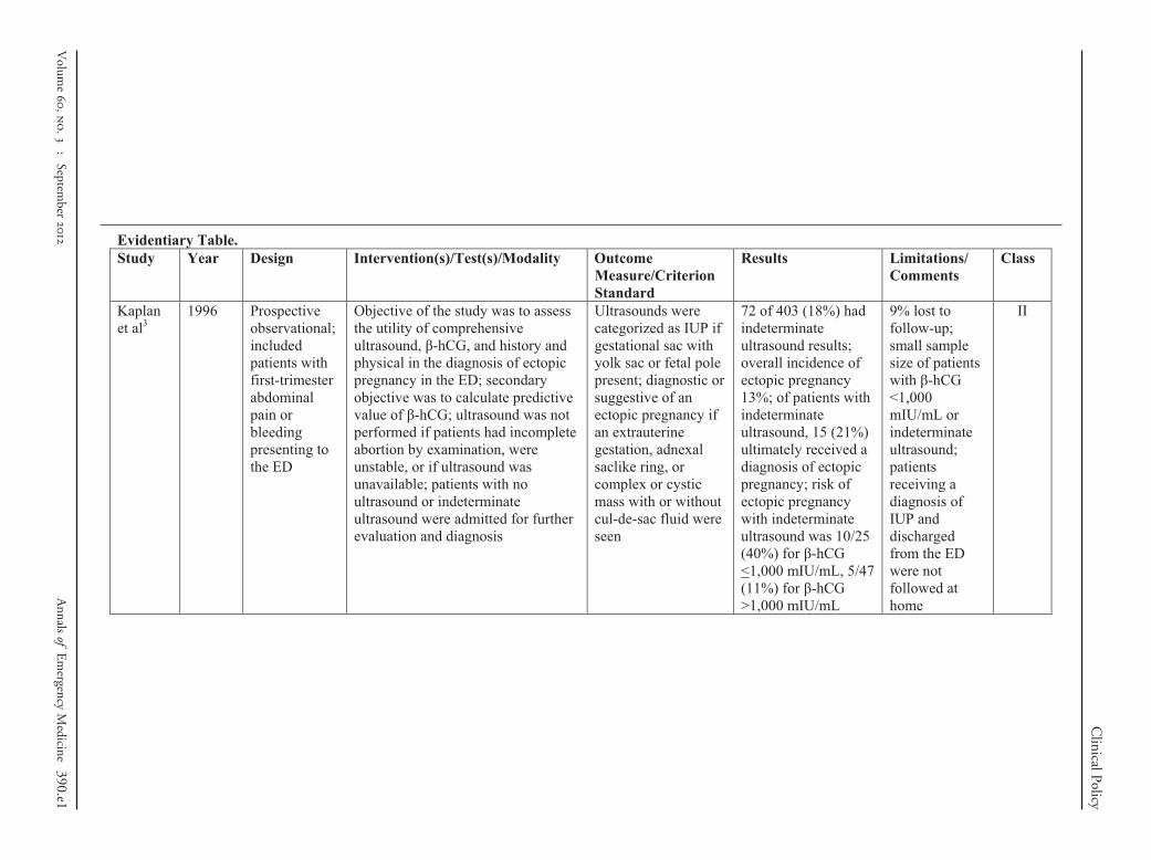
3. Kaplan BC, Dart RG, Moskos M, et al. Ectopic pregnancy:prospective study with improved diagnostic accuracy. Ann EmergMed. 1996;28:10-17.
4. Moore CL, Molina AA, Lin H. Ultrasonography in communityemergency departments in the United States: access toultrasonography performed by consultants and status ofemergency physician–performed ultrasonography. Ann Emerg Med.2006;47:147-153.
5. American College of Emergency Physicians. Policy statement:emergency ultrasound imaging criteria compendium. Ann EmergMed. 2006;48:487-510.
6. Stein JC, Wang R, Adler N, et al. Emergency physicianultrasonography for evaluating patients at risk for ectopicpregnancy: a meta-analysis. Ann Emerg Med. 2010;56:674-683.
7. Barnhart K, Mennuti MT, Benjamin I, et al. Prompt diagnosis ofectopic pregnancy in an emergency department setting. ObstetGynecol. 1994;84:1010-1015.
8. Bernaschek G, Rudelstorfer R, Csaicsich P. Vaginal sonographyversus serum human chorionic gonadotropin in early detection ofpregnancy. Am J Obstet Gynecol. 1988;158:608-612.
9. Wang R, Reynolds TA, West HH, et al. Use of a �-hCGdiscriminatory zone with bedside pelvic ultrasonography. AnnEmerg Med. 2011;58:12-20.
10. Doubilet PM, Benson CB. Further evidence against the reliabilityof the human chorionic gonadotropin discriminatory level. JUltrasound Med. 2011;30:1637-1642.
11. Saxon D, Falcone T, Mascha EJ, et al. A study of ruptured tubalectopic pregnancy. Obstet Gynecol. 1997;90:46-49.
12. Pisarska MD, Carson SA, Buster JE. Ectopic pregnancy. Lancet.1998;351:1115-1120.
13. Lipscomb GH, McCord ML, Stovall TG, et al. Predictors ofsuccess of methotrexate treatment in women with tubal ectopicpregnancies. N Engl J Med. 1999;341:1974-1978.
14. American College of Obstetricians and Gynecologists. MedicalManagement of Ectopic Pregnancy. ACOG Practice Bulletin No.94. Washington, DC:ACOG;2008.
15. Schriger DL, Cantrill SV, Greene CS. The origins, benefits, harms,and implications of emergency medicine clinical policies. AnnEmerg Med. 1993;22:597-602.
16. Nyberg DA, Mack LA, Laing FC, et al. Early pregnancycomplications: endovaginal sonographic findings correlated withhuman chorionic gonadotropin levels. Radiology. 1988;167:619-622.
17. Kadar N, Caldwell BV, Romero R. A method of screening forectopic pregnancy and its indications. Obstet Gynecol. 1981;58:162-165.
18. Shapiro BS, Escobar M, Makuch R, et al. A model-basedprediction for transvaginal ultrasonographic identification of earlyintrauterine pregnancy. Am J Obstet Gynecol. 1992;166:1495-1500.
19. Dart RG, Kaplan B, Cox C. Transvaginal ultrasound in patientswith low �-human chorionic gonadotropin values: how often is thestudy diagnostic? Ann Emerg Med. 1997;30:135-140.
20. Barnhart KT, Simhan H, Kamelle SA. Diagnostic accuracy ofultrasound above and below the beta-hCG discriminatory zone.
Obstet Gynecol. 1999;94:583-587.388 Annals of Emergency Medicine
1. Cacciatore B. Can the status of tubal pregnancy be predicted withtransvaginal sonography? A prospective comparison ofsonographic, surgical, and serum hCG findings. Radiology. 1990;177:481-484.
2. Counselman FL, Shaar GS, Heller RA, et al. Quantitative �-hCGlevels less than 1,000 mIU/mL in patients with ectopicpregnancy: pelvic ultrasound still useful. J Emerg Med. 1998;16:699-703.
3. Adhikari S, Blaivas M, Lyon M. Diagnosis and management ofectopic pregnancy using bedside transvaginal ultrasonography inthe ED: a 2-year experience. Am J Emerg Med. 2007;25:591-596.
4. Hendry JN, Naidoo Y. Delayed ultrasound in patients withabdominal pain and vaginal bleeding during the first trimester ofpregnancy. Emerg Med. 2001;13:338-343.
5. Durham B, Lane B, Burbridge L, et al. Pelvic ultrasoundperformed by emergency physicians for the detection of ectopicpregnancy in complicated first-trimester pregnancies. Ann EmergMed. 1997;29:338-347.
6. Dart RG, Burke G, Dart L. Subclassification of indeterminatepelvic ultrasonography: prospective evaluation of the risk ofectopic pregnancy. Ann Emerg Med. 2002;39:382-388.
7. Tayal VS, Cohen H, Norton HJ. Outcome of patients with anindeterminate emergency department first-trimester pelvicultrasound to rule out ectopic pregnancy. Acad Emerg Med.2004;11:912-917.
8. Mateer JR, Aiman EJ, Brown MH, et al. Ultrasonographicexamination by emergency physicians of patients at risk forectopic pregnancy. Acad Emerg Med. 1995;2:867-873.
9. Condous G, Kirk E, Lu C, et al. Diagnostic accuracy of varyingdiscriminatory zones for the prediction of ectopic pregnancy inwomen with a pregnancy of unknown location. Ultrasound ObstetGynecol. 2005;26:770-775.
0. Condous G, Okaro E, Khalid A, et al. The use of a new logisticregression model for predicting the outcome of pregnancies ofunknown location. Hum Reprod. 2004;19:1900-1910.
1. Condous G, Okaro E, Khalid A, et al. A prospective evaluation ofa single-visit strategy to manage pregnancies of unknownlocation. Hum Reprod. 2005;20:1398-1403.
2. Banerjee S, Aslam N, Woelfer B, et al. Expectant management ofearly pregnancies of unknown location: a prospective evaluationof methods to predict spontaneous resolution of pregnancy.BJOG. 2001;108:158-163.
3. Mol BW, Hajenius PJ, Engelsbel S, et al. Serum human chorionicgonadotropin measurement in the diagnosis of ectopic pregnancywhen transvaginal sonography is inconclusive. Fertil Steril. 1998;70:972-981.
4. Mateer JR, Valley VT, Aiman EJ, et al. Outcome analysis of aprotocol including bedside endovaginal sonography in patientsat risk for ectopic pregnancy. Ann Emerg Med. 1996;27:283-289.
5. Dart R, Kaplan B, Ortiz L, et al. Normal intrauterine pregnancy isunlikely in emergency department patients with either menstrualdays �38 days or �-hCG �3,000 mIU/mL, but without agestational sac on ultrasonography. Acad Emerg Med. 1997;4:967-971.
6. Dart R, Howard K. Subclassification of indeterminate pelvicultrasonograms: stratifying the risk of ectopic pregnancy. AcadEmerg Med. 1998;5:313-319.
7. Hahlin M, Thorburn J, Bryman I. The expectant management ofearly pregnancies of uncertain site. Hum Reprod. 1995;10:1223-1227.
8. Periti E, Comparetto C, Villanucci A, et al. The use of intravenousmethotrexate in the treatment of ectopic pregnancy. J Chemother.
2004;16:211-215.Volume , . : September

4
4
4
5
5
5
5
5
5
Clinical Policy
39. Ransom MX, Garcia AJ, Bohrer M, et al. Serum progesterone as apredictor of methotrexate success in the treatment of ectopicpregnancy. Obstet Gynecol. 1994;83:1033-1037.
40. Tawfiq A, Agameya A-F, Claman P. Predictors of treatment failurefor ectopic pregnancy treated with single-dose methotrexate. FertilSteril. 2000;74:877-880.
41. Rozenberg P, Chevret S, Camus E, et al. Medical treatment ofectopic pregnancies: a randomized clinical trial comparingmethotrexate-mifepristone and methotrexate-placebo. HumReprod. 2003;18:1802-1808.
42. Kumtepe Y, Kadanali S. Medical treatment of ruptured withhemodynamically stable and unruptured ectopic pregnancypatients. Eur J Obstet Gynecol Reproductive Biol. 2004;116:221-225.
43. Sowter MC, Farquhar CM, Petrie KJ, et al. A randomized trialcomparing single dose systemic methotrexate and laparoscopicsurgery for the treatment of unruptured tubal pregnancy. BJOG.2001;108:192-203.
44. Korhonen J, Stenman U-H, Ylostalo P. Low-dose oral methotrexatewith expectant management of ectopic pregnancy. ObstetGynecol. 1996;88:775-778.
45. Stovall TG, Ling FW. Single-dose methotrexate: an expandedclinical trial. Am J Obstet Gynecol. 1993;168:1759-1765.
46. Stika CS, Anderson L, Frederiksen MC. Single-dose methotrexatefor the treatment of ectopic pregnancy: Northwestern MemorialHospital three-year experience. Am J Obstet Gynecol. 1996;174:
1840-1848.Volume , . : September
7. Lipscomb GH, Bran D, McCord ML, et al. Analysis of threehundred fifteen ectopic pregnancies treated with single-dosemethotrexate. Am J Obstet Gynecol. 1998;178:1354-1358.
8. Kucera E, Schindl M, Klem I, et al. Could we treat moreunruptured ectopic pregnancies with intramuscular methotrexate?Gynecol Obstet Invest. 2000;49:6-11.
9. El-Lamie IK, Shehata NA, Kamel HA. Intramuscular methotrexatefor tubal pregnancy. J Reprod Med. 2002;47:144-150.
0. Gamzu R, Almog B, Levin Y, et al. The ultrasonographicappearance of tubal pregnancy in patients treated withmethotrexate. Hum Reprod. 2002;17:2585-2587.
1. Alshimmiri MM, Al-Saleh EA, Al-Harmi JA, et al. Treatment ofectopic pregnancy with a single intramuscular dose ofmethotrexate. Arch Gynecol Obstet. 2003;268:181-183.
2. Bixby S, Tello R, Kuligowska E. Presence of a yolk sac ontransvaginal sonography is the most reliable predictor of single-dose methotrexate treatment failure in ectopic pregnancy. JUltrasound Med. 2005;24:591-598.
3. Dilbaz S, Caliskan E, Dilbaz B, et al. Predictors of methotrexatetreatment failure in ectopic pregnancy. J Reprod Med. 2006;51:87-93.
4. Tang A, Baartz D, Khoo SK. A medical management of interstitialectopic pregnancy: a 5-year clinical study. Aust N Z J ObstetGynaecol. 2006;46:107-111.
5. Barnhart KT, Gosman G, Ashby R, et al. The medicalmanagement of ectopic pregnancy: a meta-analysis comparing“single dose” and “multidose” regimens. Obstet Gynecol.
2003;101:778-784.Annals of Emergency Medicine 389

Fatally flawed X X X
mental and control groups).
§Objective is to predict outcome including mortality and morbidity.
Clinical Policy
390 Annals of Emergency Medicine
Diagnosis‡
Prognosis§
ective cohort using a criterionndard or meta-analysis ofspective studies
Population prospective cohortor meta-analysis ofprospective studies
spective observational Retrospective cohortCase control
series Case seriesreport Case report(eg, consensus, review) Other (eg, consensus, review)
lly.
Appendix B. Approach to downgrading strength of evidence.
Downgrading
Design/Class
1 2 3
None I II III1 level II III X2 levels III X X
Appendix C. Likelihood ratios and number needed to treat.*
LR (�) LR (�)
1.0 1.0 Useless1–5 0.5–1 Rarely of value, only minimally changes
pretest probability10 0.1 Worthwhile test, may be diagnostic if
the result is concordant with pretestprobability
20 0.05 Strong test, usually diagnostic100 0.01 Very accurate test, almost always
diagnostic even in the setting of lowor high pretest probability
LR, likelihood ratio.*Number needed to treat (NNT): number of patients who need to be treated toachieve 1 additional good outcome; NNT�1/absolute risk reductionx100, whereabsolute risk reduction is the risk difference between 2 event rates (ie, experi-
Appendix A. Literature classification schema.*
Design/Class Therapy†
1 Randomized, controlled trial ormeta-analysis of randomized trials
Prospstapro
2 Nonrandomized trial Retro
3 Case series CaseCase report CaseOther (eg, consensus, review) Other
*Some designs (eg, surveys) will not fit this schema and should be assessed individua†Objective is to measure therapeutic efficacy comparing interventions.‡Objective is to determine the sensitivity and specificity of diagnostic tests.
Volume , . : September
on(s)/Test(s)/Modality Outcome Measure/Criterion Standard
Results Limitations/ Comments
Class
f the study was to assess f comprehensive
, β-hCG, and history and the diagnosis of ectopic in the ED; secondary as to calculate predictive
hCG; ultrasound was not if patients had incomplete examination, were
r if ultrasound was ; patients with no
or indeterminate were admitted for further and diagnosis
Ultrasounds were categorized as IUP if gestational sac with yolk sac or fetal pole present; diagnostic or suggestive of an ectopic pregnancy if an extrauterine gestation, adnexal saclike ring, or complex or cystic mass with or without cul-de-sac fluid were seen
72 of 403 (18%) had indeterminate ultrasound results; overall incidence of ectopic pregnancy 13%; of patients with indeterminate ultrasound, 15 (21%) ultimately received a diagnosis of ectopic pregnancy; risk of ectopic pregnancy with indeterminate ultrasound was 10/25 (40%) for β-hCG <1,000 mIU/mL, 5/47 (11%) for β-hCG >1,000 mIU/mL
9% lost to follow-up; small sample size of patients with β-hCG <1,000 mIU/mL or indeterminate ultrasound; patients receiving a diagnosis of IUP and discharged from the ED were not followed at home
II
Volum
ClinicalPolicy
Evidentiary Table. Study Year Design Interventi
Kaplan et al3
1996 Prospective observational; included patients with first-trimester abdominal pain or bleeding presenting to the ED
Objective othe utility oultrasoundphysical inpregnancy objective wvalue of β-performed abortion byunstable, ounavailableultrasoundultrasoundevaluation
e
,
.
:
September
Annals
ofE
mergency
Medicine
390.e1

Evidentiary Table (continued). Study Year Design Intervention(s)/Test(s)/Modality Outcome
Measure/Criterion Standard
Results Limitations/ Comments
Class
Barnhart et al7
1994 Prospective observational; included pregnant patients with abdominal pain or vaginal bleeding; excluded patients with hemodynamic instability, peritonitis, an open os suggestive of incomplete abortion, or a recent termination of pregnancy
Objectives of the study were to (1) determine the discriminatory threshold, (2) observe the performance of diagnostic algorithm in which patients with β-hCG level >1,500 mIU/mL had transvaginal ultrasound; if they had no IUP, they were taken to the operating room for laparoscopy or uterine curettage was performed; patients with β-hCG level <1,500 mIU/mL did not have transvaginal ultrasound but were discharged with 48-h follow-up, (3) review the characteristics of ectopic pregnancies diagnosed by above protocol
Final diagnoses were characterized as normal IUP, miscarriage, ectopic pregnancy, molar pregnancy, or lost to follow-up
The discriminatory zone, based on 68 consecutive transvaginal ultrasounds, was established to be 1,500–2,000 mIU/mL; 167 stable patients received a final diagnosis of ectopic pregnancy; 69 (41%) had a β-hCG level <1,500 mIU/mL and therefore had had ultrasound deferred; in this group, the mean time to diagnosis of ectopic pregnancy was 5.2 days
Transvaginal ultrasounds were performed by radiologists; the authors report that 5 of 85 patients not initially receiving a diagnosis of ectopic pregnancy had evidence of rupture at the time of diagnosis at follow-up, but it is not reported whether they had an ultrasound deferred because of an initial β-hCG level <1,500 mIU/mL
III
390.e2A
nnalsof
Em
ergencyM
edicineV
olume
,
.
:
September
ClinicalPolicy

Evidentiary Table (continued). Study Year Design Intervention(s)/Test(s)/
Modality Outcome Measure/Criterion Standard
Results Limitations/ Comments
Class
Wang et al9 2011 Cross-sectional study; included stable first-trimester pregnant patients presenting to the ED with symptoms of abdominal pain, vaginal bleeding, or syncope
The objective of the study was to assess the clinical utility of the discriminatory zone of β-hCG level 3,000 mIU/mL in differentiating ectopic from normal pregnancy after indeterminate bedside pelvic ultrasonography
Bedside ultrasounds included views of the uterus, adnexa, and cul-de-sac; bedside ultrasounds were categorized as (1) IUP, based on positive yolk sac or fetal pole, (2) no IUP, (3) indeterminate; final diagnosis of IUP was determined by visualization of IUP (with yolk sac) by radiology ultrasound or at 8-week follow-up interview
141 of 256 (55%) did not have an IUP diagnosed on bedside ultrasound; overall ectopic incidence was 11% (29/256); test characteristics of discriminatory threshold of β-hCG level 3,000 mIU/mL: sensitivity was 35% (95% CI 18% to 54%), specificity was 58% (95% CI 48% to 67%), positive LR 0.82 (95% CI 0.48 to 1.40), negative LR 1.13 (95% CI 0.83 to 1.50); authors attempted to identify a better discriminatory threshold but found there was no cutoff at which 100% of the intrauterine pregnancies were visualized; using a cutoff of more than 25,000 mIU/mL identified 87 of 99 (88%)
Convenience sample missed 18% of eligible patients
II
Volum
e
,
.
:
September
Annals
ofE
mergency
Medicine
390.e3
ClinicalPolicy

Evidentiary Table (continued). Study Year Design Intervention(s)/Test(s)/Modality Outcome
Measure/Criterion Standard
Results Limitations/ Comments
Class
Lipscomb et al13
1999 Retrospective case series
Chart review (N=360) of patients with ectopic pregnancy treated with methotrexate (50 mg/m2 IM), repeated weekly as needed
Resolution of ectopic pregnancy, based on β-hCG resolution and clinical follow-up
10 patients withdrew (2 cervical pregnancies, 8 elective); of the remainder, 320/350 (91%) had resolution without surgery; ruptures not reported
Treatment failure associated with higher β-hCG level at study entry
III
390.e4A
nnalsof
Em
ergencyM
edicineV
olume
,
.
:
September
ClinicalPolicy

Evidentiary Table (continued). Study Year Design Intervention(s)/Test(s)/Modality Outcome
Measure/Criterion Standard
Results Limitations/ Comments
Class
Barnhart et al20
1999 Retrospective chart review; included consecutive pregnant patients with abdominal pain or vaginal bleeding presenting to the ED
Objective of the study was to compare the diagnostic accuracy of comprehensive transvaginal ultrasounds for diagnosing ectopic pregnancy or other complications of early pregnancy in patients with a β-hCG level below and above the discriminatory zone of 1,500 mIU/mL
Transvaginal ultrasound findings were defined as IUP (“definitive gestational sac”), spontaneous miscarriage (“impressions of incomplete or complete miscarriage”), ectopic pregnancy, or nondiagnostic; final diagnosis was categorized as IUP, ectopic pregnancy (with surgical confirmation), spontaneous miscarriage, or other
Included 333 patients, 269 with β-hCG level >1,500 mIU/mL and 64 with β-hCG level <1,500 mIU/mL; overall ectopic pregnancy incidence was 8%, but it was 25% in patients with β-hCG level <1,500 mIU/mL; diagnostic performance of transvaginal ultrasounds for IUPs in group with β-hCG level <1,500 mIU/mL: sensitivity 33% (95% CI 10% to 65%), specificity 98% (95% CI 90% to 100%); diagnostic performance of transvaginal ultrasounds for ectopic pregnancies in group with β-hCG level <1,500 mIU/mL: sensitivity 25% (95% CI 5% to 57%), specificity 96% (95% CI 87% to 99%)
Transvaginal ultrasounds performed by radiologists; a relatively small number of patients with a β-hCG level <1,500 mIU/mL resulted in wide CI around the estimates of sensitivity and specificity
II
Volum
e
,
.
:
September
Annals
ofE
mergency
Medicine
390.e5
ClinicalPolicy

Evidentiary Table (continued). Study Year Design Intervention(s)/Test(s)/Modality Outcome
Measure/Criterion Standard
Results Limitations/ Comments
Class
Cacciatore21 1990 Secondary analysis of prospectively collected data from previous study comparing transabdominal ultrasound and transvaginal ultrasound, which included 380 pregnant patients with abdominal pain or vaginal bleeding; this study analyzed subgroups with ectopic pregnancy diagnosed at surgery, who had initial β-hCG level available and ultrasound within 48 h of surgery
The objective of this study was to correlate transvaginal ultrasound findings with β-hCG in patients with proven ectopic pregnancy
Ultrasound was considered diagnostic of ectopic pregnancy if complex adnexal mass or gestational saclike adnexal ring was seen, separate from the ovaries; ultrasound was “nondiagnostic” if pelvic fluid alone was seen; absence of IUP with β-hCG level >1,000 mIU/mL was considered suggestive of ectopic pregnancy
120 patients were included in this analysis, 38 of whom had a β-hCG level <1,000 mIU/mL; 32% incidence of ectopic pregnancy among original cohort of 380 patients; transvaginal ultrasound was diagnostic in 92% (95% CI 79% to 97%) with β-hCG level <1,000 mIU/mL
Appears to be a hospital-based study that includes patients referred for evaluation of possible ectopic pregnancy, with a high ectopic pregnancy prevalence; ultrasounds were originally performed by the author, and it is not stated whether they were reviewed in a blinded fashion
III
390.e6A
nnalsof
Em
ergencyM
edicineV
olume
,
.
:
September
ClinicalPolicy

Evidentiary Table (continued). Study Year Design Intervention(s)/Test(s)/Modality Outcome
Measure/Criterion Standard
Results Limitations/ Comments
Class
Counselman et al22
1998 Multicenter, retrospective chart review; included patients with the final diagnosis of ectopic pregnancy, who had an ultrasound and β-hCG testing at initial ED presentation; unstable patients were not excluded if they were stable enough for ultrasound (included patients with tachycardia, anemia, or orthostatic blood pressure)
The objective of the study was to determine whether patients with an initial β-hCG level <1,000 mIU/mL and who received a final diagnosis of ectopic pregnancy had evidence of ectopic pregnancy on comprehensive ultrasound during their initial visit
The outcome measure was a diagnostic performance of the initial comprehensive ultrasound for ectopic pregnancy; ultrasound was considered diagnostic of ectopic pregnancy if an extrauterine fetal pole with cardiac activity was identified and was considered suggestive if there was an empty uterus plus a complex adnexal mass and/or a moderate to large amount of pelvic fluid
64 patients with ectopic pregnancy were included, of whom 18 had a β-hCG level <1,000 mIU/mL; of these 18 patients, 16 had findings suggestive of ectopic pregnancy, but this included 4 patients with vital sign abnormalities; 12 of 14 stable patients with β-hCG level <1,000 mIU/mL had evidence of ectopic pregnancy on ultrasound
Presenting symptoms were not abstracted from the chart; likely had selection bias for higher-risk patients, because there was no protocol to guide who was getting ultrasound on initial visit
III
Volum
e
,
.
:
September
Annals
ofE
mergency
Medicine
390.e7
ClinicalPolicy

Evidentiary Table (continued). Study Year Design Intervention(s)/Test(s)/Modality Outcome
Measure/Criterion Standard
Results Limitations/ Comments
Class
Adhikari et al23
2007 Retrospective study; included patients with “first-trimester complications’’presenting to the ED who had transvaginal ultrasound suggestive or diagnostic of ectopic pregnancy; excluded patients with only a small amount of free fluid and an empty uterus with no other suggestive findings
Objective of the study was to describe ED diagnosis of ectopic pregnancy
Ultrasound categorized as definite (extrauterine gestation with yolk sac or fetal pole), probable (tubal ring, complex adnexal mass, or large echogenic free fluid), or possible ectopic (adnexal mass); final diagnosis determined by consulting obstetrics service
Included 74 patients; transvaginal ultrasound found definite ectopic in 6 patients (8%), probable in 28 (38%), and possible in 40 (54%); 47 (64%) of patients included received a final diagnosis of ectopic pregnancy;17 (36%) with a final diagnosis of ectopic pregnancy had a β-hCG level <1,000 mIU/mL
Does not specify that patients were stable; transvaginal ultrasounds performed by emergency physicians but included views of the adnexa and cul-de-sac, as well as the uterus
III
390.e8A
nnalsof
Em
ergencyM
edicineV
olume
,
.
:
September
ClinicalPolicy

Evidentiary Table (continued). Study Year Design Intervention(s)/Test(s)/Modality Outcome
Measure/Criterion Standard
Results Limitations/ Comments
Class
Hendry and Naidoo24
2001 Retrospective review; included patients with surgically diagnosed ectopic pregnancy who had presented to the ED in stable condition, with complaint of abdominal pain and/or vaginal bleeding in the first trimester; excluded unstable patients, defined as having major risk factors for ectopic pregnancy, vital sign abnormalities, peritoneal signs, or adnexal mass on examination
The objective of the study was to determine whether stable patients with final diagnosis of ectopic pregnancy experienced an adverse event between presentation to the ED and outpatient ultrasound at 12 to 24 h
An adverse event was defined as death or hemodynamic instability requiring a fluid bolus
Of 117 total patients with ectopic pregnancy, 37 were stable and had deferred ultrasound; the median delay from presentation to ultrasound was 14 h, and the range was 0 to 126 h; 62% waited 12 h or longer, but only 2 waited longer than 24 h; no adverse events were identified in the clinically stable group during the interval between presentation and ultrasound (95% CI 0% to 14%)
Small number of stable patients (by their definition) makes safety difficult to establish; assumed complete follow-up based on absence of other hospitals within a 100-km radius; retrospective chart review, and if no fluid bolus was reported it was assumed not to have been needed
III
Volum
e
,
.
:
September
Annals
ofE
mergency
Medicine
390.e9
ClinicalPolicy

Evidentiary Table (continued). Study Year Design Intervention(s)/Test(s)/Modality Outcome
Measure/Criterion Standard
Results Limitations/ Comments
Class
Dart et al26 2002 Prospective, observational study; included pregnant patients with abdominal pain and vaginal bleeding who presented to an ED and who had an ultrasound result that was indeterminate
The purpose of this study was to determine whether indeterminate comprehensive ultrasound results could be subclassified to risk-stratify patients; a secondary objective was to examine the predictive value of β-hCG level for ectopic pregnancy within each subclass of indeterminate ultrasound results
Ultrasound was diagnostic of IUP if a gestational sac with yolk sac or fetal pole was seen; ultrasound was considered diagnostic or suggestive of an ectopic pregnancy if it showed an extrauterine sac with or without a fetal pole or yolk sac, a complex mass discrete from the ovary, or a large amount of fluid in the cul-de-sac; all other study results were considered indeterminate; indeterminate subclassifications were empty uterus, gestational sac, nonspecific fluid, abnormal sac, echogenic material; final diagnosis was determined by a combination of follow-up with diagnostic ultrasound, serial β-hCG level measurements, and pathology
780 identified but 145 lost to follow-up; 635 patients with indeterminate ultrasound results included in analysis; overall incidence of ectopic pregnancy 7% (46 of 635); ectopic pregnancy rate with β-hCG level <1,000 mIU/mL: 15% (95% CI 11% to 20%); ectopic pregnancy rate with β-hCG level >1,000 mIU/mL: 2% (95% CI 1% to 4%)
Large number lost to follow-up
II
390.e10A
nnalsof
Em
ergencyM
edicineV
olume
,
.
:
September
ClinicalPolicy

Evidentiary Table (continued). Study Year Design Intervention(s)/Test(s)/
Modality Outcome Measure/Criterion Standard
Results Limitations/ Comments
Class
Tayal et al27
2004 Prospective, observational study; included consecutive patients presenting to the ED with first-trimester abdominal pain or vaginal bleeding who had an indeterminate transvaginal ultrasound
The objective of this study was to examine the outcome of patients with indeterminate bedside transvaginal ultrasound on initial ED visit
Bedside ultrasounds included views of the uterus, adnexa, and cul-de-sac; ultrasound diagnostic criteria: IUP defined as gestational sac with yolk sac or fetal pole; embryonic demise sac above a specific diameter without yolk sac or fetal pole; ectopic pregnancy defined as extrauterine gestational sac with chorionic ring, yolk sac, or fetal pole; indeterminate was all others, except molar pregnancies; final diagnoses were defined as follows: IUP based on appropriate increase of β-hCG level, follow-up ultrasound, or clinic visit; ectopic pregnancy based on surgery or pathology report, or follow-up after methotrexate; miscarriage based on decreasing β-hCG level
1,490 patients had transvaginal ultrasound, and 300 (20%) had indeterminate findings; overall ectopic pregnancy incidence 4.5%; in the indeterminate group, there was no difference in β-hCG level by final diagnosis: IUP 1,304 mIU/mL, embryonic demise 1,572 mIU/mL, ectopic pregnancy 1,147 mIU/mL (P=0.748); final diagnosis in patients with indeterminate ultrasound: IUP 29% (95% CI 24% to 34%), embryonic demise 53% (95% CI 47% to 58%), ectopic pregnancy 15% (95% CI 11% to 19%), unknown 3% (95% CI 1% to 5%)
May have included some patients with abnormal vital signs or peritoneal signs
II
Volum
e
,
.
:
September
Annals
ofE
mergency
Medicine
390.e11
ClinicalPolicy

Evidentiary Table (continued). Study Year Design Intervention(s)/Test(s)/
Modality Outcome Measure/Criterion Standard
Results Limitations/ Comments
Class
Mateer et al28
1995 Prospective observational study; convenience sample of pregnant patients >18 y of age presenting to the ED with abdominal pain, vaginal bleeding, orthostasis, adnexal tenderness, or risk factors for ectopic pregnancy; excluded patients with hypotension or beyond 16 weeks of gestation
The primary objective of this study was to evaluate the diagnostic accuracy of bedside transvaginal ultrasounds performed by emergency physicians
Transvaginal ultrasound diagnostic definitions: definiteIUP required a gestational sac plus yolk sac or fetal pole or double decidual sign “plus thick concentric echogenic ring”; probable abnormal IUP if large sac seen without yolk sac or fetal pole; ectopic pregnancy required extrauterine gestational sac with yolk sac or fetal pole; “no definite IUP” was none of above; final diagnosis determined by telephone contact, clinic records, surgical records, pathology report, subsequent ultrasound, or labor and delivery records
41 patients had “no definite IUP” on transvaginal ultrasound; of these 5/11 (45%) with β-hCG level >2,000 mIU/mL had an ectopic pregnancy; 8/30 (27%) with β-hCG level <2,000 mIU/mL had an ectopic pregnancy
Did not include only symptomatic patients; diagnosis of ectopic pregnancy actually required an extrauterine yolk sac or fetal pole; there was no “probably ectopic pregnancy” category
III
390.e12A
nnalsof
Em
ergencyM
edicineV
olume
,
.
:
September
ClinicalPolicy

Evidentiary Table (continued). Study Year Design Intervention(s)/Test(s)/
Modality Outcome Measure/Criterion Standard
Results Limitations/ Comments
Class
Condous et al29
2005 Secondary analysis of prospectively collected observational data; included symptomatic and asymptomatic stable patients presenting to an early pregnancy unit who had a pregnancy of unknown location after transvaginal ultrasound
The objective was to evaluate the utility of different discriminatory thresholds for predicting ectopic pregnancy (if a pregnancy of unknown location with a β-hCG level above the threshold was considered predictive of an ectopic pregnancy)
Pregnancy of unknown location was defined as no ultrasound signs of “intrauterine sac,” no “adnexal mass thought to be an ectopic pregnancy,” no hemoperitoneum on ultrasound, and no tissue within the uterus thought to be retained products of conception; final diagnosis was IUP (based on IUP on repeat ultrasound), ectopic pregnancy (at laparoscopy or on pathology), failing pregnancy of unknown location (based on no definitive ultrasound findings and decreasing β-hCG level), or persistent pregnancy of unknown location (no definitive ultrasound findings but β-hCG level failing to decrease); persistent pregnancies of unknown location were grouped with ectopic pregnancies in the results section
527 patients with pregnancy of unknown location were included in analysis; final diagnoses were failing pregnancy of unknown location 300 (57%), IUP 181 (34%), ectopic pregnancy or persistent pregnancy of unknown location 46 (9%); among patients with pregnancy of unknown location, sensitivity and specificity of various discriminatory thresholds, respectively, for ectopic pregnancy were 1,000 mIU/mL 22%, 87% 1,500 mIU/mL 15%, 93% 2,000 mIU/mL 11%, 95%; among patients with pregnancy of unknown location, the PPV and NPV of various discriminatory thresholds, respectively, for ectopic pregnancy were 1,000 mIU/mL 14%, 92% 1,500 mIU/mL 18%, 92% 2,000 mIU/mL 18%, 92%
Not an ED population; includes both symptomatic and asymptomatic (often high-risk) patients referred to the early pregnancy unit; only 75% were symptomatic
II
Volum
e
,
.
:
September
Annals
ofE
mergency
Medicine
390.e13
ClinicalPolicy

Evidentiary Table (continued). Study Year Design Intervention(s)/Test(s)/
Modality Outcome Measure/Criterion Standard
Results Limitations/ Comments
Class
Condous et al30
2004 Model derivation and prospective validation; included stable pregnant patients presenting to an early pregnancy unit with pain and with or without bleeding, poor obstetric history, or who were there to establish gestational age; only patients with pregnancy of unknown location on initial ultrasound were included
The purpose of this study was to develop a model to predict the outcome of pregnancies of unknown location using demographic and hormonal data
Pregnancy of unknown location was defined as no ultrasound signs of “intrauterine sac,” no “adnexal mass thought to be an ectopic pregnancy,” no hemoperitoneum on ultrasound, and no tissue within the uterus thought to be retained products of conception; final diagnosis was IUP (based on IUP on repeat ultrasound), ectopic pregnancy (at laparoscopy or on pathology), failing pregnancy of unknown location (based on low progesterone or decrease of β-hCG level to <5 mIU/mL), or persistent pregnancy of unknown location
189 patients with pregnancy of unknown location were used in the derivation phase and 199 in the validation phase; mean β-hCG level in derivation set (mIU/mL): IUP 781 (SD 1,323), failing IUP 595(SD 894), ectopic pregnancy 1,510 (SD 2,374); differences between ectopic pregnancy and IUP or failing IUP plus IUP were not significant; mean β-hCG level in test set (mIU/mL): IUP (38%) 640 (SD 643), failing IUP (55%) 287 (SD 457), ectopic pregnancy (6%) 567 (SD 446)
Not an ED population; includes both symptomatic and asymptomatic (often high-risk) patients referred to the early pregnancy unit; model did not incorporate simple initial β-hCG level because of poor predictive performance in the past
II
390.e14A
nnalsof
Em
ergencyM
edicineV
olume
,
.
:
September
ClinicalPolicy

Evidentiary Table (continued). Study Year Design Intervention(s)/Test(s)/
Modality Outcome Measure/Criterion Standard
Results Limitations/ Comments
Class
Condous et al31
2005 Retrospective data used for derivation and prospective data used for validation of clinical decision rule; data were collected in an early pregnancy unit on stable pregnant patients with pain and with or without bleeding and poor obstetric history, or to establish gestational age; only patients with pregnancy of unknown location on initial ultrasound were included
The purpose of this study was to derive a model to distinguish high-risk pregnancies of unknown location (high-risk ectopic pregnancy requiring management) from low-risk pregnancies of unknown location (early IUP, resolving pregnancy of unknown location, or resolving ectopic pregnancy) on the basis of a single visit with transvaginal ultrasound and β-hCG and progesterone levels
Pregnancy of unknown location was defined as no ultrasound signs of “intrauterine sac,” no “adnexal mass thought to be an ectopic pregnancy,” no hemoperitoneum on ultrasound, and no tissue within the uterus thought to be retained products of conception; final diagnosis was IUP (based on IUP on repeat ultrasound), ectopic pregnancy (at laparoscopy or on pathology), failing pregnancy of unknown location (based on low progesterone level or decrease of β-hCG level to <5 mIU/mL), or persistent pregnancy of unknown location
200 patients with pregnancy of unknown location were included in the derivation data set, and the decision rule was tested on 318 consecutive patients with pregnancy of unknown location; mean β-hCG level (mIU/mL) by final diagnosis in prospective data set: ectopic pregnancy (5%) 649 (SD 719), IUP (36%) 619 (SD 564), failing pregnancy of unknown location (59%) 329 (SD 663)
Not an ED population; includes both symptomatic and asymptomatic (often high-risk) patients referred to the early pregnancy unit; included patient data from previous publication; only data from test set are presented here to minimize overlap with data from previous publication
II
Volum
e
,
.
:
September
Annals
ofE
mergency
Medicine
390.e15
ClinicalPolicy

Evidentiary Table (continued). Study Year Design Intervention(s)/Test(s)/
Modality Outcome Measure/Criterion Standard
Results Limitations/ Comments
Class
Banerjee et al32
2001 Prospective observational; included patients with “suspected complications of early pregnancy” referred to an early pregnancy unit who had pregnancy of unknown location; excluded patients who were unstable or had products of conception visible on examination
The objective of the study was to compare 2 multi-parameter models for predicting the final diagnosis (location) of pregnancies of unknown location
Pregnancy of unknown location was defined as patients who did not have IUP, retained products, or an ectopic pregnancy; it excluded patients with “sac-like structure in the uterus, adnexal mass thought to be ectopic pregnancy, or patients with hemoperitoneum”; final diagnosis was determined when an IUP with live embryo was seen on ultrasound, ectopic pregnancy was diagnosed laparoscopically and on pathology, or pregnancy resolved with β-hCG level decreasing to <20 mIU/mL (“spontaneous resolution”)
113 of 2,114 (5%) patients received a diagnosis of pregnancy of unknown location on initial visit, and 104 with complete data were included; final diagnoses of pregnancies of unknown location: 72 (69%) spontaneous resolution, 23 (22%) normal IUP, 2 (2%) miscarriage, 7 (7%) ectopic pregnancy; there was no difference in mean initial β-hCG level among the final diagnoses (P=0.48): 320 mIU/mL (95% CI 93 to 847 mIU/mL) spontaneous resolution, 385 mIU/mL (95% CI 297 to 582 mIU/mL) normal IUP, 139 mIU/mL miscarriage, 811 mIU/mL (95% CI 542 to 1,025 mIU/mL) ectopic pregnancy; the ROC curve for β-hCG was not significantly better than chance for predicting the need for intervention in a pregnancy of unknown location (AUC 0.47; P=NS)
Not an ED population; includes both symptomatic and asymptomatic (often high-risk) patients referred to the early pregnancy unit; transvaginal ultrasounds performed in the early pregnancy unit
II
390.e16A
nnalsof
Em
ergencyM
edicineV
olume
,
.
:
September
ClinicalPolicy

Evidentiary Table (continued). Study Year Design Intervention(s)/Test(s)/
Modality Outcome Measure/Criterion Standard
Results Limitations/ Comments
Class
Mol et al33 1998 Prospective observational; included stable, consecutive pregnant patients with suspected ectopic pregnancy with 1 or more of the following: abdominal pain or vaginal bleeding, 6-week ultrasound without an IUP, risk factors for ectopic pregnancy, or D&C without villi on pathology; excluded patients who had undergone IVF and who had a complete miscarriage clinically
The objective of this study was to determine the diagnostic accuracy of initial and repeat β-hCG-level measurements in patients with an indeterminate transvaginal ultrasound
Transvaginal ultrasound (performed by obstetricians and included views of the adnexa and cul-de-sac) was considered diagnostic of IUP when an “intrauterine gestational sac” was seen; ectopic pregnancy was diagnosed only in the presence of an extrauterine gestational sac with yolk sac or fetal pole; otherwise, the transvaginal ultrasound was considered indeterminate; final diagnostic categories: IUP (by ultrasound at 12 weeks or pathology in case of miscarriage), ectopic pregnancy (at laparoscopy), nonviable pregnancies (nonviable IUPs or β-hCG level that resolved)
354 patients had an indeterminate transvaginal ultrasound; 58 patients had an adnexal mass and 14 had free fluid, 20 had both findings but were included in the indeterminate category by their definition; LR for ectopic pregnancy in patients without adnexal mass or free fluid (stratified by β-hCG level, mIU/mL): <1,000 (n=36): 0.62 (95% CI 0.5 to 0.8) 1,000-1,499 (n=2): 0.31 (95% CI 0.1 to 1.3) 1,500-1,999 (n=1): 0.63 (95% CI 0.1 to 5) >2,000 (n=24): 19 (95% CI 6.8 to 52)
Patients included were not the usual ED population; they included 34 patients suspected of having ectopic pregnancy based on negative routine ultrasound results at 6 weeks and 14 patients with negative pathology results after D&C
II
Volum
e
,
.
:
September
Annals
ofE
mergency
Medicine
390.e17
ClinicalPolicy

Evidentiary Table (continued). Study Year Design Intervention(s)/Test(s)/
Modality Outcome Measure/Criterion Standard
Results Limitations/ Comments
Class
Mateer et al34
1996 Prospective observational study; convenience sample of stable patients >18 y of age presenting to the ED with abdominal pain, vaginal bleeding, orthostasis, adnexal tenderness, and/or risk factors for ectopic pregnancy; excluded patients beyond 16 weeks of gestation
The primary objective of this study was to evaluate whether bedside transvaginal ultrasound performed by emergency physicians reduced rates of missed or ruptured ectopic pregnancy compared with previous diagnostic approach
Transvaginal ultrasound criteria were definite IUP defined as gestational sac plus yolk sac or fetal pole or double decidual sign “plus thick concentric echogenic ring”; probable abnormal IUP if large sac seen without yolk sac or fetal pole; ectopic pregnancy required extrauterine gestational sac with yolk sac or fetal pole; “no definite IUP” was none of above; final diagnosis determined by clinic follow-up records, surgical records, pathology report, subsequent ultrasound, or labor and delivery records
95 patients had indeterminate transvaginal ultrasound (“no definite IUP”); rates of ectopic pregnancy by β-hCG level: 16/28 (57%) with β-hCG level >2,000 mIU/mL; 19/67 (28%) with β-hCG level <2,000 mIU/mL
Did not include only symptomatic patients; diagnosis of ectopic pregnancy actually required an extrauterine yolk sac or fetal pole, there was no “probably ectopic pregnancy” category; of patients in the “no definite IUP” group who received an ectopic pregnancy diagnosis, 18 (51%) had an abnormal adnexal mass or free fluid, which was significantly higher than in the IUP or abortion groups; significant ancillary findings including abnormal adnexal mass or abnormal free fluid “were discussed with obstetrics/ gynecology consultants”
III
390.e18A
nnalsof
Em
ergencyM
edicineV
olume
,
.
:
September
ClinicalPolicy

Evidentiary Table (continued). Study Year Design Intervention(s)/Test(s)/
Modality Outcome Measure/Criterion Standard
Results Limitations/ Comments
Class
Dart et al35 1997 Retrospective chart review; included first-trimester pregnant patients with abdominal pain or vaginal bleeding and indeterminate ultrasound who had presented to an ED
Objective of the study was to determine whether absence of gestational sac and β-hCG level >3,000 mIU/mL and/or LMP >38 days ago excludes IUP; according to their usual protocol, if it was daytime, all patients had an ultrasound; if it was night, only patients with β-hCG level >1,000 mIU/mL had an ultrasound; patients who had an indeterminate ultrasound or a β-hCG level <1,000 mIU/mL who had no ultrasound were admitted for inpatient observation and evaluation
Indeterminate ultrasound was defined as “neither diagnostic of IUP nor suggestive of ectopic pregnancy”; gestational sac alone was not considered diagnostic of an IUP; final diagnosis of ectopic pregnancy was confirmed surgically
194 patients with indeterminate ultrasound were included; percentage of ectopic pregnancy stratified by β-hCG level: β-hCG level >3,000 mIU/mL and no gestational sac (n=74) 9%; β-hCG level >3,000 mIU/mL with gestational sac (n=11) 0%; β-hCG level <3,000 mIU/mL (n=109) 18%
22% of eligible patients were not included
III
Volum
e
,
.
:
September
Annals
ofE
mergency
Medicine
390.e19
ClinicalPolicy

Evidentiary Table (continued). Study Year Design Intervention(s)/Test(s)/
Modality Outcome Measure/Criterion Standard
Results Limitations/ Comments
Class
Dart and Howard36
1998 Retrospective review; included patients with first-trimester abdominal pain or vaginal bleeding who had an indeterminate transvaginal ultrasound and had presented to the ED; excluded patients without a final diagnosis
Primary objective of this study was to estimate risk of ectopic pregnancy for various findings on indeterminate ultrasound; according to their usual protocol, if it was daytime, all patients had an ultrasound; if it was night, only patients with β-hCG level >1,000 mIU/mL had an ultrasound; patients who had an indeterminate ultrasound were admitted for inpatient observation and evaluation
Indeterminate ultrasounds were categorized as empty uterus, anechoic intrauterine fluid, echogenic intrauterine material, abnormal gestational sac, gestational sac without yolk sac/fetal pole; ultrasound was considered suggestive of ectopic pregnancy with extrauterine sac with or without a fetal pole or yolk sac, a complex mass discrete from the ovary, moderate to large amount of anechoic fluid, any echogenic fluid; ultrasound with gestational sac plus yolk sac or fetal pole was diagnostic of IUP; final diagnosis of normal pregnancy was determined by ultrasound or at delivery, abnormal IUP determined at D&C or by β-hCG level decreasing to zero, and ectopic pregnancy was confirmed by laparoscopy and pathology
220 patients with indeterminate ultrasound were included; 32 (14%) of these patients had an ectopic pregnancy; 13/60 (22%) with β-hCG level <1,000 mIU/mL had ectopic pregnancy; 16/160 (10%) with β-hCG level >1,000 mIU/mL had ectopic pregnancy
No ultrasounds were performed at night on symptomatic patients with β-hCG level <1,000 mIU/mL, per department protocol; this may have contributed to lower number of patients in this group
III
390.e20A
nnalsof
Em
ergencyM
edicineV
olume
,
.
:
September
ClinicalPolicy

Evidentiary Table (continued). Study Year Design Intervention(s)/Test(s)/
Modality Outcome Measure/Criterion Standard
Results Limitations/ Comments
Class
Hahlin et al37
1995 Prospective observational; included stable patients with a pregnancy of unknown location; excluded patients with signs of incomplete abortion
The objective was to evaluate expectant management of pregnancies of unknown location
Final outcomes were categorized as normal pregnancy, spontaneous resolution, or requiring active management for ectopic pregnancy or spontaneous abortion
80 patients had unclear pregnancy location;16 received a diagnosis of ectopic pregnancy because they required active therapy; mean β-hCG level by final outcome (mIU/mL): spontaneous resolution (n=45) 355 (SD 446), active therapy for ectopic pregnancy (n=16)722 (SD 622), active therapy for spontaneous abortion (n=7) 783 (SD 724), normal pregnancy (n=12) 408 (SD 352); pairwise comparison P=NS
45 had spontaneous resolution of the pregnancy of unknown location and may have included undiagnosed ectopic pregnancies not requiring management
III
Periti et al38 2004 Retrospective case series
N=49 women with ectopic pregnancy received intravenous methotrexate (100 mg)
Resolution of ectopic pregnancy, based on β-hCG level resolution and clinical follow-up
35 (71%) had resolution without surgery;14 (29%) required surgery; no discussion of ruptures
Exclusions: β-hCG level >5,000 mIU/mL adnexal mass diameter >4 cm; gestational age >8 weeks
III
Volum
e
,
.
:
September
Annals
ofE
mergency
Medicine
390.e21
ClinicalPolicy

Evidentiary Table (continued). Study Year Design Intervention(s)/Test(s)/
Modality Outcome Measure/Criterion Standard
Results Limitations/ Comments
Class
Ransom et al39
1994 Retrospective case series
N=21 women with ectopic pregnancy who received methotrexate (50 mg/m2) IM, repeated after day 7 as needed
Resolution of ectopic pregnancy, based on β-hCG level resolution and clinical follow-up
15 (71%) had resolution without surgery; 6 (29%) required surgery including 4 (19%) with rupture, 4 with hemoperitoneum, and 2 with hemodynamic instability
Serum progesterone level >10 ng/mL may predict treatment failure (sensitivity 100%, specificity 54% in this series)
III
Tawfiq et al40
2000 Retrospective case series
N=60 women with ectopic pregnancy who received methotrexate (50 mg/m2) IM, repeated as needed
Resolution of ectopic pregnancy, based on β-hCG level resolution and clinical follow-up
44 (73%) had resolution without surgery; 16 (27%) required surgery including 10 (17%) with rupture; 11 patients (19%) had nausea/vomiting but none had more significant adverse effects
Treatment failure was associated with β-hCG level ≥4,000 mIU/mL (retrospectively derived cutoff; sensitivity 85%, specificity 65%)
III
Rozenberg et al41
2003 Clinical trial, multicenter, double blinded
N=212 women with ectopic pregnancy who received methotrexate (50 mg/m2) IM, repeated on day 7 as needed; in addition, 113 women received mifepristone (600 mg by mouth), whereas 99 women received placebo tablets
Resolution of ectopic pregnancy, based on β-hCG level resolution and clinical follow-up
162 (76%) had resolution without surgery; 48 (23%) required surgery; 1 patient (0.5%) had rupture; in addition, 30% had gastritis, 7% had stomatitis, and 3% had reversible alopecia
I
390.e22A
nnalsof
Em
ergencyM
edicineV
olume
,
.
:
September
ClinicalPolicy

Evidentiary Table (continued). Study Year Design Intervention(s)/Test(s)/
Modality Outcome Measure/Criterion Standard
Results Limitations/ Comments
Class
Kumtepe and Kadanali42
2004 Prospective observational
N=117 women with ectopic pregnancy received IM methotrexate (50 mg/m2), repeated on day 7 as needed; this group includes 88 patients with unruptured ectopic pregnancy and 29 hemodynamically stable patients with ruptured ectopic pregnancy
Resolution of ectopic pregnancy, based on β-hCG level resolution and clinical follow-up
71 (81%) of the patients with unruptured ectopic pregnancy and 18 (62%) of the patients with ruptured ectopic pregnancy had resolution without surgery; 17 (19%) of the patients with unruptured ectopic pregnancy and 11 (38%) of the women with ruptured ectopic pregnancy required surgery, although the number of patients who developed tubal rupture and/or hemodynamic instability after receiving methotrexate is not well described; at least 3 patients treated with methotrexate developed significant hemoperitoneum
III
Volum
e
,
.
:
September
Annals
ofE
mergency
Medicine
390.e23
ClinicalPolicy

Evidentiary Table (continued). Study Year Design Intervention(s)/Test(s)/
Modality Outcome Measure/Criterion Standard
Results Limitations/ Comments
Class
Sowter et al43
2001 Clinical trial, multicenter, unblinded
N=62 women with ectopic pregnancy who were randomized to receive methotrexate (50 mg/m2) IM, repeated on day 7 and subsequently as needed (n=34), or laparoscopic surgery (n=28)
Resolution of ectopic pregnancy, based on β-hCG level resolution and clinical follow-up
Of the 34 women randomized to methotrexate, 29 (85%) had resolution without surgery, 5 (15%) required surgery, and 3 (9%) had rupture; overall medical and psychosocial outcomes were slightly better in the methotrexate group
Exclusion criteria: β-hCG level ≥5,000 mIU/mL or adnexal mass ≥3.5 cm in diameter
I
Korhonen et al44
1996 Clinical trial N=60 women with ectopic pregnancy were randomized to receive oral methotrexate (2.5 mg daily x5) or placebo
Resolution of ectopic pregnancy, based on β-hCG level resolution and serial ultrasonography
For each treatment arm (n=30), 23 (77%) had resolution without surgery and 7 (23%) required surgery; no discussion of ruptures
Treatment with very low-dose methotrexate (total 12.5 mg orally) did not affect the outcome of ectopic pregnancy
II
Stovall and Ling45
1993 Prospective observational
Women (N=120) with early ectopic pregnancy (≤3.5 cm in greatest dimension) received methotrexate (50 mg/m2 IM), repeated on day 7 as needed
Resolution of ectopic pregnancy, based on β-hCG level resolution and clinical follow-up
113 (94.1%) had resolution without surgery; 7 (5.8%) required surgery, all for rupture
III
390.e24A
nnalsof
Em
ergencyM
edicineV
olume
,
.
:
September
ClinicalPolicy

Evidentiary Table (continued). Study Year Design Intervention(s)/Test(s)/
Modality Outcome Measure/Criterion Standard
Results Limitations/ Comments
Class
Stika et al46 1996 Retrospective case series
Women (N=50) with early ectopic pregnancy (≤3.5 cm in greatest dimension) received methotrexate (50 mg/m2 IM), repeated weekly as needed
Resolution of ectopic pregnancy, based on β-hCG level resolution and clinical follow-up
39 (78%) had resolution without surgery; 11 (22%) required surgery; ruptures not reported, but 3 patients (6%) had >250 mL hemoperitoneum noted on operative reports
Treatment failure associated with higher pretreatment β-hCG levels (1,662 versus 2,726 mIU/mL)
III
Lipscomb et al47
1998 Retrospective case series
Chart review (N=315) of patients with ectopic pregnancy treated with methotrexate (50 mg/m2 IM), repeated weekly as needed
Resolution of ectopic pregnancy, based on β-hCG level resolution and clinical follow-up
6 patients withdrew for unstated reasons; of the remainder, 287/309 (92.9%) had resolution without surgery; 9 (2.9%) required surgery, including 5 (1.6%) with ruptures
Median and maximum time to rupture were 14 and 32 days, respectively
III
Kucera et al48
2000 Retrospective case series
N=60 women with ectopic pregnancy who received IM methotrexate (50 mg/m2), repeated on day 7 as needed
Resolution of ectopic pregnancy, based on β-hCG level resolution and clinical follow-up
47 (78%) had resolution without surgery; 13 (22%) required surgery; 4 (7%) ruptured, all of whom were hemodynamically unstable
Excluded women with β-hCG levels >5,000 mIU/mL or ectopic mass >5 cm in greatest dimension
III
Volum
e
,
.
:
September
Annals
ofE
mergency
Medicine
390.e25
ClinicalPolicy

Evidentiary Table (continued). Study Year Design Intervention(s)/Test(s)/
Modality Outcome Measure/Criterion Standard
Results Limitations/ Comments
Class
El-Lamie et al49
2002 Prospective observational
Women with ectopic pregnancy (N=35) received 50 mg/m2 IM methotrexate, repeated on days 7 and 14 as needed
Resolution of ectopic pregnancy, based on β-hCG level resolution and clinical follow-up
33 (94%) had resolution without surgery; 2 (6%) required surgery; 1 (3%) ruptured; in addition, 7 (20%) were hospitalized because of pain
Treatment failure associated with adnexal mass >3.6 cm and/or β-hCG level >1,000 mIU/mL
III
Gamzu et al50
2002 Prospective observational
Women with ectopic pregnancy (N=56) received IM methotrexate (50 mg/m2); the number of patients requiring a second injection was not reported
Resolution of ectopic pregnancy, based on β-hCG level resolution and serial ultrasonography
50 (89%) had resolution without surgery; 6 (11%) required surgery; no discussion of ruptures
Extension of a previous cohort study by the same authors (Gamzu et al, Fertil Steril 2002;77:761-765), adding 6 patients during 5 months; risk factors for treatment failure: β-hCG level ≥2,000 mIU/mL; identified ectopic mass on ultrasound; very limited discussion of safety
III
Alshimmiri et al51
2003 Prospective observational
Women with ectopic pregnancy (N=77) received methotrexate 50 mg/m2 IM, repeated on day 7 as needed
Resolution of ectopic pregnancy, based on β-hCG level resolution and clinical follow-up
73 (95%) had resolution without surgery; 4 (5%) required surgery for ruptured ectopic pregnancy; in addition, 5 (6%) were hospitalized because of pain
III
390.e26A
nnalsof
Em
ergencyM
edicineV
olume
,
.
:
September
ClinicalPolicy

Evidentiary Table (continued). Study Year Design Intervention(s)/Test(s)/
Modality Outcome Measure/Criterion Standard
Results Limitations/ Comments
Class
Bixby et al52
2005 Retrospective case series
Chart review (N=62) of patients with ectopic pregnancy treated with single-dose methotrexate (50 mg/m2 IM), repeated on day 7 as needed
Resolution of ectopic pregnancy, based on β-hCG level resolution and clinical follow-up
45 (73%) had resolution without surgery; 17 (27%) required surgery; ruptures not reported; failure was associated with higher serum β-hCG level or visualization of a yolk sac or fetal heart motion on transvaginal ultrasonography
III
Dilbaz et al53
2006 Prospective observational
Women with ectopic pregnancy (N=58) received 50 mg/m2 IM methotrexate, repeated on day 7 as needed
Resolution of ectopic pregnancy, based on β-hCG level resolution and clinical follow-up
49 (84%) had resolution without surgery; 9 (16%) required surgery because of ruptured ectopic
The presence of subchorionic tubal hematoma in the ectopic gestation, visualization of an embryo on ultrasonography, and β-hCG level ≥3,000 mIU/mL were associated with rupture
III
Tang et al54 2006 Retrospective case series
N=11 women with ectopic pregnancy (interstitial location only) treated with methotrexate (300 mg) IV
Resolution of ectopic pregnancy, based on β-hCG level resolution and clinical follow-up
10 (91%) had resolution without surgery; 1 (9%) required surgery for tubal rupture
III
Volum
e
,
.
:
September
Annals
ofE
mergency
Medicine
390.e27
ClinicalPolicy

Evidentiary Table (continued). Study Year Design Intervention(s)/Test(s)/
Modality Outcome Measure/Criterion Standard
Results Limitations/ Comments
Class
Barnhart et al55
2003 Structured literature review
Structured review of 76 articles (N=1,327 patients) of women with ectopic pregnancy treated with methotrexate
Resolution of ectopic pregnancy with medical management
Single-dose methotrexate: 940/1,067 (88.1%) resolution without surgery; multiple-dose methotrexate: 241/260 (92.7%) had resolution without surgery; frequency of rupture not reported; rehospitalization rate approximately 12%, similar between groups
III
AUC, area under the curve; β-hCG, beta human chorionic gonadotropin; CI, confidence interval; D&C, dilatation and curettage; ED, emergency department; h, hour; IUP, intrauterine pregnancy; IV, intravenous; IVF, in vitro fertilization; IM, intramuscular; km, kilometer; LMP, last menstrual period; LR, likelihood ratio; mg, milligram; NPV, negative predictive value; PPV, positive predictive value; ROC, receiver operating characteristic; y, year.
390.e28A
nnalsof
Em
ergencyM
edicineV
olume
,
.
:
September
ClinicalPolicy