Embed Size (px)
Citation preview
8/14/2019 Clinical optics review
http://slidepdf.com/reader/full/clinical-optics-review 1/69
OpticsBoard Review

8/14/2019 Clinical optics review
http://slidepdf.com/reader/full/clinical-optics-review 2/69
Optics
Light behaves like wave and particle
Physical optics – wave properties of light
Geometrical optics – light as raysQuantum optics – interaction of light and
matter (wave and particle characteristics)

8/14/2019 Clinical optics review
http://slidepdf.com/reader/full/clinical-optics-review 3/69
Physical Optics
8/14/2019 Clinical optics review
http://slidepdf.com/reader/full/clinical-optics-review 4/69
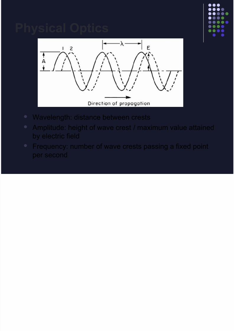
Physical Optics
Wavelength: distance between crests
Amplitude: height of wave crest / maximum value attained
by electric field
Frequency: number of wave crests passing a fixed point
per second
8/14/2019 Clinical optics review
http://slidepdf.com/reader/full/clinical-optics-review 5/69
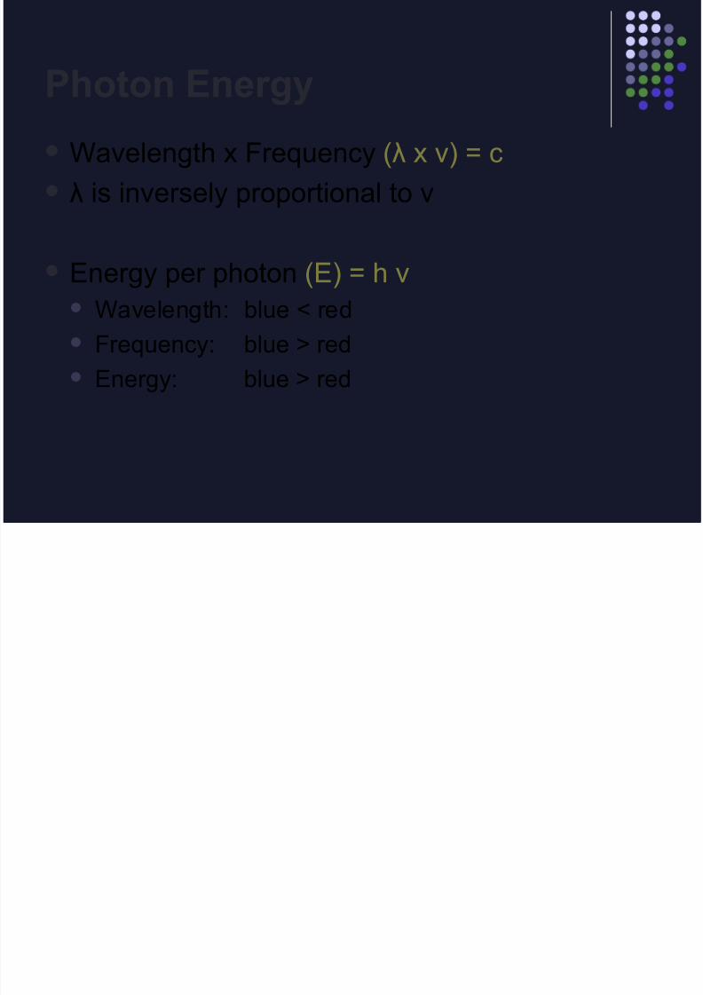
Photon Energy
Wavelength x Frequency (λ x ν) = c
λ is inversely proportional to v
Energy per photon (E) = h v Wavelength: blue < red
Frequency: blue > red
Energy: blue > red
8/14/2019 Clinical optics review
http://slidepdf.com/reader/full/clinical-optics-review 6/69
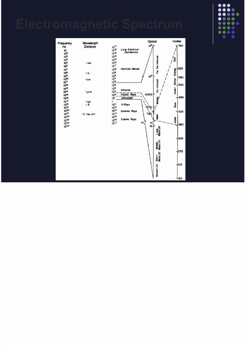
Electromagnetic Spectrum
8/14/2019 Clinical optics review
http://slidepdf.com/reader/full/clinical-optics-review 7/69
Interference
Constructive interference: crests of two waves coincide
Destructive interference: crest of one wave coincides with trough of other
wave
Coherence: measure of the ability for two light waves to
interfere

8/14/2019 Clinical optics review
http://slidepdf.com/reader/full/clinical-optics-review 8/69
Interference: Applications
Laser inferometry: Evaluates retinal function in pt w/ cataract
Laser beam split into 2 beams
Beams overlap on retina, producing interference
fringes, thus you know retina is functioning
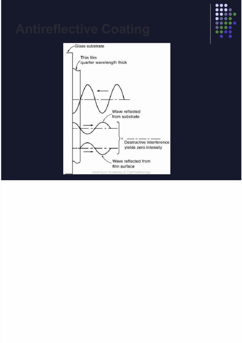
Antireflective coatings
8/14/2019 Clinical optics review
http://slidepdf.com/reader/full/clinical-optics-review 9/69
Antireflective Coating
8/14/2019 Clinical optics review
http://slidepdf.com/reader/full/clinical-optics-review 10/69
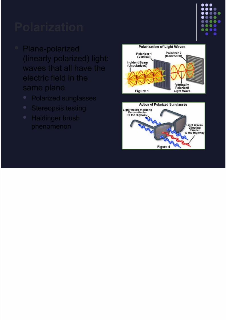
Polarization
Plane-polarized
(linearly polarized) light:
waves that all have the
electric field in thesame plane Polarized sunglasses
Stereopsis testing
Haidinger brushphenomenon
8/14/2019 Clinical optics review
http://slidepdf.com/reader/full/clinical-optics-review 11/69
Diffraction
Bending of light rays when they encounter an
obstruction
Diffraction limits visual acuity when the pupilsize is less than 2.5mm

8/14/2019 Clinical optics review
http://slidepdf.com/reader/full/clinical-optics-review 12/69
Diffraction
What is the optimal pinhole aperture?
1.2 mm
Any smaller would greatly increase diffractionand limit the amount of light into the eye
Because of diffractive effects, pinhole vision
is rarely better than 20/25
8/14/2019 Clinical optics review
http://slidepdf.com/reader/full/clinical-optics-review 13/69
Scattering
Isolated molecules absorb light and re-radiateit at same wavelength but different direction
Causes glare (cataracts, AC flare, corneal
haze)
Rayleigh Scattering
Due to scattering of very small particles Sky appears blue because of greater scattering of
shorter wavelengths

8/14/2019 Clinical optics review
http://slidepdf.com/reader/full/clinical-optics-review 14/69
Lasers
Light Amplification by Stimulated Emission of
Radiation
Which of the following features of laser lightenhance its intensity? Directionality
Coherence
Polarization
Monochromaticity

8/14/2019 Clinical optics review
http://slidepdf.com/reader/full/clinical-optics-review 15/69
Laser-Tissue Interaction
Name 3 ways lasers damage tissue: Photocoagulation (Argon)
Photodisruption (Nd:YAG)
Photoablation (Excimer)
8/14/2019 Clinical optics review
http://slidepdf.com/reader/full/clinical-optics-review 16/69
Geometrical Optics
8/14/2019 Clinical optics review
http://slidepdf.com/reader/full/clinical-optics-review 17/69
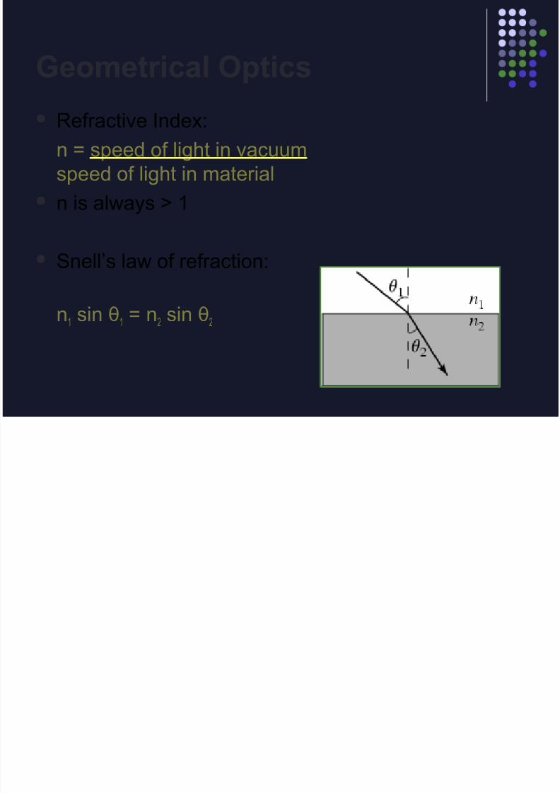
Geometrical Optics
Refractive Index:
n = speed of light in vacuum
speed of light in material
n is always > 1
Snell’s law of refraction:
n1 sin θ1 = n2 sin θ2

8/14/2019 Clinical optics review
http://slidepdf.com/reader/full/clinical-optics-review 18/69
Refraction
A fisherman attempts to
spear a fish as shown
at right.
Should he aim directly
at the fish, in front of
the fish, or behind the
fish as he sees it?
8/14/2019 Clinical optics review
http://slidepdf.com/reader/full/clinical-optics-review 19/69
Refraction
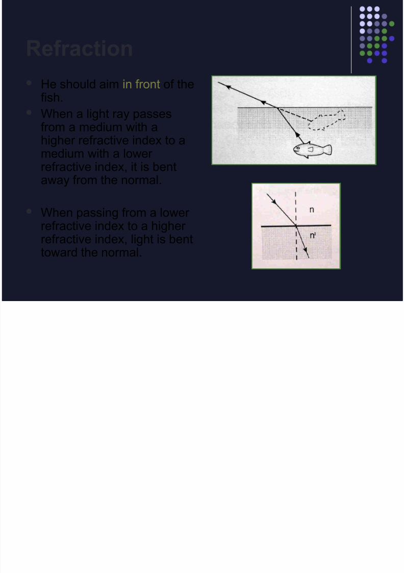
He should aim in front of thefish.
When a light ray passesfrom a medium with a
higher refractive index to amedium with a lower refractive index, it is bentaway from the normal.
When passing from a lower refractive index to a higher refractive index, light is benttoward the normal.

8/14/2019 Clinical optics review
http://slidepdf.com/reader/full/clinical-optics-review 20/69
Total Internal Reflection

8/14/2019 Clinical optics review
http://slidepdf.com/reader/full/clinical-optics-review 21/69
Vergence
A measure of the
spreading (or coming
together) of a bundle of
light rays.
8/14/2019 Clinical optics review
http://slidepdf.com/reader/full/clinical-optics-review 22/69
Vergence
The reciprocal of the distance, in meters,
from the object point or to the image point.
Units = m-1= diopters (D)
Lenses add vergence to light
8/14/2019 Clinical optics review
http://slidepdf.com/reader/full/clinical-optics-review 23/69
Vergence
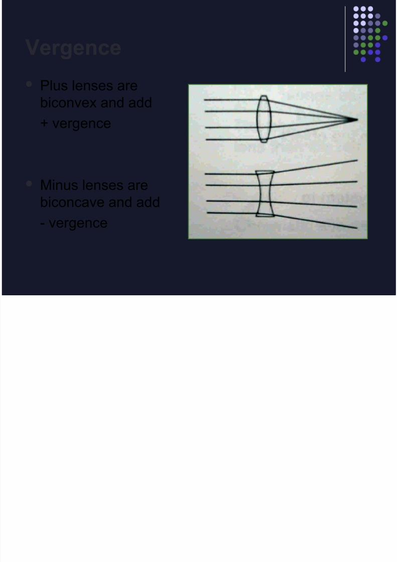
Plus lenses are
biconvex and add
+ vergence
Minus lenses are
biconcave and add- vergence
8/14/2019 Clinical optics review
http://slidepdf.com/reader/full/clinical-optics-review 24/69
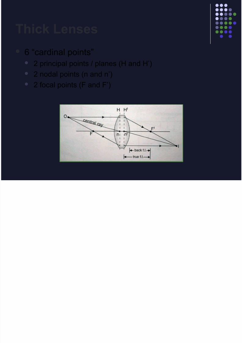
Thick Lenses
6 “cardinal points” 2 principal points / planes (H and H’)
2 nodal points (n and n’)
2 focal points (F and F’)
8/14/2019 Clinical optics review
http://slidepdf.com/reader/full/clinical-optics-review 25/69
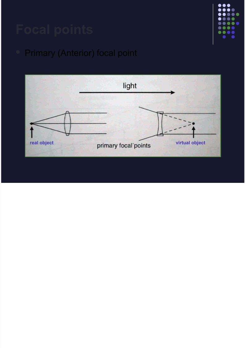
Focal points
Primary (Anterior) focal point
real object virtual object

8/14/2019 Clinical optics review
http://slidepdf.com/reader/full/clinical-optics-review 26/69
Focal points
Secondary (Posterior) focal point
real image virtual image
8/14/2019 Clinical optics review
http://slidepdf.com/reader/full/clinical-optics-review 27/69
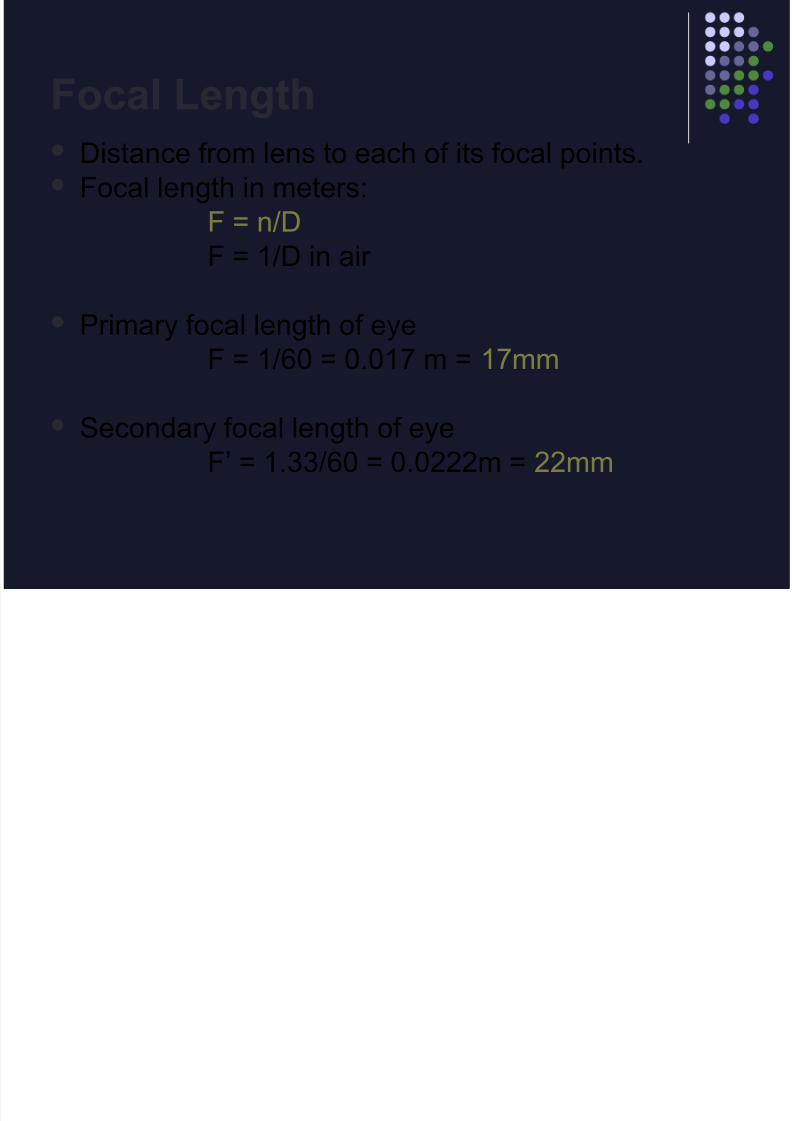
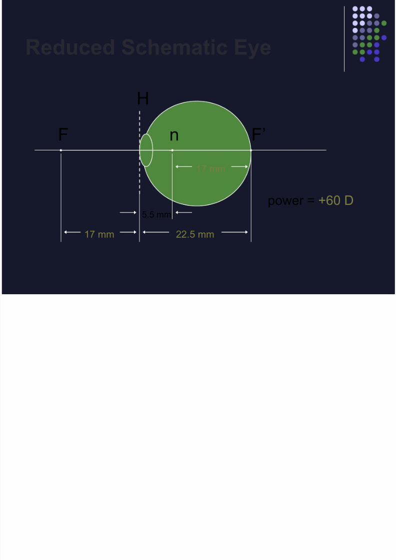
Focal Length Distance from lens to each of its focal points. Focal length in meters:
F = n/D
F = 1/D in air
Primary focal length of eye
F = 1/60 = 0.017 m = 17mm
Secondary focal length of eyeF’ = 1.33/60 = 0.0222m = 22mm
8/14/2019 Clinical optics review
http://slidepdf.com/reader/full/clinical-optics-review 28/69
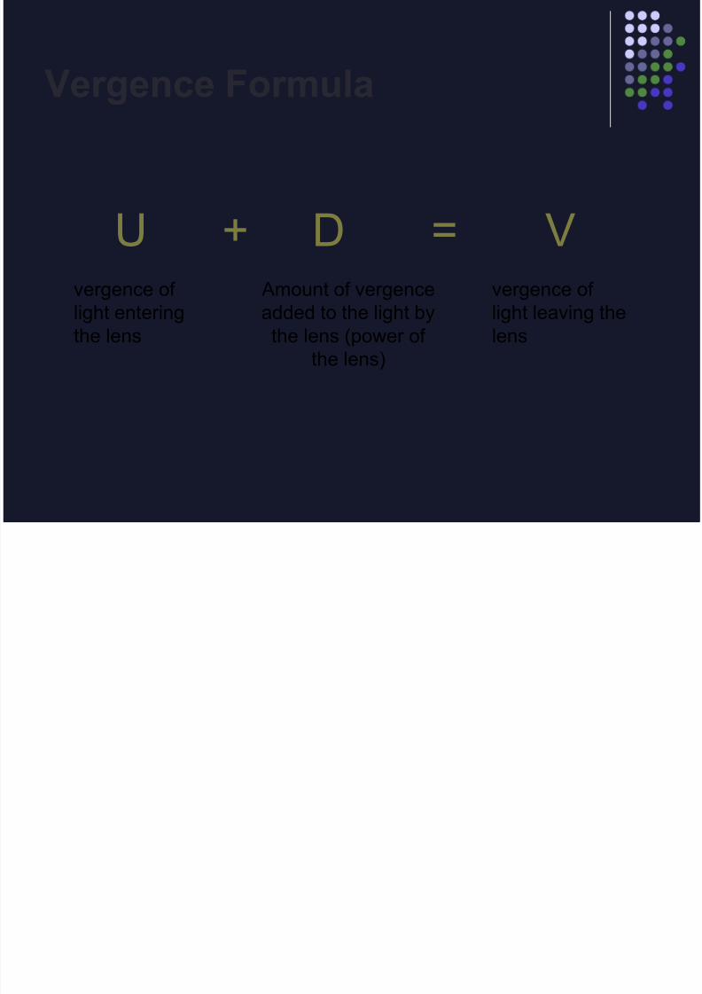
Vergence Formula
U + D = Vvergence of
light entering
the lens
Amount of vergence
added to the light by
the lens (power of
the lens)
vergence of
light leaving the
lens

8/14/2019 Clinical optics review
http://slidepdf.com/reader/full/clinical-optics-review 29/69
Real or virtual?
light
8/14/2019 Clinical optics review
http://slidepdf.com/reader/full/clinical-optics-review 30/69
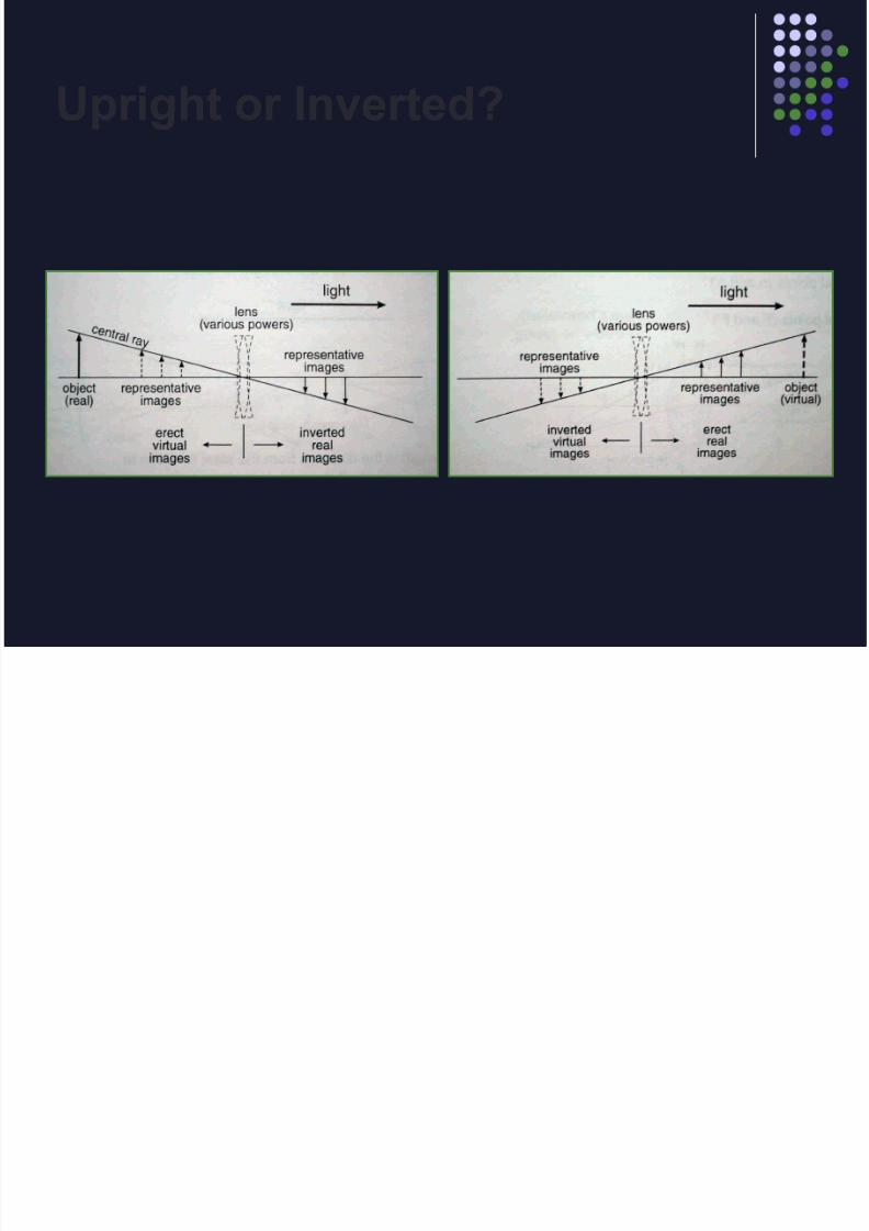
Upright or Inverted?
8/14/2019 Clinical optics review
http://slidepdf.com/reader/full/clinical-optics-review 31/69
Vergence
An object is located 20 cm to the left of a
-2.00 D lens. Where is the image located?
A) 20 cm to the right of the lens
B) 50 cm to the right of the lens
C) 33 cm to the left of the lens
D) 14 cm to the left of the lens
8/14/2019 Clinical optics review
http://slidepdf.com/reader/full/clinical-optics-review 32/69
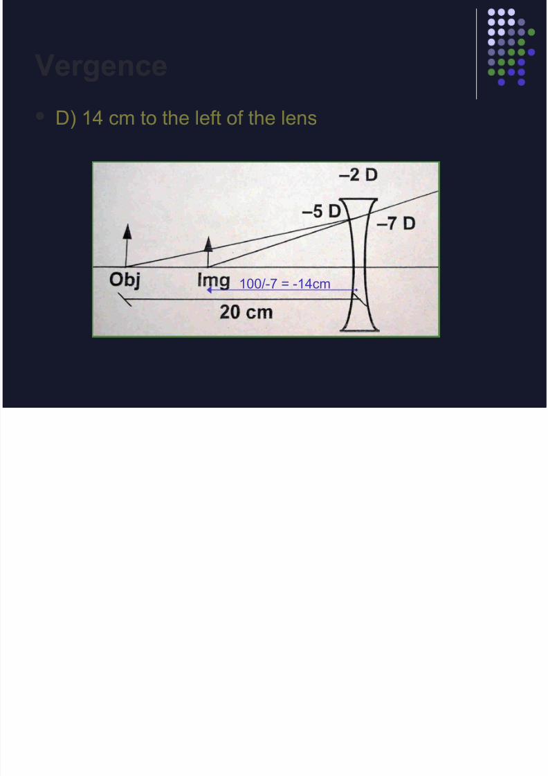
Vergence
D) 14 cm to the left of the lens
100/-7 = -14cm

8/14/2019 Clinical optics review
http://slidepdf.com/reader/full/clinical-optics-review 33/69
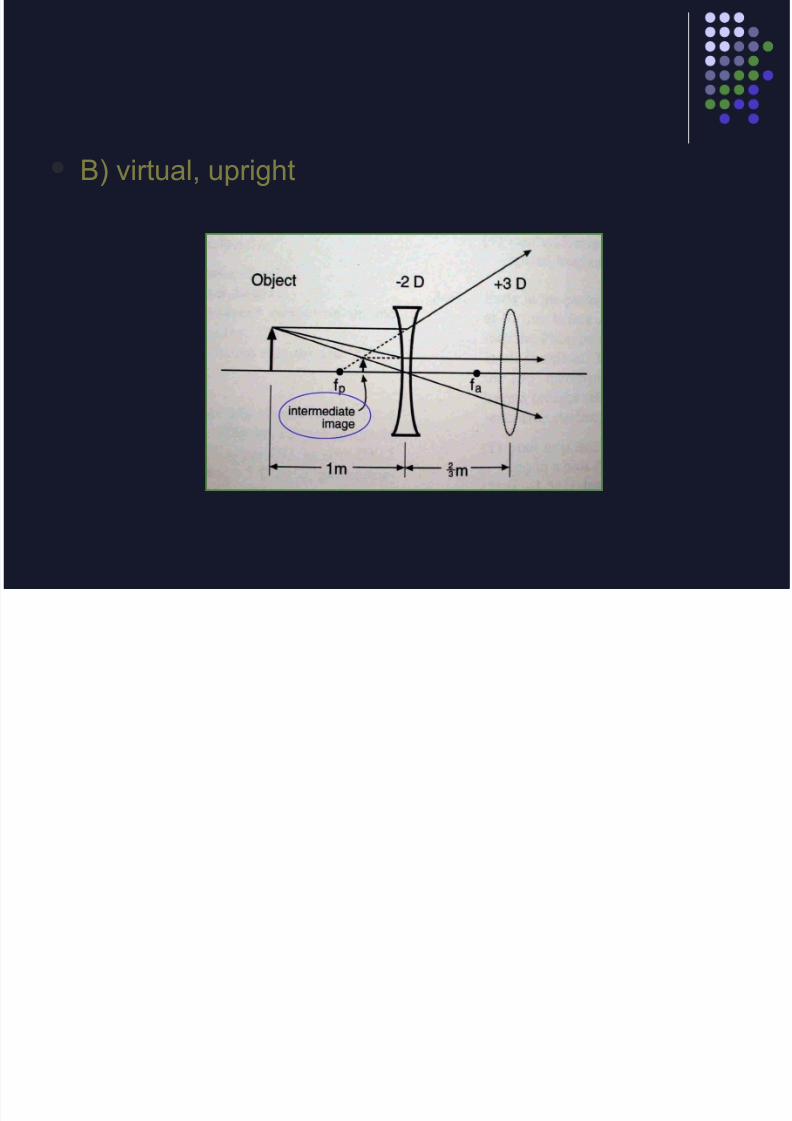
The intermediate image formed by the concave lens is
A) Real , inverted
B) Virtual, upright
C) Real, upright
D) Virtual, inverted
8/14/2019 Clinical optics review
http://slidepdf.com/reader/full/clinical-optics-review 34/69
B) virtual, upright

8/14/2019 Clinical optics review
http://slidepdf.com/reader/full/clinical-optics-review 35/69
Schematic Eye
8/14/2019 Clinical optics review
http://slidepdf.com/reader/full/clinical-optics-review 36/69
Reduced Schematic Eye
F F’n
H
5.5 mm
17 mm 22.5 mm
17 mm
power = +60 D
8/14/2019 Clinical optics review
http://slidepdf.com/reader/full/clinical-optics-review 37/69
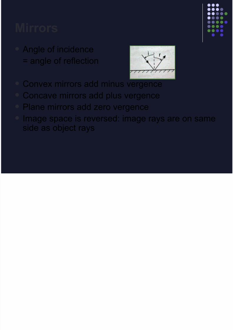
Mirrors
Angle of incidence
= angle of reflection
Convex mirrors add minus vergenceConcave mirrors add plus vergence
Plane mirrors add zero vergence
Image space is reversed: image rays are on sameside as object rays
8/14/2019 Clinical optics review
http://slidepdf.com/reader/full/clinical-optics-review 38/69
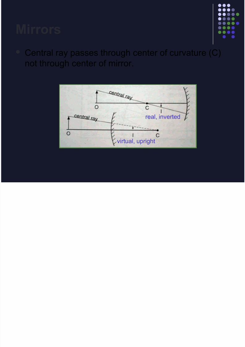
Mirrors
Central ray passes through center of curvature (C)
not through center of mirror.
real, inverted
virtual, upright

8/14/2019 Clinical optics review
http://slidepdf.com/reader/full/clinical-optics-review 39/69
Mirrors
U + D = V
F = r / 2
(r=radius of curvature)
Reflecting power
D = 1 / F = 2 / r
What is the reflecting power of cornea? 2/.008 = 250D (-250D)

8/14/2019 Clinical optics review
http://slidepdf.com/reader/full/clinical-optics-review 40/69
Magnification
Transverse Magnification
= Image height / Object height
= Image distance / Object distance
= Object vergence / Image vergence
Magtrans= U / V
For lens combinations the total magnificationis the product of the individual magnifications.
Wh i th i t di t
8/14/2019 Clinical optics review
http://slidepdf.com/reader/full/clinical-optics-review 41/69
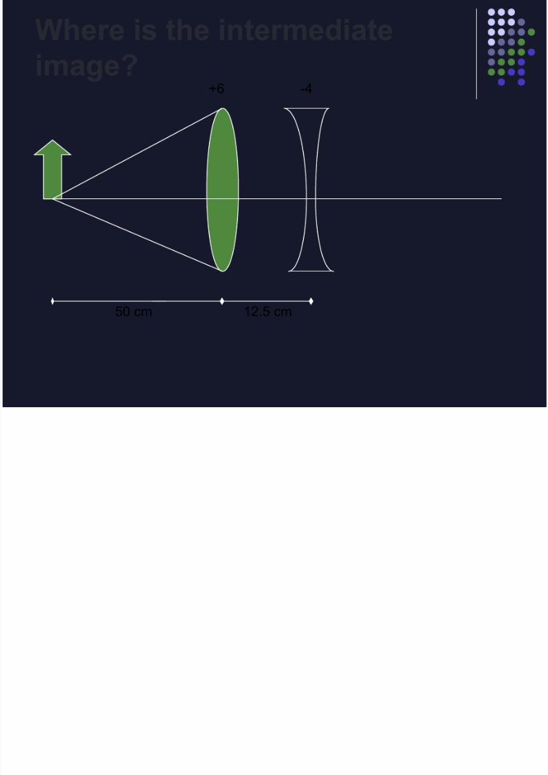
+6 -4
50 cm 12.5 cm
Where is the intermediate
image?
8/14/2019 Clinical optics review
http://slidepdf.com/reader/full/clinical-optics-review 42/69
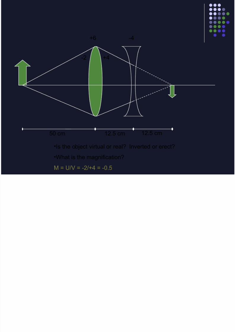
+6 -4
50 cm 12.5 cm
-2 +4
•Is the object virtual or real? Inverted or erect?
•What is the magnification?
M = U/V = -2/+4 = -0.5
12.5 cm
8/14/2019 Clinical optics review
http://slidepdf.com/reader/full/clinical-optics-review 43/69
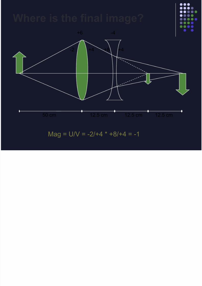
+6 -4
50 cm 12.5 cm 12.5 cm
-2 +4 +8 +4
Mag = U/V = -2/+4 * +8/+4 = -1
Where is the final image?
12.5 cm
8/14/2019 Clinical optics review
http://slidepdf.com/reader/full/clinical-optics-review 44/69
Simple Magnifiers
The (angular) magnification of a simple plus lens is
defined as the ratio of the size of the image produced by
the lens to the size of the object viewed at 25 cm
Magsimplemagnifier = D / 4
Examples:
+ 8D lens is called a 2x magnifier
+20D lens is a 5x magnifier
8/14/2019 Clinical optics review
http://slidepdf.com/reader/full/clinical-optics-review 45/69
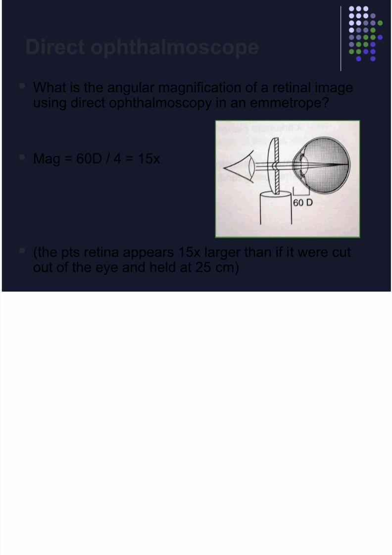
Direct ophthalmoscope
What is the angular magnification of a retinal imageusing direct ophthalmoscopy in an emmetrope?
Mag = 60D / 4 = 15x
(the pts retina appears 15x larger than if it were cutout of the eye and held at 25 cm)
8/14/2019 Clinical optics review
http://slidepdf.com/reader/full/clinical-optics-review 46/69
Telescopes
Receives parallel rays from a distant object
and projects parallel rays out.
(i.e. an afocal system)
2 lenses : objective + eyepiece
Transverse magnification is same for every
object regardless of location.
Magtelescope = Deyepiece / Dobjective

8/14/2019 Clinical optics review
http://slidepdf.com/reader/full/clinical-optics-review 47/69
Keplerian Telescope
Objective: low-power plus lens
Eyepiece: high-power plus lens
Separation: sum of focal lengths
Image: inverted, all light from objective is collected
Astronomical telescope
8/14/2019 Clinical optics review
http://slidepdf.com/reader/full/clinical-optics-review 48/69
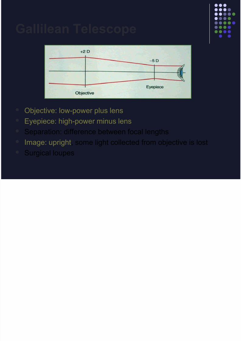
Gallilean Telescope
Objective: low-power plus lens
Eyepiece: high-power minus lens
Separation: difference between focal lengths
Image: upright, some light collected from objective is lost
Surgical loupes
8/14/2019 Clinical optics review
http://slidepdf.com/reader/full/clinical-optics-review 49/69
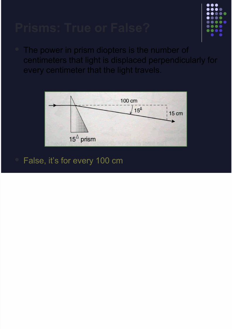
Prisms: True or False?
The power in prism diopters is the number of
centimeters that light is displaced perpendicularly for
every centimeter that the light travels.
False, it’s for every 100 cm
8/14/2019 Clinical optics review
http://slidepdf.com/reader/full/clinical-optics-review 50/69
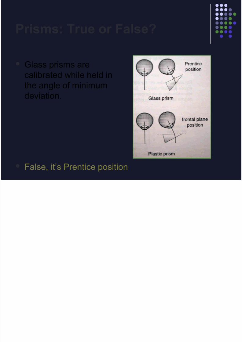
Prisms: True or False?
Glass prisms are
calibrated while held in
the angle of minimumdeviation.
False, it’s Prentice position
8/14/2019 Clinical optics review
http://slidepdf.com/reader/full/clinical-optics-review 51/69
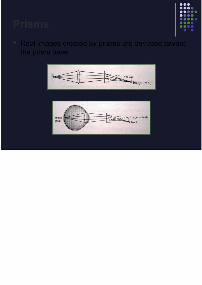
Prisms
Real images created by prisms are deviated toward
the prism base.

8/14/2019 Clinical optics review
http://slidepdf.com/reader/full/clinical-optics-review 52/69
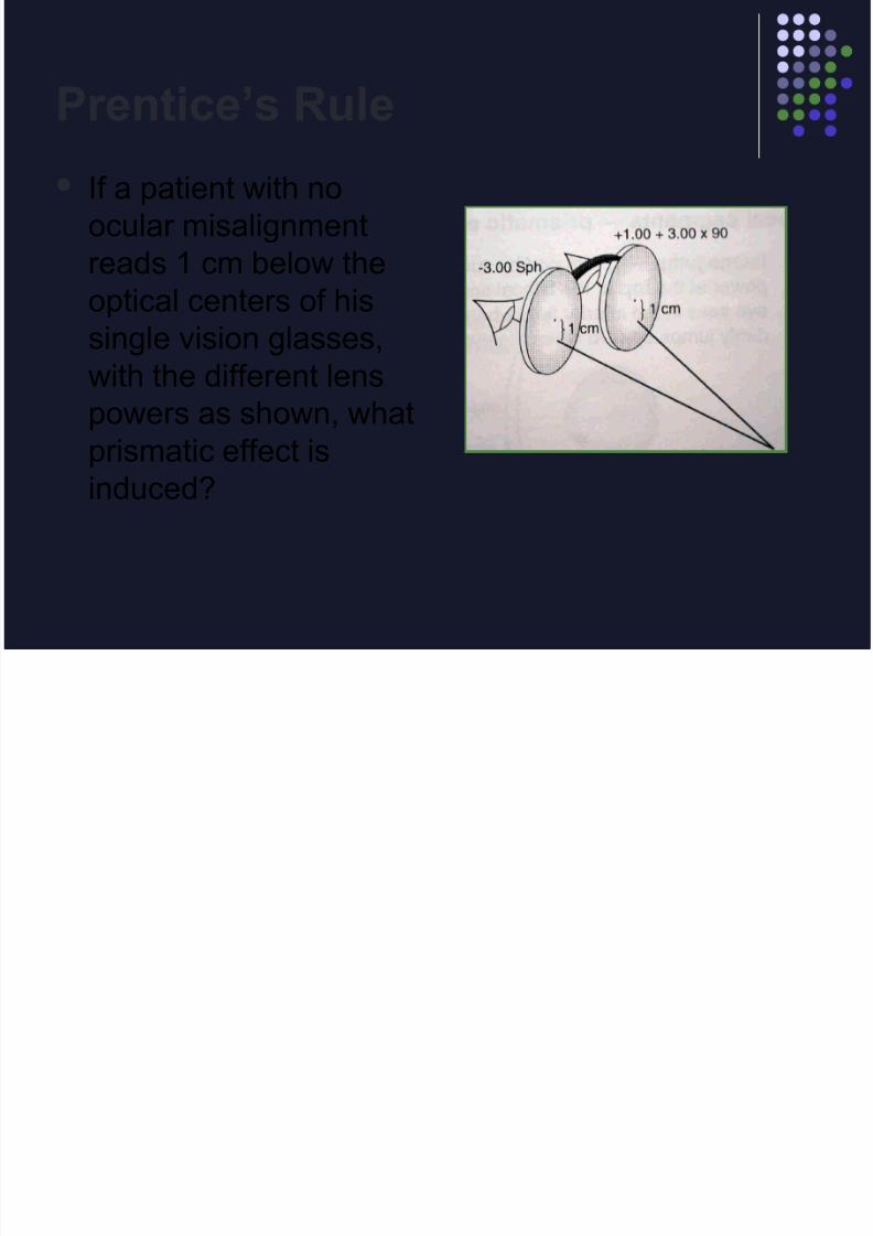
Prentice’s Rule
Except at its optical center, a spherical lens
has prism at every point on it’s surface.
Δ = h x D
Δ = prism diopters
h = distance from optical center in cm
D = diopter power of the lens
8/14/2019 Clinical optics review
http://slidepdf.com/reader/full/clinical-optics-review 53/69
Prentice’s Rule
If a patient with no
ocular misalignment
reads 1 cm below the
optical centers of hissingle vision glasses,
with the different lens
powers as shown, what
prismatic effect isinduced?
8/14/2019 Clinical optics review
http://slidepdf.com/reader/full/clinical-optics-review 54/69
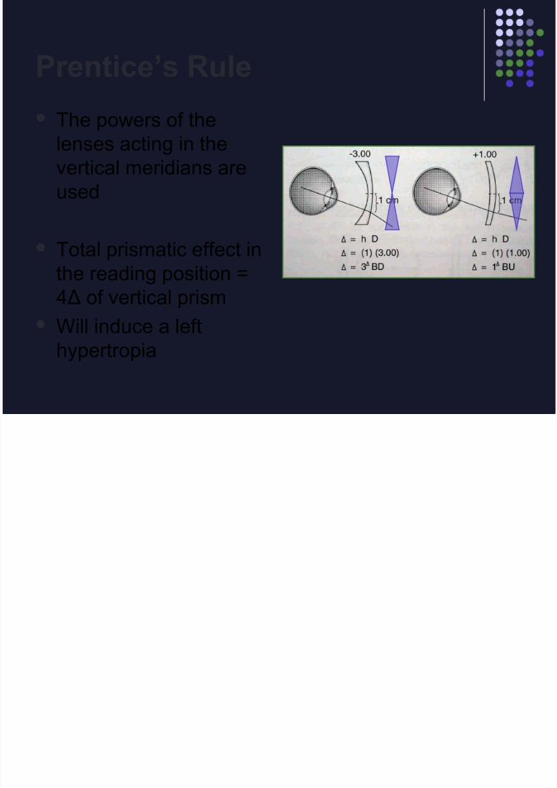
Prentice’s Rule
The powers of the
lenses acting in the
vertical meridians are
used
Total prismatic effect in
the reading position =
4Δ of vertical prism Will induce a left
hypertropia

8/14/2019 Clinical optics review
http://slidepdf.com/reader/full/clinical-optics-review 55/69
Fresnel Prisms
Fresnel prisms are equivalent to side-by-side
strips of long, narrow, thin prisms.
fresnel prism
8/14/2019 Clinical optics review
http://slidepdf.com/reader/full/clinical-optics-review 56/69
Fresnel Prisms
Used to avoid the weight of conventional
prisms.
Plastic Fresnel prisms are available as Press-
On prisms from 0.5Δ to 40Δ.
Visual acuity suffers by one or two lines with
higher power prisms because of glare and
chromatic aberration.

8/14/2019 Clinical optics review
http://slidepdf.com/reader/full/clinical-optics-review 57/69
Bifocal Segments
Image Jump – prismatic power at top of bifocalsegment Executive has no image jump
Image Displacement – total prism in readingposition
What type of add minimizes imagedisplacement with: Plus lenses? Round top
Minus lenses? Flat top
8/14/2019 Clinical optics review
http://slidepdf.com/reader/full/clinical-optics-review 58/69
Question
A patient with congenital nystagmus has a
null point measured to be 10° to the left of
fixation. The appropriate prism prescription
to rectify the induced head turn isa. 10∆ BI OS, 10∆ BO OD
b. 10∆ BI OD, 10∆ BO OS
c. 20∆ BI OS, 20∆ BI ODd. 20∆ BI OD, 20∆ BO OS
e. 20∆ BI OS, 20∆ BO OD
8/14/2019 Clinical optics review
http://slidepdf.com/reader/full/clinical-optics-review 59/69
(Regular) Astigmatism
Curvature of an astigmatic lens has minimum
and maximum values, located in meridians
90° apart.
An astigmatic surface cannot bring light rays
to a point (stigma) of focus.
Instead two focal lines are formed.
Geometric figure is formed called Conoid of Sturm.
8/14/2019 Clinical optics review
http://slidepdf.com/reader/full/clinical-optics-review 60/69
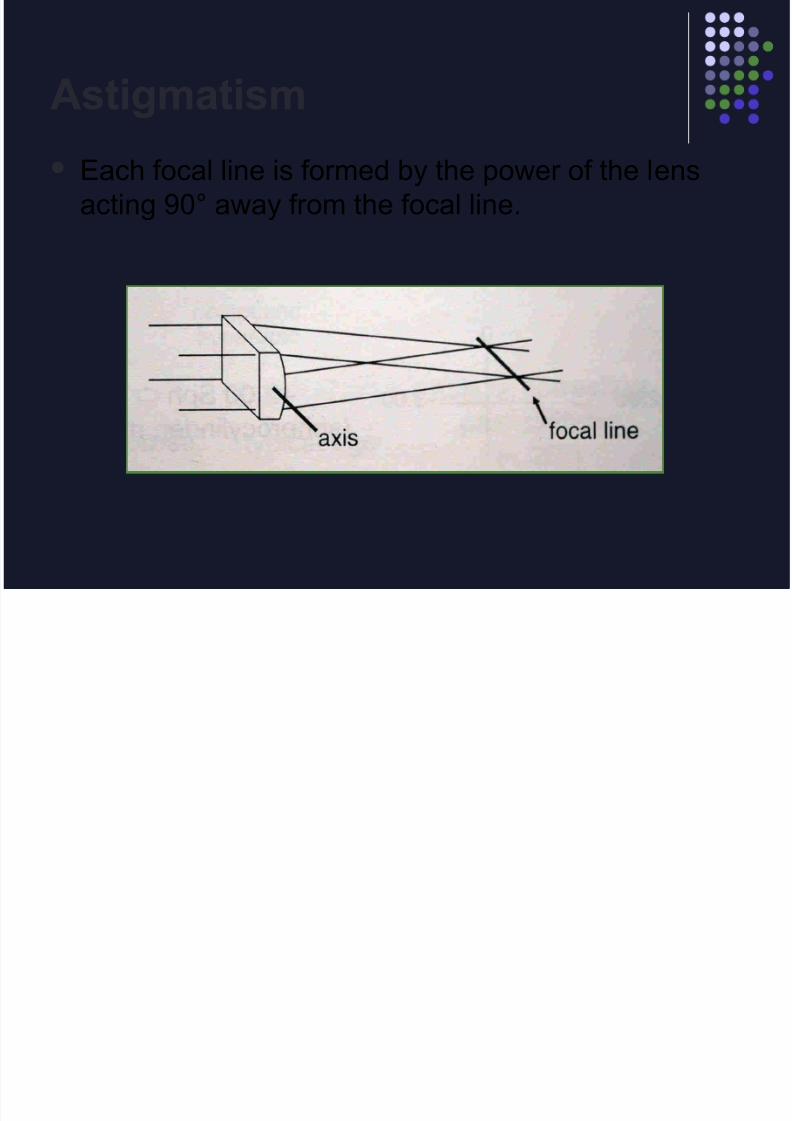
Astigmatism
Each focal line is formed by the power of the lens
acting 90° away from the focal line.
8/14/2019 Clinical optics review
http://slidepdf.com/reader/full/clinical-optics-review 61/69
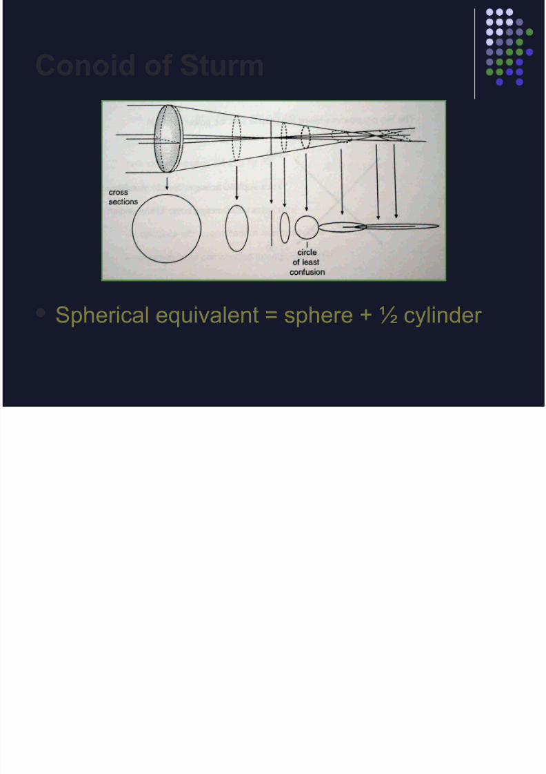
Conoid of Sturm
Spherical equivalent = sphere + ½ cylinder
8/14/2019 Clinical optics review
http://slidepdf.com/reader/full/clinical-optics-review 62/69
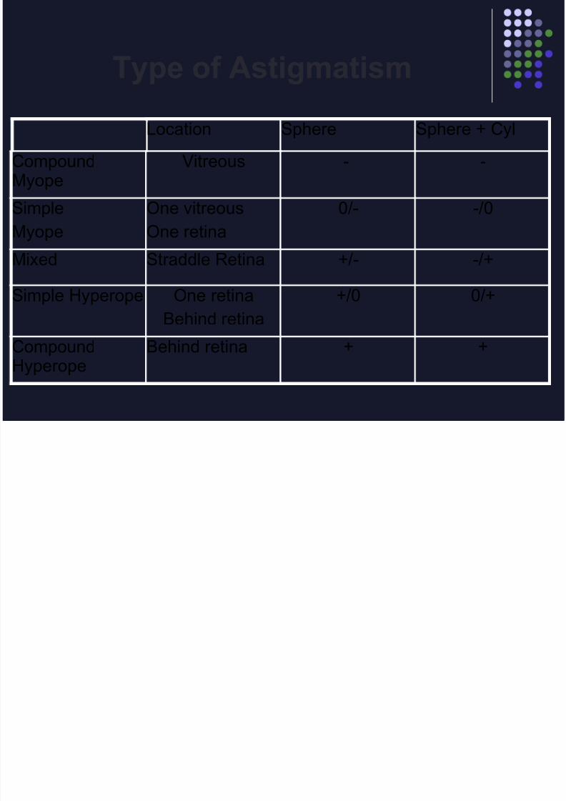
Type of Astigmatism
Location Sphere Sphere + Cyl
CompoundMyope
Vitreous - -
Simple
Myope
One vitreous
One retina
0/- -/0
Mixed Straddle Retina +/- -/+
Simple Hyperope One retinaBehind retina
+/0 0/+
CompoundHyperope
Behind retina + +
8/14/2019 Clinical optics review
http://slidepdf.com/reader/full/clinical-optics-review 63/69
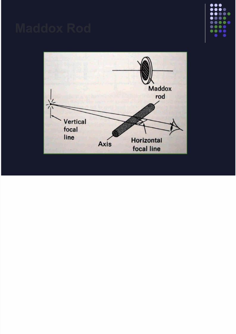
Maddox Rod

8/14/2019 Clinical optics review
http://slidepdf.com/reader/full/clinical-optics-review 64/69
8/14/2019 Clinical optics review
http://slidepdf.com/reader/full/clinical-optics-review 65/69
Accomodation
The accomodative amplitude of a 60 yr. old
healthy person is approximately:
1.50 D
Accomodative amplitude:
age 40 = 6.0 44 = 4.5 48 = 3.0
>age 48 decreases by 0.50 every 4 yrs
<age 40 increases by 1.0 every 4 yrs

8/14/2019 Clinical optics review
http://slidepdf.com/reader/full/clinical-optics-review 66/69
Kestenbaum’s Rule
A 72 yr. old patient with bilateral macular
degeneration has a distance acuity of 20/100. The
add required for this patient to read newspaper print
is:
A) +1.00
B) +3.00
C) +4.00
D) +5.00E) +10.00

8/14/2019 Clinical optics review
http://slidepdf.com/reader/full/clinical-optics-review 67/69
Contact Lenses
Obtain Refraction & K'sChoose base curve steeper than low K Usu +0.50 D steeper to form tear lens
Prevents apical touchConvert refraction to Minus cylinder formDisregard the cylinder
Convert to zero vertex distanceSubtract +0.50 spherical tear lens from the
sphere value to obtain the final RGP sphere
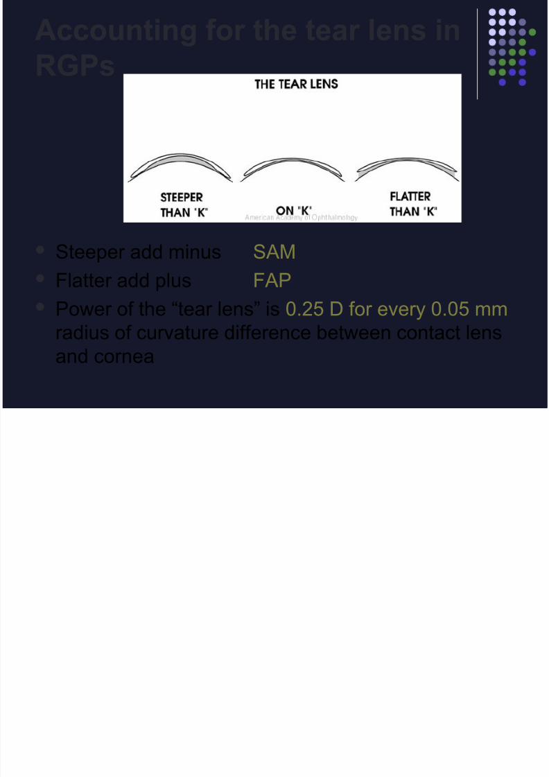
Accounting for the tear lens in
8/14/2019 Clinical optics review
http://slidepdf.com/reader/full/clinical-optics-review 68/69
Steeper add minus SAM
Flatter add plus FAP
Power of the “tear lens” is 0.25 D for every 0.05 mm
radius of curvature difference between contact lens
and cornea
Accounting for the tear lens in
RGPs

8/14/2019 Clinical optics review
http://slidepdf.com/reader/full/clinical-optics-review 69/69
The refractive error of an eye is -3.00 D, the
K measurement is 7.80 mm and the base
curve chosen for the rigid contact lens is 7.95
mm. What is the anticipated power of thecontact lens?
Power of tear lens: 7.95-7.80 = 0.15 mm = 0.75 D CL power: -3.00 D + 0.75 D = -2.25 D (FAP)