Embed Size (px)
Citation preview


Visit This Book’s Web Page / Buy Now / Request an Exam/Review CopyThis is a sample from Clinical Neurophysiology in Pediatrics: A Practical Approach to Neurodiagnostic Testing and Management
© Demos Medical Publishing
Clinical Neurophysiology in Pediatrics

Visit This Book’s Web Page / Buy Now / Request an Exam/Review CopyThis is a sample from Clinical Neurophysiology in Pediatrics: A Practical Approach to Neurodiagnostic Testing and Management
© Demos Medical Publishing

Visit This Book’s Web Page / Buy Now / Request an Exam/Review CopyThis is a sample from Clinical Neurophysiology in Pediatrics: A Practical Approach to Neurodiagnostic Testing and Management
© Demos Medical Publishing
Clinical Neurophysiology in Pediatrics
A Practical Approach to Neurodiagnostic Testing
and Management
EDITOR
Gloria M. Galloway, MD, FAANProfessor, Department of Neurology
Neuromuscular Division
Ohio State University Medical Center
Columbus, Ohio
NEW YORK

Visit This Book’s Web Page / Buy Now / Request an Exam/Review CopyThis is a sample from Clinical Neurophysiology in Pediatrics: A Practical Approach to Neurodiagnostic Testing and Management
© Demos Medical Publishing
Visit our website at www.demosmedical.com
ISBN: 9781620700457
e-book: 9781617052118
Acquisitions Editor: Beth Barry
Compositor: Exeter Premedia Services Private Ltd.
© 2016 Demos Medical Publishing, LLC. All rights reserved. This book is protected by copyright. No part
of it may be reproduced, stored in a retrieval system, or transmitted in any form or by any means, electronic,
mechanical, photocopying, recording, or otherwise, without the prior written permission of the publisher.
Medicine is an ever-changing science. Research and clinical experience are continually expanding our knowl-
edge, in particular our understanding of proper treatment and drug therapy. The authors, editors, and publisher
have made every effort to ensure that all information in this book is in accordance with the state of knowledge
at the time of production of the book. Nevertheless, the authors, editors, and publisher are not responsible for
errors or omissions or for any consequences from application of the information in this book and make no
warranty, expressed or implied, with respect to the contents of the publication. Every reader should examine
carefully the package inserts accompanying each drug and should carefully check whether the dosage sched-
ules mentioned therein or the contraindications stated by the manufacturer differ from the statements made
in this book. Such examination is particularly important with drugs that are either rarely used or have been
newly released on the market.
Library of Congress Cataloging-in-Publication Data
Clinical neurophysiology in pediatrics : a practical approach to neurodiagnostic testing and management /
[edited by] Gloria M. Galloway.
p. ; cm.
Includes bibliographical references and index.
ISBN 978-1-62070-045-7 — ISBN 978-1-61705-211-8 (e-book)
I. Galloway, Gloria M., 1960– , editor.
[DNLM: 1. Child. 2. Nervous System Diseases—diagnosis. 3. Diagnostic Techniques, Neurological.
4. Infant. 5. Nervous System Diseases—therapy. 6. Neurophysiology—methods. WS 340]
RJ486
618.92'8—dc23
2015014554
Special discounts on bulk quantities of Demos Medical Publishing books are available to corporations,
professional associations, pharmaceutical companies, health care organizations, and other qualifying
groups. For details, please contact:
Special Sales Department
Demos Medical Publishing, LLC
11 West 42nd Street, 15th Floor
New York, NY 10036
Phone: 800-532-8663 or 212-683-0072
Fax: 212-941-7842
E-mail: [email protected]
Printed in the United States of America by McNaughton & Gunn.
15 16 17 18 / 5 4 3 2 1
Visit This Book’s Web Page / Buy Now / Request an Exam/Review CopyThis is a sample from Clinical Neurophysiology in Pediatrics: A Practical Approach to Neurodiagnostic Testing and Management
© Demos Medical Publishing
v
1. EEG Monitoring in Neonatal Epilepsies 1Lekha M. Rao, MD, Joyce H. Matsumoto, MD, Jason T. Lerner, MD, and Marc R. Nuwer, MD, PhD
2. Pediatric Febrile Seizures 21Charuta N. Joshi, MBBS, FRCPC and Thoru Yamada, MD, FACNS
3. Epileptic and Nonepileptic Paroxysmal Events in Childhood 29Jorge Vidaurre, MD
4. EEG Interpretation in Childhood Epilepsies 45Charuta N. Joshi, MBBS, FRCPC and Thoru Yamada, MD, FACNS
5. The Evaluation of Pediatric Sleep Disorders 77Deborah C. Lin-Dyken, MD and Mark Eric Dyken, MD
6. Pediatric Muscular Dystrophies and Myopathies: The Role of Neurophysiology,
Genetics, and Ancillary Testing 97Russell J. Butterfield, MD, PhD and Mark B. Bromberg, MD, PhD
7. Clinical Evaluation in Pediatric Peripheral Neuropathies 127Zarife Sahenk, MD, PhD, FAAN
8. EMG Considerations in the Pediatric Patient 147Gloria M. Galloway, MD, FAAN
9. Clinical Neurophysiology in Pediatric Peripheral Neuropathy 153Gloria M. Galloway, MD, FAAN
Contents
Contributors vii
Preface ix
Visit This Book’s Web Page / Buy Now / Request an Exam/Review CopyThis is a sample from Clinical Neurophysiology in Pediatrics: A Practical Approach to Neurodiagnostic Testing and Management
© Demos Medical Publishing
vi ■ Contents
10. EMG in Pediatric Brachial Plexopathy 161Jaime R. López, MD
11. Intraoperative Considerations in the Pediatric Patient 181Gloria M. Galloway, MD, FAAN
12. Evoked Potentials in Pediatric Brainstem Lesions 187Ze Dong Jiang, MD
13. Evoked Potentials in Adolescent Idiopathic Scoliosis: Intraoperative
Neurophysiological Monitoring 215Ronald G. Emerson, MD
14. Autonomic Disorders in Children 241Nancy L. Kuntz, MD, Theresa Oswald, BS, MS, and Pallavi P. Patwari, MD
15. Clinical Neurophysiology: Future Role in Pediatric Neurologic Disorders 265Aatif M. Husain, MD
Index 275

Visit This Book’s Web Page / Buy Now / Request an Exam/Review CopyThis is a sample from Clinical Neurophysiology in Pediatrics: A Practical Approach to Neurodiagnostic Testing and Management
© Demos Medical Publishing
vii
Mark B. Bromberg, MD, PhD Professor, Department of Neurology, University of Utah,
Salt Lake City, Utah
Russell J. Butterfield, MD, PhD Assistant Professor, Departments of Neurology and
Pediatrics, University of Utah, Salt Lake City, Utah
Mark Eric Dyken, MD Director, Sleep Laboratory, Professor, Department of Neurology,
University Iowa Hospitals, Iowa City, Iowa
Ronald G. Emerson, MD Director of Intraoperative Monitoring, Department of Neurology,
Hospital for Special Surgery; Professor, Department of Neurology, Weill Cornell Medical
Center, New York, New York
Gloria M. Galloway, MD, FAAN Professor, Department of Neurology, Neuromuscular
Division, Ohio State University Medical Center, Columbus, Ohio
Aatif M. Husain, MD Professor, Department of Neurology, Duke University Medical
Center; Director, Neurodiagnostic Center, Veterans Affairs Medical Center, Durham,
North Carolina
Ze Dong Jiang, MD, PhD Professor, Department of Pediatrics, Children's Hospital, Fudan
University, Shanghai, China; Senior Researcher, Department of Pediatrics, University of
Oxford, Oxford, United Kingdom
Charuta N. Joshi, MBBS, FRCPC Clinical Professor, Pediatric Neurology; Director
of Pediatric Epilepsy, Department of Pediatrics, University of Iowa Children’s Hospital,
Iowa City, Iowa
Contributors

Visit This Book’s Web Page / Buy Now / Request an Exam/Review CopyThis is a sample from Clinical Neurophysiology in Pediatrics: A Practical Approach to Neurodiagnostic Testing and Management
© Demos Medical Publishing
viii ■ Contributors
Nancy L. Kuntz, MD Associate Professor of Pediatrics and Neurology, Departments of
Pediatrics, Neurology, Northwestern Feinberg School of Medicine; Medical Director, Mazza
Foundation Neuromuscular Disorders Program, Department of Pediatrics, Ann & Robert H.
Lurie Children’s Hospital of Chicago, Chicago, Illinois
Jason T. Lerner, MD Associate Professor, Department of Pediatrics, Division of Pediatric
Neurology, David Geffen School of Medicine at UCLA, Los Angeles, California
Deborah C. Lin-Dyken, MD Clinical Associate Professor of Pediatrics, Division of
Pediatric Neurology, Development and Behavior, Carver College of Medicine, University of
Iowa, Center for Disability and Development, Iowa City, Iowa
Jaime R. López, MD Associate Professor, Departments of Neurology and Neurological
Sciences and Neurosurgery, Stanford University School of Medicine, Stanford, California
Joyce H. Matsumoto, MD Health Science Assistant Clinical Professor, Department of
Pediatrics, Division of Pediatric Neurology, David Geffen School of Medicine at UCLA,
Los Angeles, California
Marc R. Nuwer, MD, PhD Professor and Vice Chair, Department of Neurology,
Reed Neurological Research Center, David Geffen School of Medicine at UCLA; Department
Head, Clinical Neurophysiology Department, Ronald Reagan UCLA Medical Center,
Los Angeles, California
Theresa Oswald, BS, MS Instructor in Pediatrics, Department of Pediatrics, Northwestern
Feinberg School of Medicine, Chicago, Illinois
Pallavi P. Patwari, MD Assistant Professor of Clinical Pediatrics, Director of Pediatric
Sleep Medicine Program, Interim Chief of the Division of Pediatric Critical Care Medicine,
Department of Pediatrics, University of Illinois Hospital and Health Science System,
University of Illinois College of Medicine, Chicago, Illinois
Lekha M. Rao, MD Health Sciences Assistant Clinical Professor, Department of
Pediatrics, Division of Pediatric Neurology, David Geffen School of Medicine at UCLA,
Los Angeles, California
Zarife Sahenk, MD, PhD, FAAN Director, Neuromuscular Disease Laboratory,
Neuromuscular Division, Center for Gene Therapy, Department of Pediatrics, Nationwide
Children's Hospital, Columbus, Ohio
Jorge Vidaurre, MD Assistant Professor/Director, Pediatric Clinical Neurophysiology
Program–EEG Laboratory, Pediatric Neurology Division, Nationwide Children’s
Hospital–The Ohio State University, Columbus, Ohio
Thoru Yamada, MD, FACNS Director, Neurophysiology Laboratory, University of Iowa
Hospitals, Iowa City, Iowa

Visit This Book’s Web Page / Buy Now / Request an Exam/Review CopyThis is a sample from Clinical Neurophysiology in Pediatrics: A Practical Approach to Neurodiagnostic Testing and Management
© Demos Medical Publishing
ix
The field of clinical neurophysiology has expanded with the development of new approaches,
techniques, and studies over the last two decades. In many cases, new neurophysiologic proce-
dures and interpretations have allowed more accurate diagnosis, aided in diagnosis, and set the
gold standard for diagnostic confirmation in numerous neurological disorders. Thus, clinical
neurophysiology has grown and increasingly gained respect due to its diagnostic acumen. With
this growth has come a wide diversity of subspecialty skills. Each subspecialty lends itself to
focused research and, in many cases, clinical certification. In this regard, neurophysiologists
skilled in sleep studies, for example, would find themselves unlikely to spend much of their
time evaluating complex electromyography (EMG) cases. Pediatric or adult training creates
another layer of differentiation so that the interpretation of an electroencephalogram (EEG) in
an infant or child differs greatly from that of an adult. A lack of information focused on pedi-atric clinical neurophysiology exists, with most texts written largely with the adult patient in
mind. This book uniquely bridges that gap by providing information from a pediatric perspec-
tive in various aspects of clinical neurophysiology. Contributors to this book are thought leaders
and researchers in their respective fields of clinical neurophysiology. Each has provided discus-
sion in their subspecialty area with a pediatric focus emphasizing diagnostic neurophysiologic
techniques. Each chapter emphasizes a different focused area of neurophysiology and brings
together the clinical and technical information needed for understanding. Chapters are devoted
to pediatric sleep disorders, epilepsy, febrile seizures, and nonepileptic paroxysmal disorders.
Other chapters are devoted to pediatric muscular dystrophies, EMG, brachial plexopathies,
and peripheral neuropathy. A chapter devoted to intraoperative monitoring is included along
with other chapters on evoked potentials and autonomic disorders. In several chapters, multiple
authors have contributed, each providing aspects related to their research or area of unique
expertise.
This book will serve as an excellent reference for the clinical provider as well as for train-
ees and technologists in gaining greater knowledge in the various subspecialty areas of clinical
neurophysiology.
Preface

Visit This Book’s Web Page / Buy Now / Request an Exam/Review CopyThis is a sample from Clinical Neurophysiology in Pediatrics: A Practical Approach to Neurodiagnostic Testing and Management
© Demos Medical Publishing
x ■ Preface
I want to thank the contributors of this book who, through their passion for the field of
clinical neurophysiology, devoted much time to writing and sharing their wealth of informa-
tion. Additionally, none of the research or clinical data would be possible without the patients
who entrusted their care to us. Of course, the time devoted to research and dedication to the
field of clinical neurophysiology would not be possible without the encouragement of mentors
and the support of our families. My parents’ encouragement has been invaluable throughout my
life. I dedicate this book to them and to my sons Nadeem and Corey who taught me how deep
love can be. Never stop following your dreams, my darlings.
Gloria M. Galloway, MD, FAAN

Visit This Book’s Web Page / Buy Now / Request an Exam/Review CopyThis is a sample from Clinical Neurophysiology in Pediatrics: A Practical Approach to Neurodiagnostic Testing and Management
© Demos Medical Publishing
1
1
EEG Monitoring inNeonatal Epilepsies
Lekha M. Rao, MDJoyce H. Matsumoto, MD
Jason T. Lerner, MDMarc R. Nuwer, MD, PhD
Electroencephalographers often approach neonatal studies with trepidation. Neonatal studies
vary from traditional electroencephalograms (EEGs) in both technical and visual aspects. Half
of the full electrode set is used, placed at double distance, and the recording lasts for 60 minutes
in order to catch a full sleep-wake cycle. Extra electrodes are also essential for interpreting the
recording, such as ocular leads, chin electromyogram (EMG), and cardiac and respiratory mon-
itoring. When interpreting the neonatal EEG, the paper speed is slowed to 15 mm/sec in order
to more easily recognize the slower delta frequencies, which dominate in neonatal records. The
low-frequency filter is set to 0.5 Hz in order to clearly interpret slow eye movements (1). Sensi-
tivity is often lowered below the standard 7 mv/sec, given that amplitudes are not as high and
scalp impedance is lower. Although these differences exist, with experience and knowledge of
these EEG differences, interpretation in this age group is readily accomplished.
Much of the trepidation associated with the interpretation of neonatal EEG stems from the
fact that “normal” background is somewhat of a moving target. Findings that are acceptable at
30 weeks conceptional age (CA) are grossly abnormal at 36 weeks. Therefore, neonatal EEG is
best interpreted by first noting the infant’s current CA and then recognizing the characteristics
that should be present in the EEG background of a normal neonate. CA is calculated by adding
the estimated gestational age at birth to the current chronologic age (in weeks). If not given the
correct gestational age, an age range can be estimated based on recognized patterns.

Visit This Book’s Web Page / Buy Now / Request an Exam/Review CopyThis is a sample from Clinical Neurophysiology in Pediatrics: A Practical Approach to Neurodiagnostic Testing and Management
© Demos Medical Publishing
2 ■ Clinical Neurophysiology in Pediatrics
NEONATAL EEG BACKGROUND
Neonatal EEG studies should be systematically evaluated, with interpretation phrased in terms
of several key features:
■ Continuity
■ Amplitude
■ Symmetry
■ Interhemispheric synchrony
■ Normal named patterns
In extreme prematurity, normal electrographic findings are typically discontinuous, with bursts
of continuous cerebral activity separated by intervals of relative quiescence and lower ampli-
tude. This discontinuity improves with age, with the interburst interval becoming progressively
shorter and higher in amplitude as the baby approaches full term. By 40 to 44 weeks CA, the
EEG background becomes continuous in both wake and sleep ( 2).
Differentiation between wake and sleep states initially appears around 30 weeks CA. By
definition, the infant is awake whenever his/her eyes are open and asleep when eyes are closed.
Sleep is further subdivided into active sleep (AS, characterized by irregular respirations, occa-
sional limb movements, and rapid horizontal eye movements) and quiet sleep (QS), character-
ized by deep, regular respirations and paucity of limb/trunk movement. Electrographically,
wakefulness and AS in infants more than 30 weeks CA demonstrate fairly continuous cerebral
activity, developing into a characteristic mixed frequency, moderate-amplitude activité moy-enne pattern.
Because neonatal background abnormalities may become most apparent during deeper
sleep stages, a complete assessment of the EEG background requires thorough evaluation of
QS. To this end, continuous EEG (cEEG) provides a significant advantage over routine EEG
in ensuring that a generous sample of QS is captured for review. As the invariant, nonreactive
pattern of burst suppression seen in extremely preterm infants transitions into more defined
wake-sleep stages around 30 weeks CA, the final remnants of EEG discontinuity linger in
QS. As development proceeds, QS discontinuity gradually resolves, with gradual improve-
ment in the duration and amplitude of the interburst activity. Between 30 and 32 weeks CA,
QS activity consists of a tracé discontinue pattern in which periods of cerebral activity are
separated by nearly isoelectric periods of quiescence with voltage less than 25 μV. With time,
the voltage of the interburst intervals gradually increases such that by 35 to 36 weeks CA,
QS typically transitions to a tracé alternant pattern, in which cerebral activity is consistently
maintained above 25 μV but cycles between higher-amplitude bursts and more quiescent peri-
ods. The interburst amplitude continues to increase until no periods of relative quiescence
are perceived, and a continuous slow-wave sleep pattern is fully established around 44 weeks
CA ( 3,4).
Bursts of activity appearing in one hemisphere within 1.5 seconds of the other hemi-
sphere are considered to be synchronous. Prior to 30 weeks CA, cerebral activity occurs nearly

Visit This Book’s Web Page / Buy Now / Request an Exam/Review CopyThis is a sample from Clinical Neurophysiology in Pediatrics: A Practical Approach to Neurodiagnostic Testing and Management
© Demos Medical Publishing
1: EEG Monitoring in Neonatal Epilepsies ■ 3
simultaneously in both the right and left hemispheres, a phenomenon described as a hypersyn-chron y (5). The reason for early interhemispheric hypersynchrony is unknown, though it has
been postulated to be related to prominent thalamic drivers without significant cortical input.
Following 30 weeks, occasional asynchronous bursts are seen, which progressively diminish
until 100% synchrony is reestablished around 37 weeks CA.
BACKGROUND PATTERNS
A. Excessive sharps
B. Excessive discontinuity
C. Brief ictal/interictal rhythmic/repetitive discharges (BIRDs)
D. Other patterns (depressed/undifferentiated, low voltage)
EEG background findings (Table 1.1) are also frequently employed to assess the functional
integrity of the neonatal brain and to aid in the evaluation of neurologic prognosis. At the same
time, however, many patterns are nonspecific and of uncertain clinical significance.
TABLE 1.1 EEG Background in Prematurity
CONCEPTIONAL AGE (WEEKS)
MAXIMUM INTERBURST DURATION (SEC)
EEG BACKGROUND FEATURES
24–25 60 No sleep organization or reactivity
27–30 35 Discontinuous in both wake and sleep, some
reactivity
31–33 20 Differentiation between active and quiet sleep
patterns
Wake and active sleep: mixed frequency
continuous (activité moyenne)
Quiet sleep: interburst intervals amplitude nearly
isoelectric, <25 μV (trace discontinue pattern)
34–36 10 Wake and active sleep: mixed frequency
continuous (activité moyenne)
Quiet sleep: Interburst intervals increase in
amplitude, eventually exceeding 25 μV (trace
alternant pattern)
37–40 6 Wake and active sleep: mixed frequency
continuous (activité moyenne)
Quiet sleep: Interburst intervals continue to
increase in amplitude, increasing continuity (trace
alternant) transitioning to continuous slow-wave
sleep pattern
Source: Adapted from Refs. 5, 8, 9.
Visit This Book’s Web Page / Buy Now / Request an Exam/Review CopyThis is a sample from Clinical Neurophysiology in Pediatrics: A Practical Approach to Neurodiagnostic Testing and Management
© Demos Medical Publishing
4 ■ Clinical Neurophysiology in Pediatrics
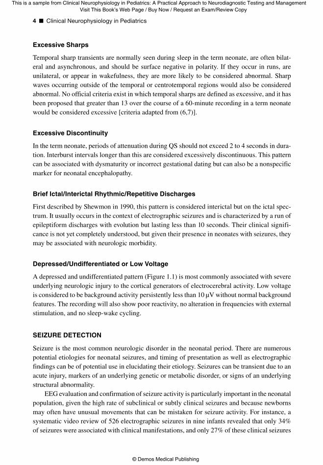
Excessive Sharps
Temporal sharp transients are normally seen during sleep in the term neonate, are often bilat-
eral and asynchronous, and should be surface negative in polarity. If they occur in runs, are
unilateral, or appear in wakefulness, they are more likely to be considered abnormal. Sharp
waves occurring outside of the temporal or centrotemporal regions would also be considered
abnormal. No official criteria exist in which temporal sharps are defined as excessive, and it has
been proposed that greater than 13 over the course of a 60-minute recording in a term neonate
would be considered excessive [criteria adapted from (6,7)].
Excessive Discontinuity
In the term neonate, periods of attenuation during QS should not exceed 2 to 4 seconds in dura-
tion. Interburst intervals longer than this are considered excessively discontinuous. This pattern
can be associated with dysmaturity or incorrect gestational dating but can also be a nonspecific
marker for neonatal encephalopathy.
Brief Ictal/Interictal Rhythmic/Repetitive Discharges
First described by Shewmon in 1990, this pattern is considered interictal but on the ictal spec-
trum. It usually occurs in the context of electrographic seizures and is characterized by a run of
epileptiform discharges with evolution but lasting less than 10 seconds. Their clinical signifi-
cance is not yet completely understood, but given their presence in neonates with seizures, they
may be associated with neurologic morbidity.
Depressed/Undifferentiated or Low Voltage
A depressed and undifferentiated pattern (Figure 1.1) is most commonly associated with severe
underlying neurologic injury to the cortical generators of electrocerebral activity. Low voltage
is considered to be background activity persistently less than 10 μV without normal background
features. The recording will also show poor reactivity, no alteration in frequencies with external
stimulation, and no sleep-wake cycling.
SEIZURE DETECTION
Seizure is the most common neurologic disorder in the neonatal period. There are numerous
potential etiologies for neonatal seizures, and timing of presentation as well as electrographic
findings can be of potential use in elucidating their etiology. Seizures can be transient due to an
acute injury, markers of an underlying genetic or metabolic disorder, or signs of an underlying
structural abnormality.
EEG evaluation and confirmation of seizure activity is particularly important in the neonatal
population, given the high rate of subclinical or subtly clinical seizures and because newborns
may often have unusual movements that can be mistaken for seizure activity. For instance, a
systematic video review of 526 electrographic seizures in nine infants revealed that only 34%
of seizures were associated with clinical manifestations, and only 27% of these clinical seizures

Visit This Book’s Web Page / Buy Now / Request an Exam/Review CopyThis is a sample from Clinical Neurophysiology in Pediatrics: A Practical Approach to Neurodiagnostic Testing and Management
© Demos Medical Publishing
1: EEG Monitoring in Neonatal Epilepsies ■ 5
(9% of overall seizures) were recognized by nursing staff. Of more concern, 73% of “seizures”
documented by the neonatal intensive care unit (NICU) nursing staff were not epileptic sei-
zures. Rather, the events marked by NICU nursing were not epileptic in nature. Instead, these
movements commonly consisted of likely nonepileptic events such as jitteriness, mouthing, and
fisting (10). Therefore neonatal seizure quantification solely by clinical observation is plagued
by both high false-positive and high false-negative rates. To ensure an accurate assessment of
seizure detection and treatment response, EEG monitoring is essential.
Subclinical Seizures
EEG confirmation of seizure cessation following anticonvulsant treatment is also recom-
mended. Neonates are particularly vulnerable to the phenomenon of electroclinical uncou-
pling, in which clinical evidence of seizure activity ceases, following the administration of
seizure medications, while subclinical electrographic seizure activity continues unabated.
Although subclinical seizures are known to occur in critically ill children and adults (11,12),
features of chloride homeostasis unique to the immature brain contribute to a high likeli-
hood of electroclinical uncoupling. The potassium-chloride cotransporter (KCC2), which
is the predominant type of chloride channel in the adult brain, transport chloride ions out-
side of neurons and have a hyperpolarizing effect. In contrast, the predominant chloride
channel in the immature brain is the sodium-potassium-chloride cotransporter (NKCC1),
FIGURE 1.1 A 38-week-old baby boy born via emergency Caesarean-section (C-section)
for polyhydramnios and nonreassuring fetal heart tracings with severe hypoxic-ischemic
encephalopathy. Background shows low voltage (<10 μV) without reactivity.

Visit This Book’s Web Page / Buy Now / Request an Exam/Review CopyThis is a sample from Clinical Neurophysiology in Pediatrics: A Practical Approach to Neurodiagnostic Testing and Management
© Demos Medical Publishing
6 ■ Clinical Neurophysiology in Pediatrics
which transports chloride ions into neurons and has a depolarizing effect. Gamma aminobu-
tyric acid (GABA), a neurotransmitter that activates chloride channels, can therefore have a
paradoxically excitatory effect in developing neurons due to the predominance of NKCC1
channel s (13). Because the transition from NKCC1 to KCC2 chloride channels occurs in a
caudal-to-rostral progression, GABA initially becomes inhibitory in subcortical structures
such as the brainstem and basal ganglia while remaining excitatory in the cortex. Commonly
used medications such as phenobarbital, which exert their effects through GABA agonist
activity, may therefore suppress brainstem motor output, while allowing electrographic sei-
zure activity to continue in the cortex.
The high risk of subclinical seizures has been well documented in the NICU populati on
(14–17). For instance, cEEG monitoring of neonates randomized to initial treatment with either
phenobarbital or phenytoin demonstrated that while 24 of 50 infants responded completely to
the first seizure medication administered, 15 of the remaining 26 neonates (58%) demonstrated
electroclinical uncoupling, with suppression of clinical seizure activity during all or the major-
ity of posttreatment electrographic seizures (18).
Neonatal Seizure Semiology
Seizure semiology in the newborn is variable but can be grouped into the following catego-
ries: clonic, tonic, and myoclonic (Table 1.2). These are focal, repetitive, and cannot be sup-
pressed by the examiner. Due to incomplete myelination, infants cannot generate generalized
tonic-clonic seizures, but they can have multifocal seizures that can appear generalized to the
untrained or inexperienced examiner. Infants can also have generalized epileptic spasms that
are hypothesized to be more subcortically driven.
Because infants often have repetitive movements which can be difficult to interpret, EEG is
often relied upon to distinguish stereotyped or rhythmic movements as epileptic or nonepilep-
tic. Oral automatisms, bicycling, roving eye movements, and other nonrhythmic but repetitive
movements are often seen in critically ill infants. Without clear electrographic correlate, these
had been previously termed clinical only seizures, but are now more commonly presumed to be
TABLE 1.2 Neonatal Seizure Types
MOVEMENT TYPE
LOCALIZATION/CLINICAL ELECTROGRAPHIC CORRELATE
Clonic Focal rhythmic jerking of an extremity
Nonsuppressible
Yes
Tonic Focal sustained extension or flexion of an extremity
Not able to overcome with external manipulation
Sustained extension of the whole body
Yes
Not usually
Myoclonic Single jerk or multiple nonrhythmic jerks of an
extremity
Usually
Spasms Focal or generalized
Flexor, extensor, or mixed flexor-extensor
Yes

Visit This Book’s Web Page / Buy Now / Request an Exam/Review CopyThis is a sample from Clinical Neurophysiology in Pediatrics: A Practical Approach to Neurodiagnostic Testing and Management
© Demos Medical Publishing
1: EEG Monitoring in Neonatal Epilepsies ■ 7
nonepileptic in nature. These movements tend to occur more often in encephalopathic infants
and are also associated with poor prognosis (19).
Role of Amplitude-Integrated EEG
The use of amplitude-integrated EEG (aiEEG) is now growing in the NICU, because it offers an
opportunity for continuous monitoring of cerebral activity in a manner that can be interpreted at
the bedside by the neonatologist rather than requiring a certified electroencephalographer. With
the growing use of therapeutic hypothermia for hypoxic-ischemic encephalopathy (HIE) in the
NICU, aiEEG has become more widely used concurrently in monitoring for seizures and change
in background activity.
aiEEG differs from conventional EEG in that it involves the use of only four electrodes and
relies on the trending of voltage and comparison between the two hemispheres. The timescale
is also broader, with the evaluation of 8 to 12 hours of data on one screen, as opposed to 20 to
30 seconds per screen of a conventional EEG.
Background activity on conventional EEG can be assessed using continuity, amplitude, and
symmetry, all of which can also be assessed on aiEEG in a different manner. Interburst interval
cannot be precisely interpreted with this method, but voltage over time is averaged in order to
give a range of activity, which can then be interpreted. This is tightly linked to amplitude, where
the peak-to-peak interval of minimum and maximum voltage ranges is represented as band-
width. If the minimum voltages are consistently less than 5 μV and maximum less than 10 μV,
this is considered a low-voltage, suppressed background. Normal activity is considered to be a
minimum voltage of greater than 5 μV and maximum voltage greater than 10 μV.
Seizures are detected on aiEEG as a relative increase in overall amplitude over a given
period of time. These can be detected by relative increases of the peak-to-peak amplitude with
narrow bandwidth. Some indication of localization can be inferred if this occurs only in one
hemisphere. Overall seizure burden can also be inferred, based on the number of peaks of
increased voltage peaks. (20)
However, a limitation of condensing this data and relying on voltage alone is that aiEEG
can be ripe with artifact. When the baby is handled and high-amplitude electrode artifact is
generated, this will appear as an amplitude spike on aiEEG. Similarly, when continuous exter-
nal artifacts such as EKG rhythm occur in the setting of a low-voltage, suppressed background,
this can be misrepresented as a normal voltage range on aiEEG.
aiEEG has been shown in studies to be sensitive, but not very specific for the identification
of an abnormal background and seizures (21). Regardless, given the ease of use, the wide-
spread availability, and the ability for bedside interpretation, aiEEG has now become part of
the standard of care during therapeutic hypothermia for HIE of the newborn (22–24). Studies
have shown that the use of aiEEG may even be beneficial in that neonates are being treated for
seizures only with electrographic confirmation, rather than purely on a clinical basis (25).
TRANSIENT OR “BENIGN” NEONATAL SEIZURES
Hypoxic-Ischemic Encephalopathy
HIE is the leading cause of seizures in the neonatal period, with an incidence of 2 to 5 per
1,000 live births. Seizures have been found in up to 80% of this population, but this may be an

Visit This Book’s Web Page / Buy Now / Request an Exam/Review CopyThis is a sample from Clinical Neurophysiology in Pediatrics: A Practical Approach to Neurodiagnostic Testing and Management
© Demos Medical Publishing
8 ■ Clinical Neurophysiology in Pediatrics
underestimation, given that continuous EEG monitoring is not routinely used. aiEEG is often
used in the NICU to fulfill the need for continuous electrographic monitoring.
Therapeutic hypothermia has also become the standard of care in the treatment of infants
with HIE and has been shown to improve neurodevelopmental background. Evaluation of back-
ground activity can be useful for prognostication in infants with HIE. Persistently abnormal
background activity without evidence of improvement over time is more likely to be associated
with a worse neurodevelopmental outcome. A normal background or improvement in back-
ground is less likely to be associated with poor neurodevelopmental outcome.
Recent studies have shown a high incidence of seizures in infants undergoing therapeutic
hypothermia for HIE, up to 40% to 60%, with 35% to 75% of these being subclinical (26,27) (Fig-
ure 1.2). The burden of seizures is highest in the first 24 to 48 hours, with a natural decline after
72 hours (28). It is presumed that a higher burden of seizures is associated with worse neurodevel-
opmental outcome; however, this is a topic of much debate, as infants with more severe HIE are
also likely to have more refractory seizures. Additionally despite advances in antiepileptic drug
development, relatively few advances have been made in the treatment of seizures due to HIE, and
many treatments also have potential unwanted side effects in the developing brain (29,30).
Benign Familial Neonatal Convulsions
Benign familial neonatal convulsions are often seen around the fifth day of life, giving them
the frequently used descriptive term of “fifth day fits.” Most are associated with a mutation in
the KCNQ2 gene coding for a voltage-gated potassium channel, which has autosomal trans-
mission, but other potassium channels as well as the sodium channel, such as SCN2A muta-
tion, have also been implicated (31). There is often a family history of neonatal seizures, and
the electrographic background is frequently normal but can show excessive discontinuity and
excessive sharp transients. These were initially termed benign because there was thought to be
no long-term consequence, although recent studies have shown that this is not always the case.
KCNQ2 mutations have also been associated with Ohtahara syndrome, and the phenotype can
be variable, with seizures persisting well beyond the neonatal period (32,33).
Stroke
Perinatal stroke is also a common cause of neurologic morbidity in the newborn period. The
majority are arterial ischemic, although at least 30% can be venous in nature (34). Seizures
are a common presentation of neonatal arterial ischemic stroke; up to 72% present with sei-
zures (35). In a neonate with persistently unilateral seizures, arterial ischemic stroke should be
strongly considered as an etiology and neuroimaging should be undertaken.
Hypoglycemia and Other Reversible Causes
Neonatal hypoglycemia is a frequent complication of infants of mothers with gestational diabetes,
but can also be seen in well neonates with poor feeding. The occipital lobes are particularly at
risk because of the high metabolic demand of the visual cortex. Persistent focal seizures can be
seen emanating from either posterior quadrant. Imaging can show diffusion restriction in the areas
affected, partly due to frequent seizures and increased local metabolism and partly due to watershed
ischemia. These areas can later undergo laminar necrosis and develop the appearance of ulegyria.

Visit This Book’s Web Page / Buy Now / Request an Exam/Review CopyThis is a sample from Clinical Neurophysiology in Pediatrics: A Practical Approach to Neurodiagnostic Testing and Management
© Demos Medical Publishing
1: EEG Monitoring in Neonatal Epilepsies ■ 9
FIGURE 1.2 A 41+1-week-old baby boy with hypoxic-ischemic encephalopathy and
meconium aspiration syndrome on selective hypothermia therapy, with seizures starting on
the first day of life. This recording shows a seizure starting at T4.

Visit This Book’s Web Page / Buy Now / Request an Exam/Review CopyThis is a sample from Clinical Neurophysiology in Pediatrics: A Practical Approach to Neurodiagnostic Testing and Management
© Demos Medical Publishing
10 ■ Clinical Neurophysiology in Pediatrics
Other electrolyte and metabolic disturbances can also precipitate seizures in the neonatal
period, similar to adults. Hypomagnesemia, hypocalcemia, hyponatremia, and hyperbilirubi-
nemia can also lead to neonatal seizures. In these instances, correction of the underlying etiol-
ogy is necessary to effectively treat the seizures (34).
CATASTROPHIC EPILEPTIC ENCEPHALOPATHIES
There are several conditions presenting in the neonatal period which have been termed “cata-
strophic,” in that they are associated with frequent seizures and severe interictal background
abnormalities which, without prompt remedy, almost inevitably result in poor neurodevelop-
mental outcome (Table 1.3).
Ohtahara Syndrome
Ohtahara syndrome, also known as early infantile epileptic encephalopathy with suppression-
burst presents in early infancy. Initial symptoms are seen within the first 3 months, frequently
within the first 2 weeks. Clinically this presents with brief (less than 10 seconds) tonic spasms
(generalized or focal), which occur independently or in clusters. Other seizure types including
focal seizures, hemiconvulsions, or tonic-clonic seizures are seen in approximately 33%. Most
cases are related to a variety of structural brain lesions, although metabolic and genetic disor-
ders have been reported. Mutations associated include syntaxin binding protein 1 (STXBP1),
Aristaless-related homeobox (ARX), sodium channel SCN2A, and KCNQ 2 (36–39).
The typical EEG pattern is a consistent (wake and sleep) “suppression-burst” pattern with
periods of diffuse amplitude suppression alternating with bursts of high amplitude spike and
polyspike discharges.
Diagnosis of Ohtahara syndrome is based on the clinical picture and EEG findings. The
prognosis is poor, with many affected children dying in infancy. Survivors have developmental
impairment and many have chronic seizures or evolve into Lennox-Gastaut or West syndrome.
Anti-seizure medications are used; however, there is no specific evidence-based therapy known.
Surgery has been performed for cases with clear focal lesions (40).
TABLE 1.3 Neonatal Epilepsy Syndromes
EPILEPSY SYNDROME INTERICTAL EEG BACKGROUND SEIZURE TYPES
Ohtahara syndrome Burst-suppression (wake and
sleep)
Tonic spasms
Focal
Tonic-clonic
Early myoclonic epilepsy of
infancy
Burst-suppression (more prominent
in sleep)
Multifocal myoclonic
Malignant migrating partial
seizures of infancy
Multifocal sharps Focal, arising from
multiple regions
Pyridoxine-dependent
epilepsy
Continuous spike-wave
Burst suppression
Infantile spasms
Multifocal myoclonic
Focal
Tonic

Visit This Book’s Web Page / Buy Now / Request an Exam/Review CopyThis is a sample from Clinical Neurophysiology in Pediatrics: A Practical Approach to Neurodiagnostic Testing and Management
© Demos Medical Publishing
1: EEG Monitoring in Neonatal Epilepsies ■ 11
Early Myoclonic Epilepsy of Infancy
Early myoclonic epilepsy of infancy (EMEI) was described shortly after Ohtahara syndrome
and there are a number of similarities between them. EMEI also begins within the first 3
years, although it can present as early as a few hours after birth. Clinically this begins with
focal myoclonus that can shift between different body parts often in an asynchronous and
random pattern. A wide range of focal seizures (anything from tonic posturing to autonomic
signs) is very common and tonic spasms are also seen. There is a range of underlying disorders
associated with EMEI including structural lesions and metabolic and genetic abnormalities. In
contrast to Ohtahara syndrome, diffuse cortical atrophy, rather than focal structural lesions, is
typically seen. A variety of metabolic abnormalities have been associated, in particular, non-
ketotic hyperglycine mia (41). Mutation of the v-erb-a erythroblastic leukemia viral oncogene
homologue 4 (ErbB4), which is associated with cortical migration, is also rel ated (42).
The typical EEG pattern of EMEI is similar to the suppression-burst pattern seen in
Ohtahara syndrome; however, in EMEI the suppression-burst pattern is not continuous and
occurs more prominently (or exclusively) in sleep. The myoclonic seizures are not generally
associated with changes on the EEG.
EMEI is also diagnosed clinically and treated with antiseizure medications. Additionally
treatment of the underlying metabolic disorder may be helpful. The prognosis of EMEI is also
very poor, with 50% of patients dying by 3 years and the survivors having severe developmental
impairment (40).
Malignant Migrating Partial Seizures of Infancy
Malignant migrating partial seizures of infancy (MMPSIs) present in the first 6 months of life
with multifocal, bilateral, independent seizures. Seizures are very difficult to control and are
associated with progressive developmental impairment and a decrease in the head circumfer-
ence. The underlying etiology is unknown; however, it is likely genetic. Mutations have been
found in a number of genes including SCN1A, phospholipase C beta 1 (PLCB1), KCNT1, and
TBC1D24.
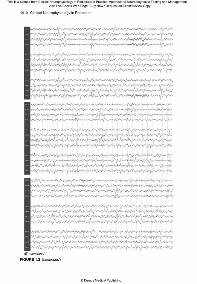
The ictal EEG shows focal seizures initiating from different locations in both hemispheres
that “migrate” from one area to another (Figure 1.3).
MMPSI is diagnosed by clinical presentation along with the typical EEG pattern and has
a poor prognosis. Status epilepticus is common and may be related to patients dying in the first
2 years of life (43).
OTHER EPILEPSY SYNDROMES PRESENTING IN NEONATES
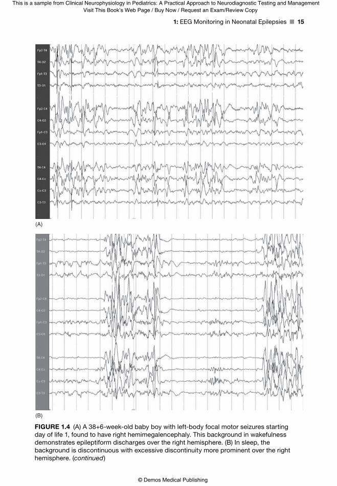
Hemimegalencephaly (HME) is a severe developmental brain anomaly characterized by the
overgrowth of one hemisphere. This is associated with epilepsy, psychomotor retardation, and
contralateral motor defect. Seizure types include focal motor seizures, asymmetric tonic or
clonic seizures, and epileptic spasms. HME is one of the causes of Ohtahara syndrome (Fig-
ure 1.4) and West syndrome and is associated with a variety of genetic abnormalities and neu-
rocutaneous syndromes; however; it may be an isolated syndrome.

Visit This Book’s Web Page / Buy Now / Request an Exam/Review CopyThis is a sample from Clinical Neurophysiology in Pediatrics: A Practical Approach to Neurodiagnostic Testing and Management
© Demos Medical Publishing
12 ■ Clinical Neurophysiology in Pediatrics
(A)
FIGURE 1.3 (A) A 40+1-week-old baby boy with seizures starting on the first day of life,
consisting of clonic movements of any extremity. This shows a seizure over the right
posterior quadrant. (continued)
Patients with West syndrome associated with HME may have a unique EEG background
called hemihypsarrhythmia (high amplitude, poorly organized with multifocal spikes over the
affected side only) (Figure 1.4).
(text continues on page 16)

Visit This Book’s Web Page / Buy Now / Request an Exam/Review CopyThis is a sample from Clinical Neurophysiology in Pediatrics: A Practical Approach to Neurodiagnostic Testing and Management
© Demos Medical Publishing
1: EEG Monitoring in Neonatal Epilepsies ■ 13
FIGURE 1.3 (continued) (B) Seizures arose from all electrodes, often with a new seizure emerging
amidst the existing seizure at a noncontiguous electrode. This demonstrates seizures occurring
independently at C3 and C4, as evidenced by nonsynchronous frequencies. (continued)
(B)
Visit This Book’s Web Page / Buy Now / Request an Exam/Review CopyThis is a sample from Clinical Neurophysiology in Pediatrics: A Practical Approach to Neurodiagnostic Testing and Management
© Demos Medical Publishing
14 ■ Clinical Neurophysiology in Pediatrics
FIGURE 1.3 (continued )
(B) (continued )
Visit This Book’s Web Page / Buy Now / Request an Exam/Review CopyThis is a sample from Clinical Neurophysiology in Pediatrics: A Practical Approach to Neurodiagnostic Testing and Management
© Demos Medical Publishing
1: EEG Monitoring in Neonatal Epilepsies ■ 15
FIGURE 1.4 (A) A 38+6-week-old baby boy with left-body focal motor seizures starting
day of life 1, found to have right hemimegalencephaly. This background in wakefulness
demonstrates epileptiform discharges over the right hemisphere. (B) In sleep, the
background is discontinuous with excessive discontinuity more prominent over the right
hemisphere. (continued)
(A)
(B)

Visit This Book’s Web Page / Buy Now / Request an Exam/Review CopyThis is a sample from Clinical Neurophysiology in Pediatrics: A Practical Approach to Neurodiagnostic Testing and Management
© Demos Medical Publishing
16 ■ Clinical Neurophysiology in Pediatrics
Diagnosis of HME is based on imaging including asymmetry of the hemispheres and ven-
tricles, loss of gray-white differentiation, neuronal heterotopia, thick cortex, and abnormalities
in the gyri, basal ganglia, and internal capsule. The clinical course and prognosis is dependent
on seizure control, the severity of the affected side, the ability of the contralateral side to com-
pensate, and early surgery (44).
FIGURE 1.4 (continued) (C) The patient had focal left-body tonic seizures, as demonstrated
here with a buildup of sharply contoured alpha over the occipital region, which then spreads
anteriorly and builds in amplitude. (continued)
(C)

Visit This Book’s Web Page / Buy Now / Request an Exam/Review CopyThis is a sample from Clinical Neurophysiology in Pediatrics: A Practical Approach to Neurodiagnostic Testing and Management
© Demos Medical Publishing
1: EEG Monitoring in Neonatal Epilepsies ■ 17
FIGURE 1.4 (continued)
METABOLIC EPILEPSIES
Pyridoxine-dependent epilepsy was first described by Hunt and colleagues in 1954. This syn-
drome is unique in that it is severe but treatable, and thus early recognition is of tantamount
importance. This syndrome has an estimated birth incidence between 1:400,000 and 1:750,000.
Seizures can be prenatal in onset and can include multiple seizure types, including infantile
spasms and focal, multifocal myoclonic, and tonic seizures. There can also be an associated
encephalopathy, which may manifest as tremulousness, irritability, or hypothermia. The base-
line EEG will show a continuous spike-wave or burst-suppression pattern. Diagnosis is estab-
lished by giving an intravenous dose of 100-mg pyridoxine during EEG monitoring, which
will often lead to the resolution of epileptiform activity and improvement of the background
(Figure 1.5). The response is often seen rapidly, although delayed responses have also been
reported. Relapses can occur after a median of 9 days if pyridoxine therapy is withheld, and
therefore patients need to remain on lifelong therapy (45).
Folinic acid–responsive seizures are another treatable cause of neonatal seizures. EEG
background features and seizure types can be similar to pyridoxine-dependent seizures, and
concurrent pyridoxine dependency can occur within individuals. Seizures respond to 2.5- to
5-mg folinic acid given twice daily, and daily doses should be added for patients with an incom-
plete response to pyridoxine treatment.
(C) (continued )

Visit This Book’s Web Page / Buy Now / Request an Exam/Review CopyThis is a sample from Clinical Neurophysiology in Pediatrics: A Practical Approach to Neurodiagnostic Testing and Management
© Demos Medical Publishing
18 ■ Clinical Neurophysiology in Pediatrics
(A)
(B)
FIGURE 1.5 (A) A 40+5–week-old baby boy who presented with “jitteriness” and
episodes of flexor spasms 3 hours after birth. Initial background was discontinuous and
asynchronous. (B) After pyridoxine administration, the background normalized, becoming
synchronous and continuous.
REFERENCES
1. American Clinical Neurophysiology Society. Guideline two: minimum technical standards for pedi-
atric electroencephalography; 2006. http://www.acns.org/pdf/guidelines/Guideline-2.pdf. Accessed
March 12, 2015.
2. Ellingson RJ, Dutch SJ, McIntire MS. EEG’s of prematures: 3–8 year follow-up study. Dev Psychobiol. 1974;7(6):529–538.
3. Clancy R, Berggvist AC, Dlugos D. Neonatal encephalography. In: Ebersole JS, Pedley T, eds.
Current Practice of Clinical Electroencephalography. 3rd ed. Philadelphia, PA: Lippincott Williams
& Wilkins; 2003:160–234.

Visit This Book’s Web Page / Buy Now / Request an Exam/Review CopyThis is a sample from Clinical Neurophysiology in Pediatrics: A Practical Approach to Neurodiagnostic Testing and Management
© Demos Medical Publishing
1: EEG Monitoring in Neonatal Epilepsies ■ 19
4. Vecchierini MF, Andre M, d’Allest AM. Normal EEG of premature infants born between 24 and
30 weeks gestational age: terminology, definitions and maturation aspects. Neurophysiol Clin.
2007;37:311–323.
5. Vecchierini MF, d’Allest AM, Verpillat P. EEG patterns in 10 extreme premature neonates with nor-
mal neurological outcome: qualitative and quantitative data. Brain Dev. 2003;25:330–337.
6. Tsuchida TN, Hahn CD, Riviello JJ, et al. ACNS standardized EEG terminology and catego-
rization for the description of continuous EEG monitoring in neonates: report of the American
Clinical Neurophysiology Society Critical Care Monitoring Committee. J Clin Neurophysiol. 2013;30(2):161–173.
7. Mizrahi EM, Hrachovy RA, Kellaway P, et al. Patterns of uncertain diagnostic significance. In: Atlas of Neonatal Electroencephalography. 3rd ed. Philadelphia, PA: Lippincott Williams & Wilkins;
2004:93–115.
8. Hahn JS, Monyer H, Tharp BR. Interburst interval measurements in the EEGs of premature infants
with normal neurological outcome. Electroencephalogr Clin Neurophysiol. 1989;73:410–418.
9. Selton D, Andre M, Hascoët JM. Normal EEG in very premature infants: reference criteria. Clin Neurophysiol. 2000;111:2116–2124.
10. Murray DM, Boylan GB, Fitzgerald AP, et al. Persistent lactic acidosis in neonatal hypoxic-isch-
aemic encephalopathy correlates with EEG grade and electrographic seizure burden. Arch Dis Child Fetal Neonatal Ed. 2008;93:183–186.
11. Abend NS, Arndt DH, Carpenter JL, et al. Electrographic seizures in pediatric ICU patients: cohort
study of risk factors and mortality. Neurology. 2013;81(4):383–391.
12. Glass HC, Wusthoff CJ, Shellhaas RA, et al. Risk factors for EEG seizures in neonates treated with
hypothermia: a multicenter cohort study. Neurology. 2014;82(14):1239–1244.
13. Staley K, Smith R. A new form of feedback at the GABAA
receptor. Nat Neurosci. 2001;4(7):674–676.
14. Nash KB, Bonifacio SL, Glass HC, et al. Video-EEG monitoring in newborns with hypoxic-isch-
emic encephalopathy treated with hyporthermia. Neurology. 2011;76:556–562.
15. Lynch NE, Stevenson NJ, Livingstone V, et al. The temporal evolution of electrographic seizure
burden in neonatal hypoxic ischemic encephalopathy. Epilepsia. 2012;53(3):549–557.
16. Glass HC, Glidden D, Jeremy RJ, et al. Clinical neonatal seizures are independently associated with
outcome in infants at risk for hypoxic-ischemic brain injury. J Pediatr. 2009;155(3):318–323.
17. Yap V, Engel M, Takenouchi T, Perlman JM. Seizures are common in term infants undergoing head
cooling. Pediatr Neurol. 2009;41(5):327–331.
18. Scher MS, Alvin J, Gaus L, et al. Uncoupling of EEG-clinical neonatal seizures after antiepileptic
drug use. Pediatr Neurol. 2003;28(4):277–280.
19. Mizrahi EM, Kellaway P. Characterization and classification of neonatal seizures. Neurology.
1987;37(12):1837–1844.
20. El-Dib M, Chang T, Tsuchida TN, Clancy RR. Amplitude-Integrated Electroencephalography in
Neonates. Pediatr Neurol. 2009;41(5):315–326.
21. Evans E, Koh S, Lerner JT, et al. Accuracy of amplitude integrated EEG in a neonatal cohort. Arch Dis Child Fetal Neonatal Ed. 2010;95(3):F169–F173.
22. Shankaran S, Pappas A, McDonald SA, et al. Predictive value of an early amplitude integrated elec-
troencephalogram and neurologic examination. Pediatrics, 2011;128:e112–e120.
23. Sarkar S, Barks JD, Bhagat I, Donn SM. Effects of therapeutic hypothermia on multiorgan dys-
function in asphyxiated newborns: whole-body cooling versus selective head cooling. J Perinatol. 2009;29:558–563.
24. Shah PS. Hypothermia: a systematic review and meta-analysis of clinical trials. Semin Fetal Neonatal Med. 2010;15(5):238–246.
25. Shellhaas RA, Barks AK. Impact of amplitude-integrated electroencephalograms on clinical care
for neonates with seizures. Pediatr Neurol. 2012;46:32–35.
26. Glass HC, Kan J, Bonifacio SL, Ferriero DM. Neonatal seizures: treatment practices among term
and preterm infants. Pediatr Neurol. 2012;46:111–115.
27. Abend NS, Gutierrez-Colina AM, Monk HM, et al. Levetiracetam for treatment of neonatal sei-
zures. J Child Neurol. 2011;26(4):465–470.
28. Lynch NE, Stevenson NJ, Livingstone V, et al. The temporal evolution of electrographic seizure
burden in neonatal hypoxic ischemic encephalopathy. Epilepsia. 2012;53(3):549–557.

Visit This Book’s Web Page / Buy Now / Request an Exam/Review CopyThis is a sample from Clinical Neurophysiology in Pediatrics: A Practical Approach to Neurodiagnostic Testing and Management
© Demos Medical Publishing
20 ■ Clinical Neurophysiology in Pediatrics
29. Bittigau P, Sifringer M, Genz K, et al. Antiepileptic drugs and apoptotic neurodegeneration in the
developing brain. Proc Natl Acad Sci USA. 2002;99(23):15089–15094.
30. Painter MJ, Scher MS, Stein AD, et al. Phenobarbital compared with phenytoin for the treatment of
neonatal seizures. N Engl J Med. 1999;341(7):485–489.
31. Zara F, Specchio N, Striano P, et al. Genetic testing in benign familial epilepsies of the first year of
life: clinical and diagnostic significance. Epilepsia. 2013;54(3):425–436.
32. Weckhuysen S, Mandelstam S, Suls A, et al. KCNQ2 encephalopathy: emerging phenotype of a
neonatal epileptic encephalopathy. Ann Neurol. 2012;71:15–25.
33. Kato M, Yamagata T, Kubota M, et al. Clinical spectrum of early onset epileptic encephalopathies
cause by KCNQ2 mutation. Epilepsia, 2013;54(7):1282–1287.
34. Ferriero DM. Neonatal brain injury. N Engl J Med. 2004;351:1985–1995.
35. Kirton A, Armstrong-Wells J, Chang T, et al. Symptomatic neonatal arterial ischemic stroke: the
international pediatric stroke study. Pediatrics. 2011;128(6):e1402–e1410.
36. Fullston T, Brueton L, Willis T, et al. Ohtahara syndrome in a family with an ARX protein trunca-
tion mutation. Eur J Hum Genet. 2010;18(2):157–162.
37. Nakamura K, Kato M, Osaka H, et al. Clinical spectrum of SCN2A mutations expanding to Ohtahara
syndrome. Neurology. 2013;81(11):992–998.
38. Saitsu H, Kato M, Shimono M, et al. Association of genomic deletions in the STXBP1 gene with
Ohtahara syndrome. Clin Genet. 2012;81(4):399–402.
39. Saitsu H, Kato M, Koide A, et al. Whole exome sequencing identifies KCNQ2 mutations in Ohtahara
syndrome. Ann Neurol. 2012;72(2):298–300.
40. Beal JC, Cherian K, Moshe SL. Early-onset epileptic encephalopathies: Ohtahara syndrome and
early myoclonic encephalopathy. Pediatr Neurol. 2012;47:317–323.
41. Wang PJ, Lee WT, Hwu WL, et al. The controversy regarding diagnostic criteria for early myoclonic
encephalopathy. Brain Dev. 1998;20(7):530–535.
42. Backx L, Ceulemans B, Vermeesch JR, et al. Early myoclonic encephalopathy caused by a disrup-
tion of the neuregulin-1 receptor ErbB4. Eur J Hum Genet. 2009;17(3):378–382.
43. DeFilippo MR, Rizzo F, Marchese G, et al. Lack of pathogenic mutations in six patients with
MMPSI. Epilepsy Res. 2014;108:340–344.
44. Honda R, Kaido T, Sugai K, et al. Long-term developmental outcome after early hemispherectomy
for hemimegalencephaly in infants with epileptic encephalopathy. Epilepsy Behav. 2013;29:30–35.
45. Pearl PL. New treatment paradigms in neonatal metabolic epilepsies. J Inherit Metab Dis. 2009;32:204–213.