Embed Size (px)
Citation preview

Clinical Guideline: Suction Authors: East of England benchmarking group members
For use in: EoE Neonatal Units
Guidance specific to the care of neonatal patients. Used by: Healthcare professionals giving direct care to neonatal patients.
Key Words: suction, endotracheal, nasopharyngeal, oropharyngeal, tracheostomy, HFOV, surfactant
Review due: July 2023
Registration No: NEO-ODN-2020-4
Approved by:
Neonatal Clinical Oversight
Group
1st July 2020
Clinical Lead Matthew James
Matthew James
Ratified by Clinical Oversight Group:
Date of meeting 1st July 2020

1. Scope For use in neonatal units in the East of England Neonatal ODN
2. Aim Airway suction is necessary to remove secretions and prevent obstruction1. Serious complications may result from the procedure. We aim to achieve a balance by helping without harming2.
3. Indications for endotracheal suctioning3,4,5
Coarse breath sounds on chest auscultation
Lack of breath sounds on chest auscultation Reduced chest movement
Increased work of breathing Apnoea
Bradycardia
Increasing end tidal CO2 Audible or visible secretions Increasing airway pressures
Decreasing minute or tidal volume Deteriorating oxygen saturation levels or blood gases Removal of thick meconium particulate under direct vision
43
The need for suction should be based on assessment of the infant's clinical status35. To help with this clinical decision making, the oxygen saturation, inspired oxygen concentration trend, blood gases, chest movement and tidal volume (Vt) can be assessed, as well as taking into consideration the amount and type of secretions removed during suction performed in the past 12 hours.
3.1 Complications of suctioning
Atelectasis38
Hypoxia6,7,8
Bradycardia9
Blood pressure changes10
Airway trauma eg necrotising tracheobronchitis11
Pneumothorax (after closed circuit suction)5,12,13
Raised intracranial pressure14,15,16
Nosocomial infection4
Discomfort17,18 Tube dislodgement Risk of CoNS colonisation
43
3.1.2 Competency Oro/nasopharyngeal suction should only be performed by registered nurses, nursery nurses & assistant practitioners who have been deemed competent to do so, following
local competencies. Staff yet to attain competence should be under the direct supervision of a nurse already competent in suction Endotracheal (ET) suction must only
be performed by a QIS (Qualified in Speciality) neonatal nurse, or by a non-QIS

registered nurse undergoing QIS training under the direct supervision of a QIS neonatal nurse.
3.2 Precautions
3.2.1 Saline Lavage Occasional use of saline when secretions are thick is acceptable even though there is
no proven benefit45. Saline lavage may nonetheless be necessary when secretions are very thick or when pulmonary haemorrhage has occluded the ET tube, but should only
be performed under medical instruction20,21.
0.25ml (<2kg baby) 0.5ml (>2kg baby) should be sufficient to loosen secretions6,19,34. Always use a plastic saline ampoule.
3.2.2 Pre-oxygenation Although there appears to be evidence suggesting that pre-oxygenation can significantly reduce hypoxemia related to ET suction, this procedure has in fact been found to have long-term adverse effects on the infant. These include retinopathy of prematurity, parenchymal lung damage and oxygen free radical damage and therefore
should not be common practice22,36.
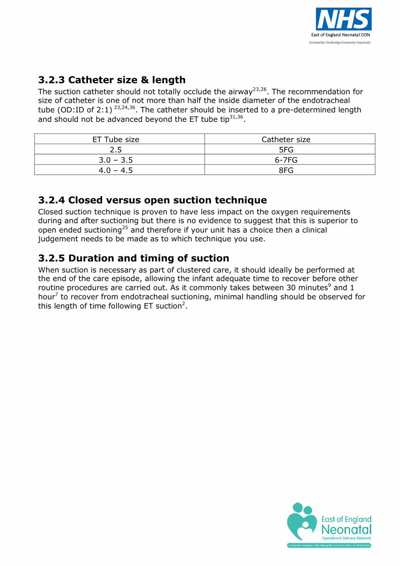
3.2.3 Catheter size & length The suction catheter should not totally occlude the airway23,26. The recommendation for size of catheter is one of not more than half the inside diameter of the endotracheal
tube (OD:ID of 2:1) 23,24,36. The catheter should be inserted to a pre-determined length
and should not be advanced beyond the ET tube tip31,36.
ET Tube size Catheter size
2.5 5FG
3.0 – 3.5 6-7FG
4.0 – 4.5 8FG
3.2.4 Closed versus open suction technique Closed suction technique is proven to have less impact on the oxygen requirements during and after suctioning but there is no evidence to suggest that this is superior to
open ended suctioning25 and therefore if your unit has a choice then a clinical judgement needs to be made as to which technique you use.
3.2.5 Duration and timing of suction When suction is necessary as part of clustered care, it should ideally be performed at the end of the care episode, allowing the infant adequate time to recover before other
routine procedures are carried out. As it commonly takes between 30 minutes9 and 1
hour7 to recover from endotracheal suctioning, minimal handling should be observed for
this length of time following ET suction2.

3.2.6 ANTT Aseptic Non-Touch Technique should be used for all suction procedures42
As well as PPE.
3.2.7 Surfactant administration via ETT & suction If pulmonary secretions are prominent prior to surfactant administration, suctioning prior to dosing may lessen the probability of mucus plugs obstructing the endotracheal tube following surfactant administration. If possible, suction should be avoided for at least one hour after surfactant administration, with the exception of life-threatening
circumstances.37
3.2.8 Assessment of need For all types of suction, an assessment of need should be undertaken: Have the physiological parameters changed?
Is the chest moving?
Has the tidal volume or minute volume decreased? What was the result of the most recent blood gas? Auscultate the chest – are the breath sounds noisy, is air entry equal? Has the oxygen requirement increased?
What were the secretions like on the last suction event? When was the last suction performed? Has the infant recently been handled?
If the parents are present, explain the reason for suctioning and the procedure.
4. Closed Suction of Endotracheal Tubes
4.1 Setting up the closed suction 1. Select the correct size of catheter, if the catheter is too large for the tube it will
occlude the airway and lead to hypoxia. As a rough guide, the French size of the
catheter should be approximately twice the diameter of the ETT. Eg A 6Fr catheter is correct for a size 2.5, 3.0 or 3.5mm ETT.
2. Select the correct size Y-adapter for the ETT. The Y-adapter stays on the ETT and
replaces ETT connector e.g. the blue connection on a Portex tube. 3. Connect the catheter to the Y-adapter.
4. Take out the ETT connector and attach the larger port to the ventilator tubing.
5. Attach the daily change sticker across the bottom of the thumb control valve, and
then lock the thumb valve by rotating the white button on the suction valve. The
catheter should be changed every 24 hours.
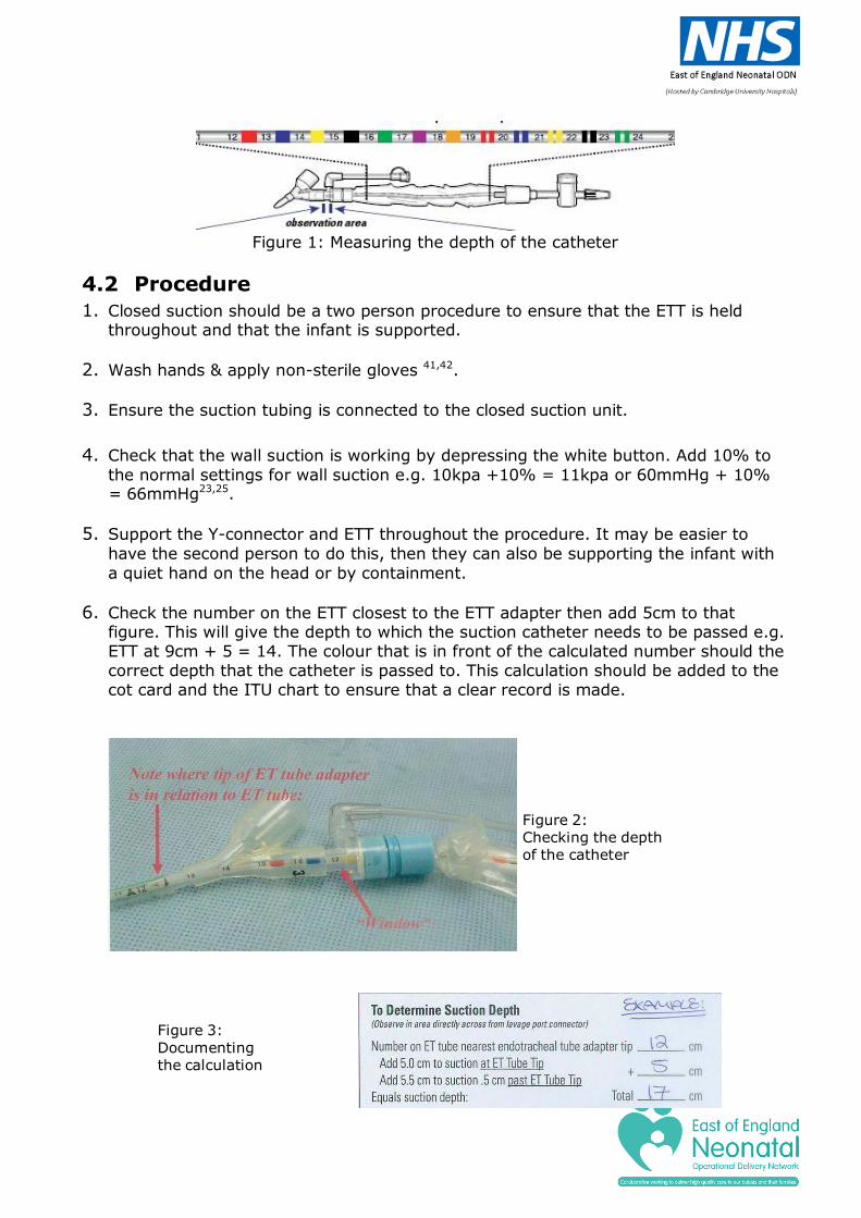
Figure 1: Measuring the depth of the catheter
4.2 Procedure
1. Closed suction should be a two person procedure to ensure that the ETT is held throughout and that the infant is supported.
2. Wash hands & apply non-sterile gloves 41,42.
3. Ensure the suction tubing is connected to the closed suction unit.
4. Check that the wall suction is working by depressing the white button. Add 10% to
the normal settings for wall suction e.g. 10kpa +10% = 11kpa or 60mmHg + 10% = 66mmHg23,25.
5. Support the Y-connector and ETT throughout the procedure. It may be easier to
have the second person to do this, then they can also be supporting the infant with
a quiet hand on the head or by containment.
6. Check the number on the ETT closest to the ETT adapter then add 5cm to that figure. This will give the depth to which the suction catheter needs to be passed e.g. ETT at 9cm + 5 = 14. The colour that is in front of the calculated number should the correct depth that the catheter is passed to. This calculation should be added to the cot card and the ITU chart to ensure that a clear record is made.
Figure 3:
Documenting the calculation
Figure 2: Checking the depth of the catheter

7. Push the catheter forward and advance it into the airway. Do not let go of the catheter but lighten the grip to allow the plastic sleeve to slide back over the catheter. The catheter is gripped by PEEP seal allowing the catheter to advance whilst the sleeve remains out of the way.
8. Slowly advance the catheter until you see the coloured strip in the cleaning port.
This position will ensure that the tip of the catheter is near the end of the ETT.
9. Apply suction. Wait for 2 seconds, if the infant tolerates it, and then withdraw
the catheter slowly back. Because the infant remains ventilated throughout the procedure the actual suction can take up to 8 seconds. If the infant does not tolerate then the procedure should be stopped and the suction catherter withdrawn.
10. Withdraw the catheter until the black tip is within the cleaning chamber. It is
also important to fully withdraw the catheter, as leaving it inside the ETT may restrict ventilation. In addition it poses the risk of inadvertently cutting the suction catheter when cutting the ETT, which could result in the cut end of the catheter
lodging in the patient’s airway. (As per Patient Safety Alert, July 2014)39
11. With the black tip inside the cleaning chamber apply suction, the ETT in a
position parallel to the cot and at the same time instil 0.5-1.0ml saline via the administration port. Secretions should be seen in the clear tubing in front of the suction valve. Use as little saline as is needed to clear the catheter, repeat if necessary until all secretions are removed from the catheter. This MUST be performed after every suction event.
12. Replace the cap over the administration port and and turn off the wall suction
when finished.
13. If saline lavage is indicated (see 3.2.1), advance the catheter tip to the infant’s lips with the ETT tilting downwards, instil the saline and suction as above.
5. Endotracheal Suction (Open)
5.1 Equipment
Stethoscope Gloves either clean or sterile.40,41,42
Appropriate sized suction catheters
Suction bottle connected to suction apparatus – pressures set 60-80mmHg (8- 10kpa)5,23 and suction connecting tube
Pre-cut tape measure for suction catheter measurement
Saline, syringe and filter needle if required Rubbish bag
Sterile water (to rinse suction tubes after the procedure) Documentation
5.2 Preparation 1. Wash hands
2. Prepare equipment
3. Using a filter needle draw up saline into syringe if required (see 3.2.1), taking
care not to touch key parts, and replace syringe back into paper packet
4. Attach suction catheter to suction tubing, leaving catheter in the protective
packaging to prevent contamination
5. Check the vacuum pressure ensuring that the maximum negative pressure does not
exceed 60-80mmHg (8-10kpa)5
6. Determine length of endotracheal tube (including any dead space and length of the
blue ETT hub). This is the length you will need to pass the suction catheter to ensure
that the catheter reaches the end of the ETT tip27,28,29. This can be achieved by observing the markings of the suction catheter or by measuring the catheter against a pre-cut tape measure which may be stuck to the inside of the incubator. The catherter should remain within its packaging to ensure sterility is maintand.
Emergency suction
For emergency suction of the ETT, step 7 of the preparation procedure may not be achievable. It may be necessary to increase the inspired oxygen
concentration in response to desaturation, but in some cases (such as blocked ETT) waiting until the oxygen saturation is >90% is not practical. Suction in this
situation should be performed to prevent further desaturation or bradycardia.
7. Auscultate the chest prior to suctioning to have baseline information on which to compare post suctioning auscultation
8. Position the head in the mid-line if possible to reduce changes to cerebral blood
flow. 9. Where possible organize another person/parent to help with the procedure to allow
the infant to be contained during the suctioning as it has been shown to aid
recovery.
5.3 Procedure
1. Wash hands and apply gloves42. 2. If preferred, put two gloves onto the dominant hand. Double gloving means that
the top glove can be removed to contain the catheter after the suctioning is

complete. 3. If only using I pair of gloves then ensure that the suction catheter is placed
directly into your rubbish bag after use
4. Withdraw suction catheter from protective packaging and hold in the sterile double gloved hand.
5. Silence ventilator alarm with other hand.
6. Detach ETT from ventilation tubing with non-dominant hand
7. Steady the ET tube with the non-dominant hand and insert the suction catheter
down the ETT to the pre-determined length with the gloved hand. The catheter
should not advance beyond the end of the ETT11,31, as doing so can cause damage to the carina.
8. Apply suction
9. Withdraw catheter, whilst applying suction. It should take no longer than 4 - 5
seconds to completely withdraw catheter. 10. Re-attach ventilation tubing to the ETT.
11. Assess tolerance of the procedure by observing oxygen saturation, colour, heart
rate and activity.
12. Adjust the FiO2 to stabilise the oxygen saturations of the baby and reset to baseline
requirements when the baby reaches pre-suction apex and oxygen saturation levels 13. Auscultate the chest and repeat suction as necessary
14. If further suction is needed, repeat the procedure from step 3 using a new suction
catheter each time. Usually 1-2 attempts are sufficient to clear ET secretions
15. Routine use of saline isn’t recommended (see section 3.2.1), but if there are
indications, (such as thick secreations or blood from pulmonary haemorrahage occluding the tube), for using saline, instil this into the ETT prior to Step 2 above.
Reconnect the ventilator for a minimum of 5 breaths to ensure that the saline has thoroughly moistened the ETT.
16. Observe and document colour, quantity and type of secretions in the suction
catheter. 17. Continue containment if the infant has not tolerated the procedure well.

5.4 Following the Procedure 1. Remove the glove from the dominant hand by inverting it over the used catheter
2. Dispose of waste in clinical waste bin
3. Use water to clean through suction tubing and turn suction off
4. Wash hands 5. Document the procedure, including amount, type and colour of secretions.
If fresh blood is obtained, report to medical staff immediately. Tolerance and effectiveness of the suctioning should also be documented.
5.5 Collecting a Specimen
Additional equipment needed
Mucus trap Ampoule of sterile water/saline
Procedure If a specimen is required, a mucus trap should be attached between the suction
tubing and the suction catheter. ET suction should then be performed in the same way as detailed in the guideline.
Following suction the sterile water should be suctioned through the catheter into the mucus trap before it is sealed.
6. Additional Information
6.1 HFOV and suctioning The procedure for ET suction is exactly the same for infants receiving HFOV or conventional ventilation. After instilling the saline, it may be necessary to oscillate for a few seconds, as there is no great pressure shift to get the saline into the ETT. In some situations the mean airway pressure may need to be briefly increased by 1-
2cm to re-recruit alveoli and stabilise ventilation, but care must be taken to reduce back to initial settings within a specific time frame (discuss on ward round with consultant) to avoid any over-distension of already damaged lungs.

7. Oropharyngeal or nasopharyngeal suction
7.1 Preparation 1. Wash hands
2. Prepare equipment needed for nasopharyngeal suction. Catheter size will vary based on baby’s size and gestation (typically 6Fr – 10Fr, though a Yankauer may be
needed for thick meconium).
3. Attach suction catheter to suction tubing, leaving catheter in the protective
packaging to prevent contamination
4. Check the vacuum pressure ensuring that the maximum negative pressure does not
exceed 60-80mmHg (8-10kpa)5. 5. Observe pre-suctioning saturation, apex beat and blood pressure (if monitored) and
ensure the infant is well oxygenated prior to procedure without "pre-oxygenating" 6. Where possible organize another person/parent to help with the procedure to allow
the infant to be contained during the suctioning as it has been shown to aid recovery.
7.2 Procedure 7.3 Nasal suction
1. Wash Hands and apply gloves
2. Measure the catheter from the mouth to the suprasternal notch to estimate the length required for insertion30
3. It may be necessary to lubricate the tip of the catheter in some saline/water to prevent trauma to the lining of the nose.
4. Introduce the catheter gently into the nostril and ease it to the back of the pharynx
to the predetermined length. 5. Apply suction and gently withdraw the catheter not taking more than 10 seconds.
Suction should not be applied whilst inserting the catheter as this causes mucosal irritation or damage and can potentially lead to hypoxia.
7.4 Oral suction
1. Wash hands and apply gloves

2. Measure the catheter from the mouth to the suprasternal notch to estimate the
length required for insertion
3. Gently insert the catheter into the mouth in an upward and backward direction, if
the infant has a gag reflex he/she may cough
4. Apply suction and gently withdraw the catheter not taking more than 10 seconds.
Suction should not be applied whilst inserting the catheter as this causes mucosal irritation, damage and can potentially lead to hypoxia.
8. Tracheostomy suction 1. The suction equipment should be pre-set at 60-80 mmHg (or 8-10kpa)5 to minimise
the risk of trauma and atelectasis.
2. Measure the depth to which the suction tube needs to be passed against an
identical tracheostomy tube33. Suction catheter diameter should be less than half the size of the tracheostomy tube to reduce the potential for hypoxia. As a guide, practitioners should double the size of the tracheostomy tube to obtain the appropriate size catheter. Eg. Size 4.0 tracheostomy tube = size 8fg suction
catheter38. 3. Wash hands, dry and apply alcohol hand rub to reduce the risk of infection
4. Open the suction catheter and attach to the tubing, leaving the rest of the catheter
in the packet to keep the catheter as clean as possible
5. Put on two non-sterile gloves on your dominant hand to minimise the risk of
infection. The gloves should be non-powdered to prevent the introduction of powder
into the airway40,41,42.
6. Saline instillation is not recommended and pre-suction saline nebulisers are
preferred to loosen secretions32,33. (see 3.2.1)
7. Insert the catheter into the tracheostomy tube. DO NOT pass catheter beyond end
of tracheostomy tube (check the length by measuring against another tube).
8. To minimise irritation of the mucous membranes apply suction to the side port only
as the catheter is gently removed. Do not rotate the catheter as it is withdrawn32.
9. Any pass of the suction catheter should not take longer than 10 seconds32. 10. Reassess the infant to determine whether further suctioning is necessary, ensuring
infant has recovery time between each pass. Use a new sterile catheter on each occasion.
11. Observe for recovery of oxygenation, heart rate, respiration - altering FiO2 if

necessary.
12. Disconnect and dispose of the catheter. Clear the suction tubing with water. Attach
a new catheter ready for the next use. 13. Record the suctioning event on the ITU chart, indicating the amount, colour and
consistency. Secretions are likely to be blood stained in the first 24 hours.
9. Signs that suctioning has been effective Reduced work of breathing
Reduced respiratory rate in the unventilated infant Increased oxygen saturation
Chest movement improves Mv/Vt improves Apnoea & bradycardia events lessen
Visible evidence of secretion removal Absence of audible/visible secretions in the upper airway
Improving blood gases
References 1. Levene, Tudehope and Thearle (2000) Essentials of neonatal medicine 3rd Ed. p
123 Blackwell Science Ltd. Oxford and London. [III]
2. Czarnecki ML, Kariac CL. (1999) Infant basal-pharyngeal suctioning; is it
beneficial? Pediatric Nursing. March/April 25(2):193-6,218. [III]
3. Day T, Wainwright SP, Wilson-Barnett J. (2001) An evaluation of a teaching
intervention to improve the practice of endotracheal suctioning in intensive care units. Journal of Clinical Nursing. September;10(5):682-696. [IIb]
4. Jolly,E and Summers, D. (2010) Management of respiratory disorders. In Boxwell, G. Neonatal intensive care nursing. 3rd Ed. Abingdon: Routledge. [IV]
5. Gardner, SL, Enzman-Hines, M and Dickey, LA. (2011) Respiratory diseases. In
Gardner, SL, Carter, BS, Enzman-Hines, MI and Hernandez, JA. Merenstein and
Garner’s handbook of neonatal intensive care. 7th Ed. Missouri: Elsevier. [IV]
6. Kinloch D (2000) Installation of normal saline during endotracheal suctioning:
effects on mixed venous oxygen saturation. American Journal of Critical Care. January;9(1):78-9. [III]
7. Evans JC. (1991) Incidence of hypoxemia associated with care-giving in premature
infants. Neonatal Network. September;10(2):17-24.[III]
8. Evans JC. (1992) Reducing the hypoxemia, bradycardia and apnea associated with suctioning in low birth weight infants. Journal of Perinatology. June;12(2):137-42.
[III]
9. Tan AM, Gomez JM, Mathews J, Williams M, Paratz J, Rajadurai VS. (2005) Closed versus partially ventilated endotracheal suction in extremely preterm neonates: physiological consequences. Intensive Critical Care Nursing. August;21(4):234-42. Epub. [Ib]
10. Simbruner G, Coradello H, Fodor M, Havelec L, Lubec G, Pollak A (1981) Effect of
tracheal suction on oxygenation, circulation and lung mechanics in newborn infants. Archives of Disease in Childhood. 56 (5):326-330 [Ib]
11. Kleiber C, Krutzfeld N, Rose EF. (1988) Acute histologic changes in the
tracheobronchial tree associated with different suction catheter insertion
techniques. Heart Lung. January;17(1):10-14. [IIa]
12. Thakur A, Buchmiller T, Atkinson J. (2000) Bronchial perforation after closed- tube endotracheal suction. Journal of Pediatric Surgery. September;35(9):1353-5.
[III]
13. Garcia-Aparico L, Castanon M, Tarrado X, Rodriguez L, Iriondo M, Morales L. (2002) Bronchial complication of a closed-tube endotracheal suction catheter.
Journal of Pediatric Surgery. October;37(10):1483-4. [III]
14. Durand M, Sangha B, Hoppenbrouwers T, Hodgman JE. (1989) Cardiopulmonary
and intracranial pressure changes related to endotracheal suctioning in preterm infants. Critical Care Medicine. June;17(6):506-10. [III]
15. Bernert G et al. (1997) The effect of behavioural states on cerebral oxygenation
during endotracheal suctioning of preterm babies. Neuropediatrics. April;28(2):111-5. [IIb]
16. Kaiser JR, Gauss CH, Williams DK. (2008) Tracheal suctioning is associated with
prolonged disturbances of cerebral haemodynamic in very low birth weight infants.
Journal of Perinatology. 28:34-41 [III]
17. Anand KJS, Barton RA, McIntosh N, Lagercrantz H, Pelausa E, Young TE, Vasa R. (1999) Analgesia and sedation in preterm neonates who require ventilatory support: results from the NOPAIN trial. Archives of Pediatric and Adolescent Medicine. April;153(4):331-8. [Ib]
18. Ward-Larson, C, Horn, RA, Gosell, F. (2004) The efficacy of facilitated tucking
for relieving procedural pain of endotracheal suctioning in very low birth weight infants. MCN. The American Journal of Maternal/Child Nursing. May-June 29 (3), 151-158. [Ib]
19. Puchalski, ML. (2007) Should normal saline be used when suctioning the
endotracheal tube of the neonate? Medscape Nurses. www.medscape.com/viewarticle/552862
20. Shorten D, Byrne P, Jones R (1991) Infant responses to saline instillations and
endotracheal suctioning. Journal of Obstetric, Gynecological and Neonatal Nursing. 20 (6): 464 - 469 [IIa]
21. Beeram MR, Dhanireddy R (1992) Effects of saline instillation during tracheal
suction on lung mechanics in newborn infants. Journal of Perinatology 12 (2):120- 123 [IIb]
22. Pritchard M, Flenady V, Woodgate P (2010) Pre-oxygenation for tracheal
suctioning in intubated, ventilated newborn infants. The Cochrane Database of
Systematic Reviews (1) CD000427. [Ia]
23. Hodge D (1991) Endotracheal suctioning and the infant: A nursing care protocol to decrease complications. Neonatal Network. 9 (5):7-15 [IV]
24. Singh NC, Kissoon N, Frewen T, Tiffin N. (1991) Physiological responses to
endotracheal and oral suctioning in paediatric patients: the influence of

endotracheal tube sizes and suction pressures. Clinical Intensive Care. 2(6):345-
50. [III]
25. Taylor, JE, Hawley, G, Flenady, V, Woodgate, PG. (2011) Tracheal suction without disconnection in intubated ventilated neonates. Cochrane Database
of Systematic Reviews. (12) CD003065. [Ia]
26. Kiraly NJ, Tingay DG, Mills JF, Morley CJ, Copnell B. (2008) Negative tracheal pressure during neonatal endotracheal suction. Pediatric Research.
July;64(1):29- 33. [III]
27. Howard F. (1994) Endotracheal suctioning and the neonate. Pediatric
Nursing.6(7):14-17. Cited in: Wallace JL (1998) Suctioning - a two edged sword: reducing the theory-practice gap. Journal of Neonatal Nursing.4(6):12,14-17.
[IV]
28. Gillies, D, Spence, K. (2011) Deep versus shallow suction of endotracheal tubes in ventilated neonates and young infants. Cochrane Database of Systematic Reviews (7) CD003309. [1a]
29. Youngmee A, Yonghoon J. (2003) The effects of the shallow and the deep
endotracheal suctioning on oxygen saturation and heart rate in high-risk infants. International Journal of Nursing Studies.40:97-104. [IIa]
30. Dixon, M. (2010) Suctioning. In Trigg, E and Mohammed, TA. Practices in
children’s nursing: guidelines for hospital and community. 3rd Ed. Edinburgh: Churchill Livingstone. [Ia]
31. Gillies D, Spence K. (2011) Deep versus shallow suction of endotracheal tubes in
ventilated neonates and young infants. Cochrane Database of Systematic Reviews 2011, Issue 7. Art. No.: CD003309. DOI: 10.1002/14651858.CD003309.pub2.
32. Wilson, M. (2005) Paediatric tracheostomy. Paediatric Nursing 17(3), 38-44.
[IV]
33. Glasper, A, McEwing, G and Richardson, J. (2007) Oxford handbook of
children’s and young people’s nursing. Oxford: Oxford University Press.
[IV]
34. Fidment, S. (2010) Tracheostomy care. In Trigg, E and Mohammed, TA.
Practices in children’s nursing: guidelines for hospital and community. 3rd
Ed. Edinburgh: Churchill Livingstone. [Ia]
35. Cordero, L, Sananes, M and Ayers, LW. (2001) A comparison of two airway suctioning frequencies in mechanically ventilated, very-low-birthweight
infants. Respiratory Care, 46, 783-788.

36. Gardner, DL and Shirland, L. (2009) Evidence-based guideline for suctioning
the intubated neonate and infant. Neonatal Network 28(5), 281-302.
37. Chiesi (2014): Curosurf (poractant alfa) User’s Guide. Obtained 24.04.2015 from http://www.curosurf.com/UI/pdfs/CurosurfUsersGuide.pdf
38. Morrow et al (2006) Effect of endotracheal suction on lung dynamics in
mechanically-ventilated paediatric patients. Australian Journal of Physiotherapy 52,121-126
39. Patient Safety Alert, (July 2014) Stage One: Warning: Risk of inadvertently
cutting in-line (or closed) suction catheters. www.england.nhs.uk/patientsafety
40. Morrow, B et al (2008) A Comprehensive Review of Pediatric Endotracheal Suctioning: Effects, Indications, and Clinical Practice. Pediatric Critical Care
Medicine 9(5):465-477
41. Centers for Disease Control & Prevention (2003) Guidelines for Preventing Health-Care--Associated Pneumonia: Recommendations
of CDC and the Healthcare Infection Control Practices Advisory Committee. March 26, 2004 / 53(RR03);1-36
42. Loveday,HP et al (2014) epic3: National Evidence-Based Guidelines for Preventing
Healthcare-Associated Infections in NHS Hospitals in England Journal of Hospital Infection 86S1: S1–S70
43. Chiruvolu, A et al (2018) Delivery room management of meconium-stained
newborns and respiratory support. Pediatrics 2018 vol 142 no6
44. Boo, NY et al (2015) Frequent nasopharyngeal suctioning as a risk factor
associated with neonatal coagulase-negative staphylococcal colonisation and sepsis. Singapore medical journal March 2015 vol 56 no.3 p 164-168
45. McKinley D F, et al (2018) long-term effects of saline instilled during endotracheal suction in pediatric intensive care:A randomized trial. American
Journal of critical care Nov 2018 vol 27 no 6 p486 – 494

Monitoring compliance with and effectiveness of the Guideline
Individual units should periodically monitor that the standards set out in this guideline
are being met, by review of case notes and charts and an observational audit of infants cared for on the neonatal unit.
This can be achieved through annual benchmarking activity and consequent action
planning. Poor scores may necessitate more frequent audits to ensure progress is being made.
Disclaimer It is your responsibility to check against the electronic library that this printed out copy is the most recent issue of this document.

All Rights Reserved. The East of England Neonatal ODN withholds all rights to the maximum extent allowable under law. Any unauthorised broadcasting, public performance, copying or re-recording will constitute infringement of copyright. Any reproduction must be authorised and consulted with by the holding organisation (East of England Neonatal ODN).
The organisation is open to share the document for supporting or reference purposes but appropriate authorisation and discussion must take place to ensure any clinical risk is mitigated. The document must not incur alteration that may pose patients at potential risk. The East of England Neonatal ODN accepts no legal responsibility against any unlawful reproduction. The document only applies to the East of England region with due process followed in agreeing the content.

Exceptional Circumstances Form
Form to be completed in the exceptional circumstances that the Trust is not able to follow ODN approved guidelines.
Details of person completing the form:
Title: Organisation:
First name: Email contact address:
Surname: Telephone contact number:
Title of document to be excepted from:
Rationale why Trust is unable to adhere to the document:
Signature of speciality Clinical Lead:
Date:
Signature of Trust Nursing / Medical Director:
Date:
Hard Copy Received by ODN (date and sign):
Date acknowledgement receipt sent out:
Please email form to: [email protected] requesting receipt. Send hard signed copy to: Mandy Baker
EOE ODN Executive Administrator Box 93 Cambridge University Hospital Hills Road Cambridge CB2 0QQ