Embed Size (px)
Citation preview

Jpn. J. Infect. Dis., 71, 177–183, 2018
177
Original Article
Clinical Evaluation of Hepatocarcinogenesis and Outcome Using a Novel Glycobiomarker Wisteria floribunda Agglutinin-Positive Mac-2 Binding Protein
(WFA+-M2BP) in Chronic Hepatitis C with Advanced FibrosisTakako Inoue1, Yuji Tsuzuki2, Etsuko Iio3, Noboru Shinkai4, Kayoko Matsunami3, Kei Fujiwara3,
Kentaro Matsuura3, Shunsuke Nojiri3, and Yasuhito Tanaka1,4*1Department of Clinical Laboratory Medicine, 2Clinical Laboratory, Nagoya City University Hospital, Nagoya 467-8602; 3Department of Gastroenterology and Metabolism, and 4Department of Virology and Liver Unit, Nagoya City University
Graduate School of Medical Sciences, Nagoya 467-8601, Japan
SUMMARY: This study aimed to assess the association between the serum glycobiomarker Wisteria floribunda agglutinin-positive Mac-2 binding protein (WFA+-M2BP) for liver fibrosis and outcomes and carcinogenesis of hepatocellular carcinoma (HCC) in chronic hepatitis C (CHC) patients with advanced fibrosis. Serum WFA+-M2BP levels were measured in 128 consecutive CHC patients including 49 with HCC histopathologically diagnosed with advanced fibrosis (44 with fibrosis stage F3 and 84 with fibrosis stage F4) in our hospital. The median WFA+-M2BP level was significantly higher in F4 than in F3 patients (6.9 vs. 2.3 cutoff index [COI], respectively; p < 0.001). The difference in WFA+-M2BP levels between patients with and without HCC was not significant. The respective 5-/8-yr survival rates of patients without HCC at enrollment with high (≥ 4 COI, n = 39), intermediate (1–4 COI, n = 33), and low WFA+-M2BP (< 1 COI, n = 7) levels were 78%/48%, 100%/82%, and 100%/100%, respectively. The differences in sur-vival rates between groups were significant (p = 0.0041). Patients with high WFA+-M2BP levels had a sig-nificantly higher incidence of HCC than those with low WFA+-M2BP levels (p = 0.0019). Cumulative 5-yr carcinogenesis rates in patients with high, intermediate, and low WFA+-M2BP levels were 48.7%, 16.9%, and 0%, respectively; the differences between groups were significant (p = 0.002). Serum WFA+-M2BP lev-els might allow the prediction of carcinogenesis and outcome in CHC patients with advanced fibrosis.
INTRODUCTION
Hepatitis C virus (HCV) affects 130–210 million people worldwide, and HCV infection is a major risk factor for cirrhosis and hepatocellular carcinoma (HCC) (1). Nota-bly, the risk of HCC is 20-fold higher in patients with HCV than in those without HCV (2), indicating a major carcinogenic role for chronic HCV infection. Factors such as older age, male sex, and liver fibrosis have been re-ported as predictors of the risk of hepatocarcinogenesis in patients with chronic hepatitis C (CHC) (3). Specifically, because the presence of advanced liver fibrosis or cirrho-sis before treatment is widely recognized as a major risk factor for HCC development, even after a sustained viro-logical response (SVR) is achieved (4), the assessment of fibrosis stage is of great importance for CHC patients (3). The histological assessment of a liver biopsy specimen is the mainstay for the diagnosis and staging of chronic liver disease (5). However, the liver biopsy procedure is not without problems, including a risk of mortality and sample variability (5). Therefore, several alternative labo-ratory methods for the diagnosis and staging of liver
fibrosis have been proposed, including the aspartate: 2- oxoglutarate aminotransferase (AST)-to-platelet ratio index (APRI) (6) and the fibrosis-4 (FIB-4) index (7). The limitations of these methods stem from a lack of specific markers for liver fibrosis. Either an increase or decrease in the APRI and FIB-4 indices can markedly affect the results for reasons independent of liver fibrosis. Therefore, noninvasive and reliable methods that evaluate liver fibro-sis and can predict the progression of liver disease are still needed. Glycoprotein-based biomarkers (glycobiomarkers) are novel recently developed biomarkers of disease. In 2013, the Mac-2 binding protein glycosylation isomer (also called Wisteria floribunda agglutinin-positive Mac-2 bind-ing protein [WFA+-M2BP]) was introduced as a novel, noninvasive, rapidly assayed serological glycobiomarker for evaluating liver fibrosis (8). M2BP has been found to have multibranching and sialylated N-glycans. WFA is thought to recognize the GalNAc residues of N-glycans and O-glycans and the clustered LacNAc structure (9). An automatic and high-throughput assay can measure WFA+-M2BP levels in 10-μL serum samples (8). The WFA+-M2BP level in CHC patients was found to increase with the progression of liver fibrosis (10) and was found to be a predictor of HCC (11). Hence, predicting the development of HCC and the outcomes in high-risk patients, such as those with ad-vanced fibrosis, is important. The aim of this study was to assess the association between the novel serum glycobio-marker for fibrosis, WFA+-M2BP, and hepatocarcino-genesis and outcomes in CHC patients with advanced
Received October 20, 2017. Accepted December 11, 2017.J-STAGE Advance Publication February 28, 2018.DOI: 10.7883/yoken.JJID.2017.459* Corresponding author: Mailing address: Department of Virol-ogy & Liver Unit, Nagoya City University Graduate School of Medical Sciences, Kawasumi, Mizuho, Nagoya 467-8601, Japan. Tel: +81-52-853-8191, Fax: +81-52-842-0021, E-mail: [email protected]

178
fibrosis.
MATERIALS AND METHODS
Ethical standards: Written informed consent was ob-tained from each patient. The study protocol was ap-proved by the Institutional Review Board of Nagoya City University and was in accordance with the Declaration of Helsinki (acceptance numbers 00000657-3 and 00000866-2). Patients: From January 1998 to August 2014, 128 con-secutive HCV-infected patients at Nagoya City University Hospital were enrolled in this study. The diagnosis of chronic HCV infection was based on continuous positivity for serum HCV RNA in a polymerase chain reaction assay. Every study patient underwent a liver biopsy for diagnostic or therapeutic purposes, and patients either had never received interferon (IFN)-based therapy or did not achieve an SVR to IFN-based therapy. Either F3 (bridging fibrosis plus lobular distortion) or F4 (liver cirrhosis), based on the new Inuyama classification system, which is the standard evaluation system in Japan, had been diag-nosed in the enrolled patients (12). All F4 patients were diagnosed with Child-Pugh class A. F3 was found in 44 (with/without HCC = 14/30) and F4 in 84 (with/without HCC = 35/49) patients. Exclusion cri-teria for this study were positivity for hepatitis B surface antigen, positivity for human immunodeficiency virus RNA, alcoholic steatosis, nonalcoholic steatohepatitis, and autoimmune hepatitis and/or primary biliary cirrhosis. Demographic data and laboratory tests: All serum samples were stored at −80°C until use. Demographic data, including sex and age at the initiation of therapy, were collected. The levels of AST, alanine 2-oxoglutarate aminotransferase (ALT), serum albumin (Alb), and total bilirubin as well as prothrombin activity time (PT) and platelet count (PLT) were included in the basic laboratory data. Levels of protein induced by vitamin K absence or antagonist-II (PIVKA-II), α-fetoprotein (AFP), and Lens culinaris agglutinin-reactive fraction of AFP (AFP-L3) were measured in the serum samples obtained at enroll-ment. Routine measurements of serum HCV RNA were performed using the Cobas AmpliPrep/Cobas TaqMan HCV test version 2.0 (Roche Diagnostics K.K., Tokyo, Japan). Serum WFA+-M2BP was quantified using a lectin-anti-body sandwich immunoassay on a fully automatic HISCL-2000i immunoanalyzer (Sysmex Co., Kobe, Japan) (8). In this study, according to a previous report (13), serum WFA+-M2BP levels were classified into 3 groups: high (≥ 4 cutoff index [COI]), intermediate (1–4 COI), or low (< 1 COI) levels. Study design: The associations between serum WFA+-M2BP level and the progression of liver fibrosis and he-patocarcinogenesis were evaluated in the study patients. Among the 128 enrolled patients, 79 patients without HCC at enrollment were analyzed for survival and rates of carcinogenesis during a median follow-up period of 51 months (1–195). The observation periods to analyze the survival rates and the cumulative carcinogenesis rates were, respective, 5 and 8 yr and 5 yr, respectively. HCC was definitively diagnosed using ultrasonography (US), dynamic computed tomography (CT), and/or mag-netic resonance imaging (MRI). Diagnostic imaging that
included US, CT, and/or MRI were performed at least once every 6 months during the observation period. A single tumor of ≤ 5 cm in diameter or a maximum of 3 tumors of ≤ 3 cm without evidence of vascular invasion obtained from diagnostic imaging was considered to be newly diagnosed HCC during the observation period in those patients without HCC at enrollment. The findings were considered to be early-stage HCC based on the Milan criteria (14). Statistical analysis: Continuous variables are expressed as median values with the range and discrete variables as absolute and relative frequencies. The Kaplan-Meier method was used to estimate cumulative survival and rates of hepatocarcinogenesis; differences between the groups were evaluated using the log-rank test. A 2-tailed probability (p) value of < 0.05 was considered significant. All statistical analyses were performed using SPSS version 2.0 (SPSS Inc., Chicago, IL, USA) or Ekuseru-Toukei 2012 software version 1.02 (Social Survey Re-search Information Co. Ltd., Tokyo, Japan).
RESULTS
Patient characteristics: The characteristics of the 128 patients at enrollment are summarized in Table 1. The median age of the patients was 65 (29–85) yr. There were 74 (57.8%) male and 54 (42.2%) female patients. All pa-tients were histopathologically diagnosed with advanced fibrosis (F3/F4). F3 was found in 44 and F4 in 84 pa-tients. Significant differences were found between the me-dian values of Alb, PT, PLT, AFP, and WFA+-M2BP of patients diagnosed with F3 vs. those with F4 (p = 0.01, < 0.001, < 0.001, < 0.001, and < 0.001, respectively) (Table 1). The characteristics of the 44 patients diagnosed with F3 at enrollment are shown in Table 2. There were 14 and 30 F3 patients with and without HCC, respectively. Differ-ences between the median ages and AFP levels of the F3 patients with and without HCC were significant (p = 0.002 and 0.04, respectively). The characteristics of the 84 patients diagnosed with F4 at enrollment are shown in Table 3. There were 35 and 49 F4 patients with and without HCC, respectively Differ-ences in the median ages and levels of PIVKA-II and AFP of the F4 patients with and without HCC were sig-nificant (p = 0.007, 0.027, and 0.029, respectively). The median serum level of WFA+-M2BP of all study patients (n = 128) was 4.7 COI (0.5–39.3), and the levels in F3 (n = 44) and F4 (n = 84) were 2.3 COI (0.5–14) and 6.9 COI (0.5–39.3), respectively (Table 1). The median serum levels of WFA+-M2BP in F3 patients with and without HCC were 3.5 COI (1.0–8.4) and 2.0 COI (0.5–14.0), respectively (Table 2) and in F4 patients with and without HCC were 5.7 COI (0.5–39.2) and 7.2 COI (0.9–19.3), respectively (Table 3). The serum level of WFA+-M2BP was significantly higher in F4 patients than in F3 patients (p = 0.001) (Table 1). Notably, differences be-tween the serum levels of both F3 and F4 patients with and without HCC were not significant (p = 0.054 and 0.11, respectively) (Tables 2 and 3). Cumulative survival rate of patients without HCC at enrollment (n = 79): To identify the variables that affect-ed the cumulative survival rate of CHC patients, we ex-amined the features of the patients without HCC at enroll-

WFA+-M2BP for Evaluation of Outcome and HCC
179
ment (Table 4). The patients who survived during the observation period had higher Alb (4.1 g/dL, p = 0.001), lower AFP (8.0 ng/mL, p = 0.002), and lower serum WFA+-M2BP (3.1 COI, p = 0.001) levels than the patients who died. As shown in Fig. 1A, Kaplan-Meier analysis revealed that the respective 5-/8-yr survival rates of patients with high WFA+-M2BP (≥ 4 COI, n = 39) and intermediate WFA+-M2BP (1–4 COI, n = 33) levels were 77%/49% and 100%/88%, respectively. None of the patients with low WFA+-M2BP (< 1 COI, n = 7) levels died. The differences in survival rates between the 3 groups were significant (p = 0.0041), suggesting that a high serum WFA+-M2BP level is a possible risk factor for increased mortality. Among the patients without HCC at enrollment, the 7 patients who died within a 5-yr period all received a histopathological diagnosis of F4 fibrosis. Their median
serum WFA+-M2BP level was 16.3 (7.7–18.9) COI, and the median survival time was 29 (3–58) months. Among 14 patients with high serum WFA+-M2BP levels who died within 8 yr, the causes of death were HCC (n = 7), liver failure (n = 6), and sepsis (n = 1). Among 4 patients with intermediate serum levels of WFA+-M2BP who died with-in 8 yr, the causes of death were HCC (n = 3) and liver failure (n = 1). Cumulative carcinogenesis rate of patients without HCC at enrollment (n = 79): To identify the variables that affected the cumulative carcinogenesis rate of CHC patients, we examined the features of the patients without HCC at enrollment (Table 5). The patients who developed HCC showed higher AST (69 IU/L, p = 0.040), higher AFP (19.1 ng/mL, p = 0.009), and higher serum WFA+-M2BP (6.8 COI, p = 0.021) levels than the patients who did not develop HCC.
Table 1. Characteristics of the 128 study patients at enrollment
CharacteristicAll patients(n = 128)
Patients diagnosed with F3 (n = 44)
Patients diagnosed with F4 (n = 84)
P value
Gender (M/F) 74/54 21/23 53/31 0.09Age (yr) 65 (29–85) 64 (29–75) 67 (41–85) 0.25HCC (–/+) 79/49 30/14 49/35 0.28AST (IU/L) 57 (19–209) 56 (23–209) 58 (19–196) 0.73ALT (IU/L) 59 (6–260) 58 (14–183) 59 (6–260) 0.16Alb (g/dL) 4.0 (2.8–5.0) 4.2 (3.3–4.8) 3.9 (2.8–5.0) 0.01T-Bil (mg/dL) 0.8 (0.3–1.9) 0.7 (0.3–1.9) 0.8 (0.3–1.9) 0.10PT (%) 85.4 (57–126) 92 (71–122) 83 (57–126) < 0.001PLT (× 104/mm3) 10.8 (3.9–29.2) 14.2 (6.6–29.2) 9.1 (3.9–20.8) < 0.001PIVKA-II (mAU/mL) 17 (0–13,700) 17 (0–522) 19 (6–13,700) 0.21AFP (ng/mL) 12.7 (2.5–4,329) 8.0 (2.5–2,667) 19.0 (2.7–4,329) < 0.001AFP-L3 (%) 4.8 (0.4–63.1) 4.8 (0.4–63.1) 4.9 (0.4–62.5) 0.66WFA+-M2BP (COI) 4.7 (0.5–39.3) 2.3 (0.5–14) 6.9 (0.5–39.3) < 0.001
HCC, hepatocellular carcinoma; AST, aspartate 2-oxoglutarate aminotransferase; ALT, alanine 2-oxo-glutarate aminotransferase; Alb, serum albumin; T-Bil, total bilirubin; PT, prothrombin activity time; PLT, platelet count; PIVKA-II, protein induced by vitamin K absence or antagonist-II; AFP, alpha fetoprotein; AFP-L3, Lens culinaris agglutinin-reactive fraction of alpha fetoprotein; WFA+-M2BP, Wisteriafloribunda agglutinin-positive Mac-2 binding protein.
Table 2. Characteristics of the patients diagnosed with F3 at enrollment
CharacteristicPatients diagnosed with F3
with HCC (n = 14)Patients diagnosed with F3
without-HCC (n = 30)P value
Gender (M/F) 10/4 11/19 0.07Age (yr) 69 (59–75) 63 (29–70) 0.002Number of HCCs (1/2/3) 8/5/1 N/A N/AAST (IU/L) 56 (23–131) 59 (26–209) 0.43ALT (IU/L) 54 (14–155) 86 (25–183) 0.15Alb (g/dL) 4.0 (3.3–4.8) 4.2 (3.7–4.6) 0.26T-Bil (mg/dL) 0.6 (0.3–8.8) 0.8 (0.4–1.9) 0.51PT (%) 91.5 (77.8–107.5) 92.0 (70.8–122.0) 0.39PLT (× 104/mm3) 15.3 (8.2–29.2) 13.8 (6.6–28.0) 0.43PIVKA-II (mAU/mL) 21 (7–522) 16 (0–31) 0.12AFP (ng/mL) 17.7 (2.5–2,667) 7.3 (2.6–44.9) 0.04AFP-L3 (%) 4.1 (0.4–63.1) 4.9 (0.4–10.5) 0.51WFA+-M2BP (COI) 3.5 (1.0–8.4) 2.0 (0.5–14.0) 0.054
HCC, hepatocellular carcinoma; AST, aspartate 2-oxoglutarate aminotransferase; ALT, alanine 2-oxo glutarate aminotransferase; Alb, serum albumin; T-Bil, total bilirubin; PT, prothrombin activity time; PLT, platelet count; PIVKA-II, protein induced by vitamin K absence or antagonist-II; AFP, alpha fetoprotein; AFP-L3, Lens culinaris agglutinin-reactive fraction of alpha fetoprotein; WFA+-M2BP, Wisteriafloribunda agglutinin-positive Mac-2 binding protein; N/A, not applicable.

180
As shown in Fig. 1B, Kaplan-Meier analysis revealed that the 5-yr cumulative carcinogenesis rates of patients with high serum WFA+-M2BP (≥ 4 COI, n = 39) and in-termediate serum WFA+-M2BP (1–4 COI, n = 33) levels were 49% and 18%, respectively. None of the patients with a low serum WFA+-M2BP (< 1 COI, n = 7) level de-veloped HCC within the observation period. The differ-ences in cumulative carcinogenesis rates between groups were significant (p = 0.0019), suggesting that a high se-rum WFA+-M2BP level is a possible risk factor for devel-oping HCC.
DISCUSSION
In our study, we demonstrated that serum WFA+-M2BP was a unique, reliable glycobiomarker for the assessment of liver fibrosis, hepatocarcinogenesis, and outcome in CHC patients with a histopathological diagnosis of ad-vanced fibrosis (F3 or F4). To our knowledge, this is the first study to report on the relationship of serum WFA+-M2BP levels with clinical outcomes that was focused on CHC patients with advanced fibrosis. Biopsy of the liver has long been considered the gold standard for the staging of liver fibrosis (15,16). However, liver biopsy has limitations, because various states of
Table 3. Characteristics of the patients diagnosed with F4 fibrosis at enrollment
CharacteristicPatients diagnosed with F4
with HCC (n = 35)Patients diagnosed with F4
without-HCC (n = 49)P value
Gender (M/F) 26/9 27/22 0.07Age (yr) 72 (49–85) 64 (41–80) 0.007Number of HCCs (1/2/3) 22/6/7 N/A N/AAST (IU/L) 58 (22–154) 57 (19–196) 0.53ALT (IU/L) 51 (6–166) 64 (12–260) 0.27Alb (g/dL) 3.9 (2.8–4.2) 3.9 (2.9–5.0) 0.13T-Bil (mg/dL) 0.8 (0.3–1.8) 0.8 (0.3–1.9) 0.99PT (%) 84.6 (60.5–116.7) 78.8 (57.0–126.0) 0.06PLT (× 104/mm3) 9.5 (5.5–20.8) 8.9 (3.9–18.3) 0.51PIVKA-II (mAU/mL) 26 (6–13,700) 15 (7–92) 0.027AFP (ng/mL) 26.2 (2.7–4,329.0) 15.0 (3.0–130.1) 0.029AFP-L3 (%) 4.5 (0.4–62.5) 5.6 (0.4–57.3) 0.51WFA+-M2BP (COI) 5.7 (0.5–39.2) 7.2 (0.9–19.3) 0.11
HCC, hepatocellular carcinoma; AST, aspartate 2-oxoglutarate aminotransferase; ALT, alanine 2-oxo glutarate aminotransferase; Alb, serum albumin; T-Bil, total bilirubin; PT, prothrombin activity time; PLT, platelet count; PIVKA-II, protein induced by vitamin K absence or antagonist-II; AFP, alpha fetoprotein; AFP-L3, Lens culinaris agglutinin-reactive fraction of alpha fetoprotein; WFA+-M2BP, Wisteriafloribunda agglutinin-positive Mac-2 binding protein; N/A, not applicable.
Table 4. Survival and death rates during the observation period of the patients without HCC at enrollment
CharacteristicPatients without
HCC at enrollment (n = 79)
Outcome
Survival(n = 61)
Non-survival(n = 18)
P value
Gender (M/F) 38/41 29/32 9/9 0.854Age (yr) 63 (29–80) 63 (29–80) 63 (52–78) 0.874Fibrosis stage (F3/F4) 30/49 25/36 5/13 0.310IFN treatment (–/+) 38/41 25/36 13/5 0.020SVR/Non-SVR 16/25 16/20 0/5 0.056Observation period (mo) 72 (0–195) 72 (0–195) 73 (3–134) 0.520AST (IU/L) 57 (19–209) 55 (19–209) 62 (22–183) 0.636ALT (IU/L) 67 (12–260) 63 (21–260) 80 (12–151) 0.838Alb (g/dL) 4.0 (2.9–5.0) 4.1 (2.9–5.0) 3.8 (3.0–4.4) 0.001T-Bil (mg/dL) 0.8 (0.3–1.9) 0.7 (0.4–1.9) 0.9 (0.3–1.9) 0.292PT (%) 86.3 (57–126) 86.5 (63–122) 73.1 (57–126) 0.476PLT (× 104/mm3) 11.0 (3.9–28.0) 11.2 (3.9–28.0) 9.5 (4.8–22.9) 0.066PIVKA-II (mAU/mL) 15 (0–92) 15 (0–64) 19 (10–92) 0.236AFP (ng/mL) 9.0 (2.6–130.1) 8.0 (2.6–116) 27.0 (3.0–130) 0.002AFP-L3 (%) 5.0 (0.4–57.4) 5.2 (0.4–10.6) 5.0 (0.4–57.3) 0.420WFA+-M2BP (COI.) 3.9 (0.5–19.3) 3.1 (0.5–19.3) 7.2 (2.0–18.9) 0.001
HCC, hepatocellular carcinoma; IFN, interferon; SVR, sustained virological response; AST, aspartate 2-oxoglutarate aminotransferase; ALT, alanine 2-oxoglutarate aminotransferase; Alb, serum albumin; T-Bil, total bilirubin; PT, prothrombin activity time; PLT, platelet count; PIVKA-II, protein induced by vitamin K absence or antagonist-II; AFP, alpha fetoprotein; AFP-L3, Lens culinaris agglutinin-reactive fraction of alpha fetoprotein; WFA+-M2BP, Wisteriafloribunda agglutinin-positive Mac-2 binding protein.
WFA+-M2BP for Evaluation of Outcome and HCC
181
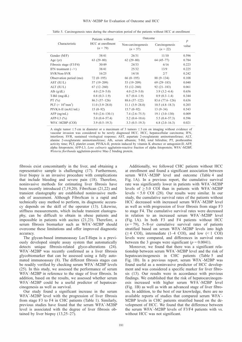
Table 5. Carcinogenesis rates during the observation period of the patients without HCC at enrollment
CharacteristicPatients without
HCC at enrollment (n = 79)
OutcomeP
valueNon-carcinogenesis (n = 57)
Carcinogenesis (n = 22)
Gender (M/F) 38/41 26/31 11/10 0.596Age (yr) 63 (29–80) 62 (29–80) 64 (45–77) 0.784Fibrosis stage (F3/F4) 30/49 24/33 6/16 0.223IFN treatment (–/+) 38/41 25/32 13/9 0.225SVR/Non-SVR 16/25 14/18 2/7 0.242Observation period (mo) 72 (0–195) 66 (0–195) 88 (9–134) 0.108AST (IU/L) 57 (19–209) 53 (19–209) 69 (29–183) 0.040ALT (IU/L) 67 (12–260) 53 (12–260) 92 (21–183) 0.061Alb (g/dL) 4.0 (2.9–5.0) 4.0 (2.9–5.0) 3.9 (3.2–4.4) 0.656T-Bil (mg/dL) 0.8 (0.3–1.9) 0.7 (0.4–1.9) 0.9 (0.3–1.4) 0.344PT (%) 86.3 (57–126) 88.6 (57–122) 83.6 (77.6–126) 0.636PLT (× 104/mm3) 11.0 (3.9–28.0) 11.1 (3.9–28.0) 10.5 (4.8–18.3) 0.203PIVKA-II (mAU/mL) 15 (0–92) 15.7 (0–92) 13 (9–34) 0.543AFP (ng/mL) 9.0 (2.6–130.1) 7.4 (2.6–75.5) 19.1 (3.0–130) 0.009AFP-L3 (%) 5.0 (0.4–57.4) 5.2 (0.4–10.6) 5.5 (0.4–57.3) 0.596WFA+-M2BP (COI) 3.9 (0.5–19.3) 3.3 (0.5–19.3) 6.8 (2.0–16.3) 0.021
A single tumor ≤ 5 cm in diameter or a maximum of 3 tumors ≤ 3 cm on imaging without evidence of vascular invasion was considered to be newly diagnosed HCC. HCC, hepatocellular carcinoma; IFN, interferon; SVR, sustained virological response; AST, aspartate 2-oxoglutarate aminotransferase; ALT, alanine 2-oxoglutarate aminotransferase; Alb, serum albumin; T-Bil, total bilirubin; PT, prothrombin activity time; PLT, platelet count; PIVKA-II, protein induced by vitamin K absence or antagonist-II; AFP, alpha fetoprotein; AFP-L3, Lens culinaris agglutinin-reactive fraction of alpha fetoprotein; WFA+-M2BP, Wisteriafloribunda agglutinin-positive Mac-2 binding protein.
fibrosis exist concomitantly in the liver, and obtaining a representative sample is challenging (17). Furthermore, liver biopsy is an invasive procedure with complications that include bleeding and severe pain (18). Therefore, noninvasive methods for estimating liver fibrosis have been recently introduced (7,19,20). FibroScan (21,22) and transient elastography are established noninvasive meth-ods of assessment. Although FibroScan is a rapid and technically easy method to perform, its diagnostic accura-cy depends on the skill of the operator (13). Likewise, stiffness measurements, which include transient elastogra-phy, can be difficult to obtain in obese patients and impossible in patients with ascites (21,23). Therefore, a serum fibrosis biomarker such as WFA+-M2BP should overcome these limitations and offer improved diagnostic accuracy. The glycan-based immunoassay LecT-Hepa is a previ-ously developed simple assay system that automatically detects unique fibrosis-related glyco-alterations (24). WFA+-M2BP was recently confirmed as a liver fibrosis glycobiomarker that can be assessed using a fully auto-mated immunoassay (8). The different fibrosis stages can be readily verified by checking serum WFA+-M2BP levels (25). In this study, we assessed the performance of serum WFA+-M2BP in reference to the stage of liver fibrosis. In addition, based on the results, we assessed whether serum WFA+-M2BP could be a useful predictor of hepatocar-cinogenesis as well as survival. Our study found a significant increase in the serum WFA+-M2BP level with the progression of liver fibrosis from stage F3 to F4 in CHC patients (Table 1). Similarly, previous studies have found that the serum WFA+-M2BP level is associated with the degree of liver fibrosis ob-tained by liver biopsy (13,25–27).
Additionally, we followed CHC patients without HCC at enrollment and found a significant association between serum WFA+-M2BP level and outcome (Table 4 and Fig. 1A). In a previous report, the cumulative survival rate was significantly lower in patients with WFA+-M2BP levels of ≥ 5.0 COI than in patients with WFA+-M2BP levels < 5.0 COI (28). Our results were similar. In our study, the cumulative survival rates of the patients without HCC decreased with increased serum WFA+-M2BP level as well as with progression of liver fibrosis from stage F3 to stage F4. The cumulative survival rates were decreased in relation to an increased serum WFA+-M2BP level (Fig. 1A). In both F3 and F4 patients without HCC (n = 79), 5-/8-yr cumulative survival rates of patients stratified based on serum WFA+-M2BP levels into high (≥ 4 COI), intermediate (1–4 COI), and low (< 1 COI) levels were compared, and differences in survival rates between the 3 groups were significant (p = 0.0041). Moreover, we found that there was a significant rela-tionship between serum WFA+-M2BP level and the risk of hepatocarcinogenesis in CHC patients (Table 5 and Fig. 1B). In a previous report, serum WFA+-M2BP was found useful as a noninvasive predictor of HCC develop-ment and was considered a specific marker for liver fibro-sis (13). Our results were in accordance with previous findings. We established that the risk of hepatocarcinogen-esis increased with higher serum WFA+-M2BP level (Fig. 1B) as well as with an advanced stage of liver fibro-sis. In addition, to the best of our knowledge, there are no available reports of studies that compared serum WFA+-M2BP levels in CHC patients stratified based on the de-velopment of HCC. We found that the difference between the serum WFA+-M2BP levels of F3/F4 patients with vs. without HCC was not significant.

182
Fig. 1. Cumulative survival rates and cumulative carcinogenesis rates in CHC patients without HCC (n = 79). A) Cumu-lative survival rates in CHC patients without HCC. The cumulative survival rates were calculated by the Kaplan-Meier method; differences between the groups were evaluated with the log-rank test. A two-tailed probability (p) value of < 0.05 was considered to be significant. In F3 or F4 patients without HCC (n = 79), the respective 5-/8-yr survival rates of patients with high WFA+-M2BP (≥ 4 COI, n = 39) and intermediate WFA+-M2BP (1–4 COI, n = 33) levels were 77%/49% and 100%/88%, respectively. None of the patients with low WFA+-M2BP (< 1 COI, n = 7) levels died. The differences in survival rates between the 3 groups were significant (p = 0.0041). B) Cumulative carcinogenesis rates in CHC patients without HCC. The cumulative hepatocarcinogenesis rates were calculated by the Kaplan-Meier method; differences between the groups were evaluated with the log-rank test. A two-tailed probability (p) value of < 0.05 was considered to be significant. The cumulative carcinogenesis rate in patients with high serum WFA+-M2BP (≥ 4, n = 39) was 49% after 5 yr, in contrast to 18% in patients with inter mediate WFA+-M2BP (1–4, n = 33) and 0% in patients with low WFA+-M2BP (< 1, n = 7) levels. The cumulative carcinogenesis rate differed significantly among the 3 groups (p = 0.0019). CHC, chronic hepatitis C; HCC, hepatocellular carcinoma; WFA+-M2BP, Wisteria floribunda agglutinin-positive Mac-2 binding protein.

WFA+-M2BP for Evaluation of Outcome and HCC
183
This study has several strengths. First, all the patients underwent a liver biopsy and had been definitively diag-nosed with F3 or F4 fibrosis. Second, this study included patients with and without HCC. Third, this cohort of 128 patients was analyzed prospectively, and we established that the serum WFA+-M2BP level was a predictor of he-patocarcinogenesis and outcome. However, this study has some limitations. First, changes in serum WFA+-M2BP levels after eradication of HCV should have been examined prospectively. Second, the effects of different HCV genotypes on serum levels of WFA+-M2BP should have been analyzed. Finally, because all of the study patients were Japanese, the applicability of the findings in this study to other populations remains to be evaluated. In conclusion, we revealed that a high serum WFA+-M2BP level was strongly associated with not only the stage of liver fibrosis but also hepatocarcinogenesis and outcome in CHC patients with advanced fibrosis. Hence, measurement of serum WFA+-M2BP levels might allow the prediction of carcinogenesis and outcome in CHC patients with advanced fibrosis.
Acknowledgments The study was supported in part by a grant-in-aid from the Research Program on Hepatitis from the Japan Agency for Medical Research and Development (JP17fk0210201h0002.) and the Ministry of Education, Culture, Sports, Science and Technology (15K09015).
Conflict of interest Takako Inoue is currently supported re-search grant by Gilead Sciences. Yasuhito Tanaka is currently conducting research sponsored by Chugai Pharmaceutical Co., Ltd., Bristol-Myers Squibb, Janssen Pharmaceutical K.K., AbbVie Inc. MSD K.K., and Gilead Sciences. The other authors declare no conflict of interest.
REFERENCES 1. Lee MH, Yang HI, Yuan Y, et al. Epidemiology and natural history
of hepatitis C virus infection. World J Gastroenterol. 2014;20:9270-80.
2. Sun CA, Wu DM, Lin CC, et al. Incidence and cofactors of hepatitis C virus-related hepatocellular carcinoma: a prospective study of 12,008 men in Taiwan. Am J Epidemiol. 2003;157:674-82.
3. Ikeda M, Fujiyama S, Tanaka M, et al. Risk factors for development of hepatocellular carcinoma in patients with chronic hepatitis C after sustained response to interferon. J Gastroenterol. 2005;40:148-56.
4. Yoshida H, Shiratori Y, Moriyama M, et al. Interferon therapy re-duces the risk for hepatocellular carcinoma: national surveillance program of cirrhotic and noncirrhotic patients with chronic hepatitis C in Japan. IHIT Study Group. Inhibition of Hepatocarcinogenesis by Interferon Therapy. Ann Intern Med. 1999;131:174-81.
5. Karanjia RN, Crossey MM, Cox IJ, et al. Hepatic steatosis and fibrosis: non-invasive assessment. World J Gastroenterol. 2016;22: 9880-97.
6. Wai CT, Greenson JK, Fontana RJ, et al. A simple noninvasive index can predict both significant fibrosis and cirrhosis in patients with chronic hepatitis C. Hepatology. 2003;38:518-26.
7. Vallet-Pichard A, Mallet V, Nalpas B, et al. FIB-4: an inexpensive and accurate marker of fibrosis in HCV infection. Comparison with liver biopsy and fibrotest. Hepatology. 2007;46:32-6.
8. Kuno A, Ikehara Y, Tanaka Y, et al. A serum “sweet-doughnut” pro-tein facilitates fibrosis evaluation and therapy assessment in patients with viral hepatitis. Sci Rep. 2013;3:1065.
9. Sasaki R, Yamasaki K, Abiru S, et al. Serum Wisteria floribunda agglutinin-positive Mac-2 binding protein values predict the devel-
opment of hepatocellular carcinoma among patients with chronic hepatitis C after sustained virological response. PLoS One. 2015;10: e0129053.
10. Ito K, Kuno A, Ikehara Y, et al. LecT-Hepa, a glyco-marker derived from multiple lectins, as a predictor of liver fibrosis in chronic hep-atitis C patients. Hepatology. 2012;56:1448-56.
11. Nagata H, Nakagawa M, Nishimura-Sakurai Y, et al. Serial measure-ment of Wisteria floribunda agglutinin positive Mac-2-binding pro-tein is useful for predicting liver fibrosis and the development of hepatocellular carcinoma in chronic hepatitis C patients treated with IFN-based and IFN-free therapy. Hepatol Int. 2016;10:956-64.
12. Iio E, Ocho M, Togayachi A, et al. A novel glycobiomarker, Wiste-ria floribunda agglutinin macrophage colony-stimulating factor re-ceptor, for predicting carcinogenesis of liver cirrhosis. Int J Cancer. 2016;138:1462-71.
13. Yamasaki K, Tateyama M, Abiru S, et al. Elevated serum levels of Wisteria floribunda agglutinin-positive human Mac-2 binding protein predict the development of hepatocellular carcinoma in hepatitis C patients. Hepatology. 2014;60:1563-70.
14. Mazzaferro V, Regalia E, Doci R, et al. Liver transplantation for the treatment of small hepatocellular carcinomas in patients with cirrho-sis. N Engl J Med. 1996;334:693-9.
15. Freeman AJ, Law MG, Kaldor JM, et al. Predicting progression to cirrhosis in chronic hepatitis C virus infection. J Viral Hepat. 2003;10:285-93.
16. Bravo AA, Sheth SG, Chopra S. Liver biopsy. N Engl J Med. 2001; 344:495-500.
17. Bedossa P, Dargère D, Paradis V. Sampling variability of liver fibro-sis in chronic hepatitis C. Hepatology. 2003;38:1449-57.
18. Cadranel JF, Rufat P, Degos F. Practices of liver biopsy in France: results of a prospective nationwide survey. For the group of epide-miology of the French association for the study of the liver (AFEF). Hepatology. 2000;32:477-81.
19. Yoneda M, Mawatari H, Fujita K, et al. Type IV collagen 7s domain is an independent clinical marker of the severity of fibrosis in pa-tients with nonalcoholic steatohepatitis before the cirrhotic stage. J Gastroenterol. 2007;42:375-81.
20. Guha IN, Parkes J, Roderick P, et al. Noninvasive markers of fibro-sis in nonalcoholic fatty liver disease: validating the European liver fibrosis panel and exploring simple markers. Hepatology. 2008;47: 455-60.
21. Sandrin L, Fourquet B, Hasquenoph JM, et al. Transient elastogra-phy: a new noninvasive method for assessment of hepatic fibrosis. Ultrasound Med Biol. 2003;29:1705-13.
22. Crespo G, Fernandez-Varo G, Mariño Z, et al. ARFI, FibroScan, ELF, and their combinations in the assessment of liver fibrosis: a prospective study. J Hepatol. 2012;57:281-7.
23. Castéra L, Vergniol J, Foucher J, et al. Prospective comparison of transient elastography, Fibrotest, APRI, and liver biopsy for the as-sessment of fibrosis in chronic hepatitis C. Gastroenterology. 2005; 128:343-50.
24. Kuno A, Ikehara Y, Tanaka Y, et al. LecT-Hepa: a triplex lectin- antibody sandwich immunoassay for estimating the progression dynamics of liver fibrosis assisted by a bedside clinical chemistry analyzer and an automated pretreatment machine. Clin Chim Acta. 2011;412:1767-72.
25. Toshima T, Shirabe K, Ikegami T, et al. A novel serum marker, gly-cosylated Wisteria floribunda agglutinin-positive Mac-2 binding pro-tein (WFA+-M2BP), for assessing liver fibrosis. J Gastroenterol. 2015;50:76-84.
26. Ura K, Furusyo N, Ogawa E, et al. Serum WFA+-M2BP is a non-invasive liver fibrosis marker that can predict the efficacy of direct-acting anti-viral-based triple therapy for chronic hepatitis C. Aliment Pharmacol Ther. 2016;43:114-24.
27. Toyoda H, Kumada T, Tada T, et al. Serum WFA+-M2BP levels as a prognostic factor in patients with early hepatocellular carcinoma un-dergoing curative resection. Liver int. 2016;36:293-301.
28. Hanai T, Shiraki M, Ohnishi S, et al. Impact of serum glycosylated Wisteria floribunda agglutinin positive Mac-2 binding protein levels on liver functional reserves and mortality in patients with liver cir-rhosis. Hepatol Res. 2015;45:1083-90.