Embed Size (px)
Citation preview

Clinical DataCaucasian woman 56 yo complain precordial chest burning pain, of sudden onset, occurring in the anterior chest, pleuritic in nature with referral to the left trapeziusridge. It is worse during inspiration, when lying flat, or during swallowing and with body motion.The fabled Prof W. Proctor Harvey wore a long time ago: “Suspect acute pericarditiswhen a patient says, “I have pain in my chest when I am lying down, but I can get relief if I sit up and get in a certain position”
Physical Minimal fever ( 37,2 °C.), tachypnea, and regular tachycardia (110bpm.). A pericardial friction triphasic rub: an atrial systolic rub that precedes S1, a ventricular systolic rub between S1 and S2 and coincident with the peak carotid pulse, and an early diastolic rub after S2. The rub has a scratching, grating sound similar to leather rubbing against leather and is best auscultated with the diaphragm of the stethoscope over the third left border using the diaphragm chest piece of the stethoscope pressed firmly against the chest wall with the patient sitting upright, breath held in deep expiration.
Right Joint infection with Celso triad, is observed.

Dados clínicosAnamnese:Mulher caucasiana 56 anos queixa-se dor ardente precordial, do início repentino, de caráter pleurítico, localizado na parede anterior do tórax irradiado ao cume esquerdo do trapézio. A dor piora durante a inspiração, na posição horizontal deitada, ou com os movimentos do corpo. O lendário Prof. W. Proctor Harvey escrevera há muito tempo: “A pericardite aguda é suspeita quando um paciente diz, “tenho a dor em meu peito quando estou deitado, mas melhoro em certa posição”
Exame Físico Paciente com febre baixa (37.2 °C.), taquipnéica, e com taquicardia regular (110bpm.). Uma atrito trifásico de fricção pericárdica é auscultado: um atrito sistólico atrial que precede a B1, um atrito sistólico ventricular entre o B1 e o B2 coincidente com o pulso carotídeo máximo, e um atrito diastólico após B2. O atrito assemelha-se com a fricção de coro e se ausculta melhor sobre o terceiro espaço intercostal na borda esquerda do esterno usando a campana do estetoscópio pressionado firmemente à parede torácica com o paciente sentado e com a respiração prendida na expiração profunda.Um processo inflamatório com a tríade de Celso, no joelho esquerdo foi observado.

ECG analysis: Sinus rhythm, heart rate 115bpm, (Sinus tachycardia), diffuse ST-segment elevation (<5mm) of superior concavity. ST segment changes are EXTENSIVE AND NOT TOO INTENSE, noticeable in several leads simultaneously (circumferential), excluding aVR and V1. Note especially in V6 that, unlike the “early repolarization pattern”, the J-point level almost equals the height of the T wave. (In early repolarization pattern or early repolarization variant, the J-point level is usually less than 25% of the T-Wave height). Reciprocal ST segment depression is observed in aVR and minimally in V1. These leads exhibit PR-segment elevation.
ECG Conclusion: first ECG phase or stage of acute pericarditis.
Clinical diagnosis: Purulent bacterial pericarditis secondary to staphylocococcal joint infection.
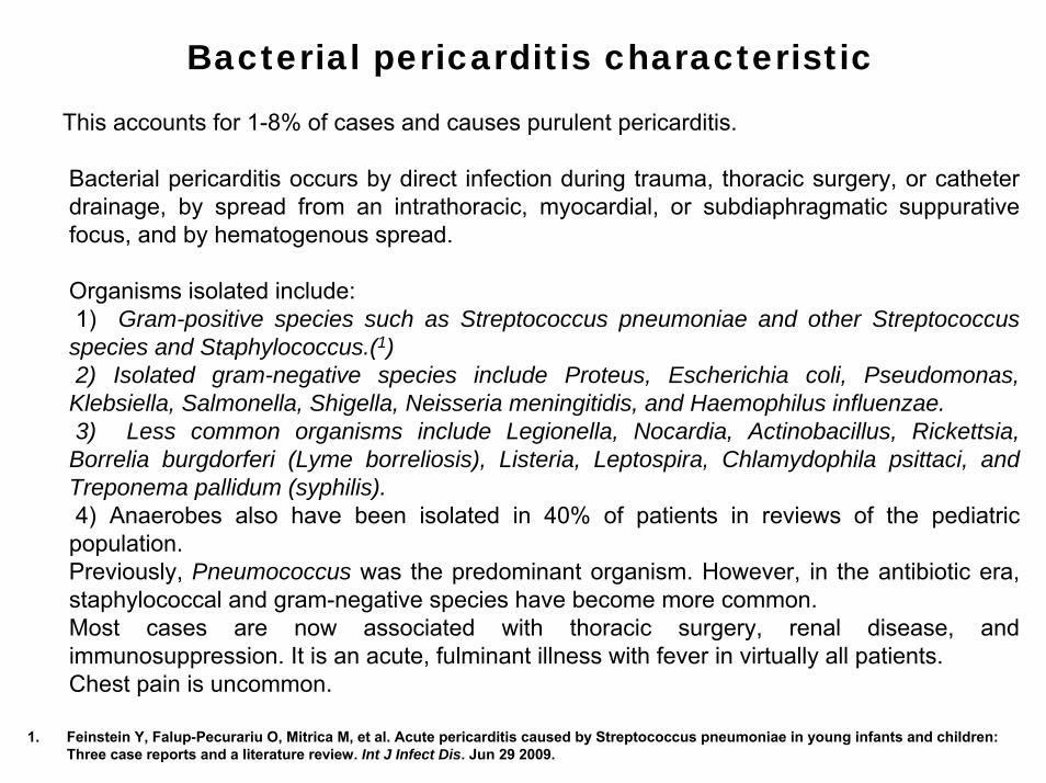
Bacterial pericarditis characteristic
This accounts for 1-8% of cases and causes purulent pericarditis.
Bacterial pericarditis occurs by direct infection during trauma, thoracic surgery, or catheter drainage, by spread from an intrathoracic, myocardial, or subdiaphragmatic suppurativefocus, and by hematogenous spread.
Organisms isolated include:1) Gram-positive species such as Streptococcus pneumoniae and other Streptococcus species and Staphylococcus.(1)2) Isolated gram-negative species include Proteus, Escherichia coli, Pseudomonas,Klebsiella, Salmonella, Shigella, Neisseria meningitidis, and Haemophilus influenzae. 3) Less common organisms include Legionella, Nocardia, Actinobacillus, Rickettsia, Borrelia burgdorferi (Lyme borreliosis), Listeria, Leptospira, Chlamydophila psittaci, and Treponema pallidum (syphilis). 4) Anaerobes also have been isolated in 40% of patients in reviews of the pediatric population. Previously, Pneumococcus was the predominant organism. However, in the antibiotic era, staphylococcal and gram-negative species have become more common. Most cases are now associated with thoracic surgery, renal disease, and immunosuppression. It is an acute, fulminant illness with fever in virtually all patients. Chest pain is uncommon.
1. Feinstein Y, Falup-Pecurariu O, Mitrica M, et al. Acute pericarditis caused by Streptococcus pneumoniae in young infants and children: Three case reports and a literature review. Int J Infect Dis. Jun 29 2009.

Purulent pericarditis can develop from previous aseptic pericarditis and is associated with a poorer prognosis, with a mortality rate nearing 85% for untreated persons and a mortality rate of 12-40% for treated patients. A high percentage of patients develop constrictive pericarditis. Death is mostly due to cardiac tamponade, systemic toxicity, cardiac decompensate, and constriction. Urgent pericardial drainage, combined with appropriate antibiotics for at least 4 weeks and drainage of pericardial fluid. Intravenous antibacterial therapy (e.g. vancomycin 1g twice daily, ceftriaxone 1-2g twice daily, and ciprofloxacin 400 mg/day) is mandatory in purulent pericarditis. Irrigation with urokinase or streptokinase, using large catheters, may liquefy the purulent exudates, but open surgical drainage is preferable. Intrapericardial fibrinolysis can be a useful treatment to assist with drainage of thick, loculated fluid, but open surgical drainage is preferred. Occasionally, patients require partial to total pericardiectomy(1).
1. Pankuweit S, Ristić AD, Seferović PM, Maisch B. Bacterial pericarditis: diagnosis and management. Am J Cardiovasc Drugs.2005;5:103-112.

MAIN ECG FEATURES IN ACUTE PERICARIDIS
ECG changes in acute pericarditis mainly indicates inflammation of the epicardium (the layer directly surrounding the heart), since the fibrous pericardium is electrically inert. For example, in uremia, there is no inflammation in the epicardium, only fibrin deposition, and therefore the ECG in uremic pericarditis will be normal.
An useful clue in differentiating acute pericarditis from early repolarization pattern (ERP) or variant (ERV) is the ST/T ratio in lead V6. This is calculated by dividing the millimeters of ST-segment elevation by the millimeters to the tallest point of the T wave. Each value is measured from the isoelectric point.
An ST/T ratio of greater than 0.25 in lead V6 suggests acute pericarditis.(1). In early repolarization, the J-point level is usually less than 25% of the T-wave height.
1. Ginzton LE, Laks MM. The differential diagnosis of acute pericarditis from the normal variant: new electrocardiographic criteria. Circulation 1982;65: 1004-1009.

Typical ECG changes in acute pericarditis includes 4 ECG stages or phases described in pericarditis(1;2), present in only 50% of cases:
1. FIRST PHASE: ST segment elevation (<5mm) of superior concavity with concordance of T waves. It is observed only two hours before chest pain and it lasts for several days. ST segment changes are EXTENSIVE (diffuse concave-upward) AND NOT TOO INTENSE(1), normally noticeable in several leads simultaneously, excluding V1 and aVR. Absence of reciprocal ST-segment changes with exception of ST-segment depression in aVRor V1 (reciprocal alterations )(2;3). Eventual PR-segment depression in aVR and V1 and elevation of PR segment in others leads mainly V5-V6 indicates atrial injury. Eventual low QRS voltage. This stage may last up to twoweeks.
1. SECOND PHASE: normalization of ST and PR deviations ST segment returns to baseline and flat T wave. This stage may last from days to several weeks.
2. THIRD PHASE: diffuse T wave inversion, (may not be present in all patients) with no formation of Q wave. Usually begins at the end of the second or third week.
3. FOURTH PHASE: ECG normalization with gradual reversion of T wave inversion or T waves may be indefinitely inverted.
1) Kasasbeh ES, Iskandar SB, Fahrig SA.Diffuse ST segment elevation: what's your diagnosis? Acute pericarditis. Tenn Med. 2009 Apr;102:39-40.
2) Dehmer GJ, O'Meara JJ 3d. Update on acute pericarditis. Hosp Med 1995;31:39-44.3) Pericarditis. In: Chou TC. Electrocardiography in clinical practice. 3d ed. Philadelphia: Saunders, 1991:219-234.

ECG IN ACUTE PERICARDITIS(AP) VERSUS ACUTE ISCHEMIA(AI)
1. PR-segmentAP: Frequent depression in aVR and V1 and elevation in others leads mainly V5-V6 indicate
atrial injury. AI: Rarely.
2. Abnormal Q wavesAP: None unless with infarctionAI: Frequently present with MI.
3. J point and ST segmentAP: Diffuse ST-segment elevation of superior concavity (circumferential), excluding aVR and V1.AI: Segmental or localized, convex to the top. Mirror image in presence of MI.
4. T wavesAP: Positive in first phase, flat in second phase, diffuse T wave inversion in third phase and
gradual reversion of T wave inversion in fourth phase. T waves may be indefinitely inverted.AI: Symmetrical with broad base and inverted while ST segment still elevated.
5. Conduction disorders AP: None in absence of heart disease.AI: Frequent.
6. ArrhythmiasAP: None.AI: Frequent.1. Spodick DH. Acute pericarditis: classic electrocardiogram.Am J Geriatr Cardiol. 2003 Jul-Aug;12:266.2. Spodick DH. Acute pericarditis: current concepts and practice. JAMA. 2003 Mar 5; 289:1150-1153.

AP ERV AMI Type 1 BrS
ST segment appearance
Diffuse concave to the top, evoluttive
Concave to the top stable
Convex to the top. Segmental.
Convex to the top in right precordial leads
Pathological Q waves
Absent Absent Frequently present Absent
Mirror image Possible in aVR Possible in aVR Present Eventually present
Leads involved Diffuse All Conspicuous in precordial leads V4-
V6. Eventually inferior.
Segmentar Right precordial
PR interval Non-affected Eventually prolonged in athletes
Eventually prolonged Prolonged ≈ 50% of cases
R voltage Non-affected Eventually augmented Lost Non affected
ST/T ratio in lead V6
<0.25 >0.25 Not applicable. Not applicable.
AP: Acute pericarditis; ERP: Early Repolarization Pattern; AMI: Acute Myocardial infarction; Type 1 BrS: Type 1 Brugada ECG patter.

CONDITIONING FACTORS OF ECG MANIFESTATIONS IN PERICARDITIS
Three causes are acknowledged as responsible for ECG modifications in pericarditis:• Accumulation of liquid between the two sheets: responsible for QRS complexes low voltage
and electrical alternance. It is considered pathognomonic of cardiac tamponade, and is characterized by changes in P wave, QRS complex and T wave voltages from beat to beat. The most frequent type of alternance is the one that affects only QRS. When it includes the three waves, it is called total alternance, observed in case of cardiac tamponade, characterized by Beck’s triad (jugular venous distension, hypotension and hypophoneticnoises).
• Electrical alternance is the result from a heart that wavers in an extensive effusion. • Lesion of epicardial region of myocardium by liquid or fibrin pressure: responsible for ST
segment elevation, depression in V1 and aVR and PR segment depression (STa) and elevation in aVR.
• Superficial myocarditis (epicarditis): changes in T wave.
EVOLUTIONARY CLASSIFICATION OF PERICARDITIS1. Acute: < 6 weeks (fibrinous with pericardial effusion).
2. Sub-acute: 6 weeks to 6 months (constrictive and pericardial effusion).
3. Chronic: >6 months (it divides into constrictive, pericardial effusion and adhesive without constriction).

COMMENTARIES FORUM COLLEAGES
Dear Andrés,These symptoms are similar to those of a mammal. Does it have four legs and does it meow?The ECG has ST-T elevation of the circumferential epicardial type, with sinus tachycardia, and
pericardial friction. This is pericardial irritation until the contrary is proven. The problem is the etiology: viral, by TB or metastasis; this is the first thing that should be established by a differential diagnosis:
1. If the symptoms persist, it could be pericardial infarction that produced a hemopericardium.2. If it is persistent, it could be a hydatid cyst, with pericardial reaction. 3. It could be autoimmune disease. The problem is that the woman should sit down to relieve the pain. The problem with tamponades in patients is relieved when patients lie down and raise their
arms to increase diastolic flow. But Andrés, you probably have something hidden, if you presented this case.
Warm regards,
Samuel Sclarovsky

Español (Spanish)Querido Andrés este cuadro es parecido a decir que es un animal mamífero, que con 4 patas y dice miu?El ECG muestra una elevación del ST-T del tipo circunferencial epicárdico, con taquicardia sinusal y fricción pericárdica en el examen físico. Este conjunto indica irritación pericárdica hasta que se demuestre lo contrario. El problema es determinar la etiologia si viral, tuberculosa o metastásica. La etiogia es lo primero que hay que establecer en el diagnóstico diferencial
1. Si el cuadro es persistente puede ser un infarto pericardial que produjo un hemo pericardio
2. Si es persistente podria ser un quiste hidatídico con reacción pericárdica3. Podria ser una enfermedad autoinmune(colagenopatia).
La referencia que la mujer debe sentarse para aliviar el dolor puede sugerir tamponamiento. Em esta circunstancia los pacientes se acuestan y levantan los brazos para aumentar el flujo diastólico.
Pero Andrés, sospecho que usted debe tener algo guardado debajo del poncho, para presentar este caso
Con abrazo fraternal
Samuel Sclarovsky

EnglishDear Friends
ECG analysis:
• Sinus rhythm at 110 bpm• PR-segment depression in leads I and inferior leads • PR-segment elevation in aVR• ST-segment elevation with up concave shape in leads I, aVL, inferior leads and V3 to
V6
ID: Pericarditis. Probable origin: septic emboli from the joint? Some of these cases are also MYOPERICARDITIS by involvement of the myocardium during the process. In such cases, troponins are usually elevated too, despite normal coronary arteries.I do not think that this is septic embolus to an specific coronary artery, given diffuse ST-segment elevations.
Happy Holidays, best for all!
Thanks Andrés for another challenging case
Adrian Barackchuk

PortuguêsDr. Pérez Riera:Análise detalhada não posso oferecer mas farei o que meu pouco saber permite:1. Ritmo sinusal regular(a cada onda P segue-se um complexo QRS) 2. Freqüência cardíaca: 115 bpm3. Duração de P: 0,08"(II) 4. SÂP: Aproximadamente em 60º .Concordo com alterações de P descritas pelo Prof.
Adrian Compatíveis com pericardite. 5. Duração de PR: 0,16"Duração de QRS: 0,06“6. SÂQRS: Aproximadamente em 55º7. Segmento ST supra desnivelado em todas derivações do plano frontal exceto aVR e
supra a partir de V2 nas precordiais 8. Chama minha atenção o entalhe na rampa descendente da R em I, aVF e V6.
Não deixaria de pensar em embolia pulmonar e pericardite com este processo no joelho esquerdo embora não haja menção de insuficiência venosa e Lúpus entraria em consideração.
Dr. Adail Paixão - Bahia - Brasil

English Detailed analysis I cannot offer but I will make what mine little to know it allows: 1) Rhythm: Regular sinusal rhythm (each wave P is followed by a QRS complex) 2) Heart rate: 115 bpm (a bit higher than the estimation by Prof. Adrian) 3) P wave characteristics: P duration: 0,08" (II), SÂP: Approximately in 60º. I agree to
described alterations of P for Prof. Adrian Compatible with pericardite, 4) PR interval: Duration 0,16" 5) QRS complex: QRS duration 0,06, “ SÂQRS: approximately in 55º6) ST segment: Segment ST elevation in all leads of the frontal plane except aVR and fsince
V2 in precordials leads. My attention is drawn to the notch in the descending slope of R in I, aVF and V6.
7) Diagnosis hypothesis: I agree with the P alterations described by Prof. Adrian. They are compatible to pericarditis, but I would not rule out pulmonary embolism and pericarditis with this process in the left knee, although there is no mention of venous insufficiency and lupus should be considered.
Happy Holidays, best for all!
Adail Paixão Bahia Brazil (Vitória da Conquista)

Differential Diagnosis of Acute Pericarditis by ECG
In cases of acute non-traumatic chest pain the interpretation of the ECG by prehospitalemergency doctors give to unsatisfactory results. Additional training in ECG interpretation may be a critical component of the education of physicians who care for patients presenting with acute coronary syndrome(ACS)(1). Acute pericarditis exhibits characteristic changes on ECG that usually enable one to make the diagnosis readily. There are other conditions that the clinician needs to consider in the differential diagnosis of acute pericarditis by ECG. However, findings on the history and physical examination and on laboratory assessment usually narrow the diagnostic possibilities. Two conditions that are commonly confused with acute pericarditis include ACS and early repolarization pattern (ERP). ERP is usually a normal variant that occurs commonly in young males, especially blacks, and does not evolve with the stages of acute pericarditis. ERP is distinguished by ST-segment elevation limited to the precordial leads, elevation of the ST segment in V1, an isoelectric ST segment in lead V6, notching of the terminal aspect of the QRS complex, and a shift to baseline of the ST segments with exercise.
1. Ohlow MA, Schreiber M, Lauer B. Prehospital assessment and treatment decisions of a suspected acute coronary syndrome: what are the problems? Results of the "Emergency Doctor and Acute Myocardial Infarction" study (NAAMI)] Dtsch Med Wochenschr. 2009 Oct;134:1984-1989.

Patients with pericarditis can be safely managed on an outpatient basis without a thoroughdiagnostic evaluation unless a specific cause is suspected or the patient has high-risk features, or both. A targeted etiological search should be directed to the most common cause on the basis of the clinical background, epidemiological issues or specific presentations.
In developed countries the clinicians should rule out neoplastic, tuberculous, and purulent pericarditis, as well as pericarditis related to a systemic disease.
Urgent coronary angiography is commonly performed in patients with acute pericarditis, particularly those with ST-segment elevation, typical myocardial infarction symptoms, and elevated troponin T values. Coronary artery disease is present angiographically in ≈ 30% of patients undergoing the procedure. Although patients with ST-segment elevation myocardial infarction must receive prompt reperfusion, clinicians must also consider the diagnosis of pericarditis to avoid unneeded coronary angiography(1).
1. Salisbury AC, Olalla-Gómez C, Rihal CS, Bell MR, Ting HH, Casaclang-Verzosa G, Oh JK.Frequency and predictors of urgent coronary angiography in patients with acute pericarditis. Mayo Clin Proc. 2009; 84:11-15.

ISOLATED PERICARDITIS VERSUS MYOPERICARDITIS
Myopericarditis are common in clinical practice: up to 15% of acute pericarditis have a significant myocardial involvement as assessed by biological markers. Between May 2005 and September 2007, 103 patients hospitalised for acute pericarditis were prospectively enrolled by Machado et al (1). Physical examination, ECG, echocardiography, biological screening and cardiac MRI, in case of myopericarditis defined as acute pericarditis with troponin I elevation, were performed. Between December 2007 and July 2008, patients were contacted for new clinical and MRI evaluation. Among the initial population of 103 patients admitted for acute pericarditis, 14 myopericarditis and 38 pericarditis were included. Compared with pericarditis, the myopericarditis group was associated with the following features: •Younger age•ST-segment elevation •Higher troponin I •Lower systemic inflammation.In the case of myopericarditis, infectious etiologies were predominant and patients stayed longer in hospital Follow-up showed no difference in terms of functional status and globalcomplications between paired myopericarditis and pericarditis. Nevertheless, cardiac mortality was higher for myopericarditis. MRI follow-up showed myocardial sequelae without clinical impact. The author concluded that myopericarditis significantly distinguished from pericarditis. Three years follow-up showed no difference in terms of global complications but a higher cardiac mortality for myopericarditis. MRI myocardial lesions did not develop into symptomatic sequelae.
1. Machado S, Roubille F, Gahide G, et al. Can troponin elevation predict worse prognosis in patients with acute pericarditis? Ann Cardiol Angeiol (Paris). 2009 Aug 13. [Epub ahead of print]

CLUES TO SIGNIFICANT MYOCARDITIS IN PATIENT WHIT ACUTE PERICARDITIS
1. Sinus tachycardia out the proportion to fever2. Any acute ECG QRS chage, especially if localized or with atypical evolution3. Convex ST segment elevation during acute phase4. Atrioventricular or intraventricular block 5. Pulmonary edema6. Postpericardiocentesis cardiac failure 7. Any significant arrhythmia, especially ventricular8. Evidence of myocardial dysfunction in absence of other heart disease9. Sinus tachycardia out of proportion to fever10. Pericarditis with transudative effusion fluid11. Postpericardiocentesis abnormal S2(in absence of constrictive pericarditis)12. Wall motion abnormalities13. Elevated serum levels of cardiac enzymes, especially in presence of elevated serum
myoglobin and troponyn.14. Positive ant myosin –indium 111 scintigraphy15. Myocardial production of tumor necrosis factor in absence of septic shock and
congestive heart failure16. Positive 99mtechnetium pyrophosphate imaging17. Positive gadolinium(Gd)-67 scintigraphy: Gd-DTPA-enhanced magnetic resonance
imaging18. Positive fibrinogen polymerization test.
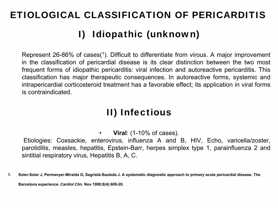
ETIOLOGICAL CLASSIFICATION OF PERICARDITIS
I) Idiopathic (unknown)
Represent 26-86% of cases(1). Difficult to differentiate from virous. A major improvement in the classification of pericardial disease is its clear distinction between the two most frequent forms of idiopathic pericarditis: viral infection and autoreactive pericarditis. This classification has major therapeutic consequences. In autoreactive forms, systemic and intrapericardial corticosteroid treatment has a favorable effect; its application in viral forms is contraindicated.
II) Infectious
• Viral: (1-10% of cases). Etiologies: Coxsackie, enterovirus, influenza A and B, HIV, Echo, varicella/zoster, parotiditis, measles, hepatitis, Epstein-Barr, herpes simplex type 1, parainfluenza 2 and sintitial respiratory virus, Hepatitis B, A, C.
1. Soler-Soler J, Permanyer-Miralda G, Sagristà-Sauleda J. A systematic diagnostic approach to primary acute pericardial disease. The
Barcelona experience. Cardiol Clin. Nov 1990;8(4):609-20.

• Bacterial/(Suppurative-purulent) other than tuberculosis)Streptococcus pneumoniae, Staphylococcus, Proteus, Escherichia coli, Pseudomonas, Klebsiella, Salmonella, Shigella, Neisseria meningitidis, and Haemophilus influenzae. Anaerobic: present in almost 40% of children with AP. Tuberculosis: ≈ 4% of cases Tuberculosis was a relatively common cause of pericarditis in the past, but this is now rare.
• Mycotic or FungalHistoplasmosis, blastomycetes, coccidioides, aspergillosis and candida.
• Rickettsial• Spirochetal
• Parasitic• Spirillum
• Micoplasma pneumoniae• Leptospira
• Listeria• Lynhogranuloma venereum• Psittacosis Chlamydiaceae
• Parasitic

IV) Colagenotic and related to An Underlying Systemic Disease
1. Lupic: ≈ 25% of patients with SLE develop AP.They rarely evolve into tamponade and constrictive pericarditis
2. Scleroderma3. Rheumatoid arthritis: 11-50% of patients with rheumatoid arthritis develop silent AP. The
diagnosis is made in only 2-6% of cases4. Sarcoidois(1)5. Sjögren's syndrome(2;3)6. Dermatomyositis/Polymyositis7. Polyarteritis nodosa8. Thrombohemolytic thrombocytopenic purpura9. Vasculitis10. Ankylosing spondylitis11. Behçet syndrome12. Whipple syndrome13. Wegener granulomatosis14. Churg-Strauss syndrome15. Hypocomentlemic uremic vasculitis syndrome16. Other collagenopathies.
1. Marie I, Lahaxe L, Guilbert M, Dacher JN, Levesque H. Pericarditis revealing a relapse of sarcoidosisSouth Med J. 2009 Dec;102: 1275-1277.2. Maisch B, Ristic AD (2002). "The classification of pericardial disease in the age of modern medicine". Curr Cardiol Rep 4 (1): 13–21.3. Chen HA, Chen CH, Cheng HH. Hemolytic uremic syndrome and pericarditis as early manifestations of primary Sjögren's syndrome. Clin
Rheumatol. 2009 Jun;28 Suppl 1:S43-46.

V) Pericarditis in Disease of Contiguous Structures
1. Myocardial infarction• Acute post-infarction: approximately 24 h after transmural AMI, fibrinous pericardial exudate
arises. Before the thrombolytic era, nearly 20% of patients with AMI developed acute pericarditis. After adopting the thrombolytic therapy and angioplasty, this incidence dropped 5-8%. Effusions may occur, but they rarely evolve into tamponade. Pericardial involvement does not contraindicate thrombolytic or anticoagulant therapy 3 to 5 days after transmural infarction.
• Dressler’s syndrome or postmyocardial infarction syndrome: it appears at the 2º or 3ºweek after AMI. typically occurs 1 to 4 weeks after the MI occurred, but may occur up to two years after the infract. The syndrome consists of a persistent low-grade fever, chest pain(usually pleuritic in nature), a pericardial friction rub, and /or a pericardial effusion.. In the setting of myocardial infarction, Dressler's syndrome occurs in about 7% of cases. This differentiates Dressler's syndrome from the much more common post myocardial infarction pericarditis that occurs in 17 to 25% of cases of acute myocardial infarction and occurs between days 2 and 4 after the infarction. Dressler's syndrome also needs to be differentiated from pulmonary embolism, another identifiable cause of pleuritic (and non-pleuritic) chest pain in people who have been hospitalized and/or undergone surgical procedures within the preceding weeks.
1. Dissecting aortic aneurysm2. Pleural and Pulmonary disease• Pneumonia• Pulmonary embolism• Pleuritis.

IV) NeoplasticSecondary (Metastatic tumor): they are responsible for 5-17% of cases. Neoplastic is the
most common cause of pericarditis(2): ≈ 35%. Dyspnea is the most common symptom. Primary malignant tumors: primary pericardial mesothelioma, sarcoma, fibroma, lipoma.
V) Metabolic1. Uremia: 6%. Chronic or acute Kidney failure caused by the buildup of certain toxins in the
body also can lead to pericarditis.2. Dialysis pericarditis3. Gout4. Hypothyroidism: ≈ 4% of cases of AP. Great effusions may develop. Evolution into
tamponade is rare.5. Scurvy.
VI) Actinicby radiation(1): pericarditis is the most frequent manifestation of post-radiotherapy cardiac
toxicity. It may evolve with pericardial effusion or chronic constrictive pericarditis.
1. Hamza A, Tunick PA, Kronzon I. Echocardiographic manifestations of complications of radiation therapy.Echocardiography. 2009 Jul; 26: 724-728.

VII) Traumatic- Penetrating chest injury- Esophageal rupture-Gastric perforation
Closed- Postpericardiotomy (PPS) is a febrile illness secondary to an inflammatory reaction involving the pleura and pericardium. It is more common in patients who have undergone surgery that involves opening the pericardium. However, postpericardiotomy syndrome has also been described following myocardial infarction (Dressler syndrome) and as an unusual complication after percutaneous procedures such as coronary stent implantation, after implantation of epicardial pacemaker leads and transvenous pacemaker leads, and following blunt trauma, stab wounds, and heart puncture. Pericardial effusions often accompany the syndrome and may develop into early or late postoperative cardiac tamponade and even recurrent cardiac tamponade. PPS is also characterized by pericardial or pleuritic pain, friction rubs, pleural effusions, pneumonitis, and abnormal ECG and radiography findings Pathophysiology similar to Dressler’s syndrome, however with incidence of 10-40% ≈ 1% develops tamponade- Pancreatic-pericardial fistula- Epicardial ablation- Catheter ablation for arrhythmias- Percutaneous Tranluminal Coronary Angioplasty- Epicardial pacemaker.

VIII) By hypersensitivity• Reaction to drugs: Some medications can trigger an immune response that causes pericarditis. These medications include the antibiotic penicillin, doxorubicin, cyclophosphamide, procainamide the hypertension and heart failure medicine hydralazine (Apresoline®), (1),methyldopa (2), the antituberculosis medicine isoniazid (4) (Nydrazid®), antiarrhythmic agent procainamide(5) (Procanbid®, Pronestyl®), and the seizure medication phenytoin(6) (Dilantin®), sulfasalazine (7) for ulcerative colitis and mesalazine(8).• Serum sicknessis is an example of the type III, or immune complex–mediated, hypersensitivity disease. Historically, the term serum sickness connotes a self-limited immune complex disease caused by exposure to foreign proteins or haptens. Immune complex formation is a common event and does not typically cause symptoms. However, an immune reaction can occur, as in the case of serum sickness. • Immunopathies.
1) Franssen CF, el Gamal MI, Gans RO, Hoorntje SJ. Hydralazine-induced constrictive pericarditis.Neth J Med. 1996 May;48:193-197.2) Rakotoson JL, Randriamanana D, Rakotomizao JR, Andrianasolo R, Rakotoarivelo R, Andrianarisoa AC. Severe systemic lupus
erythematosus induced by isoniazide. Rev Pneumol Clin. 2009 Dec;65:361-364.3) Harrington TM, Davis DE. Systemic lupus-like syndrome induced by methyldopa therapy. Chest. 1981 Jun;79:696-697. 4) Jouneau S, Volatron AC, Brinchault G, Morel V, Belleguic C, Delaval P. Isoniazid-induced pleuro-pericarditis.Rev Pneumol Clin. 2003
Dec;59:357-359.5) Spodick DH. Pericarditis in systemic diseases. Cardiol Clin. 1990 Nov;8:709-716. 6) Ebaugh L, Fleet WF 3rd, Morgan HJ. Pericarditis following antiarrhythmic therapy.J Tenn Med Assoc. 1990 Apr;83:190.7) Atwater BD, Ai Z, Wolff MR. Fulminant myopericarditis from phenytoin-induced systemic lupus erythematosus.WMJ. 2008 Sep;107:298-
300.8) Perrot S, Aslangul E, Szwebel T, et al. Sulfasalazine-induced pericarditis in a patient with ulcerative colitis without recurrence when
switching to mesalazine. Int J Colorectal Dis. 2007 Sep;22(9):1119-1121. 9) García-Morán S, Sáez-Royuela F, Pérez-Alvarez JC, Gento E, Téllez J.Myopericarditis and mitral insufficiency associated with ulcerative
colitis treated with mesalazine.Inflamm Bowel Dis. 2006 Apr;12:334-335.