Embed Size (px)
Citation preview

Clinical Connections

Clinical Connection: Spina Bifida
• Defective closure of the caudal neural tube
• Variabilility in severity– Spina bifida oculta– Spina bifida cystica– Spina bifida with myelomeningocele– Spina bifida with meningomyelocele– Spina bifida with myeloschisis

Clinical Connection: Spina Bifida
• Often occurs with other congenital anomalies
• Incidence declining with early prenatal detection and dietary supplement of folic acid

Image spina bifida

Clinical Connection: Arnold-Chiari Malformation
• Associated often with spina bifida with meningomyelocele
• Congenital anomaly• Medulla and posterior cerebellum
elongate into foramen magnum• May be asymptomatic • May result in hydrocephalus and
other symptoms

Clinical Connection: Anencephaly
• Congenital anomaly• Failure of rostral neuropore closure • Large portions of scalp, cranial
bones, and cerebral hemispheres are absent
• Most die in utero, virtually all by first postnatal week

Clinical Application Exercise
• Discuss the stories of Maria Rodriquez and Jonathan Perry
• Mechanism of injury• For Maria, do you predict motor loss, sensory loss or
both? Why? Both, Ant/post• Predict her ‘motor picture’ mid trunk & up• Will she walk? No• For Jonathan, which part of the cord has the tumor
affected? Posterior

Clinical Connection: Nervous System Pathology
• Retrograde transport of certain toxins and viruses from the environment to the CNS
• Examples– Clostridium tetani bacterium– Rabies– Herpes simplex virus– Poliomyelitis

Clinical Connection: Multiple Sclerosis
• Autoimmune disorder
• Proteins expressed by oligodendrocytes are erroneously recognized as foreign by immune system
• Loss of myelin in brain and spinal cord

Clinical Connection:Parkinson's Disease (PD)
• In PD there is a degeneration of dopamine (DA) producing cells and a substantial reduction in the synthesis of DA
• Most common treatment strategy is replacement therapy of DA

Clinical Connection:Parkinson's Disease (PD)
• DA cannot cross blood–brain barrier, but precursor L-DOPA does
• L-DOPA stimulates surviving DA cells to increase synthesis of DA
Clinical Connection:Biologic Depression
• May be due to a deficiency in brain of amine transmitters – norepinephrine (NE) and serotonin (5-HT)
• Monoamine oxidase is an enzyme that breaks done NE and 5-HT
• Treatment historically included MAO inhibitors
• MAO inhibitors have unwanted side effects
Clinical Connection: Syndrome of the Anterior Spinal Artery
• Usually acute onset due to ischemia of lower thoracic or upper lumbar spinal cord
Clinical Connection: Syndrome of the Anterior Spinal Artery
• Characterized initially by– Flaccid paraplegia in both legs– Bilateral deficits in STT sensibilities
– Loss of bowel and bladder control– Sparing of DCML sensibilities
• Following spinal shock, spastic paraplegia may develop

Clinical Connection: Herniation of the Vertebral Disc
• Displacement of disc tissue from normal position between vertebral bodies– Nuclear protrusion mild– Disc prolapse more severe

Disc Herniations

Clinical Connection: Herniation of the Vertebral Disc
• May compress the spinal cord against the vertebral canal of the spinal nerves against the intervertebral foramina
• Most herniations are in a posterolateral direction
• Common levels for herniations correspond to areas where vertebral column moves freest– L5/S1, C5/C6, C6/C7

Clinical Connection: Vertebral Fractures and Neurotoxic Spinal Cord Injury
• With vertebral dislocations or fracture dislocations the spinal cord can be damaged immediately by trauma or subsequently through secondary neurochemical injury
• Endogenous neurotransmitters cause excitotoxic neuronal cell death
• Initial trauma causes changes in ions leading to cytotoxic edema

Clinical Connection: Cervical Spondylosis
• Degeneration of the discs and surrounding ligaments, osteophytic changes
• Lower cervical vertebra particularly vulnerable

Clinical Connection: Cervical Spondylosis
• Results in compression and compromised blood supply of spinal cord
• Characterized by neck pain and stiffness, hand numbness, and spastic leg weakness

Spondylosis

Clinical Connection: Control of the Level of Consciousness
• The portion of the RF that contributes to the level of consciousness is called the ascending reticular activating system (ARAS)
• Sleep is a part of this, epithalamus is main contributor of cycles (melatonin)

Clinical Connection: Control of the Level of Consciousness
• Levels of consciousness– Attention– Alertness– Drowsiness– Stupor– Coma
Clinical Connection: Thalamic Syndrome
• Typically caused by occlusion of thalamogeniculate branches of the PCA supplying posterior thalamus
• Symptoms contralateral to lesion– Hemianesthesia, ataxia, excruciating neurogenic pain
Symptoms in diff proportions in diff pts
• Unresponsive to analgesics

Clinical Connection: Hypothalamic Syndromes
• Diabetes INSIPIDUS• Problems with regulation of thirst, hunger, and body temperature; menstrual and sleep–wakefulness cycles

Clinical Connection: Cerebellar Dysfunction
• Variety of causes– Acute/chronic alcohol intoxication, developmental disorders, stroke, trauma
• Characteristic symptoms– Hypotonia, incoordination, intention tremor, ataxia, nystagmus
Clinical Connection: Involuntary Movements Attributed to Basal Ganglia Dysfunction
• Tremor-rhythmic, alternating movement of a body part
• Athetosis – slow, sinuous, writhing movements
• Chorea – brisk, graceful, complex movements (caudate)
• Ballismus – forceful, flinging movements (subthalamic)

Clinical Connection: Occlusion of the MCA
• Occlusion of the superior division of the MCA– Structures affected: BAs 4, 3, 1, 2, 8, 44, 45
– Related symptoms include contralateral motor and sensory deficits, eye movement deficits, and motor aphasia if dominant hemisphere occluded
– Problem with language production

Clinical Connection: Occlusion of the MCA
• Occlusion of the inferior division of the MCA– Structures affected: BAs 22, 39, 40
– Related symptoms include sensory aphasia if dominant hemisphere occluded, visuospatial deficits if nondominant hemisphere occluded

Figure 7-20 (a) Area of the left cerebral hemisphere infarcted by occlusion of the left superior division of the MCA. Note that the damage is confined to the frontal lobe and postcentral gyrus of the parietal lobe. (b) Area of the left
hemisphere infarcted by occlusion of the inferior division of the MCA. Note that the damage is confined to the posterior portion of the parietal lobe and the
temporal lobe. Abbreviation: BA, Brodmann's area. (Adapted from Bowman, J. P., and Giddings, D. F. Strokes: An Illustrated Guide to Brain Structure, Blood Supply, and
Clinical Signs. Prentice Hall,New Jersey, 2003.)
Clinical Connection: Occlusion of the ACA
• Less common than occlusion of the MCA or IC
• Structures damaged include the medial aspects of BA 4, 3, 1, 2
• Related symptoms include contralateral motor and sensory deficits that are most severe in the lower extremity

Occlusion of the ACA
Clinical Connection: Occlusion of the IC
• Carotid border syndrome occurs with occlusion of 70% or more of the IC
• Blood flow to the distal territories is decreased, but not eliminated
• Symptoms include contralateral numbness and weakness
Clinical Connection: Occlusion of the PCA
• Structures affected include BA 17, the primary visual cortex
• Related symptoms include loss of vision in the contralateral one-half of the visual field of each eye
• Because a branch of MCA perfuses the cortex that receives afferents from the fovea, there may be sparing of foveal vision (macular sparing)

Clinical Connection: Motor Signs
• Positive signs (release phenomena)– An excess of neural activity– Hyperreflexia, hypertonicity, Babinski sign
• Negative signs– Motor deficits due to the loss of function of damaged neural structures
– Weakness, loss of speed of movement, loss of dexterity

Clinical Connections
• Peripheral neuropathies typically impair somatosensation and autonomic functions
• Herpes zoster (shingles) is a viral infection of the somatosensory ganglia of the peripheral and cranial nerves (ophthalamic

Clinical Connections: Disorders of Peripheral Nerves
• Neuropathy – disease of the peripheral nerves– Mononeuropathy – affecting one nerve
– Polyneuropathy – affecting many nerves
– Radiculopathy – affecting spinal nerve roots

Clinical Connections: Disorders of Peripheral Nerves
• Diabetes mellitus (DM) is the most common cause of polyneuropathy– Characterized by distal, bilateral sensory losses

Clinical Connection: Brown-Sequard Syndrome
• Lesion of one-half of the spinal cord and all fibers entering the cord via the dorsal root producing – Ipsilateral zone of somatosensory anesthesia at the damaged dermatomes
– Ipsilateral loss of DC-ML modalities below the lesion
– Contralateral loss of STT modalities below the lesion

Clinical Connection: Syringomyelia
• Disease process in the center of the spinal cord, usually over a few cervical segments
• Disrupts pain and temperature fibers of the STT in the ventral white commissure
• Characterized by bilateral loss or pain and temperature sensibility, usually in the hands and arms

Clinical Connection: Subacute Combined Degeneration
• Disorder that typically first involves the fibers of the dorsal columns
• Related to lack of Vitamin B12 absorption
• Early symptoms include symmetrical parathesias of fingers and toes and loss of proprioception
• Person may demonstrate unsteady, ataxic gait

Clinical Connection: LMN Syndrome
• Damage to the LMN cell body or axon• Characterized by:– Hyporeflexia– Hypotonia– Paralysis (loss) or paresis (weakness)– Atrophy = rapid and severe– Denervation• Fasciculations = can happen in tired, healthy people or LMN damage = visible twitch• Fibrillations = similar to fasciculation, smaller, need EMG, not visible twitch

Clinical Connection: Diseases of the Motor Unit
• Four sites of the motor unit can be attacked by specific disease entities– The cell body of the LMN, its axon, the neuromuscular junction, or the muscle fibers
• Neurogenic-affecting cell body or axons– (e.g., polio, neuropathies)
• Myopathic-affecting striated muscle– (e.g., myasthenia gravis)

Clinical Connection: Polio
• Viral infection, prevented by vaccination
• Attacks cell bodies of LMNs• Cardinal clinical signs: weakness/paralysis, atrophy, and decreased reflexes
• Postpolio syndrome may appear 20–30 years after the acute infection– Remaining neurons overworked after normal neuron apoptosis

Clinical Connection: Neuropathies
• Pathological changes of the peripheral nerves– Segmental degeneration– Axonal degeneration– Wallerian degeneration
• Diabetes mellitus (DM) • Acute inflammatory demyelinating polyneuropathy (AIDP) = aka guillanne barre’s

Clinical Connection: Myasthenia Gravis
• Autoimmune disorder of neuromuscular transmission resulting in weakness
• Typically eye muscles are affected first, resulting in ptosis and diplopia
• Muscles of facial expression, mastication, swallowing, and speech are typically affected next, resulting in altered facial expression, dysphagia, and dysarthria

Clinical Connection: Myopathies
• Progressive hereditary diseases of skeletal muscle
• Duchenne muscular dystrophy– Defective gene that codes for protein dystrophin
– Sex-linked recessive trait transmitted from mother to male children

Clinical Connection: Myopathies
• Duchenne muscular dystrophy– Lack of dystrophin renders sarcolemma vulnerable
– Weakness, tearing, and loss of muscle fibers
– Shortens life expectancy to late adolescence or early adulthood

Duchenne Muscular Dystrophy

Clinical Connection: Amyotrophic Lateral Sclerosis
• Also called Lou Gehrig's disease
• Most frequent motor system disease
• Characterized by – amyotrophy (weakness, denervation, atrophy)
– lateral sclerosis (upper motor neuron signs)

Clinical Connection: Amyotrophic Lateral Sclerosis
• Relentlessly progressive
• Some 50% of cases progress to death within 2–3 years of diagnosis
• Role of the DPT
Clinical Connection: Evaluation of Reflexes
• Is the reflex present?
• If present, is status altered (grading)? – Hyporeflexia– Hyperreflexia
• Are pathological reflexes present?– Babinski or stepping response in adults (normal in infants)

Clinical Connection: Knee Stability
• Anterior cruciate ligament (ACL) prevents femur from sliding posteriorly on the tibia, preventing hyperextension of the knee
• The hamstring muscles play a critical role in knee stability by protecting the ACL

Clinical Connection: Knee Stability
• If knee moderately hyperextended, hamstring stretch reflexes will cause contraction of the hamstrings and relieve the strain on the ACL
• Two phases SLR and MLR • MLR latency associated with ‘giving way’ in patients post ACL rupture and surgical repair

Spasticity
• Motor disorder characterized by a velocity-dependent increase in tonic stretch reflexes (muscle tone) and exaggerated tendon jerks
• Spasticity often develops in clinical disorders with UMN damage such as stroke, multiple sclerosis, and spinal cord injury
Spinal Shock and the Emergence of Spasticity
• Spinal shock – following complete spinal cord transection or compression, the initial response is one of transient hypotonia and hyporeflexia– All sensation below the lesion is lost
– All voluntary movement below the lesion is lost
– Reflex activity below lesion is lost

Spinal Shock and the Emergence of Spasticity
• In addition to motor and sensory losses, bowel and bladder function are lost, leading to fecal and urinary incontinence

Emergence of Spasticity
• After the period of spinal shock, the intrinsic spinal cord circuits may begin to display autonomous activity
• Minimal reflex activity and initially weak flexor responses to painful stimuli– Mass reflex, triple flexion

Emergence of Spasticity
• Eventually and gradually, extensor tone also increases
• Due to altered suprasegmental influences

Triple Flexion Reflex

Pattern of Involvement of Muscles Weakness of LMN versus
UMN Damage• LMN damage– Flaccid paralysis or flaccid paresis of individual muscles or groups of muscles
– Ipsilateral to the lesion

Pattern of Involvement of Muscles Weakness of LMN versus
UMN Damage• UMN damage– Spastic paresis of synergistic muscle groups
– Contralateral to lesion if damage is rostral to decussation
– Ipsilateral to lesion if damage is caudal to the decussation

Clinical Connection: Pathological Reflexes
• Babinski
• Flexion
• Clasp-knife
• Clonus

Clinical Connection: Babinski Reflex
• Elicited in healthy babies as a bilateral plantar response during first year of life
• When elicited beyond infancy, is a reliable sign of corticospinal tract damage
• Elicited by stroking sole of foot• Abnormal response is dorsiflexion of the great toe and abduction of the other toes

Figure 11-16 (a) The normal response to stroking the sole of the foot. (b) The abnormal Babinski sign.

Clinical Connection: Flexion Reflex
• Flexion reflex normally elicited with noxious stimuli
• Following UMN damage, the flexion response may be seen with innocuous stimuli
• Due to disruption of descending suprasegmental inhibition

Clinical Connection: Clasp-Knife Phenomenon
• Sometimes accompanies spasticity following UMN damage
• Elicited with passive movement of a limb

Clinical Connection: Clasp-knife Phenomenon
• Limb moves freely for a short distance, followed by a rapid increase in resistance, followed by a sudden giving way to movement
• Reflects the length dependence of hyperreflexia

Clinical Connection: Clonus
• A clinical sign of spasticity
• Elicited with abrupt and sustained ankle dorsiflexion
• Series of rhythmic involuntary muscle contractions

Figure 11-18 Ankle clonus in an individual with spasticity consists of rhythmic contraction–relaxation cycles of the ankle extensor muscles (plantar flexors of the foot) in response to their sustained stretch (i.e.,
stretch of the Achilles tendon).

Clinical Connection: Combined Sensory and Motor Damage
• Brown-Sequard syndrome or spinal cord hemisection
• Syringomyelia
• Subacute combined degeneration

Clinical Connection:Brown-Sequard Syndrome
• Rarely encountered lesion involving only one-half of the spinal cord

Clinical Connection:Brown-Sequard Syndrome
• Characterized by – Ipsilateral flaccid paralysis at the level of lesion
– Ipsilateral spastic paralysis below the level of lesion
– Ipsilateral loss of DCML sensibilities below the level of lesion
– Contralateral losses of STT sensibilities below the level of lesion

Figure 11-19 The Brown-Sequard syndrome results from injury to one-half of the spinal cord, extending over several spinal cord segments (shaded). (a) Degeneration (broken lines) associated with spinal cord hemisection. The ventral white commissure is represented as a tube to illustrate the oblique crossing of
spinothalamic tract afferents before they enter the anterolateral funiculus on the opposite side of the cord.

Figure 11-19 The Brown-Sequard syndrome results from injury to one-half of the spinal cord, extending over several spinal cord segments (shaded). (b) Motor and somatosensory losses associated
with the Brown-Sequard syndrome.

Clinical Connection: Syringomyelia
• Rare and chronic disorder characterized by a progressive development of fluid-filled cavity in the spinal cord, most commonly in lower cervical or upper thoracic regions

Clinical Connection: Syringomyelia
• Typically a slow progression beginning with interruption of pain and temperature fibers crossing in the ventral white commissure
• Hallmark symptom bilateral loss of pain and thermal sensation
• Occasionally cavity extends rostrally into brainstem, resulting in syringobulbia

Figure 11-20 The cavity in syringomyelia often interrupts spinothalamic afferents crossing in the ventral
white commissure.

Clinical Connection: Subacute Combined Degeneration
• Pernicous anemia results from an inability to absorb vitamin B12
• Unclear mechanisms lead to degeneration of peripheral and optic nerves, brain, and spinal cord
• Spinal cord is the first and most commonly affected region

Clinical Connection: Subacute Combined Degeneration
• Posterior and lateral funiculi degeneration is characteristic, leading to symmetrical parathesias, loss of dorsal column sensation, and ataxia
• Progresses to lateral column signs of loss of strength and UMN signs

Figure 11-21 Myelin stained sections from the lower cervical spinal cord. (a) Normal. (b) Lesions in subacute combined
degeneration involve degeneration of the white matter in the posterior and lateral funiculi of the spinal cord. The
degenerated fibers lose their myelin and, hence, are unstained.

Clinical Connections: Primary Orthostatic Hypotension
• In primary orthostatic hypotension blood pressure falls suddenly upon standing from a recumbent position
• May be due to degeneration of postganglionic sympathetic fibers and sparing of parasympathetic fibers or degeneration of the intermediolateral cell column of the thoracic cord
Clinical Connections: Primary Orthostatic Hypotension
• Many other possible causes and conditions that cause low blood pressure

Clinical Connections: Bladder Dysfunction
• Interruption of reflex connections of the bladder produces neurogenic bladder
• Reflex neurogenic bladder caused by bilateral spinal cord lesion above T12, resulting in UMN paralysis, spasticity, and sudden and reflexive emptying

Clinical Connections: Bladder Dysfunction
• Nonreflex neurogenic bladder caused by bilateral lesion of sacral spinal cord, resulting in LMN paralysis, flaccidity, and bladder leakage

Clinical Connections: Horner's Syndrome
• Combination of symptoms: – Miosis- small pupil– Ptosis- drooping eyelid– Enopthalmos (apparent) - retraction of eyeball

Clinical Connections: Horner's Syndrome
• Occurs as a result of lesion to preganglionic sympathetic fibers emerging from T1 and T2 – Causative peripheral lesions include tumor of superior cervical ganglion, cervical lymph lodes, and surgical trauma to sympathetic chain
– Causative central lesions include tumor or stroke of the brainstem, and syringobulbia

Horner’s Syndrome

Clinical Connections: Acute Autonomic Paralysis
• Complete lesion of the cervical spinal cord interrupts all suprasegmental control of the sympathetic and parasympathetic divisions by the ANS

Clinical Connections: Acute Autonomic Paralysis
• Causes spinal shock with autonomic effects of– Paralysis of bowel and bladder– Anhidrosis– Loss of piloerection and sexual function
– Potentially severe hypotension

Clinical Connections: Autonomic Dysreflexia (AD)
• Potentially life-threatening condition associated with spinal cord lesions above the sixth thoracic segment
• Characterized by high blood pressure, severe headaches, goose bumps, and sweating

Autonomic Dysreflexia

Clinical Connections: Autonomic Dysreflexia (AD)
• Elicited by a noxious stimuli below level of injury triggering sympathetic-driven increase in blood pressure
• Disruption of the descending parasympathetic fibers unable to modulate
• Treated by immediately sitting person with AD upright

Clinical Evaluation of the Olfactory Nerve
• Test each nostril separately with a familiar scent
• Olfaction, emotion, and memory
• Anosmia (inability to smell) may occur with a common cold, trauma, and some degenerative diseases such as Parkinson's and Alzheimer's
Disorders of Eye Movement
• Opthalmoplegia – paralysis of one or more of the extraocular muscles
• Strabismus – inability to direct both eyes to the same object– Lateral – due to paralysis of CN III– Medial-due to paralysis of CN VI
• Diplopia – double vision• Ptosis – weakness of levator palpebrae superioris muscle

Clinical Evaluation of the Trigeminal Nerve
• Corneal reflex
• Sensory tests for DCML and STT modalities
• Palpate muscles of mastication, resist jaw opening
Lesions of the Trigeminal Nerve: Trigeminal Neuralgia (tic douloureux)
• No motor or sensory losses
• Excruciating bursts of pain, usually in one of the three sensory distributions of CN V
• Triggered by use of jaw, yawning, hot and cold, light breeze on face

Lesions of the Trigeminal Nerve: Herpes Zoster Ophthalmicus
• Inflammatory and infectious disease
• 2–3 days of severe pain along distribution of the opthalmic division of CN V
• Rash follows in this distribution
• Can result in corneal damage
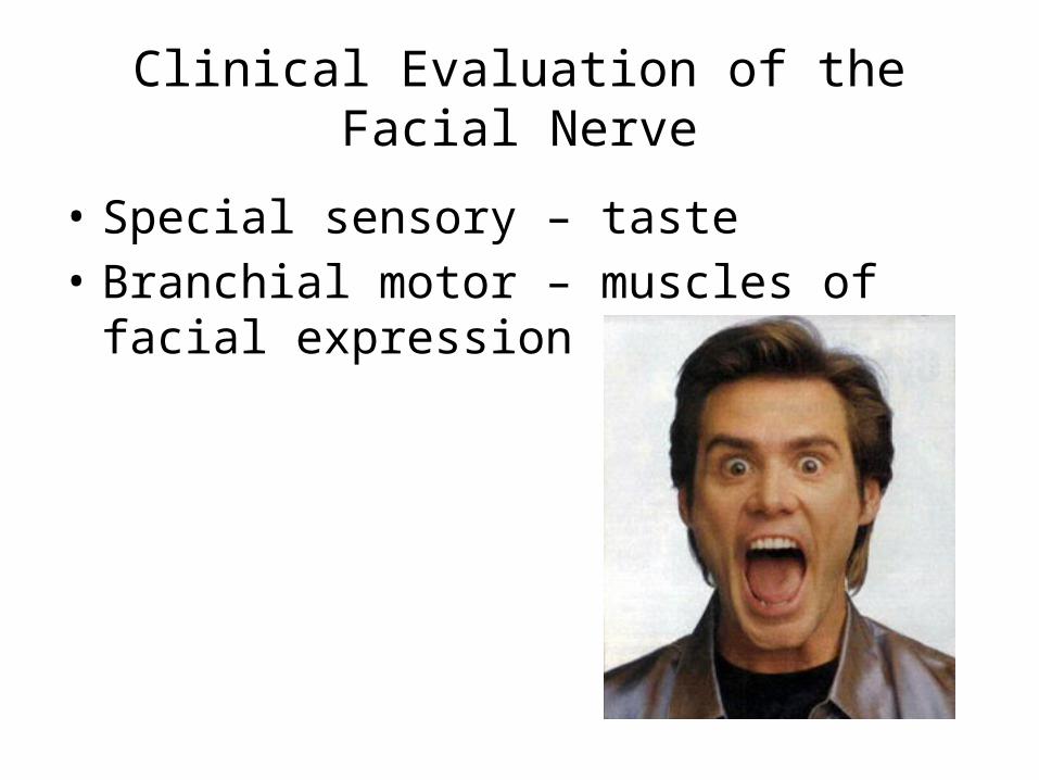
Clinical Evaluation of the Facial Nerve
• Special sensory – taste• Branchial motor – muscles of facial expression

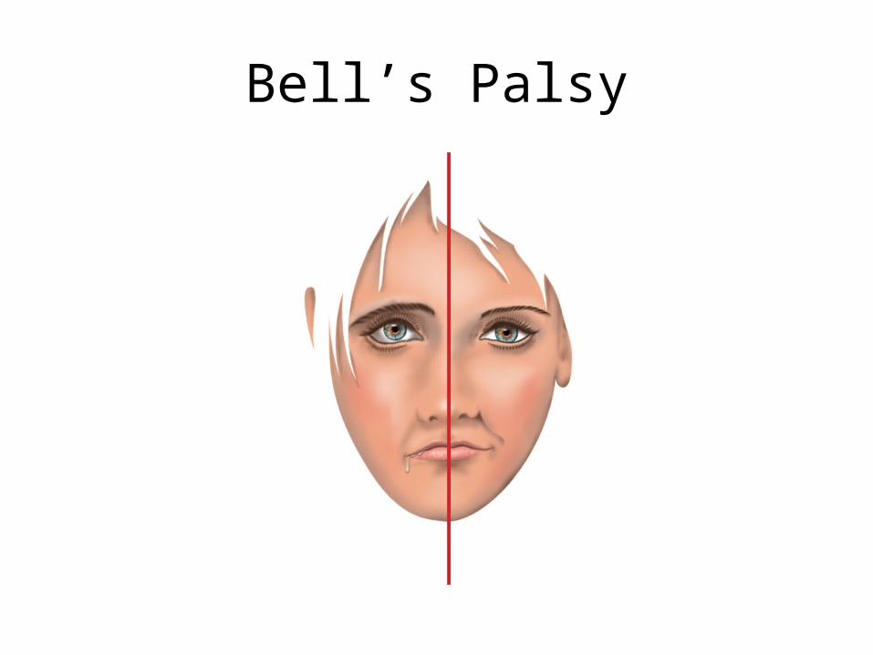
Lesions of the Facial Nerve: Bell's Palsy
• Most common disease affecting facial nerve
• Often caused by herpes simplex virus
• Acute onset• Characterized by paralysis of facial muscles, impaired corneal blink reflex, and hyperacusis
Bell’s Palsy

Vestibulocochlear Nerve (VIII)
• Special sensory• Conveys vestibular (equilibrium) and cochlear (hearing/auditory) information
• Nuclei located in caudal pons and rostral medulla
• Clinical evaluation includes tests of the ability to coordinate eye–head movements and of hearing

Clinical Evaluation and Lesions of the Glossopharyngeal Nerve
• Testing of gag reflex somewhat unreliable as pharyngeal wall innervated by CN IX and CN X
• Isolated lesions of CN IX are rare

Clinical Evaluation and Lesions of the Vagus Nerve
• Observation of movements of the soft palate
• Observation of vocal quality
• Lesions of CN X can produce hoarseness of speech and difficulty with swallowing

Palate drooped and deviated to Unaffected side
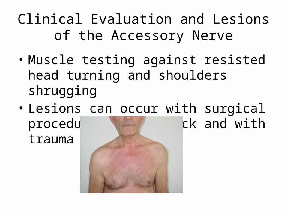
Clinical Evaluation and Lesions of the Accessory Nerve
• Muscle testing against resisted head turning and shoulders shrugging
• Lesions can occur with surgical procedures of the neck and with trauma

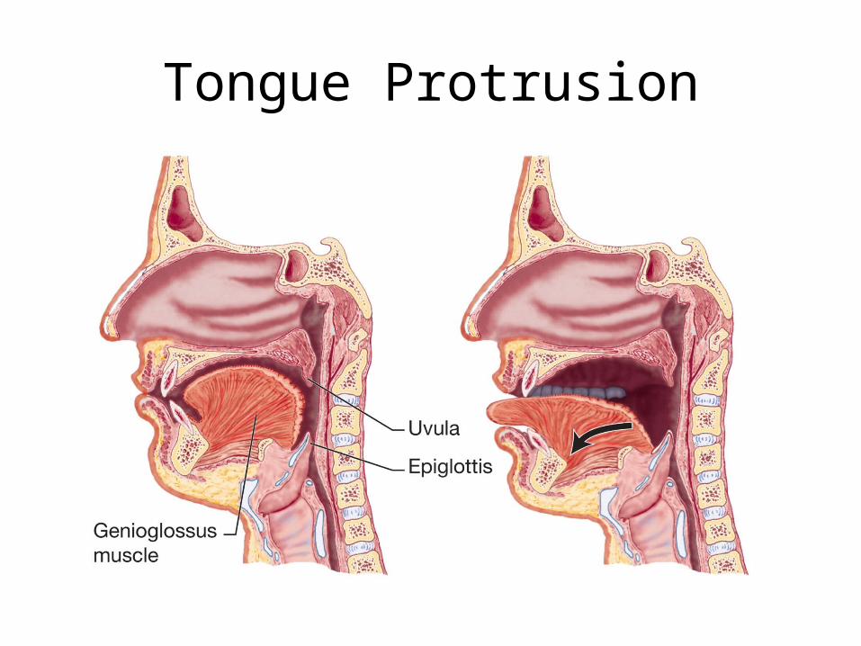
Clinical Evaluation and Lesions of the Hypoglossal Nerve
• Observation of tongue protrusion and speech articulation
• Lesions result in atrophy of the tongue and dysarthria – try saying “late night downtown”
Tongue Protrusion

Clinical Connection: Trigeminal Neuralgia
• Also called "tic douloureux"• Disease of PNS affecting the trigeminal ganglion or nerve
• Characterized by brief attacks of excruciating pain in one or more divisions of the trigeminal nerve
• Mechanisms and cause largely unknown

Trigeminal Neuralgia

Clinical Connection: Wallenberg's Syndrome
• Also called lateral medullary syndrome
• Typically caused by a vascular lesion, occlusion of the posterior inferior cerebellar artery
• Characterized by loss of pain and thermal sense in ipsilateral face and contralateral body due to damage of ascending spinal trigeminal and STT tracts

Wallenberg’s and Hypoglossal Syndromes
Clinical Connection: Dysarthria
• Motor deficits in the production of articulated speech
• Follows damage to UMNs or LMNs subserving the muscles of articulation
• Could be caused by a variety of pathologies including stroke, trauma, disease processes

Clinical Connection: Progressive Bulbar Palsy
• Motor system disease (combined UMN and LMN signs) dominated by weakness of the orofacial muscles
• Characterized by dysarthria, impaired chewing and swallowing, atrophy and fasciculations of tongue
• May present with pathological laughing and crying

Clinical Connection: Pseudobulbar Palsy
• Also called spastic bulbar paralysis
• Often caused by bilateral lesions of internal capsule affecting corticobulbar tracts
• Pathological laughing and crying along with bilateral bulbar signs

Figure 15-17 Midbrain syndromes. (a) Occlusion of the paramedian penetrating branches of the PCA results in superior alternating hemiplegia (Weber's syndrome). (b) Occlusion of the penetrating branches of the PCA that supply the tegmentum results in Benedikt's syndrome.

Clinical Connection: Weber's Syndrome
• Also called superior alternating hemiplegia
• Often caused by infarction of paramedian branches of the PCA

Clinical Connection: Weber's Syndrome
• Structures damaged– Descending tracts in cerebral peduncle, nucleus for CN III
• Related symptoms– Spastic hemiparesis of contralateral body and lower half of face, ipsilateral oculomotor palsy

Clinical Connection: Benedikt's Syndrome
• Caused by occlusion of tegmental branch of the PCA
• Structures damaged– Nucleus for CN III, cerebellothalamic tract
• Related symptoms– Ipsilateral oculomotor palsy and contralateral ataxia of the extremities

Medullary syndromes

Weber’s Syndrome