Embed Size (px)
Citation preview
© 2013 Ramenaden and Raiji. This work is published by Dove Medical Press Ltd, and licensed under Creative Commons Attribution – Non Commercial (unported, v3.0) License. The full terms of the License are available at http://creativecommons.org/licenses/by-nc/3.0/. Non-commercial uses of the work are permitted without any further
permission from Dove Medical Press Ltd, provided the work is properly attributed. Permissions beyond the scope of the License are administered by Dove Medical Press Ltd. Information on how to request permission may be found at: http://www.dovepress.com/permissions.php
Clinical Ophthalmology 2013:7 2113–2122
Clinical Ophthalmology Dovepress
submit your manuscript | www.dovepress.com
Dovepress 2113
R e v i e w
open access to scientific and medical research
Open Access Full Text Article
http://dx.doi.org/10.2147/OPTH.S37809
Clinical characteristics and visual outcomes in infectious scleritis: a review
emeline Radhika Ramenadenveena Rao RaijiDepartment of Ophthalmology, George washington University, washington, DC, USA
Correspondence: emeline Radhika Ramenaden Department of Ophthalmology, George washington University, 2150 Pennsylvania Ave, Nw, Suite 2A, washington, DC 20037, USA Tel +1 202 741 2800 Fax +1 202 741 2805 email [email protected]
Abstract: Infection is a very important but rare cause of scleritis, occurring in about 5%–10%
of all patients presenting with scleral inflammation. However, due to the similarity of its
presentation, infectious scleritis is often initially managed as autoimmune, potentially further
worsening its outcome. The overall visual outcome in infectious scleritis is generally worse
than its autoimmune counterparts, perhaps because of the delay in diagnosis or because of
the aggressive nature of associated microbes. Thus, there is a definite need for insight into the
diagnostic approach and treatment options for this ocular disease process. Several studies and
case reports have been published in recent years that have provided useful information regarding
the presenting clinical features and etiologic microbial agents in infectious scleritis. This review
summarizes the important findings in the literature that may aid in differentiating infectious
scleritis from other etiologies, including predisposing factors, microbe-specific characteristics,
diagnostic tools, treatment modalities, and outcomes.
Keywords: infectious scleritis, Pseudomonas, necrotizing scleritis, abscess
BackgroundScleritis is a state of ocular inflammation with a wide spectrum of clinical presentations
and etiologic factors (Table 1). Specific etiologies of scleritis, varying from idiopathic
to autoimmune to infectious, portend variable disease severity and outcome. Infection
is an important but rare cause of the scleritis, occurring in about 5%–10% of all cases.1
However, due to the similarity of its presentation, infectious scleritis is often initially
managed as autoimmune, potentially worsening its outcome. The overall visual outcome
in infectious scleritis is generally worse than its autoimmune counterparts, perhaps
because of this delay in diagnosis or because of the aggressive nature of associated
microbes. Recent studies have identified specific inciting factors for infectious scleritis.
Not surprisingly, these factors are typically surgery, most commonly pterygium surgery
but also excisions of conjunctival neoplasms, cataract surgery, vitreoretinal surgeries,
and glaucoma surgeries.2–4 Paula et al and Hodson et al report that the use of concomi-
tant radiation or mitomycin C is also a risk factor.2,5 Trauma, especially with introduction
of organic material to the ocular surface or self-inoculation from a distant site on the
body is also a significant inciting factor.6 In isolated cases, immunosuppression due to
human immunodeficiency virus or chemotherapy, may be a risk factor for spontane-
ous cases of infectious scleritis.2,7,8 Interestingly, Meyer et al reported an individual
case of spontaneous infectious scleritis without any such prior history that revealed
Pseudomonas aeruginosa resistant to 4th generation fluoroquinolones, indicating the
importance of considering infectious etiologies despite the lack of leading history.9

Clinical Ophthalmology 2013:7submit your manuscript | www.dovepress.com
Dovepress
Dovepress
2114
Ramenaden and Raiji
While postoperative worsening of scleritis has been well
reported in those with preexisting autoimmune scleritis, a
concomitant infectious etiology is much less common. The
similarities in presentation between autoimmune and infec-
tious scleritis often delays diagnosis. Infectious scleritis
after pterygium surgery with adjunctive β-radiation and
mitomycin C has been well established.2,11,12 Several reasons
may exist for this association. Meallet proposed in 2006 that
the use of adjunctive therapies such as these are likely to com-
promise the integrity of episcleral conjunctival vessels and
underlying tissue, thus inhibiting adequate wound healing and
leaving the sclera vulnerable to infection. He further hypoth-
esizes that surgical techniques such as leaving bare sclera
exposed or excessive use of cautery, as well as poor contact lens
hygiene or nonhealing epithelial defects may all contribute.12
P. aeruginosa, the most common agent in infectious scleritis,
utilizes neutrophil-activated collagenases to destroy tissue,
especially when sclera is left bare. Other microbes have
similar biochemical mechanisms by which they are able to
infiltrate ocular tissues.12 Furthermore, traditional antibiotic
regimens, both topical and systemic, remain inadequate due
to the avascularity of the sclera and the dense structure of
collagen fibers, limiting tissue penetration.12 Additionally,
the misdiagnosis of infectious scleritis as a postoperative
inflammatory complication leads to initial aggressive local
and systemic corticosteroid therapy that further worsens their
infection and may lead to poor visual outcomes.
Another known characteristic of infectious scleritis is
its sometimes delayed presentation, occurring months to
years after ocular surgery or trauma. Jain et al proposed that
conjunctival and tear film alterations may expose underly-
ing necrotic scleral collagen to microorganisms and allow
localization, adherence, colonization and invasion.1 Microbes
can then remain dormant within the sclera for years without
inciting an inflammatory response, making the diagnosis of
infectious scleritis in these cases quite difficult. It remains
unclear why these infections may be reactivated after such
long periods of latency.12
According to one large retrospective study by Hodson
et al, the median age of the patients at diagnosis of infec-
tious scleritis is 70 years old.2 Most studies reveal an older
population, suggesting that age may hold some significance
in the diagnosis of infectious scleritis.2,13,14 Additionally, the
time from an identifiable inciting event to the presentation of
scleritis can range in 0 days up to 36 years, generally longer
in postsurgical patients than the post-nonsurgical trauma
patients, with the longest latency period reported occurring
after a history of pterygium surgery.2,10
In all cases of infectious scleritis, P. aeruginosa is the
most common infectious agent.1,2 Additional identified
microbes include Nocardia, Aspergillus, Fusarium, Candida,
Beauveria, Stenotrophomonas, Streptococcus, Staphylococ-
cus aureus (especially of the methicillin-resistant type),
Serratia, Enterobacter, Achromobacter, Propionibacterium,
Haemophilus, Alcaligenes, mycobacteria, and herpes viridae.
In some cases, coinfections have been reported.2,15 Although
P. aeruginosa as well as other bacterial and viral etiologies
are most common in developed countries, fungi and Nocardia
are relatively more prevalent in developing countries, perhaps
due to humid climates and increased exposure to soil.1
Retinal surgeries such as vitrectomy, scleral buckling
with intrascleral fixation, and epiretinal membrane peel
procedures have been associated with acid-fast organisms,
such as Mycobacterium chelonae, more so than anterior seg-
ment procedures.2,16 These retinal surgeries have additionally
been associated with Pseudomonas, methicillin-resistant
Staphylococcus aureus, and coagulase-negative Staphylo-
coccus albus.3,17 The use of sub-Tenon’s triamcinolone ace-
tonide injection has also been associated with the development
of infectious scleritis in two cases, which were culture positive
for Staphylococcus epidermidis and Nocardia.18,19
PresentationInfectious scleritis may present with certain clinical patterns
that might facilitate more efficient diagnosis when recognized.
Table 1 Types of scleritis
Anterior-diffuse Greater than 50% contiguous scleral involvement. Associated with systemic disease. Generally in women 40–70 years old or men 30–60 years old. Rare in the young and the very old.
Anterior-nodular Distinct nodules present. Associated with isolated disease. Both sexes generally involved, 40–60 years old.
Anterior-necrotizing (with inflammation)
Scleral necrosis with scleromalacia. Associated with systemic disease. Generally in women in their 40s.
Anterior-necrotizing (without inflammation)
Scleral necrosis without scleromalacia. Usually associated with systemic disease. Generally in women, 35–75 years old.
Posterior scleritis Usually associated with anterior scleritis. Generally in women in their 60s. Diagnosis made using clinical and ultrasound findings.
infectious scleritis Generally necrotizing. Can have anterior and posterior involvement. Scleral ulcers with calcific plaque at the base and microabscesses. Tissue extension beyond obvious clinical findings.*
Notes: *Though infectious scleritis can present in many ways, these are the unique clinical findings found most commonly in this review. Data from Raiji et al,45 and watson and Hayreh.46
Clinical Ophthalmology 2013:7 submit your manuscript | www.dovepress.com
Dovepress
Dovepress
2115
Clinical characteristics and visual outcomes in infectious scleritis
Furthermore, because the prevalence of certain microbes in
infectious scleritis varies by environment, inciting event, or
sometimes even by symptomology, knowledge of this infor-
mation may allow faster microbial identification.
Similar to patients with autoimmune scleritis, patients
with infectious scleritis commonly present with symptoms
of redness, pain, and epiphora.13 Infectious scleritis after
retinal surgery tends to be associated with more rapid and
extreme pain than anterior segment surgeries.3 The most
common clinical signs at presentation in a retrospective
study of 55 patients with infectious scleritis included epis-
cleral and conjunctival hyperemia (98%) and scleral necro-
sis (93%),2 noted consistently in other studies as well.1,12,13
In this large study, 19 eyes had isolated scleritis whereas
37 involved adjacent ocular structures, including the cornea
and extraocular muscles, both spreading primarily from the
sclera to elsewhere or secondarily to the sclera.2 An anterior
chamber reaction of greater than 1+ cell is usually present
at the initial encounter.20,21 Endophthalmitis has also been
documented as the presenting finding.1,11,13,22 Not surpris-
ingly, retinal surgeries predisposed patients with infectious
scleritis to posterior scleral thickening and retinal or choroidal
detachments.3,16,17,21
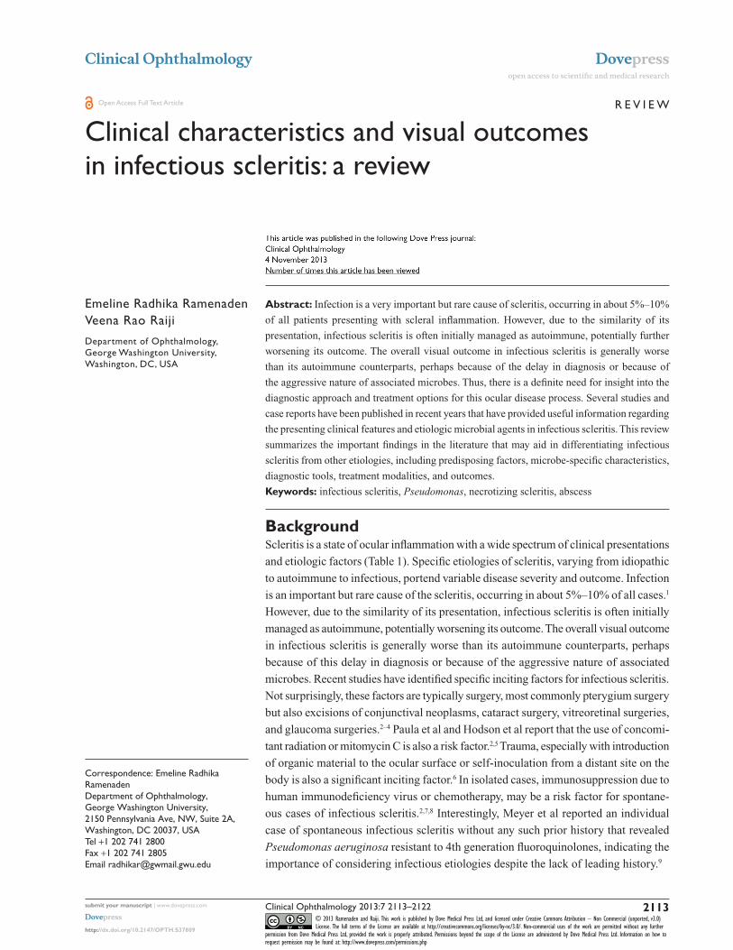
More notably, patients with infectious scleritis may
present with a scleral ulcer at sites of prior tissue pen-
etration, at times including satellite abscesses or involving
extraocular muscles, which may mimic orbital inflammatory
syndrome.1,13,21 Calcified plaques are often found at the base
of scleral ulcers (Figure 1).1,21 Unifocal or multifocal scleral
abscesses are consistently reported, appearing as distinctly
yellowish nodules under intact conjunctiva (Figure 2).1,13,23
Abscesses are characteristically scattered superiorly or infe-
riorly along an arc of 3–4 mm from the limbus (Figure 3).21
A black arc-shaped band from the initial ulcer site through
subsequent abscesses has been identified in some patients after
resolution, indicating a track of disseminated infection.21
Necrotizing scleritis tends to be the most common
presentation of infectious scleritis. A necrotizing pattern is
also seen in association with autoimmune scleritis with or
without vasculitis or metabolic disorders, all of which require
immunosuppression for treatment and would worsen visual
outcome in infectious scleritis (Figure 4).1,15 Thus, infectious
etiologies should be suspected in any case of progressive
indolent necrosis with suppuration, refractory to anti-
inflammatory regimens.1 Mild to moderate pain is generally
associated with diffuse and nodular scleritis whereas severe
pain is more frequently associated with necrotizing scleritis,24
Figure 1 Scleral ulcer with calcific plaque.Notes: (A) At presentation, there was a calcific plaque over the scleral ulcer bed (arrowhead). The ulcer progressed and became contiguous with a corneal infiltrate (arrow). (B) Multiple new nodules (arrowheads) from which P. aeruginosa was cultured, appeared on the 22nd day of hospitalization. ©1998 BMJ Publishing Group. Reproduced with permission from Hsiao CH, Chen JJ, Huang SC, Ma HK, et al. intrascleral dissemination of infectious scleritis following pterygium excision. Br J Ophthalmol. 1998;82(1):29–34.21
Figure 2 Slit lamp pictures depicting different clinical presentation.Notes: (A) Case no 2: multiple scleral abscess. (B) Case no 12: single scleral abscess. (C) Case no 4: necrotic ulcer (post cataract surgery). (D) Case no 3: two punched out ulcers. Reproduced with permission from Kumar SS, Das S, Sharma S et al. Clinico-microbiological profile and treatment outcome of infectious scleritis: experience from a tertiary eye care eye center of india. Int K Inflam. 2012:753560.13

Clinical Ophthalmology 2013:7submit your manuscript | www.dovepress.com
Dovepress
Dovepress
2116
Ramenaden and Raiji
suggesting that pain out of proportion to examination findings
may be indicative of a possible infectious etiology.
Other ocular complications noted at the time of presenta-
tion of infectious scleritis are decreased vision, peripheral
keratitis presenting as interstitial keratitis or thinning with
ulceration, or glaucoma.2,20
For the purposes of this review, infectious agents that have
not been shown in the literature to cause primary scleritis
were excluded.
DiagnosisIn two large retrospective reviews done in the United States
and Taiwan, the majority of cases of infectious scleritis were
found to be bacterial, with fewer cases being fungal.2,22 In
developing countries, however, fungal etiologies are more
common, likely related to differences in climate and envi-
ronmental exposures.1,13
In general, fungal, nocardial and mycobacterial infec-
tions take longer to diagnose than bacterial infections.2 It
is recommended that a complete diagnostic work-up of
infectious etiologies include scleral scrapings and culture
on blood and chocolate agar, brain–heart infusion broth,
thioglycolate broth, nonnutrient agar with E. coli overlay, and
Sabouraud dextrose agar.1 Diagnosis of viral etiologies like
herpes-related scleritis can be confirmed by scleral biopsy
and the use of immunofluorescence, as well as positive titers
or signs of chronic herpetic infection like corneal hypoes-
thesia.14,20 Kumar et al advocate scleral scrapings in every
case of suspected infectious scleritis that does not respond
to initial antibiotics, not only for the purpose of diagnosis
but also to debulk necrotic tissue and improve antimicrobial
penetration.13
In addition to cues from clinical presentation, history, and
slit-lamp examination, the use of ancillary testing has been
proposed to be of diagnostic value in infectious scleritis.
Nguyen and Yiu comment on the utility of ultrasound bio-
microscopy in allowing early detection of scleritis-associated
retinal and choroidal detachments.25 Ultrasound biomicros-
copy is further used to demonstrate anterior ciliary body rota-
tion and elimination of the ciliary sulcus in infectious scleritis
along with a diffuse lacy-appearing choroidal thickening.25
Additionally, they suggest that optical coherence tomography
allows the visualization of small vitreoid opacities and abnor-
mal subretinal deposits, which may represent lipofuscin-laden
macrophages in infectious scleritis (Figure 5).25
Su et al performed a prospective study in which they found
that elevated erythrocyte sedimentation rate and C-reactive
protein were noted in 9 of 12 patients with infectious scleritis.10
While these general inflammatory parameters can be elevated in
any case of inflammation, in these patients, both of these values
returned to normal with treatment of the infection.10 Addition-
ally, these patients underwent a thorough laboratory work-up
to document the lack of any associated underlying autoimmune
state that could have explained these laboratory findings.
Microbe-specific findingsBacterial etiologies comprise the majority of cases of infec-
tious scleritis.5 Bacterial proteins may contribute to tissue
damage via proteases and activation of tissue complement
pathways.5 Pseudomonas aeruginosa represents about 85%
of cases of bacterial scleritis.5 Thus, most findings discussed
Figure 3 Case 1: Scleral thinning from original ulcer through subsequent abscess extended in an arc shape.Note: © 1998 BMJ Publishing Group. Reproduced with permission from Hsiao CH, Chen JJ, Huang SC et al. Intrascleral dissemination of infectious scleritis following pterygium excision. Br J Ophthalmol. 1998;82(1):29–34.21
Before
0.00000
After
Diffuse Nodular Infectiousnecrotizing
Noninfectiousnecrotizing
Before After Before After Before After
1.00000
2.00000
BC
VA
(lo
gM
AR
)
Figure 4 Visual acuities before and after treatment.Notes: Patients with infectious necrotizing scleritis had the poorest BCVA before and after treatment. The data are presented as the mean ± standard deviation. Reproduced with permission from Ahn SJ, Oh JY, Kim MK, Lee JH, Wee WR. Clinical features, predisposing factors, and treatment outcomes of scleritis in the Korean population. Korean J Ophthalmol. 2010;24(6):331–335.15
Abbreviations: BCVA, best corrected visual acuities; logMAR, logarithmic value of the minimal angle of resolution.

Clinical Ophthalmology 2013:7 submit your manuscript | www.dovepress.com
Dovepress
Dovepress
2117
Clinical characteristics and visual outcomes in infectious scleritis
in relevant studies are largely in the context of Pseudomonas
scleritis. Though these findings are generally shared by most
infectious etiologies, there are certain characteristics or out-
comes associated with microbes other than Pseudomonas
that may be indicative of other etiologies.
Staphylococcus aureus is also an established cause of
bacterial scleritis. As reported by Arora et al, S. aureus
scleritis presented as a pyomyositis with an inverse hypopyon
and localized scleral thinning in a patient with a large thigh
abscess.26 Lee et al reported a case of methicillin-resistant
S. aureus 6 months after pterygium excision that was initially
misdiagnosed clinically as Pseudomonas and was unrespon-
sive to systemic amikacin and ceftazidime as well as topical
ciprofloxacin. They suggest that MRSA should be suspected
when typical postoperative antibiotics are ineffective.27
Streptococcus pneumoniae is a common infectious
agent in children and the elderly with concomitant joint,
cardiovascular, gastrointestinal, and genitourinary infections.
Recently, this bacteria has been associated with infectious
scleritis after pterygium surgery with concurrent use of
β-irradiation or mitomycin C.5,11 Paula et al reported a case
of S. pneumoniae scleritis that presented with mucopurulent
discharge, vascular congestion, and three scleral abscesses
with necrosis and a small aqueous fistula in the upper sclera
at the site of pterygium excision. This case responded well
to topical ciprofloxacin and intravenous cephatholin as well
as oral diclofenac.5 Altman et al discussed four cases of S.
pneumoniae scleritis that were treated with intravenous and
topical antibiotics, two of which had full resolution and good
visual outcome and two that had more devastating outcomes.
One of these latter patients developed endophthalmitis
requiring enucleation and one developed scleral perforation
requiring a corneoscleral graft with resulting count fingers
visual outcome.11 Therefore, S. pneumoniae represents a
potentially aggressive form of infectious scleritis.
Stenotrophomonas maltophilia is a rare aerobic nonfer-
mentative Gram-negative bacterium that is difficult to treat.
Lin et al reported a case of S. maltophilia-associated infec-
tious scleritis that occurred 18 years after pterygium surgery.28
Ramos-Esteban and Jeng reported a case of S. maltophilia
scleritis in which ultrasound biomicroscopy revealed a
dome-shaped mass over an area of a partial-thickness scleral
laceration after minor ocular trauma.29 In one particular case,
full resolution seemingly occurred after fortified topical and
systemic antibiotics as well as surgical debridement and patch
grafting. Interestingly, however, 5 months after resolution,
this patient presented with intrascleral dissemination of
the same infection, indicating the importance of long-term
follow-up and the possibility of microinvasion greater than
clinically detectable.28
Mycobacterium tuberculosis scleritis is rare. The myco-
bacteria itself can cause an immune-mediated inflammatory
microangiopathy and may indirectly lead to an inflamma-
tory scleritis.1 Kesen et al reported a case of a 54-year-old
woman with 6 months of eye pain and nodular scleritis
unresponsive to steroids and methotrexate. She was found
to be QuantiFERON® gold–positive with hilar granulomas
on CT, but due to initial lack of response to quadruple
tuberculosis therapy and a bronchoalveolar lavage negative
for acid fast bacilli, the regimen was abruptly discontinued
and immunosuppression was restarted. She later presented
with scleral rupture and polymerase chain reaction of the
specimen at that time revealed the M. tuberculosis genome.30
Mycobacterial infections other than tuberculosis (MOTT)
have a low incidence in humans but present in a disseminated
form in immunocompromised patients. These microbes
are known to be difficult to isolate by common laboratory
techniques and are easily confused with other pathogens.
Therefore, they should be suspected in immunocompromised
patients with +AFB (acid-fast bacilli) or granulomatous
noncaseating inflammation.31 M. chelonae is among these
MOTT species and has been shown to cause scleritis.31 M.
Figure 5 Advanced imaging studies of complications of infectious scleritis.Notes: (A) 35 MHz immersion ultrasound biomicroscopy demonstrating shallow anterior chamber, thickened and anteriorly rotated ciliary body (arrow), and elimination of the ciliary sulcus. (B) Thickened episcleral, scleral, and choroidal tissues are evident in the magnified view. (C) 10 MHz B-scan ultrasonography showing double retinal and choroidal detachment, L-12 view, (D) L-mac view, and (E) spectral optical coherence tomography showing vitreous clumps (dotted arrows and circles), subretinal fluid, and subretinal precipitates (solid arrow).Reproduced with permission from Nguyen P, Yiu SC. Imaging studies in a case of infectious scleritis after pterygium excision. Middle East Afr J Ophthalmol. 2012;19(3): 337–339.25
Abbreviations: L-12, longitudinal-12 view; L-mac, longitudinal-macular view.

Clinical Ophthalmology 2013:7submit your manuscript | www.dovepress.com
Dovepress
Dovepress
2118
Ramenaden and Raiji
chelonae is a quick-growth mycobacteria easily confused
with Nocardia or M. fortuitum that requires several tissue
samples to adequately differentiate, thereby delaying diag-
nosis and treatment. It is known to be resistant to typical
antituberculous drugs. Treatment must be continued for 4
weeks to 6 months after full resolution of clinical signs,
although due to the rarity of this diagnosis, the exact time
of treatment has not yet been established.31
Lepromatous leprosy, another mycobacterium-related
disease, has been reported to present with unilateral ocular
involvement as interstitial keratitis and granulomatous ante-
rior uveitis. In one report by Poon et al, an initial response was
achieved using antileprotics and anti-inflammatory agents;
however, the patient went on to develop recurrent episodes
of scleritis that necessitated multiple scleral patch grafts
for scleral thinning, subsequently requiring enucleation.
Histology failed to demonstrate lepromatous infection, but
showed chronic nongranulomatous scleritis.32
Toxoplasma gondii, an obligate intracellular parasitic
protozoan, is usually associated with posterior intraocular
inflammation; however, it has been reported by Schuman
et al to cause scleritis in five cases.33 Two of those patients
were diagnosed clinically and treated successfully with
medical therapy. Three of the patients, two of whom were
immunosuppressed, went undiagnosed without treatment
for toxoplasmosis, and progressed to eventually require
enucleation. Thus, T. gondii, while rare, should be suspected
in scleritis with associated retinochoroiditis, especially in
those who are immunosuppressed.33
Although herpes infections of the eye, both HSV and VZV,
have predominantly been thought of in relation to the cornea
and ocular adnexa, they have also been reported in cases of
infectious scleritis.14 Gonzalez-Gonzalez et al conducted a
large retrospective study of patients with herpetic scleritis
and found a predominance of middle-aged females with
unilateral findings and moderate to severe pain.20 The acu-
ity of the presentation of herpes scleritis can be variable.
While one study reports acute onset,20 another reports
chronic herpes-associated inflammation with an average of
3.2 years of symptoms prior to diagnosis.14 Herpetic scleritis
is more likely to be of the diffuse anterior type than of the
nodular or necrotizing type.20 Perilimbal devascularization
with peripheral corneal thinning along with a significant
amount of associated uveitis are findings associated with
herpetic scleritis, with significantly greater vision loss noted
in herpes than in idiopathic cases of scleritis.14 In general,
herpes infections are not associated with posterior scleritis,
vitreoretinal involvement, or scleromalacia perforans.14,20
Herpetic scleritis has similar rates of increased intraocular
pressure when compared to idiopathic types.20 Interestingly,
systemic symptoms are generally not present in patients with
herpetic scleritis, and the central cornea tends to be spared.14
This lack of typical corneal findings in herpetic scleritis
generally delays its diagnosis; therefore, a high index of sus-
picion should exist when the previously discussed symptoms
and signs are noted.20 Definitive diagnosis can be made by
immunohistopathological analysis of scleral biopsies, clinical
evidence of dendritic or stromal keratitis, or positive anti-HSV
or anti-VZV titers.20 Herpetic scleritis typically responds
quickly to acyclovir treatment, within 3–8 weeks,14,20 with
the inflammation generally lasting from 5–32 months.14 The
initial dosage of acyclovir used in these studies was 800 mg
five times daily,14,20 with most patients requiring a lower
maintenance dose of acyclovir to prevent recurrence, which
typically occurs at the same site as previous inflammation.14
Previous treatment with immunosuppressive therapy can
prolong and worsen herpetic scleral inflammation.14
Fungal scleritis, unlike bacterial and viral scleritis, has
worse overall outcomes (Table 2).1,34 Jain et al report that
fungal scleritis is more commonly encountered in hot, humid
climates, generally following surgical or accidental trauma.1
In cases of fungal scleritis, full thickness corneal inflamma-
tion contiguous with scleral lesions is quite common.1 In
their study, patients with fungal scleritis progressed despite
treatment, to develop rapidly progressive cataracts, serous
retinal or choroidal detachments, phthisis, or endophthalmi-
tis, of which a majority progressed to evisceration.1
Jain et al further report that Aspergillus and Nocardia
are the most common fungal agents in infectious scleritis.1
Fincher and Fulcher recovered Aspergillus fungal hyphae
from a biopsy of a scleral nodule in one patient with a
known history of intravenous drug abuse.35 This infection
was successfully treated with intravenous caspofungin as
Table 2 Comparison of fungal cases with other infectious scleritis cases
Fungal scleritis Others
Total number 8 13Associated corneal infiltration
3/8 (40%) 4/13 (30%)
Multifocal scleral abscess 3/8 (40%) 0endophthalmitis 3/8 (40%) 0eviscerated 4/8 (50%) 0Useful vision 1/8 (13%)
(95% CI, 0%–36%)6/13 (46%) (95% CI, 19%–63%)
Note: Microbial scleritis – experience from a developing country. © 2008 Nature Publishing Group. Reproduced with permission from: Jain V, Garg P, Sharma S. Microbial scleritis-experience from a developing country. Eye (Lond). 2009;23(2):255–261.1

Clinical Ophthalmology 2013:7 submit your manuscript | www.dovepress.com
Dovepress
Dovepress
2119
Clinical characteristics and visual outcomes in infectious scleritis
well as repeated surgical drainage of all emerging scleral
abscesses.35 Nocardia is much more common in agricultural
societies, where exposure to soil or plant matter is prevalent,
as compared with other infectious etiologies.1,34 Generally,
Nocardia does not cause fulminant infection; however, in
the presence of immunosuppression such as oral steroids
the presentation can be devastating, and can progress
to ocular infection and even involve the central nervous
system.36 In addition to the common findings in scleritis
of ocular pain, redness, and blurred vision, patients with
Nocardia-related scleritis have nodular lesions that present
as pointed abscesses.34 Nocardia should be suspected when
scleral inflammation does not respond to intensive first-line
antibiotic therapy.36 Direct smear is usually unrevealing;
diagnosis of Nocardia is more easily made with Gram stain
or acid-fast stain.34 One case of Nocardia scleritis showed
full resolution with debridement and systemic trimethoprim/
sulfamethoxazole,37 but it has also been shown to have very
good sensitivity to amikacin.36
Other rare fungi have been documented to cause scleritis.
Pseudallescheria boydii, a saprophytic fungus frequently
isolated from agricultural soil and polluted water, has been
reported to present with extensive involvement of the pos-
terior sclera with abscesses.38 Paecilomyces lilacinus is an
environmental mold found in soil and vegetation that rarely
causes human infection. Chung et al discussed a case of
P. lilacinus that presented as multiple scleral abscesses with
fibrinoid anterior chamber reaction that then progressed
to involve the cornea despite topical and oral antifungals.
Debridement and intracameral injection of amphotericin B
was required for resolution 4 months later, although it resulted
in phthisis and light perception vision.39 Lastly, Scedosporium
prolificans, an emerging opportunistic fungus, was found
to cause late-onset scleritis with a median presentation of
7.6 years after pterygium excision. Jhanji et al reported that
voriconazole is the most effective medical therapy for S.
prolificans scleritis, although it only allowed resolution in six
of nine cases.40 Thus, it is evident that overall visual prog-
nosis in fungal scleritis is generally poor, possibly because
of delayed diagnosis, poor penetration of antifungals into
avascular sclera, nonavailability of fungicidal agents, or the
ability of organisms to persist in avascular scleral tissue for
long periods of time without inciting inflammatory response,
allowing progressive worsening.1
TreatmentIt has been suggested repeatedly that prevention of infectious
scleritis is most important for patient outcome. Avoiding
overuse of cautery and adjunctive therapy during surgical
procedures may spare the episcleral blood flow and allow bet-
ter wound healing and resistance to infection.21 Most studies
advocate the avoidance of bare sclera techniques that leave
the ocular surface vulnerable to infection. However, in one
study by Tittler et al the use of amniotic membrane over an
area of debridement was discouraged, suggesting that it pro-
vides maximum exposure for topical antibiotics and prevents
the incubated microbes from staying at the site, with adequate
re-epithelialization occurring shortly after debridement.41 If
a bare sclera technique is chosen, close follow-up should be
undertaken with examination for scleral ischemia, the first
sign of which should prompt conjunctival autografting or
other scleral reinforcement.21
One large study of patients with infectious scleritis by
Hodson et al employed various antimicrobial treatment
regimens: 95% topical, 77% oral, and 11% intravitreal
therapy. Despite a mean treatment duration of 50 days,
medical therapy was adequate as the sole treatment in only
18% of patients, with most requiring surgical debridement.2
A higher rate of enucleation or evisceration was present in
those treated solely with medical methods.42 Cryotherapy,
lamellar or penetrating corneoscleral grafts, or removal of
hardware in addition to intensive antibiotics improves overall
outcomes in patients.2,43 Early and repetitive surgical debride-
ment is heavily advocated in multiple studies.1,13,22,41,42 Tittler
et al showed a 100% globe preservation rate, best corrected
vision of 20/120 to 20/400, fewer complications, and shorter
hospital stays with prompt surgical debridement at diagnosis
(within 2.5 days).41 Most significantly, it has been shown in
multiple studies that the inflamed area is often found intra-
operatively to be much larger than clinically judged by slit
lamp exam (Figure 6).1,13,21,22 Furthermore, there is sometimes
a notable tunnel lesion of scleral ulcer and small pockets
of abscess within necrotic sclera that require very careful
exploration. Some patients require a scleral patch graft due
to the large area of resulting debridement.1,13
The use of a fascia lata grafts in combination with an amni-
otic membrane graft after surgical debridement for infectious
scleritis was used successfully by Zheng et al in two patients,
one with Pseudomonas and one with fungal scleritis. Fascia
lata was thought to be ideal because of its bioadaptability in
size and thickness as well as its good cosmetic appearance.
Prior to grafting, necrotic scleral tissue was removed and the
surface was irrigated with ceftazidime. The fascia lata was
then dissected from the biceps femoris in the leg and placed
over the defect with an overlying amniotic membrane or
pedicle conjunctival flap, with subconjunctival injection of

Clinical Ophthalmology 2013:7submit your manuscript | www.dovepress.com
Dovepress
Dovepress
2120
Ramenaden and Raiji
ceftazidime followed by oral cefdinir for 4 days. This was the
only group to report the use of both a fascia lata and amni-
otic membrane grafts, and both patients showed evidence of
resolution.44
Not unlike fascia lata grafts, tectonic scleral reinforce-
ment with preserved pericardium and donor corneal tissue
was used successfully in a patient with nocardial scleritis
that progressed to scleral perforation and uveal prolapse.
This technique along with topical and oral antibiotics
allowed salvaging of the eye and a final Snellen visual
acuity of 20/70.36
A variety of medical modalities have been suggested as
good adjunctive treatments to surgical debridement. These
include systemic and topical antibiotics, subconjunctival
injections of antibiotic at both ends of the scleral lesion, and
wound irrigation with antibiotic solution one to two times a
day followed by normal saline after improvement.22 Intraocular
antibiotics should be used in all cases of endophthalmitis, and
topical steroids should always be initiated only when antibiot-
ics have been reliably used for several days.13
Finally, Meallet proposed the use of continuous
subpalpebral lavage antibiotics for the treatment of infectious
scleritis, reporting resolution of all six cases of scleritis in
this study after this treatment.12 However, this technique was
complicated by hyphema and cataract formation as well as
corneal thinning or dense corneal scar requiring corneal
transplants, indicating that important adjustments have yet
to be made to this technique. Additionally, the question of
systemic absorption of this continuous antibiotic regimen
was not adequately addressed.12
OutcomesAs may be expected after the discussion in this review,
infectious scleritis has worse visual acuity after remission
and longer resolution time as compared to noninfectious
scleritis.15 Not surprisingly, isolated infectious scleritis has
better prognosis than sclerokeratitis. Hodson et al reported
that 50% of eyes in their study with infectious scleritis lost
functional vision, indicating best corrected Snellen visual
acuity less than 20/200.2
Poor prognostic indicators in infectious scleritis include a
presenting vision worse than 20/200 or concomitant keratitis
or endophthalmitis, which tend to require enucleation or
evisceration more commonly.2 Additionally, fungal etiologies
tend to have worse prognosis, likely secondary to delays in
diagnosis.15 However, there is no difference in duration of
treatment or need for surgical debridement when comparing
bacterial versus fungal cases.2 The degree of final vision loss
does not significantly correlate with any specific inciting
factor, infectious organism, nor the amount of time from
symptom initiation to diagnosis. Rather, visual outcome only
significantly correlates with low presenting vision.2
Hodson et al report that the median time between com-
mencing treatment and resolution of scleritis was 46 days
with 50% of eyes losing functional vision.2 Only one case of
recurrence was noted after cessation of treatment in this study
after apparent full resolution.2 Though recurrences are noted
commonly as new nodules or necrotic areas during active
infectious scleritis, the appearance of such recurrences after
achieving full resolution of scleritis is generally rare.2,41
Surgical debridement repeatedly resulted in much greater
overall improvement in outcome. Sahu et al showed an improve-
ment in vision of greater than two lines in 60% of patients with
use of concurrent debridements as needed.13 Reversible causes
of vision loss include cataract, fibrotic pupillary membranes,
repairable retinal detachments, and corneal opacities. Irrevers-
ible causes include chronic retinal and choroidal detachments,
glaucoma, or the need for enucleation or evisceration.2,13
ConclusionInfectious scleritis, though rare, is an important and immi-
nently vision-threatening cause of scleritis. Therefore, it
is critical for eye care professionals to maintain infectious
etiologies in their differential diagnosis for all patients
with presenting symptoms of scleral hyperemia and sig-
nificant ocular pain. Of particular interest are patients with
Figure 6 Scleral ulcer before and after debridementNotes: (A) The scleral ulceration located at the 9 o’clock position. Severe congestion and subconjunctival abscess were noted at 10 o’clock position under the biomicroscope. (B) After the surgical debridement, the scleral wound was left open. in comparison with (A), the involved area was more extensive. (C) Ten days later, the debrided area was healing. © 1997 BMJ Publishing Group. Reproduced with permission from: Lin CP, Shih MH, Tsai MC. Clinical experiences of infectious scleral ulceration: a complication of pterygium operation. Br J Ophthalmol. 1997;81(11):980–983.22

Clinical Ophthalmology 2013:7 submit your manuscript | www.dovepress.com
Dovepress
Dovepress
2121
Clinical characteristics and visual outcomes in infectious scleritis
a history of ocular surgery or trauma, regardless of how
remote the event, as infections appear to remain dormant
within the sclera for months to years before reactivation.
In addition, patients’ refractory to treatment for presumed
autoimmune scleritis should be promptly considered for
infectious etiologies, as their immunosuppressive therapy
will inevitably worsen their infection. Scleral abscesses and
necrosis are common initial signs in infectious scleritis,
though many patients will present with more advanced
sequelae such as retinal or choroidal detachments and
glaucoma. Bacterial, viral, and fungal etiologies exist for
infectious scleritis, with Pseudomonas aeruginosa being
the most common overall cause and fungi having the worst
overall prognosis. Appropriate diagnosis generally requires
scleral scrapings with thorough culturing and immunohis-
tochemical staining. However, empiric treatment may be
necessary when these laboratory tests return inconclusive.
The characteristics of presentation for specific microbes
discussed in this review, along with ancillary tests such
as ultrasound and optical coherence tomography, may
facilitate appropriate diagnosis and therapy in these
cases. Effective treatment of infectious scleritis requires
both aggressive medical and surgical methods. In addi-
tion to microbe-specific medical therapy, repeat drainage
and debridement of scleral abscesses and necrotic tissue
with the use of various tissue grafts for scleral reinforce-
ment is necessary for resolution. Interestingly, the area of
involved sclera is consistently larger intraoperatively than
is initially judged on clinical exam, further supporting
the importance of surgical exploration. About half of all
patients with infectious scleritis maintain functional vision
after resolution. Recurrences rarely occur, but highlight the
importance of continued close observation for an extended
period of time even after clinical resolution in patients with
infectious scleritis. With proactive prevention, efficient
diagnosis, prompt initiation of aggressive treatment, and
close observation, patients with infectious scleritis may
have better outcomes.
DisclosureThe authors report no conflicts of interest in this work.
References1. Jain V, Garg P, Sharma S. Microbial scleritis-experience from a developing
country. Eye (Lond). 2009;23(2):255–261.2. Hodson KL, Galor A, Karp CL, et al. Epidemiology and visual outcomes
in patients with infectious scleritis. Cornea. 2013;32(4):466–472.3. Rich RM, Smiddy WE, Davis JL. Infectious scleritis after retinal surgery.
Am J Ophthalmol. 2008;145(4):695–699.
4. Cunningham MA, Alexander JK, Matoba AY, Jones DB, Wilhemus KR. Management and outcome of microbial anterior scleritis. Cornea. 2011;30(9):1020–1023.
5. Paula JS, Simão ML, Rocha EM, Romão E, Velasco Cruz AA. Atypical pneumococcal scleritis after pterygium excision: case report and litera-ture review. Cornea. 2006;25(1):115–117.
6. Maskin SL. Infectious scleritis after a diabetic foot ulcer. Am J Ophthalmol. 1993;115(2):254–255.
7. Hwang YS, Chen YF, Lai CC, Chen HS, Hsiao CH. Infectious scleri-tis after use of immunomodulators. Arch Ophthalmol. 2002;120(8): 1093–1094.
8. Moreno Honrado M, del Campo Z, Buil JA. A case of necrotizing scleritis resulting from Pseudomonas aeruginosa. Cornea. 2009;28(9): 1065–1066.
9. Meyer JJ, Espandar L, Marx DP, Vitale A, Moshirfar M. Pseudomonas scleritis resistant to fourth-generation fluoroquinolones in a patient without prior trauma or surgery. Ocul Immunol Inflamm. 2008;16(3): 127–129.
10. Su CY, Tsai JJ, Chang YC, Lin CP. Immunologic and clinical manifestations of infectious scleritis after pterygium excision. Cornea. 2006;25(6):663–666.
11. Altman AJ, Cohen EJ, Berger ST, Mondino BJ. Scleritis and Streptococcus pneumoniae. Cornea. 1991;10(4):341–345.
12. Meallet MA. Subpalpebral lavage antibiotic treatment for severe infectious scleritis and keratitis. Cornea. 2006;25(2):159–163.
13. Kumar Sahu S, Das S, Sharma S, Sahu K. Clinico-microbiological profile and treatment outcome of infectious scleritis: experience from a tertiary eye care center of India. Int J Inflam. 2012;2012:753560.
14. Bhat PV, Jakobiec FA, Kurbanyan K, Zhao T, Foster CS. Chronic herpes simplex scleritis: characterization of 9 cases of an underrecognized clinical entity. Am J Ophthalmol. 2009;148(5):779–789. e2.
15. Ahn SJ, Oh JY, Kim MK, Lee JH, Wee WR. Clinical features, predisposing factors, and treatment outcomes of scleritis in the Korean population. Korean J Ophthalmol. 2010;24(6):331–335.
16. Margo CE, Pavan PR. Mycobacterium chelonae conjunctivitis and scleritis following vitrectomy. Arch Ophthalmol. 2000;118(8): 1125–1128.
17. Lyne AJ, Lloyd-Jones D. Necrotizing scleritis after ocular surgery. Trans Ophthalmol Soc U K. 1979;99(1):146–149.
18. Gharaee H, Khalife M, Poor SS, Abrishami M. Infectious scleritis after subtenon triamcinolone acetonide injection. Ocul Immunol Inflamm. 2011;19(4):284–285.
19. Seth RK, Gaudio PA. Nocardia asteroides necrotizing scleritis associated with subtenon triamcinolone acetonide injection. Ocul Immunol Inflamm. 2008;16(4):139–140.
20. Gonzalez-Gonzalez LA, Molina-Prat N, Doctor P, Tauber J, Sainz de la Maza MT, Foster CS. Clinical features and presentation of infectious scleritis from herpes viruses: a report of 35 cases. Ophthalmology. 2012;119(7):1460–1464.
21. Hsiao CH, Chen JJ, Huang SC, Ma HK, Chen PY, Tsai RJ. Intrascleral dissemination of infectious scleritis following pterygium excision. Br J Ophthalmol. 1998;82(1):29–34.
22. Lin CP, Shih MH, Tsai MC. Clinical experiences of infectious scleral ulceration: a complication of pterygium operation. Br J Ophthalmol. 1997;81(11):980–983.
23. Riono WP, Hidayat AA, Rao NA. Scleritis: a clinicopathologic study of 55 cases. Ophthalmology. 1999;106(7):1328–1333.
24. Sainz de la Maza M, Molina N, Gonzalez-Gonzalez LA, Doctor PP, Tauber J, Foster CS. Clinical characteristics of a large cohort of patients with scleritis and episcleritis. Ophthalmology. 2012;119(1): 43–50.
25. Nguyen P, Yiu SC. Imaging studies in a case of infectious scleritis after pterygium excision. Middle East Afr J Ophthalmol. 2012;19(3): 337–339.
26. Arora R, Shroff D, Narula R, Chauhan D, Mehta DK. Inverse hypopyon as the presenting feature of infectious scleritis in a case of tropical pyomyositis. Can J Ophthalmol. 2006;41(6):769–771.

Clinical Ophthalmology
Publish your work in this journal
Submit your manuscript here: http://www.dovepress.com/clinical-ophthalmology-journal
Clinical Ophthalmology is an international, peer-reviewed journal covering all subspecialties within ophthalmology. Key topics include: Optometry; Visual science; Pharmacology and drug therapy in eye diseases; Basic Sciences; Primary and Secondary eye care; Patient Safety and Quality of Care Improvements. This journal is indexed on
PubMed Central and CAS, and is the official journal of The Society of Clinical Ophthalmology (SCO). The manuscript management system is completely online and includes a very quick and fair peer-review system, which is all easy to use. Visit http://www.dovepress.com/ testimonials.php to read real quotes from published authors.
Clinical Ophthalmology 2013:7submit your manuscript | www.dovepress.com
Dovepress
Dovepress
Dovepress
2122
Ramenaden and Raiji
27. Lee JE, Oum BS, Choi HY, Lee JS. Methicillin-resistant Staphylococcus aureus sclerokeratitis after pterygium excision. Cornea. 2007;26(6): 744–746.
28. Lin HC, Ma DH, Chen YF, Yeh LK, Hsiao CH. Late-onset intrascleral dissemination of Stenotrophomonas maltophilia scleritis after pterygium excision. Cornea. 2011;30(6):712–715.
29. Ramos-Esteban JC, Jeng BH. Posttraumatic Stenotrophomonas maltophilia infectious scleritis. Cornea. 2008;27(2):232–235.
30. Kesen MR, Edward DP, Rao NA, Sugar J, Tessler HH, Goldstein DA. Atypical infectious nodular scleritis. Arch Ophthalmol. 2009;127(8):1079–1080.
31. Metta H, Corti M, Brunzini R. Disseminated infection due to Mycobacterium chelonae with scleritis, spondylodiscitis and spinal epidural abscess. Braz J Infect Dis. 2008;12(3):260–262.
32. Poon A, MacLean H, McKelvie P. Recurrent scleritis in lepromatous leprosy. Aust N Z J Ophthalmol. 1998;26(1):51–55.
33. Schuman JS, Weinberg RS, Ferry AP, Guerry RK. Toxoplasmic scleritis. Ophthalmology. 1988;95(10):1399–1403.
34. DeCroos FC, Garg P, Reddy AK, et al; Hyderabad Endophthalmitis Research Group. Optimizing diagnosis and management of nocardia keratitis, scleritis, and endophthalmitis: 11-year microbial and clinical overview. Ophthalmology. 2011;118(6):1193–1200.
35. Fincher T, Fulcher SF. Diagnostic and therapeutic challenge of Aspergillus flavus scleritis. Cornea. 2007;26(5):618–620.
36. Ramos-Esteban JC, Servat JJ, Silva RS, Ambrósio R, Tauber S, Bia F. Necrotizing nocardial scleritis after combined penetrating keratoplasty and phacoemulsification with intraocular lens implantation: a case report and review of the literature. Arq Bras Oftalmol. 2007;70(2):355–359.
37. Maruo H, Shiraishi A, Hara Y, Maruo Y, Ohashi Y. Necrotizing nocar-dial scleritis successfully treated with surgical debridement and topi-cal polyvinyl alcohol iodine and antibiotics. J Ocul Pharmacol Ther. 2011;27(4):415–418.
38. Taravella MJ, Johnson DW, Petty JG, Keyser RB, Foster CS, Lundberg BE. Infectious posterior scleritis caused by Pseudallescheria boydii. Clini-copathologic findings. Ophthalmology. 1997;104(8):1312–1316.
39. Chung PC, Lin HC, Hwang YS, et al. Paecilomyces lilacinus scleritis with secondary keratitis. Cornea. 2007;26(2):232–234.
40. Jhanji V, Yohendran J, Constantinou M, Sheorey H, Vajpayee RB. Scedosporium scleritis or keratitis or both: case series. Eye Contact Lens. 2009;35(6):312–315.
41. Tittler EH, Nguyen P, Rue KS, et al. Early surgical debridement in the management of infectious scleritis after pterygium excision. J Ophthalmic Inflamm Infect. 2012;2(2):81–87.
42. Reynolds MG, Alfonso E. Treatment of infectious scleritis and keratoscleritis. Am J Ophthalmol. 1991;112(5):543–547.
43. Alfonso E. Surgical intervention in infectious keratoscleritis. Arch Ophthalmol. 1994;112(8):1017–1018.
44. Zheng X, Kodama T, Goto T, Ohashi Y. Autologous fascia lata grafts for scleral repair in eyes with infectious necrotizing scleritis. Arch Ophthalmol. 2011;129(9):1225–1227.
45. Raiji VR, Palestine AG, Parver DL. Scleritis and systemic disease association in a community-based referral practice. Am J Ophthalmol. 2009;148(6):946–950.
46. Watson PG, Hayreh SS. Scleritis and episcleritis. Br J Ophthalmol. 1976;60(3):163–191.
![Bilateral scleritis and sclerokeratitis associated with ...2Fs12348-012-0069-7.pdfand episcleritis [12, 14]. In IgAN, the deposits of IgA are frequently associated with complement](https://img.dokumen.tips/doc/110x75/5e357911c3b03016c37f790a/bilateral-scleritis-and-sclerokeratitis-associated-with-2fs12348-012-0069-7pdf.jpg)
![A case of scleritis associated rheumatoid arthritis ...syphilis, caused by nodular infectious uveitis [9, 10]. Biswas et al. reported a case of tuberculous uveitis associated with](https://img.dokumen.tips/doc/110x75/60997293e4fd5e2ef7072fd8/a-case-of-scleritis-associated-rheumatoid-arthritis-syphilis-caused-by-nodular.jpg)


![Hepatic angiosarcoma with an associated focal nodular ... · vascular channels [1,2]. Focal nodular hyperplasia (FNH), on the other hand, is a benign hepatic lesion displaying hepatocytic](https://img.dokumen.tips/doc/110x75/5f05ab797e708231d4141d25/hepatic-angiosarcoma-with-an-associated-focal-nodular-vascular-channels-12.jpg)