Embed Size (px)
Citation preview
ORIGINAL ARTICLE
Clinical and genetic characteristics of Japanese nephronophthisispatients
Keisuke Sugimoto1 • Tomoki Miyazawa1 • Takuji Enya1 • Hitomi Nishi1 •
Kohei Miyazaki1 • Mitsuru Okada1 • Tsukasa Takemura1
Received: 30 August 2015 / Accepted: 4 October 2015 / Published online: 23 October 2015
� Japanese Society of Nephrology 2015
Abstract
Background Nephronophthisis (NPH) accounts for
4–5 % of end-stage renal disease occurring in childhood.
Method We investigated the clinical context and char-
acteristics of renal and extrarenal symptoms, as well as the
NPHP genes, in 35 Japanese patients with clinical and
histologic features suggesting NPH.
Results NPH occurred fairly uniformly throughout Japan
irrespective of region or gender. In three families, NPH
affected siblings. The median age of patients was
12.5 years. Renal abnormalities attributable to NPH dis-
covered through mass screening, such as urine tests in
school. However, NPH accounted for less than 50 % of
children with abnormal findings, including incidentally
discovered renal dysfunction during evaluation of extra-
renal symptoms or during routine check-ups. Typical
extrarenal manifestations leaded to discovery including
anemia and delayed physical development. The urine often
showed low gravity specific density and low molecular
weight proteinuria. Frequent renal histologic findings
included cystic dilation of tubules, mainly in the medulla,
and irregularity of tubular basement membranes. Geneti-
cally abnormalities of NPHP1 were not common, with
large deletions frequently noted. Compound heterozygotes
showing single abnormalities in each of NPHP1, NPHP3,
and NPHP4 were observed.
Conclusions Our findings resemble those reported in
Western populations.
Keywords End-stage renal disease � Renal cysts � NPHPgenes � Children � Renal tubules
Introduction
Nephronophthisis (NPH) is a disease characterized by renal
medullary cyst formation. Additional histologic findings
include tubulointerstitial nephritis accompanied by pro-
gressive sclerosis and hyaline glomeruli. Although NPH
characteristically shows autosomal recessive inheritance, it
may occur sporadically [1]. NPH accounts for approximately
4–5 % of end-stage renal disease (ESRD) in childhood.
Disease subtypes include: infantile NPH (NPH2), which
progresses to ESRD around the age of 5 years; juvenile NPH
(NPH1), which develops from early childhood to school age
and usually progresses to ESRD by an age of about 13 or
14 years; and adolescent NPH (NPH3), with development of
ESRD at an average age of 19 years. Juvenile NPH is
reported to be the most common subtype [1].
NPHP1, the gene most often responsible for juvenile
nephronophthisis, encodes the nephrocystin-1 molecule.
This gene has an extent of approximately 11 kbp, and is
located on chromosome 2q12-13 [2]. The nephrocystin-1
protein consists of 677 amino acids and includes three
coiled domains; two highly acidic negatively charged
glutamic acid-rich domains; and an Src-homology 3
domain. Nephrocystin-1 has a molecular weight of 83 kD.
As this protein is located in the transition zone of primary
cilia of renal tubular epithelial cells, its abnormalities
typically cause dysfunction of these primary cilia (cil-
iopathy) [1, 2].
NPHP4, whose abnormalities cause a second form of
NPH1, is located on chromosome 1p36 and encodes the
nephrocystin-4 (nephroretinin) molecule. Nephrocystin-4
& Keisuke Sugimoto
1 Department of Pediatrics, Kinki University Faculty of
Medicine, 377-2 Ohno-higashi, Osaka-Sayama 589-8511,
Japan
123
Clin Exp Nephrol (2016) 20:637–649
DOI 10.1007/s10157-015-1180-5

has been shown to carry out signal transmission between
renal tubular epithelial cells, in cooperation with nephro-
cystin-1 [3].
NPHP2, the gene responsible for infantile NPH (NPH2),
is located on 9q22-31 [4]. NPHP2 encodes a protein termed
inversin (INVS). An abnormality in INVS can cause situs
inversus, pancreatic islet-cell dysplasia, cardiovascular
abnormalities, and hepato-biliary disorders. In addition,
INVS abnormalities can cause cyst formation resembling
that in juvenile nephronophthisis. However, the renal
prognosis is worse progression to ESRD in early childhood.
The gene responsible for adolescent NPH (NPH3),
NPHP3 is located on chromosome 3q21-22 [5]. NPHP3 is
believed to encode a protein involved in signal
Fig. 1 Genomic DNA extraction, PCR, and determination of NPHP1, 2, 3 and 4 gene sequence. PCR primers were prepared to amplify
approximately 200–300 bp fragments based on NPHP 1–4 gene sequences registered in GenBank, the following primers were used as shown
638 Clin Exp Nephrol (2016) 20:637–649
123

Fig. 1 continued
Clin Exp Nephrol (2016) 20:637–649 639
123

transmission in renal tubular epithelial cells, such as sig-
naling involving diacylglycerol kinase-zeta and receptor-
like tyrosine kinase. Abnormalities of the protein disrupt
urinary concentrating ability and the structure of cilia of
renal tubules, as in the other types of NPH.
Previous reports describe occurrence of NPHP1 muta-
tions in approximately 30–50 % of juvenile nephronoph-
thisis patients in Western countries [1, 6], where genetic
analysis of NPHP1 is performed initially when juvenile
NPH is suspected. If mutation is detected, kidney biopsy
usually is deferred [7]. Genetic diagnosis is made less
frequently in Japan; so kidney biopsy often is performed to
obtain a definitive diagnosis. Not infrequently, NPH is
discovered in the advanced or end stage in many Japanese
patients, in whom treatment no longer can slow progres-
sion. Unfortunately, symptoms typically seen in early
stages are incompletely characterized.
In the present study, we investigated clinical, histologic,
and genetic features in 35 Japanese patients clinically and
histologically suspected to have NPH, aiming to promote
early diagnosis. We studied many exons as many as 13
NPHP genes. Since such genetic analysis involves signif-
icant cost and time, we also screened biopsy specimens by
immunohistologic methods employing antibodies against
relevant peptides.
Methods
Patient registration and informed consent
Our subjects included 35 patients with clinicopathologic
findings suggestive of NPH who were referred to our
department from various regions of Japan. The study was
performed following approval by the Ethics Committee of
Kinki University Faculty of Medicine and acquisition of
written informed consent from patients or their parents
(Actual state of Japanese juvenile nephronophthisis
patients and identification of gene aberrations; approval
number 20–99).
Genomic DNA extraction, polymerase-chain
reaction (PCR), and determination of NPHP gene
sequence.
After approximately 5 mL of peripheral blood was col-
lected from patients into tubes containing Na-EDTA,
genomic DNA was extracted using NucleoSpin for Blood
(TaKaRa Bio Inc, Shiga, Japan). Human genomic DNA
(TaKaRa Clontech, code 636401; Shiga, Japan) was used
as a control. Patient samples and control genomic DNA
were diluted with sterile water to prepare 10 ng/lL solu-
tions. PCR was performed using these as templates and
TaKaRa PCR Thermal Cycler Dice Gradient (TaKaRa Bio
Inc, Shiga, Japan). To determine extent of deletions and
identify break points, PCR primers were prepared to
amplify approximately 200–300 bp fragments based on
NPHP gene sequences registered in GenBank (Fig. 1). For
PCR, annealing temperatures and times were 63 �C and
15 s for NPHP1 and NPHP3; 60 �C and 15 s for NPHP2;
and 60 �C and 20 s for NPHP4, respectively. For sequence
analysis, PCR products were purified by an enzyme reac-
tion, and templates for sequencing were prepared. The
sequencing reaction was carried out using the prepared
template DNA and a BigDye Terminator v.3.1 Cycle
Sequencing Kit (Applied Biosystems, CA, USA),
employing the dye terminator method. Reaction products
were purified by gel filtration, and sequence analysis was
performed using a capillary-type sequencer, ABI3730xl
(Applied Biosystems, CA, USA). The algorithm estab-
lished by Salomon et al. [8]. was adapted for use in our
analytical procedure. In children with renal dysfunction
Fig. 2 Percentage of NPH
patients with NPHP gene
mutation. NPHP gene mutation
was detected in 19 patients. No
NPHP gene aberration detected
within the sequences analyzed
in the other 16 patients with
suspicion of NPH
clinicopathologically
640 Clin Exp Nephrol (2016) 20:637–649
123

Table
1C
har
acte
rist
ics
of
pat
ien
tsfo
un
dto
hav
eN
PH
Pg
ene
mu
tati
on
s
Ag
e/g
end
erM
oti
ve
of
dis
cov
ery
BU
N/
Orn
(mg
/
dL
)a
UP
Uri
nar
y
LM
P
Lo
w
gra
vit
y
uri
ne
Ex
trar
enal
sym
pto
m
Dia
gn
osi
sat
firs
tb
iop
sy
NPHP
/mu
tati
on
Th
eo
ther
NPHP
mu
tati
on
Co
nse
ng
uin
eou
s
mar
riag
e
Fam
ily
his
tory
of
ren
ald
isea
se
14
yea
rs/M
An
emia
49
/3.3
(-)
(-)
(-)
n.f
n.d
Lar
ge
del
atio
n
([2
.0k
bp
)
(-)
(-)
13
yea
rs/F
No
ctu
rnal
enu
resi
s2
7/1
.3(-
)(-
)(-
)n
.fn
.dL
arg
ed
elat
ion
(-)
NP
H(y
ou
ng
er
bro
ther
)
11
yea
rs/M
Sib
lin
gw
ith
NP
H1
7/0
.6(-
)(?
)(?
)n
.fn
.dL
arg
ed
elat
ion
(-)
NP
H(e
lder
sist
er)
15
yea
rs/F
Pro
tein
uri
a(s
cho
ol
uri
nal
ysi
s)
21
.3/1
.3(±
)(-
)(-
)n
.fT
IN(-
)D
19
80
G
(NPHP4
,
het
ero
)
(-)
(-)
15
yea
rs/F
Pro
tein
uri
a(s
cho
ol
uri
nal
ysi
s)
89
.6/
11
.6
(-)
(?)
(?)
RP
NP
HP
arti
ald
elat
ion
(=3
00
bp
)
(-)
Acu
te
glo
mer
ulo
nep
hri
tis
(mo
ther
)
14
yea
rs/M
Ch
ance
dis
cov
ery
of
the
RD
(hea
tstr
ok
e)
17
/0.9
(±)
(?)
(?)
n.f
n.d
n.d
L9
39
(NPHP4
,
het
ero
)
(-)
(-)
11
yea
rs/F
En
ure
sis,
po
lyu
ria
74
/5.2
(1?
)(?
)(?
)S
S(-
2.5
SD
)N
PH
E6
77
Q(h
eter
o)
E6
42
L
(NPHP4
,
het
ero
)
(-)
(-)
18
yea
rs/F
En
ure
sis,
po
lyd
ipsi
a8
2/8
.1(±
)(?
)(?
)S
S(-
1.8
SD
)N
PH
Gln
54
7(h
eter
o)
S8
0L
(NPHP3
,
het
ero
)
(-)
Pro
tein
uri
a(f
ath
er)
8y
ears
/MG
lyco
suri
a(s
cho
ol
uri
nal
ysi
s)
15
9/
11
.1
(2?
)(?
)(?
)n
.fS
imil
arN
PH
E6
77
Q(h
eter
o)
NPHP4
(-)
(-)
(-)
14
yea
rs/M
Gly
cosu
ria
(sch
oo
l
uri
nal
ysi
s
48
/5.0
(±)
(?)
(?)
RP
NP
HL
arg
ed
elat
ion
(-)
NP
H(y
ou
ng
ersi
ster
)
13
yea
rs/F
Sib
lin
gw
ith
NP
H5
8/2
.7(1?
)(?
)(?
)R
PN
PH
Lar
ge
del
atio
n(-
)N
PH
(eld
erb
roth
er)
20
yea
rs/F
Ch
ance
dis
cov
ery
of
the
RD
(med
ical
exam
inat
ion
)
44
.6/2
.4(1?
)(?
)(?
)n
.fn
.d(-
)L
93
9Q
(NPHP4
,
ho
mo
)
(-)
(-)
8y
ears
/FC
han
ced
isco
ver
yo
f
the
RD
32
.2/1
.4(-
)(?
)(?
)n
.fN
PH
Lar
ge
del
atio
n(-
)(-
)
15
yea
rs/F
Pro
tein
uri
a(s
cho
ol
uri
nal
ysi
s)
37
/2.6
(-)
(?)
(?)
n.f
Tu
bu
lar
enla
rgem
ent
med
ull
ary
cyst
s
Lar
ge
del
atio
n(-
)(-
)
7y
ears
/FC
han
ced
isco
ver
yo
f
the
RD
(uri
ne
trac
t
infe
ctio
n)
40
/3.0
(1?
)(?
)(?
)Jo
ub
ert
syn
dro
me
n.d
(-)
AA
/OO?
AG
/
OT
(ex
on
26
/ex
on
20
)
(-)
(-)
19
yea
rs/F
Ch
ance
dis
cov
ery
of
the
RD
(bro
nch
itis
)
90
.3/8
.4(1?
)(?
)(?
)n
.fn
.d(-
)A
15
0V
(NPHP3
,
het
ero
)
(-)
(-)
Clin Exp Nephrol (2016) 20:637–649 641
123
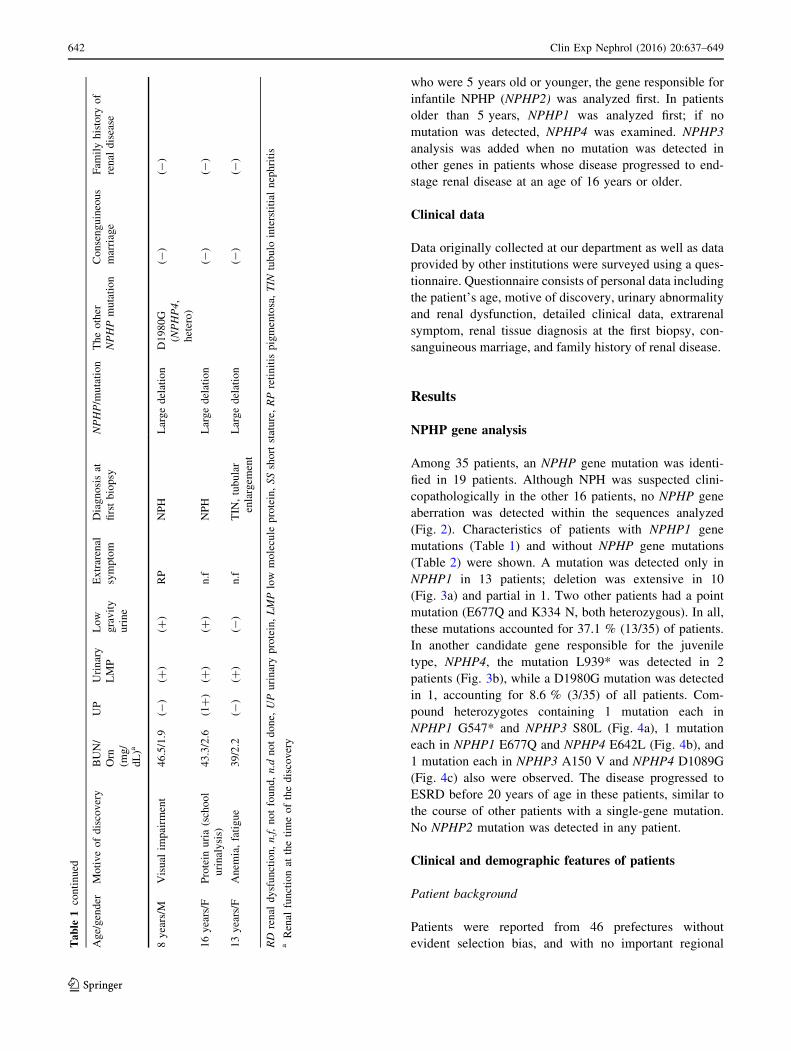
who were 5 years old or younger, the gene responsible for
infantile NPHP (NPHP2) was analyzed first. In patients
older than 5 years, NPHP1 was analyzed first; if no
mutation was detected, NPHP4 was examined. NPHP3
analysis was added when no mutation was detected in
other genes in patients whose disease progressed to end-
stage renal disease at an age of 16 years or older.
Clinical data
Data originally collected at our department as well as data
provided by other institutions were surveyed using a ques-
tionnaire. Questionnaire consists of personal data including
the patient’s age, motive of discovery, urinary abnormality
and renal dysfunction, detailed clinical data, extrarenal
symptom, renal tissue diagnosis at the first biopsy, con-
sanguineous marriage, and family history of renal disease.
Results
NPHP gene analysis
Among 35 patients, an NPHP gene mutation was identi-
fied in 19 patients. Although NPH was suspected clini-
copathologically in the other 16 patients, no NPHP gene
aberration was detected within the sequences analyzed
(Fig. 2). Characteristics of patients with NPHP1 gene
mutations (Table 1) and without NPHP gene mutations
(Table 2) were shown. A mutation was detected only in
NPHP1 in 13 patients; deletion was extensive in 10
(Fig. 3a) and partial in 1. Two other patients had a point
mutation (E677Q and K334 N, both heterozygous). In all,
these mutations accounted for 37.1 % (13/35) of patients.
In another candidate gene responsible for the juvenile
type, NPHP4, the mutation L939* was detected in 2
patients (Fig. 3b), while a D1980G mutation was detected
in 1, accounting for 8.6 % (3/35) of all patients. Com-
pound heterozygotes containing 1 mutation each in
NPHP1 G547* and NPHP3 S80L (Fig. 4a), 1 mutation
each in NPHP1 E677Q and NPHP4 E642L (Fig. 4b), and
1 mutation each in NPHP3 A150 V and NPHP4 D1089G
(Fig. 4c) also were observed. The disease progressed to
ESRD before 20 years of age in these patients, similar to
the course of other patients with a single-gene mutation.
No NPHP2 mutation was detected in any patient.
Clinical and demographic features of patients
Patient background
Patients were reported from 46 prefectures without
evident selection bias, and with no important regionalTable
1co
nti
nu
ed
Ag
e/g
end
erM
oti
ve
of
dis
cov
ery
BU
N/
Orn
(mg
/
dL
)a
UP
Uri
nar
y
LM
P
Lo
w
gra
vit
y
uri
ne
Ex
trar
enal
sym
pto
m
Dia
gn
osi
sat
firs
tb
iop
sy
NPHP
/mu
tati
on
Th
eo
ther
NPHP
mu
tati
on
Co
nse
ng
uin
eou
s
mar
riag
e
Fam
ily
his
tory
of
ren
ald
isea
se
8y
ears
/MV
isu
alim
pai
rmen
t4
6.5
/1.9
(-)
(?)
(?)
RP
NP
HL
arg
ed
elat
ion
D1
98
0G
(NPHP4
,
het
ero
)
(-)
(-)
16
yea
rs/F
Pro
tein
uri
a(s
cho
ol
uri
nal
ysi
s)
43
.3/2
.6(1?
)(?
)(?
)n
.fN
PH
Lar
ge
del
atio
n(-
)(-
)
13
yea
rs/F
An
emia
,fa
tig
ue
39
/2.2
(-)
(?)
(-)
n.f
TIN
,tu
bu
lar
enla
rgem
ent
Lar
ge
del
atio
n(-
)(-
)
RD
ren
ald
ysf
un
ctio
n,n.f,
no
tfo
un
d,n.d
no
td
on
e,UP
uri
nar
yp
rote
in,LMP
low
mo
lecu
lep
rote
in,SS
sho
rtst
atu
re,RP
reti
nit
isp
igm
ento
sa,TIN
tub
ulo
inte
rsti
tial
nep
hri
tis
aR
enal
fun
ctio
nat
the
tim
eo
fth
ed
isco
ver
y
642 Clin Exp Nephrol (2016) 20:637–649
123

Table
2C
har
acte
rist
ics
of
pat
ien
tsw
ith
ou
tap
par
ent
NP
HP
gen
em
uta
tio
ns
Ag
e/g
end
erM
oti
ve
of
dis
cov
ery
BU
N/
Orn
(mg
/
dL
)a
UP
Uri
nar
y
LM
P
Lo
w
gra
vit
y
uri
ne
Ex
trar
enal
sym
pto
m
Dia
gn
osi
sat
firs
t
bio
psy
NP
HP
1
mu
tati
on
NP
HP
3
mu
tati
on
NP
HP
4
mu
tati
on
Co
nsa
ng
uin
eou
s
mar
riag
e
Fam
ily
his
tory
of
ren
ald
isea
se
6y
ears
/ML
agg
ing
ph
ysi
cal
dev
elo
pm
ent
34
/1.2
(-)
(-)
(-)
SS
(-1
.3S
D)
NP
H(-
)n
.d(-
)(-
)(-
)
12
yea
rs/M
SS
,fa
tig
ue
48
/1.3
(-)
(?)
(-)
Sen
sory
dea
fnes
s
Ch
ron
icin
ters
titi
al
nep
hri
tis,
glo
mer
ulo
scle
rosi
s
(-)
n.d
(-)
(-)
(-)
26
yea
rs/M
Ch
ance
dis
cov
ery
of
the
RD
(med
ical
exam
inat
ion
)
21
/1.6
(-)
(?)
(-)
n.f
Inte
rsti
tial
nep
hri
tis
(-)
(-)
(-)
(-)
Ren
al
dy
sfu
nct
ion
(fat
her
,y
ou
ng
sist
er)
11
yea
rs/M
Pal
lor,
anem
ia2
7.4
/1.5
(-)
(?)
(?)
SS
(-2
.2S
D)
n.d
(-)
n.d
(-)
(-)
(-)
17
yea
rs/M
SS
34
/1.5
(±)
(?)
(?)
SS
(-3
.8S
D)
n.d
(-)
n.d
(-)
(-)
(-)
22
yea
rs/M
Hy
per
ten
tio
n3
2.5
/1.5
(1?
)(?
)(-
)R
PT
IN(-
)(-
)(-
)(-
)(-
)
11
yea
rs/F
po
lyd
ipsi
a,p
oly
uri
a1
5.4
/0.7
(-)
(?)
(?)
n.f
n.d
(-)
n.d
(-)
(-)
(-)
12
yea
rs/F
Ch
ance
dis
cov
ery
of
the
RD
(pro
tein
uri
aat
3
yea
rso
ld)
32
/1.6
(2?
)(?
)(?
)n
.fT
IN,
tub
ula
r
enla
rgem
ent
(-)
(-)
(-)
(-)
(-)
14
yea
rs/M
Pro
tein
uri
a(s
cho
ol
uri
nal
ysi
s)
58
/4.2
(1?
)(?
)(?
)n
.fT
IN,
tub
ula
r
enla
rgem
ent
(-)
(-)
(-)
(-)
(-)
26
yea
rs/M
Hy
per
ten
tio
n(m
edic
al
exam
inat
ion
)
38
.7/1
.5(-
)(?
)(?
)n
.fT
IN,
glo
mer
ulo
scle
rosi
s
(-)
(-)
(-)
( -)
(-)
10
yea
rs/M
Pal
lor,
anem
ia5
3.6
/1.0
(1?
)(?
)(?
)n
.fN
PH
(-)
n.d
(-)
(-)
(-)
28
yea
rs/F
Ch
ance
dis
cov
ery
of
the
RD
anem
ia
47
.5/2
.9(1?
)n
.d(?
)n
.fT
IN,
sim
ilar
NP
H(-
)(-
)(-
)(-
)(-
)
11
yea
rs/F
Pal
lor,
po
lyd
ipsi
a,
po
lyu
ria
27
.4/1
.5(-
)(?
)(?
)S
S(-
2.2
SD
)n
.d(-
)n
.d(-
)(-
)(-
)
18
yea
rs/M
Cru
d,
fati
gab
ilit
y4
9.6
/4(±
)(?
)(?
)S
pec
ific
com
ple
xio
n
Sim
ilar
NP
H(-
)(-
)(-
)(-
)N
PH
(yo
un
g
sist
er)
16
yea
rs/F
Sib
lin
gw
ith
NP
H3
8.3
/1.8
(-)
(?)
(-)
Sp
ecifi
c
com
ple
xio
n
Sim
ilar
NP
H(-
)(-
)(-
)(-
)N
PH
(eld
er
bro
ther
)
46
yea
rs/F
Pro
tein
uri
a,h
emat
uri
a
(at
30
yea
rso
ld)
33
/1.8
(1?
)(?
)(-
)S
enso
ry
dea
fnes
s
Ch
ron
icin
ters
titi
al
nep
hri
tis,
glo
mer
ulo
scle
rosi
s
(-)
(-)
(-)
(-)
NP
H(e
lder
bro
ther
)
RD
ren
ald
ysf
un
ctio
n,n.f
no
tfo
un
d,n.d
no
td
on
e,UP
uri
nar
yp
rote
in,LMP
low
mo
lecu
lep
rote
in,SS
sho
rtst
atu
re,RP
reti
nit
isp
igm
ento
sa,TIN
tub
ula
rin
ters
titi
aln
eph
riti
sa
Ren
alfu
nct
ion
atth
eti
me
of
the
dis
cov
ery
Clin Exp Nephrol (2016) 20:637–649 643
123

differences (Fig. 5). The male:female ratio was 16:19, with
evident gender difference. Ages of patients ranged from 2
to 38 years (median; 12.5). Familial occurrence was noted
in 3 families. Other occurrences were solitary, with no
family member showing a urinary abnormality, a diagnosis
of NPH, or any renal dysfunction of unknown cause.
Initial abnormality deletion
NPH sometimes was discovered following an abnormal
urinary finding by mass screening, such as proteinuria
detected in a urine test at school (18 %), or renal dys-
function discovered incidentally in working up other
medical symptoms, or during medical check-ups (23 %).
Approximately 20 % of cases were discovered because of
urinary tract symptoms such as polyuria with or without
polydipsia, enuresis (often nocturnal), or mellituria. Some
38 % were discovered because of either extrarenal mani-
festations such as lagging physical development, dwarfism,
anemia, pallor, hypertension, or visual disturbance arising
from pigmentary retinal degeneration; a prior diagnosis of
NPH in a sibling; or both (Fig. 6).
Urinary findings
Urine specific gravity frequently was low (not greater than
1.010); approximately 75 % of cases. Low molecular
weight proteinuria, such as b2-microgloburinuria, also was
common (85 %), even though inclusion of renal function
shown such as between blood urea nitrogen and serum
creatinine was relatively mild at that time.
Renal histologic findings
Renal biopsy was performed in 25 patients (71 %). These
included 13 patients demonstrated to have an NPHP gene
mutation and in 12 with no NPHP gene mutation identi-
fied (suspected cases). Histologic findings included sus-
pected NPH; interstitial nephritis, renal tubular dilation,
and glomerulosclerosis. Cystic dilation of renal tubules
and irregular contours of tubular basement membranes
were observed in most patients, mainly in the renal
medulla (Fig. 7a). Sclerotic glomeruli, inflammatory cell
infiltration in the renal tubules and interstitium, and
fibrosis were frequent, although not seen in all patients
(Fig. 7b).
Discussion
Renal tubular epithelial cells are attached to the basement
membrane through integrin cross-linking, which transmits
extracellular signals to the cell nucleus [2]. Nephrocystin
acts importantly in signal transmission between tubular
epithelial cells and between these epithelial cells and the
extracellular matrix functioning as a docking protein.
Nephrocystin also is involved in cell adhesion, together
with N-cadherin, catenin, and b-catenin [2, 8]. Further-
more, nephrocystin influences actin cytoskeleton structure
together with b-tubulin, contributing to maintenance of the
cytoskeleton and determination of cell polarity. Nephro-
cystin forms a complex with Crk-associated substrate,
which promotes phosphorylation of Pyk2 and transmits
intracellular information through a Pyk2-dependent path-
way [2]. Furthermore, nephrocystin is present on primary
cilia, where it functions in cooperation with a-tubulin;
nephrocystin also is involved in signal transmission in
organelles [9]. Accordingly, abnormalities in the nephro-
cystin molecule disrupt signal transmission between cells
Fig. 3 Analysis of deletion in NPHP1 (a) and analysis of NPHP4
(b). In a lane 1 and lanes 6 and 7, contain PCR products of regions
within and outside NPHP1, respectively; Lane 2 contains PCR
products from the junction between NPHP1 and the adjacent MALL
gene. Lanes 3–5 show the PCR products of NPHP1 obtained with
primers amplifying fragments of approximately 300 bp. NPHP1 was
nearly completely deleted (1.2 kbp deletion). In b, substitution of
TAG for TTG formed a stop codon, prematurely terminating peptide
synthesis
644 Clin Exp Nephrol (2016) 20:637–649
123

and the extracellular matrix, intercellular adhesion,
cytoskeletal integrity, cell polarity, primary cilia function,
and intracellular signal transmission to the nucleus.
Structural and functional disorders involving the renal
tubular epithelium result.
An NPHP gene mutation was detected in about 54 % of
all patients, but no mutation was noted within the sequences
analyzed in the other 46 %. However, nephronophthisis was
suspected clinically and histologically, suggesting possible
mutation in some other NPHP gene. An NPHP1 mutation
was most frequent among our Japanese patients, most often
representing a large deletion rather than a point mutation.
Frequency of an NPHP1 mutation was similar to that
reported in Western populations [10].
On the other hand, mutation in the gene responsible for
the infantile type, NPHP2, a patient in a compound
heterozygous stable with another abnormal NPHP gene
such as that responsible for NPH3 recently has been
Fig. 4 Compound
heterozygotes with
heterozygous mutations in
different NPHP genes. In a, a
compound heterozygote has one
heterozygous mutation
involving each of NPHP1
(G547*) and NPHP3 (S80L). In
b, a compound heterozygote has
one heterozygous mutation
involving each of NPHP1
(E677Q) and NPHP4 (E642L).
In c, a compound heterozygote
has one heterozygous mutation
involving each of NPHP3
(A150 V) and NPHP4
(D1089G)
Clin Exp Nephrol (2016) 20:637–649 645
123

reported [11, 12]. We also found compound heterozygosity
across multiple NPHP genes in some of our Japanese
nephronophthisis patients. In NPHP4, L939* (IVS-
20T[A) was detected in two geographically distant
patients who were not consanguineous. The mutation
formed a stop codon by substituting TAG for TTG in exon
21, terminating peptide synthesis. This might prove to be a
‘hot spot’ among Japanese patients.
We detected in three patients with two mutations in
either NPHP1, NPHP3, or NPHP4 in this study. As similar
to the results of the other studies [13, 14], the age of the
initial discovery of this disease and the course of pro-
gression to end-stage renal disease were not significantly
different from those of the patients having mutation in
single NPHP gene. An analysis of patient backgrounds
revealed that NPH was distributed fairly evenly Japan,
including the suspected cases where no causative mutation
was identified. Heterozygotes carrying NPHP gene muta-
tions also were rather evenly distributed nationwide. No
gender difference was evident from our analysis. Although
the median age at time of disease discovery was 12.5 years,
individual presentation ranged from infantry to adulthood.
Frequency of disease discovery in mass screening pro-
grams, such as school urine tests, was low, as previously
reported [15]. Incidental discovery of renal dysfunction
during diagnostic workup of possibly unrelated symptoms,
or during routine check-ups, accounted for less than 50 %
of cases. Often symptoms that led to the discovery of NPH
represented extrarenal manifestations such as incomplete
physical development reported previously [15]. In partic-
ular, currently used urine test strips, intended mainly to
detect albuminuria, are insensitive to this disease.
Fig. 5 Demographic features of patients in Japan. Regional distribution of study subjects within Japan, show as a dot for each patient
646 Clin Exp Nephrol (2016) 20:637–649
123

Development in siblings was noted in three families, sug-
gesting autosomal recessive inheritance. However, many
cases appear to be sporadic. Familial genetic analysis
centering on patients, parents is needed.
In contract to albuminuria, urinary findings such as low
specific gravity and low-molecular-weight proteinuria are
relatively helpful in early discovery. According to the
results of this study, we suggest that the findings of the
low-molecular weight proteinuria and hypotonic urine
reflecting renal tubular disorder coupled with the histologic
abnormalities involving cystic dilation of renal tubules and
the irregularity of tubular basement membrane could be a
convincing diagnostic criterion of this disease. Extrarenal
manifestations, such as short stature, delayed physical
development, and anemia also were frequent. Unfortu-
nately, these tended to coincide with were progression of
renal dysfunction rather than early NPH. Nonetheless, NPH
needs to be considered in children with such presentations.
Some patients have been reported to show somewhat dis-
tinctive extrarenal manifestations [13] such as pigmentary
retinal degeneration (Senior-Loken syndrome), ocular
dysmetria (Cogan’s syndrome), cerebral ataxia, hepatic
fibrosis, and skeletal and facial abnormalities [13, 16, 17].
Even the most frequent of these extrarenal manifestations,
pigmentary retinal degeneration, was present only in some
patients and not in others, even among children showing
the same NPHP1 deletion. Similar lesions also have been
reported in Jeune, Joubert, oro-facial-digital (OFD1), and
Meckel syndromes [13, 18, 19]. NPHP1 mRNA is
expressed predominantly in a wide range of extrarenal
tissues including pituitary gland, spine, testis, lymph nodes,
and thyroid [14]. Expression also is high in the central
nervous system, which could account for associated cere-
bellar ataxia. However, associated symptoms may develop
in organs with low NPHP1 expression, such as hepatic
fibrosis. The role of nephrocystin in extrarenal manifesta-
tions remains poorly understood. The 11 kb interval
between the 30 end of NPHP1 and an inverted repeat
containing the distal deletion breakpoint was found to
contain the first exon of a second gene, MALL [20].
Although the detail of the MALL gene function has not
been clarified, recent report suggested the involvement of
the age-related macular degeneration (AMD) [21]. Inter-
estingly, associations have also been reported between
AMD and chronic kidney disease [22]. Since pigmentary
retinal degeneration is the most common extrarenal
Fig. 6 Clinical suspicion and motivation to discover for NPH.
Proteinuria is detected in a urine test at school (18 %), renal
dysfunction discovered incidentally (23 %), urinary tract symptoms
such as polyuria with or without polydipsia, enuresis, or mellituria
(approximately 20 %). Some 38 % were discovered because of either
extrarenal manifestations such as lagging physical development,
dwarfism, anemia, pallor, hypertension, or visual disturbance arising
from pigmentary retinal degeneration
Fig. 7 Pathologic findings in the kidney in nephronophthisis patients.
In a irregularity (arrow) of the renal tubular basement membrane was
evident (methenamine silver stain, 9200). In b, Inflammatory cell
infiltration involved the renal tubular interstitium, and sclerotic
glomeruli (arrow) were present (periodic acid-Schiff stain, 9100)
Clin Exp Nephrol (2016) 20:637–649 647
123

manifestation of NPH, similar to AMD, MALL gene may
involve the pathogenesis of this eye disorder found in NPH
patients as the contiguous gene syndrome.
No truly effective treatment currently is available for
NPH. Dietary therapy and administration of ion exchange
resins and bicarbonate are carried out to manage hypona-
tremia, hyperkalemia, or metabolic acidosis. Studies pos-
sibly relevant to drug therapy have been conducted in
various animals, even protozoa [23, 24]. Previous studies
reported that renal cyst expression was inhibited by stim-
ulating the G-protein-coupled calcium sensing receptor and
elevating Ca2? and cAMP in the renal tubular epithelial
cells of pcy mice. Morphology and function of cilia in
zebrafish with ciliopathy may be improved by the admin-
istration of rapamycin and rescovitine [25, 26]; however,
applicability to human NPHP is unknown. Living-donor
kidney transplantation was found to have favorable out-
come in many reports including the North American
Pediatric Renal Trials and Collaborative Studies
(NAPRTCS) [27].
Acknowledgments This study was performed after approval by the
Ethics Committee of Kinki University Faculty of Medicine. Written
informed consent was obtained from the patient’s guardian for genetic
examination. We thank Ai Itoh for technical support in tissue staining
and manuscript preparation.
Compliance with ethical standards
Conflicts of interest This study was partly supported by a Grant-in-
Aid for Scientific Research from Morinaga Hoshikai to Tsukasa
Takemura (2013–2014) and from Ministry of Health, Labour and
Welfare Japan (grant number: 26070201, Representative investigator:
Kazumoto Iijima, Pediatrics, Kobe University School of Medicine).
The authors declare that they have no competing interests involving
this work.
References
1. Hildebrandt F, Otto E. Molecular genetics of the nephronoph-
thisis-medullary cystic disease complex. J Am Soc Nephrol.
2000;11:1753–61.
2. Donaldson JC, Dise RS, Ritchie MD, Hanks SK. Nephrocystin-
converted domains involved in targeting to epithelial cell-cell
functions, interaction with filaments, and establishing cell
polarity. J Biol Chem. 2002;277:29028–35.
3. Mollet G, Salomon R, Gribouval O, Silbermann F, Bacq D,
Landthaler G, Milford D, Nayir A, Rizzoni G, Antignac C,
Saunier S. The gene mutated in juvenile nephronophthisis type 4
encodes a novel protein that interacts with nephrocystin. Nat
Genet. 2002;32:300–5.
4. Otto EA, Schermer B, Obara T, O’Toole JF, Hiller KS, Mueller
AM, Ruf RG, Hoefele J, Beekmann F, Landau D, Foreman JW,
Goodship JA, Strachan T, Kispert A, Wolf MT, Gagnadoux MF,
Nivet H, Antignac C, Walz G, Drummond IA, Benzing T,
Hildebrandt F. Mutations in INVS encoding inversin cause
nephronophthisis type 2, linking renal cystic disease to the
function of primary cilia and left-right axis determination. Nat
Genet. 2003;34:413–20.
5. Omran H, Fernandez C, Jung M, Haffner K, Fargier B, Vil-
laquiran A, Waldherr R, Gretz N, Brandis M, Ruschendorf F,
Reis A, Hildebrandt F. Identification of a new gene locus for
adolescent nephronophthisis, on chromosome 3q22 in a large
Venezuelan pedigree. Am J Hum Genet. 2000;66:118–27.
6. Broyer M, Kleinknecht C. Structural tubulointerstitial disease:
nephronophthisis. In: Morgan SH, Grunfeld JP, editors. Inherited
disorders of the kidney. Investigation and management. Oxford:
Oxford University Press; 1998. p. 340–8.
7. Hildebrandt F, Rensing C, Betz R, Sommer U, Birnbaum S, Imm
A, Omran H, Leioldt M, Otto E. Arbeitsgemeinschaft fur Pae-
diatrische Nephrologie (APN) Study Group. Establishing an
algorithm for molecular genetic diagnostics in 127 families with
juvenile nephronophthisis. Kidney Int. 2001;59:434–45.
8. Salomon R, Saunier S, Niaudet P. Nephronopthisis. Pediatr
Nephrol. 2009;24:2333–44.
9. Hurd TW, Hildebrandt F. Mechanisms of nephronophthisis and
related ciliopathies. Nephron Exp Nephrol. 2011;118:e9–14.
10. Wolf MT. Nephronophthisis and related syndromes. Curr Opin
Pediatr. 2015;27:201–11.
11. Tory K, Rousset-Rouviere C, Gubler MC, Moriniere V, Paw-
towski A, Becker C, Guyot C, Gie S, Frishberg Y, Nivet H,
Deschenes G, Cochat P, Gagnadoux MF, Saunier S, Antignac C,
Salomon R. Mutations of NPHP2 and NPHP3 in infantile
nephronophthisis. Kidney Int. 2009;75:839–47.
12. Hoefele J, Wolf MT, O’Toole JF, Otto EA, Schultheiss U,
Deschenes G, Attanasio M, Utsch B, Antignac C, Hildebrandt F.
Evidence of oligogenic inheritance in nephronophthisis. J Am
Soc Nephrol. 2008;18:2789–95.
13. Benzing T, Schermer B. Clinical spectrum and pathogenesis of
nephronophthisis. Curr Opin Nephrol Hypertens. 2012;21:272–8.
14. Hildebrandt F, Zhou W. Nephronophthisis-associated cil-
iopathies. J Am Soc Nephrol. 2007;18:1855–71.
15. Hirano D, Fujinaga S, Ohtomo Y, Nishizaki N, Hara S, Murakami
H, Yamaguchi Y, Hattori M, Ida H. Nephronophthisis cannot be
detected by urinary screening program. Clin Pediatr (Phila).
2013;52:759–61.
16. Ronquillo CC, Bernstein PS, Baehr W. Senior-Løken syndrome:
a syndromic form of retinal dystrophy associated with
nephronophthisis. Vision Res. 2012;75:88–97.
17. Deacon BS, Lowery RS, Phillips PH, Schaefer GB. Congenital
ocular motor apraxia, the NPHP1 gene, and surveillance for
nephronophthisis. J AAPOS. 2013;17:332–3.
18. Valente EM, Dallapiccola B, Bertini E. Joubert syndrome and
related disorders. Handb Clin Neurol. 2013;113:1879–88.
19. Bredrup C, Saunier S, Oud MM, Fiskerstrand T, Hoischen A,
Brackman D, Leh SM, Midtbø M, Filhol E, Bole-Feysot C,
Nitschke P, Gilissen C, Haugen OH, Sanders JS, Stolte-Dijk-
stra I, Mans DA, Steenbergen EJ, Hamel BC, Matignon M,
Pfundt R, Jeanpierre C, Boman H, Rødahl E, Veltman JA,
Knappskog PM, Knoers NV, Roepman R, Arts HH. Cil-
iopathies with skeletal anomalies and renal insufficiency due to
mutations in the IFT-A gene WDR19. Am J Hum Gene.
2011;89:634–43.
20. Hildebrandt F, Otto E, Rensing C, Nothwang HG, Vollmer M,
Adolphs J, Hanusch H, Brandis M. A novel gene encoding an
SH3 domain protein is mutated in nephronophthisis type 1. Nat
Genet. 1997;17:149–53.
21. Meyer KJ, Davis LK, Schindler EI, Beck JS, Rudd DS, Grundstad
AJ, Scheetz TE, Braun TA, Fingert JH, Alward WL, Kwon YH,
Folk JC, Russell SR, Wassink TH, Stone EM, Sheffield VC.
Genome-wide analysis of copy number variants in AMD. Hum
Genet. 2011;129:91–100.
22. Cheung CM, Wong TY. Is age-related macular degeneration a
manifestation of systemic disease? New prospects for early
intervention and treatment. J Intern Med. 2014;276:140–53.
648 Clin Exp Nephrol (2016) 20:637–649
123

23. Sugiyama N, Kohno M, Yokoyama T. Inhibition of the p38
MAPK pathway ameliorates renal fibrosis in an NPHP2 mouse
model. Nephrol Dial Transplant. 2012;27:1351–8.
24. Gattone VH 2nd, Sinders RM, Hornberger TA, Robling AG. Late
progression of renal pathology and cyst enlargement is reduced
by rapamycin in a mouse model of nephronophthisis. Kidney Int.
2009;76:178–82.
25. Chen NX, Moe SM, Eggleston-Gulyas T, Chen X, Hoffmeyer
WD, Bacallao RL, Herbert BS, Gattone VH 2nd. Calcimimetics
inhibit renal pathology in rodent nephronophthisis. Kidney Int.
2011;80:612–9.
26. Wang S, Dong Z. Primary cilia and kidney injury: current
research status and future perspectives. Am J Physiol Renal
Physiol. 2013;305:F1085–98.
27. Hamiwka LA, Midgley JP, Wade AW, Martz KL, Grisaru S.
Outcomes of kidney transplantation in children with
nephronophthisis: an analysis of the North American Pediatric
Renal Trials and Collaborative Studies (NAPRTCS) Registry.
Pediatr Transplant. 2008;12:878–82.
Clin Exp Nephrol (2016) 20:637–649 649
123