Embed Size (px)
Citation preview
1
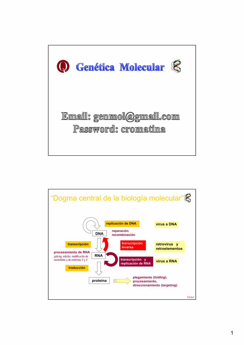
“Dogma central de la biología molecular”
DNA
RNA
transcripcióninversa
transcripción y replicación de RNA
replicación de DNA virus a DNA
retrovirus y retroelementos
virus a RNA
transcripción
traducción
proteínaplegamiento (folding), procesamiento, direccionamiento (targeting)
procesamiento de RNAsplicing, edición, modificación de nucleótidos y de extremos 5´y 3´
Víctor R ki
reparación recombinación
2
ácidos nucleicos
estructura, estabilidad, hibridación…
Alternativas de
tipos de ácidos nucleicos:
DNA o RNA
doble o simple cadena: ds, ss
lineal o circular
híbridos DNA:RNA
3
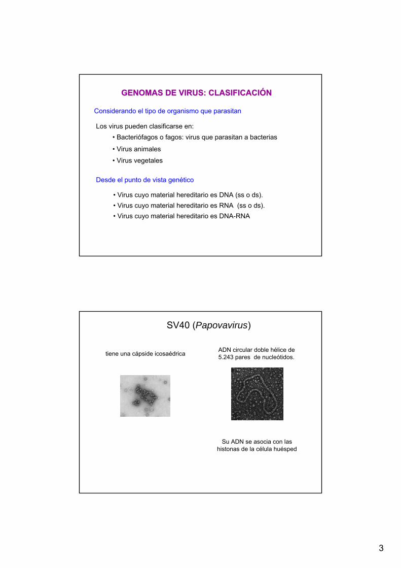
• Virus cuyo material hereditario es DNA (ss o ds).• Virus cuyo material hereditario es RNA (ss o ds).• Virus cuyo material hereditario es DNA-RNA
GENOMAS DE VIRUS: CLASIFICACIGENOMAS DE VIRUS: CLASIFICACIÓÓNN
Los virus pueden clasificarse en: • Bacteriófagos o fagos: virus que parasitan a bacterias
• Virus animales
• Virus vegetales
Considerando el tipo de organismo que parasitan
Desde el punto de vista genético
SV40 (Papovavirus)
tiene una cápside icosaédrica ADN circular doble hélice de 5.243 pares de nucleótidos.
Su ADN se asocia con las histonas de la célula huésped

4
El fago λ posee una cápside icosaédrica con una cola. Dentro de la cápside existe una molécula de ADN doble hélice lineal con 48.000 pares de bases con extremos cohesivos > circularización.
fago λ
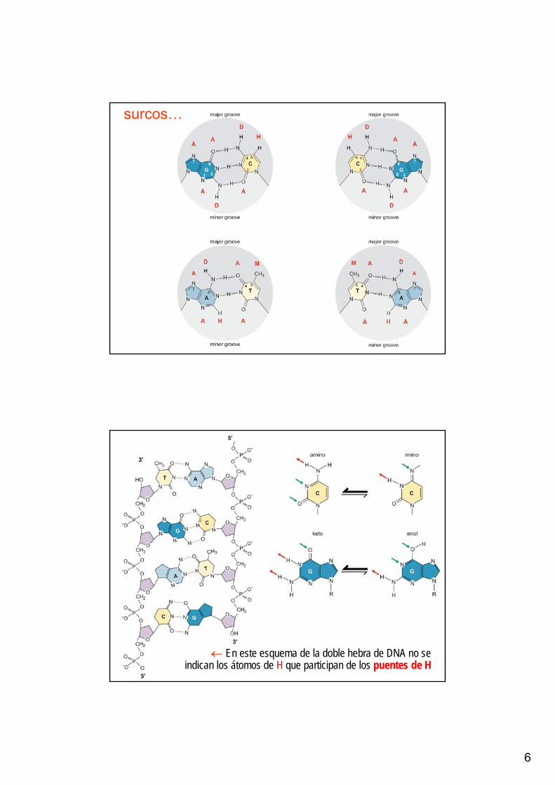
• El apareamiento de las dos cadenas genera un surco mayor y un surco menor en la superficie de la doble hèlice

5
Doble hélice con un surco mayor y un surco menor
Doble hélice con un surco mayor y un surco menor
6
surcos…
← En este esquema de la doble hebra de DNA no se indican los átomos de H que participan de los puentes de H
7
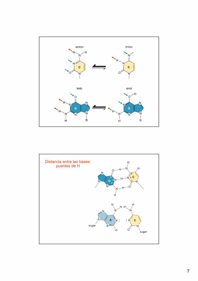
Distancia entre las bases: puentes de H

8
Descubra el error…
Descubra el error…
9
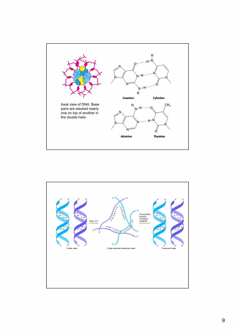
Axial view of DNA. Base pairs are stacked nearlyone on top of another in the double helix.
10
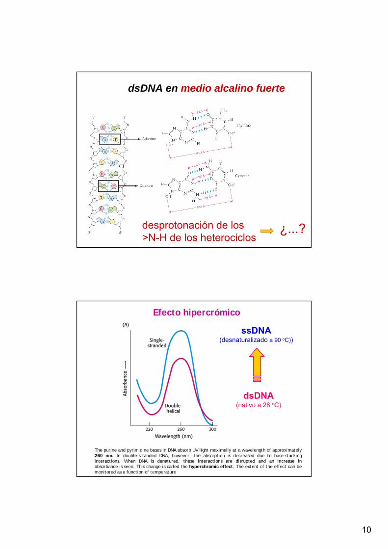
dsDNA en medio alcalino fuerte
desprotonación de los >N-H de los heterociclos
¿...?
ssDNA(desnaturalizado a 90 oC))
dsDNA(nativo a 28 oC)
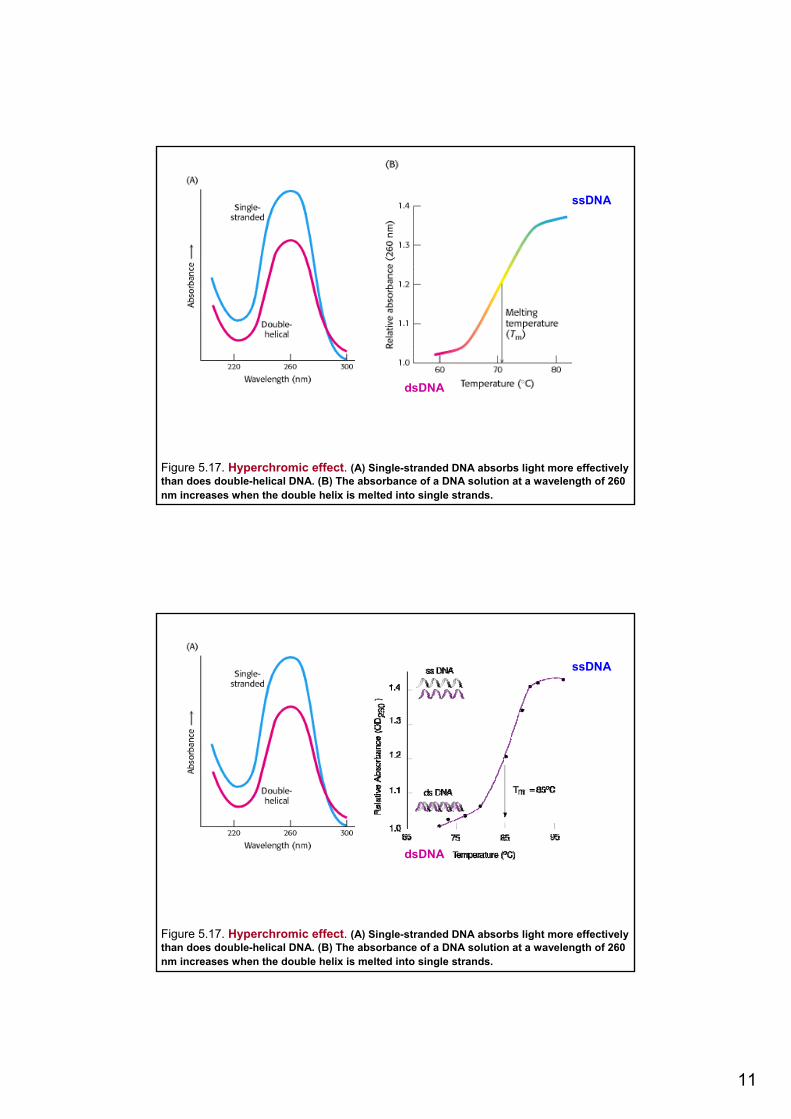
Efecto hipercrómico
The purine and pyrimidine bases in DNA absorb UV light maximally at a wavelength of approximately 260 nm. In double-stranded DNA, however, the absorption is decreased due to base-stacking interactions. When DNA is denatured, these interactions are disrupted and an increase in absorbance is seen. This change is called the hyperchromic effect. The extent of the effect can be monitored as a function of temperature
11
Figure 5.17. Hyperchromic effect. (A) Single-stranded DNA absorbs light more effectivelythan does double-helical DNA. (B) The absorbance of a DNA solution at a wavelength of 260 nm increases when the double helix is melted into single strands.
ssDNA
dsDNA
Figure 5.17. Hyperchromic effect. (A) Single-stranded DNA absorbs light more effectivelythan does double-helical DNA. (B) The absorbance of a DNA solution at a wavelength of 260 nm increases when the double helix is melted into single strands.
ssDNA
dsDNA
12
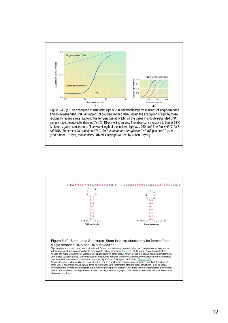
Figure 8-40. (a) The absorption of ultraviolet light of 260-nm wavelength by solutions of single-strandedand double-stranded DNA. As regions of double-stranded DNA unpair, the absorption of light by thoseregions increases almost twofold. The temperature at which half the bases in a double-stranded DNA sample have denatured is denoted Tm. (b) DNA melting curves. The absorbance relative to that at 25°Cis plotted against temperature. (The wavelength of the incident light was 260 nm.) The Tm is 69°C for E. coli DNA (50 percent GC pairs) and 76°C for Pseudomonas aeruginosa DNA (68 percent GC pairs). (Part b from L. Stryer, Biochemistry, 4th ed. Copyright © 1995 by Lubert Stryer.)
Figure 5.19. Stem-Loop Structures. Stem-loop structures may be formed fromsingle-stranded DNA and RNA molecules. The simplest and most common structural motif formed is a stem-loop, created when two complementary sequenceswithin a single strand come together to form double-helical structures (Figure 5.19). In many cases, these doublehelices are made up entirely of Watson-Crick base pairs. In other cases, however, the structures include mismatched orunmatched (bulged) bases. Such mismatches destabilize the local structure but introduce deviations from the standarddouble-helical structure that can be important for higher-order folding and for function (Figure 5.20).Single-stranded nucleic acids can adopt structures more complex than simple stem-loops through the interaction ofmore widely separated bases. Often, three or more bases may interact to stabilize these structures. In such cases, hydrogen-bond donors and acceptors that ordinarily participate in Watson-Crick base pairs may participate in hydrogenbonds of nonstandard pairings. Metal ions such as magnesium ion (Mg2+) often assist in the stabilization of these more elaborate structures.
13
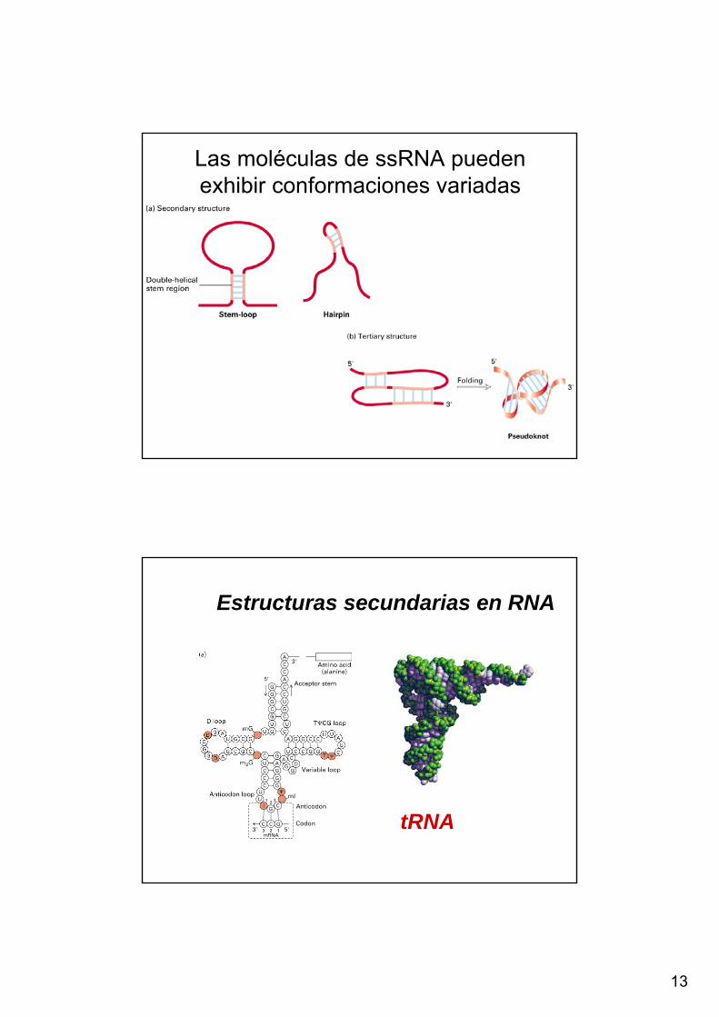
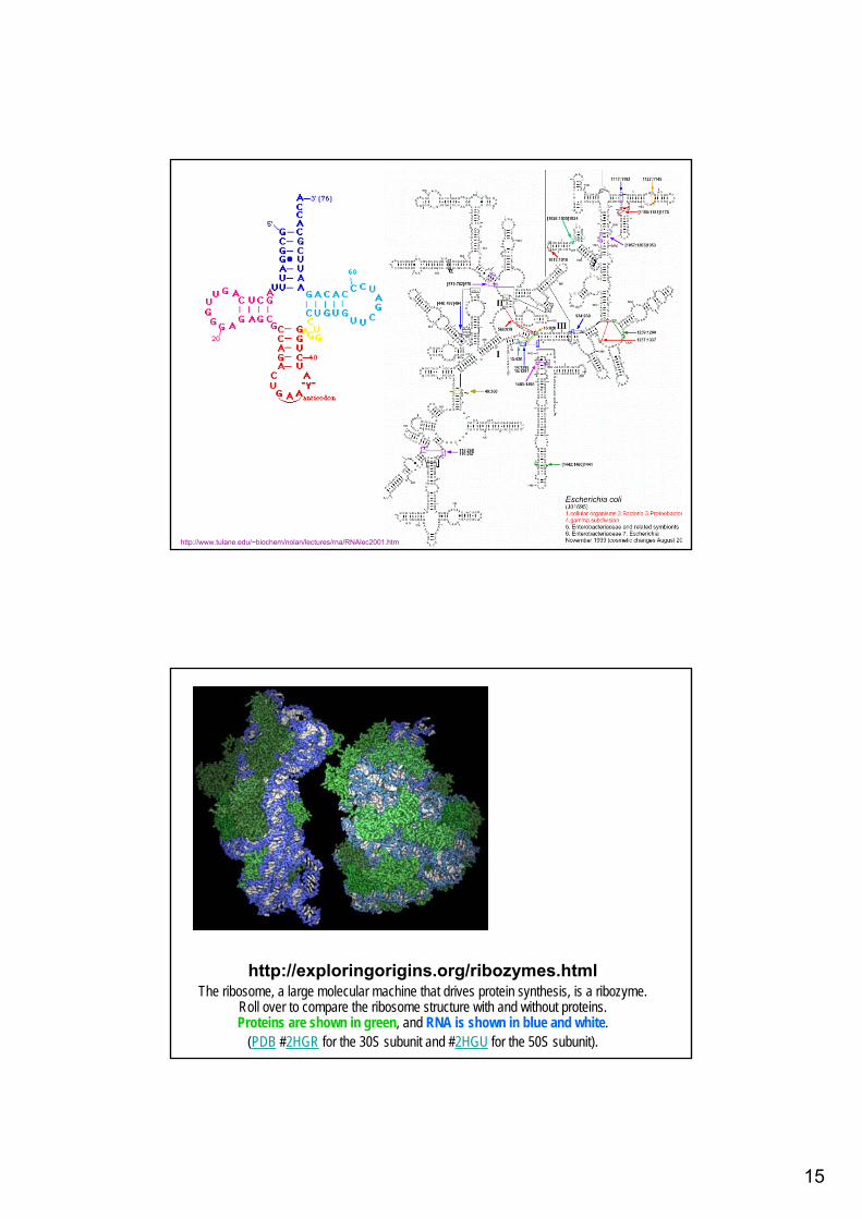
Las moléculas de ssRNA puedenexhibir conformaciones variadas
Estructuras secundarias en RNA
tRNA
14
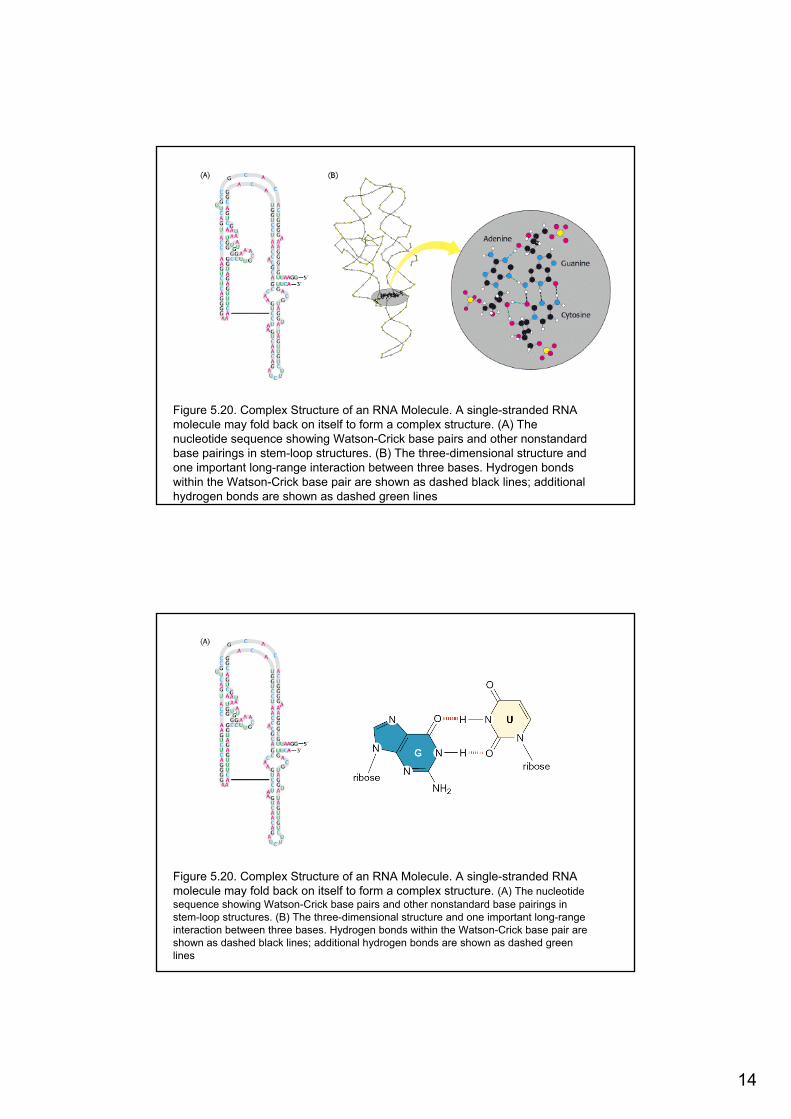
Figure 5.20. Complex Structure of an RNA Molecule. A single-stranded RNA molecule may fold back on itself to form a complex structure. (A) Thenucleotide sequence showing Watson-Crick base pairs and other nonstandardbase pairings in stem-loop structures. (B) The three-dimensional structure andone important long-range interaction between three bases. Hydrogen bondswithin the Watson-Crick base pair are shown as dashed black lines; additionalhydrogen bonds are shown as dashed green lines
Figure 5.20. Complex Structure of an RNA Molecule. A single-stranded RNA molecule may fold back on itself to form a complex structure. (A) The nucleotidesequence showing Watson-Crick base pairs and other nonstandard base pairings in stem-loop structures. (B) The three-dimensional structure and one important long-rangeinteraction between three bases. Hydrogen bonds within the Watson-Crick base pair are shown as dashed black lines; additional hydrogen bonds are shown as dashed greenlines
15
http://www.tulane.edu/~biochem/nolan/lectures/rna/RNAlec2001.htm
http://exploringorigins.org/ribozymes.htmlThe ribosome, a large molecular machine that drives protein synthesis, is a ribozyme.
Roll over to compare the ribosome structure with and without proteins. Proteins are shown in green, and RNA is shown in blue and white.
(PDB #2HGR for the 30S subunit and #2HGU for the 50S subunit).

16
> % A T
estudios termodinámicos (temperatura de desnaturalización = Tm),
relacionados con estructura (ss, ds) y composición (%GC)
estudios cinéticos (velocidad de renaturalización),
relacionados con la complejidad de la secuencia del dsDNA (Cot1/2).
Aplicaciones: hibridación de sondas…

17
Parámetros que intervienenen en la desnaturalización del DNAy renaturalización o reasociación de hebras ssDNA con secuencias complementarias (annealing, hibridación)
Parámetro Efecto sobre Tm Efecto sobre la velocidad de renaturalización
Composición de bases
Incremento de Tmcon el aumento del %G-C
No ejerce efecto
Longitud<150 bp; incremento de Tm con el incremento de longitud; >500 bp no hay efecto
Incrementa la velocidad con la longitud
Fuerza iónica Incremento de Tmcon el aumento de [Na+]
Optimo a 1.5 M Na+
% bp mismatch Disminuye Tmcon el aumento de % mismatch
Disminuye la velocidad con el aumento de % mismatch
Concentración No ejerce efecto Aumenta la velocidad con el aumento de [DNA]
Agentes denaturalizantes
disminuye Tm con el aumento de [formamida], [urea]
Optimo a 50% formamida
Temperatura - Optima a 20°C por debajo de Tm

18
Renaturalización• La desnaturalización es un proceso reversible• Reannealing – reasociación de las cadenas de DNA
Nucleación2º orden
Cierre1er orden
- dCdt
= k2.C2

19
DNA reassociation (renaturation)Double-stranded DNA
Denatured,single-strandedDNA
Slower, rate-limiting,second-order process offinding complementarysequences to nucleatebase-pairing
k2
Faster,zipperingreaction toform longmoleculesof double-strandedDNA
Complexity of chromosomal DNA
Reasociación de hebras complementarias
tkCcc
00 11
+=
C0t ½ : valor de Cot cuando se reasoció un 50%
1)1(21
0 =+ tkCtkC01
121
+= 2)1( 0 =+ tkC
1120 =−=tkC
ktC 1
2/10 =
Definición de Cot1/2: función inversa de la constante de velocidad (k)
20
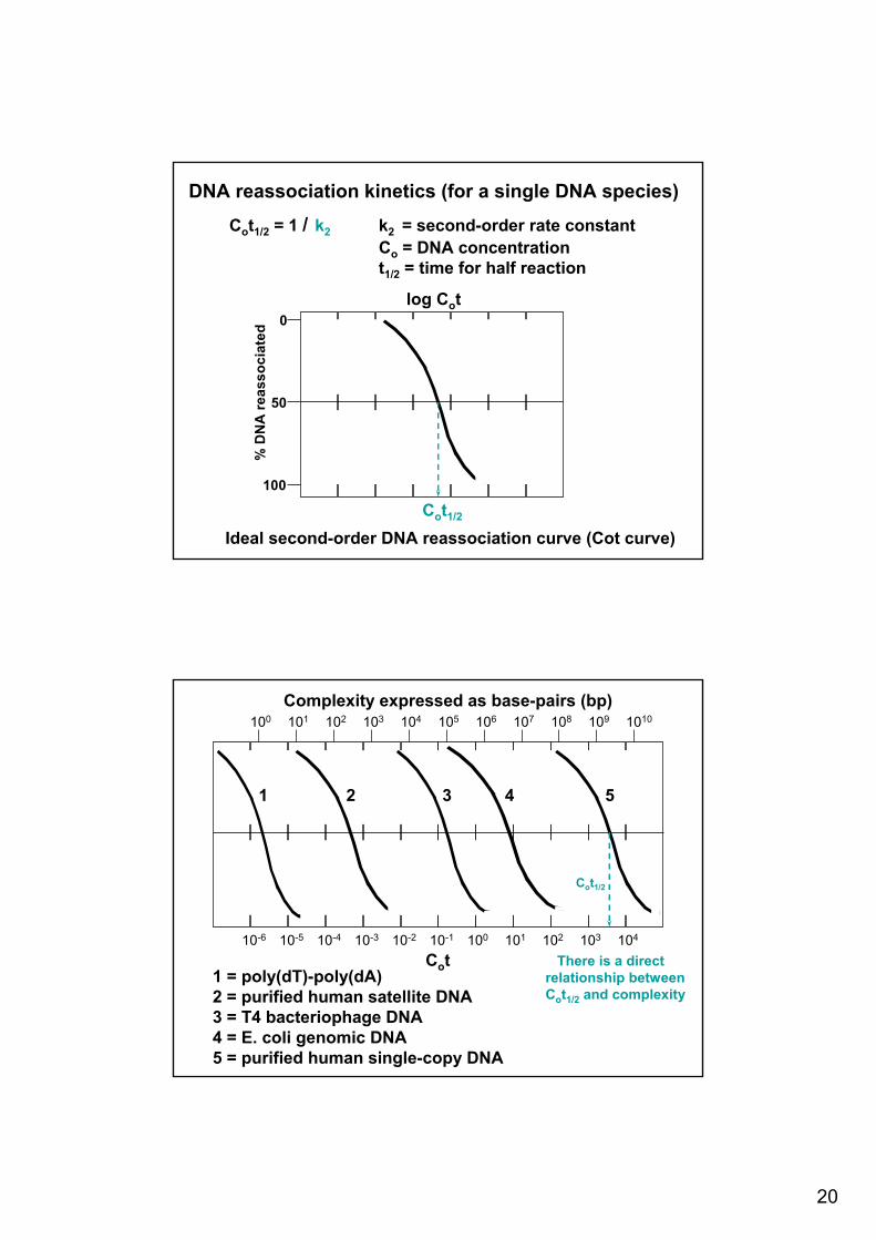
DNA reassociation kinetics (for a single DNA species)
Cot1/2 = 1 / k2 k2 = second-order rate constantCo = DNA concentrationt1/2 = time for half reaction
Cot1/2
50
100
0
% D
NA
reas
soci
ated
log Cot
Ideal second-order DNA reassociation curve (Cot curve)
10-5 10010-4 10-3 10-1 10410310210-6 10-2 101
101 106102 103 105 1010109108100 104 107
Complexity expressed as base-pairs (bp)
Cot
1 2 3 4 5
1 = poly(dT)-poly(dA)2 = purified human satellite DNA3 = T4 bacteriophage DNA 4 = E. coli genomic DNA5 = purified human single-copy DNA
Cot1/2
There is a directrelationship betweenCot1/2 and complexity
21
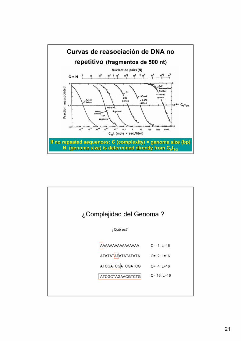
Curvas de reasociación de DNA no repetitivo (fragmentos de 500 nt)
If no repeated sequences: C (complexity) = genome size (If no repeated sequences: C (complexity) = genome size (bpbp))N (genome size) is determined directly from CN (genome size) is determined directly from C00tt1/21/2
C = NC = N
(N)(N)
106
repeats
3 genes
200 genes ≈ 4.000
genes
≈ 10.000 genes
CC00tt1/21/2
¿Complejidad del Genoma ?
¿Qué es?
AAAAAAAAAAAAAAAA
ATATATATATATATATA
ATCGATCGATCGATCG
C= 1; L=16
C= 2; L=16
C= 4; L=16
ATCGCTAGAACGTCTG C= 16; L=16

22
Reasociación de DNA de eucariotes
≈ 25 % moderadamente repetitivo350 copias
≈ 55% copia única
≈ 20 % altamente repetitivo: 2x10⁶ copias
Tamaños de genomas

23
Tamaños de genomas
Virus a DNA
1 Cell length 2 μm or 2x10⁻⁶ m
2 Cell diameter 0.8 μm or 0.8x10⁻⁶ m
3 Cell total volume 1x10-15 L
Escherichia coli genome: 4,639,221 bp4,377 genes
4,290 of these genes encode proteins; the rest RNAs

24
Tamaños de genomas
25
virusesplasmids
bacteriafungi
plantsalgae
insects
mollusks
reptiles
birds
mammals
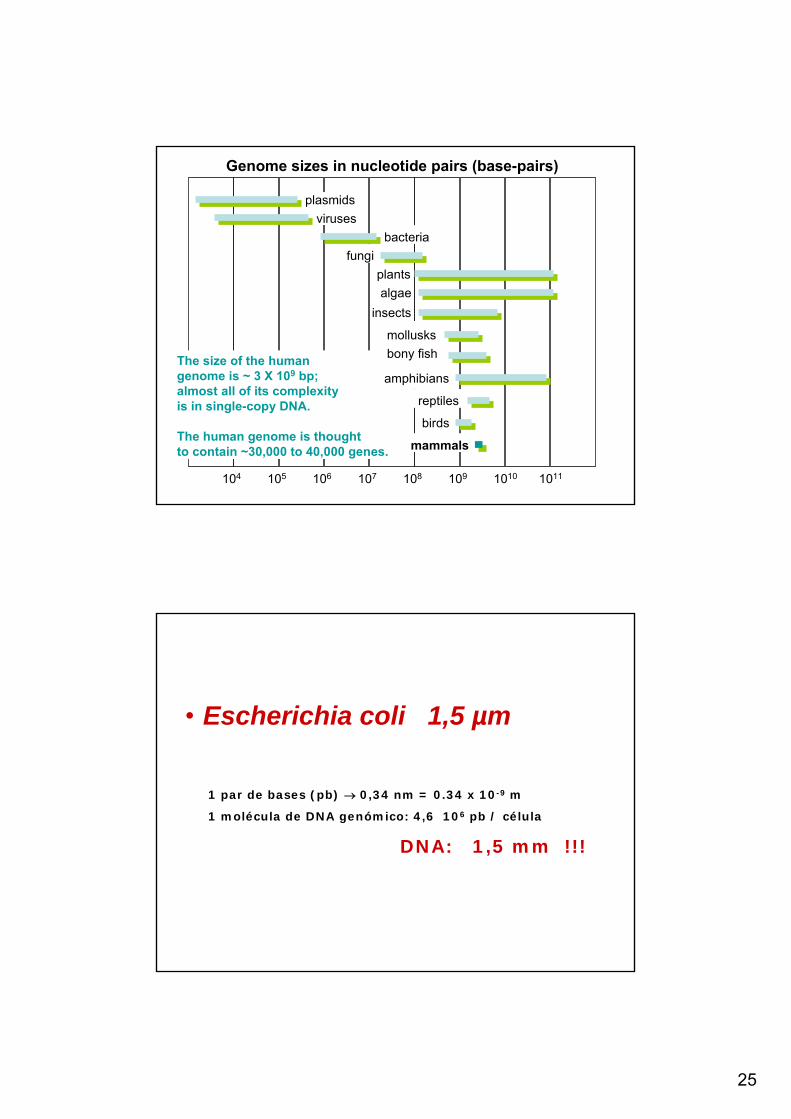
Genome sizes in nucleotide pairs (base-pairs)
104 108105 106 107 10111010109
The size of the humangenome is ~ 3 X 10⁹ bp;almost all of its complexityis in single-copy DNA.
The human genome is thoughtto contain ~30,000 to 40,000 genes.
bony fish
amphibians
• Escherichia coli 1,5 µm
1 par de bases (pb) → 0,34 nm = 0.34 x 10⁻⁹ m
1 molécula de DNA genómico: 4,6 106 pb / célula
DNA: 1,5 mm !!!
• Cromosoma y plásmidos
26
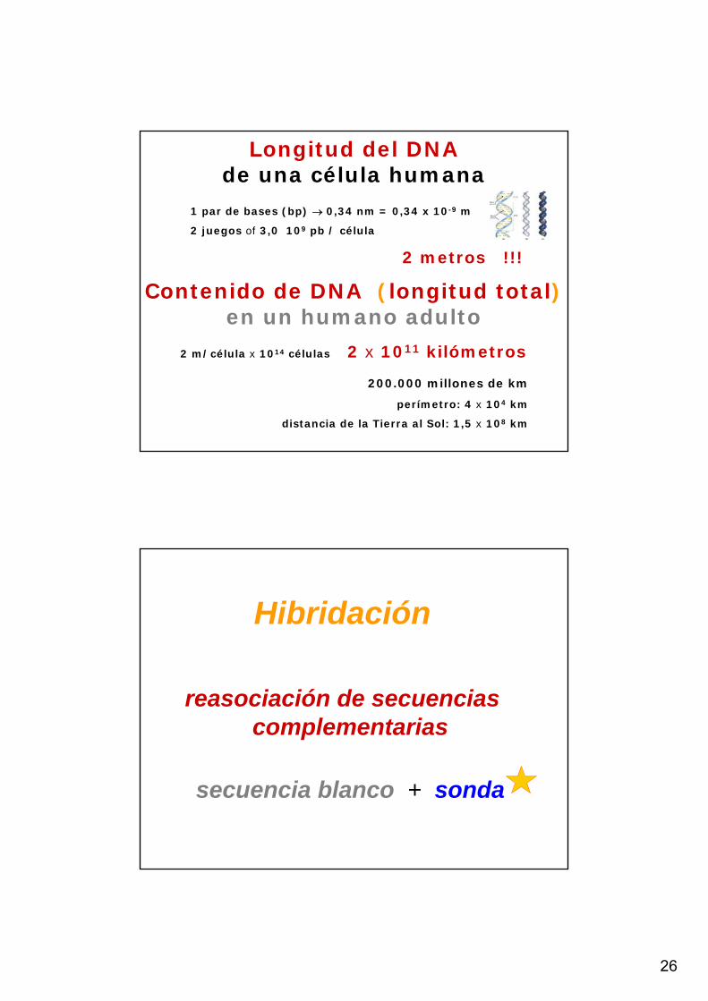
Longitud del DNAde una célula humana
1 par de bases (bp) → 0,34 nm = 0,34 x 10⁻⁹ m
2 juegos of 3,0 109 pb / célula
2 metros !!!
Contenido de DNA (longitud total)en un humano adulto
2 m/célula x 1014 células 2 x 10¹¹ kilómetros
200.000 millones de km
perímetro: 4 x 10⁴ km
distancia de la Tierra al Sol: 1,5 x 10⁸ km
Hibridación
reasociación de secuencias complementarias
secuencia blanco + sonda

27
Sonda marcada
Acido nucleico inmovilizado
Sondasfluorescentes

28
Sondas con grupos detectables porafinidad con anticuerpos (DIG) y otras
proteínas (avidina o estreptavidina)
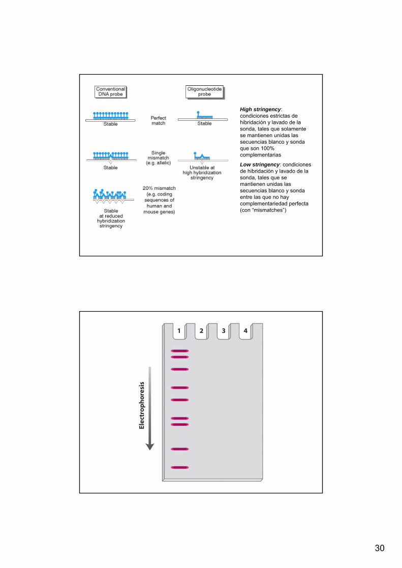
High stringency: condiciones estrictas de hibridación y lavado de la sonda, tales que solamente se mantienen unidas las secuencias blanco y sonda que son 100% complementarias
Low stringency: condiciones de hibridación y lavado de la sonda, tales que se mantienen unidas las secuencias blanco y sonda entre las que no hay complementariedad perfecta (con “mismatches”)
29
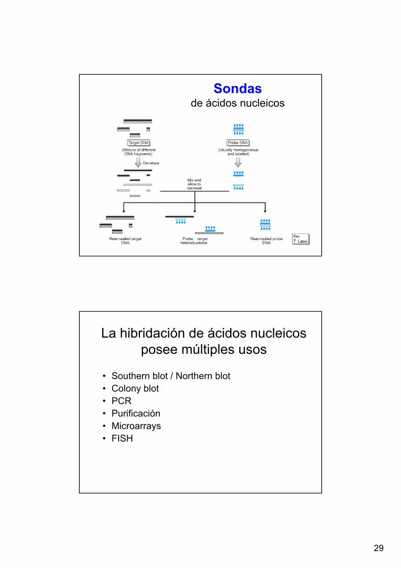
Sondasde ácidos nucleicos
La hibridación de ácidos nucleicos posee múltiples usos
• Southern blot / Northern blot• Colony blot• PCR• Purificación• Microarrays• FISH
30
High stringency: condiciones estrictas de hibridación y lavado de la sonda, tales que solamente se mantienen unidas las secuencias blanco y sonda que son 100% complementarias
Low stringency: condiciones de hibridación y lavado de la sonda, tales que se mantienen unidas las secuencias blanco y sonda entre las que no hay complementariedad perfecta (con “mismatches”)
31
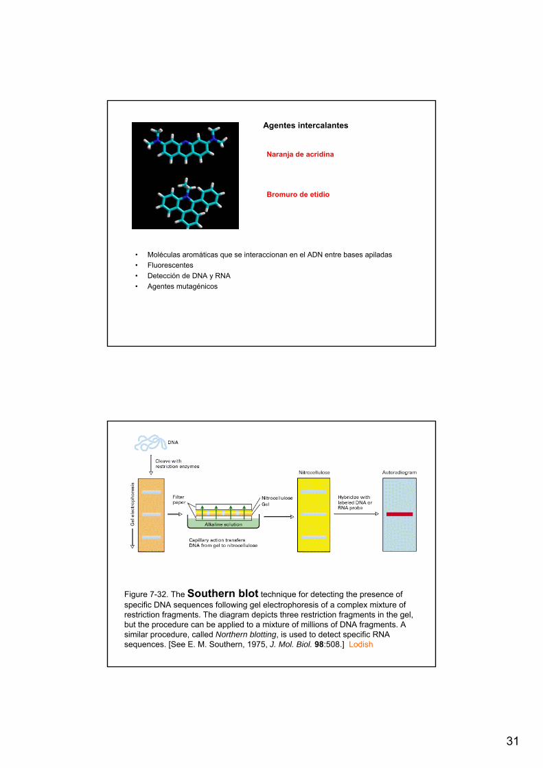
Agentes intercalantes
• Moléculas aromáticas que se interaccionan en el ADN entre bases apiladas• Fluorescentes• Detección de DNA y RNA• Agentes mutagénicos
Naranja de acridina
Bromuro de etidio
Figure 7-32. The Southern blot technique for detecting the presence ofspecific DNA sequences following gel electrophoresis of a complex mixture ofrestriction fragments. The diagram depicts three restriction fragments in the gel, but the procedure can be applied to a mixture of millions of DNA fragments. A similar procedure, called Northern blotting, is used to detect specific RNA sequences. [See E. M. Southern, 1975, J. Mol. Biol. 98:508.] Lodish

32
Northern blotting detects specific mRNAs
Figure 7-33
PCR: the polymerase chain reaction
Figure 7-38

33
PCR