Embed Size (px)
Citation preview

C RSECardiovascular and Interventional Radiological Society of Europe
Rnewscongress
Dear colleagues,
It was almost ten years ago that we set foot in
Copenhagen for a CIRSE Annual Meeting, which
welcomed around 4,800 attendees. Since then,
not only our congress attendance has grown;
when we look back over this decade, we see a
strong evolution of interventional radiology,
both in size and reputation. Thanks to
advances in technology and techniques, game-
changing interventional radiology research
in fields such as neurology and oncology has
meant that a number of minimally invasive
therapies have become a mainstay in medical
guidelines and interventional radiologists have
positioned themselves as key members of
multidisciplinary teams. On this note,
there have been exciting developments in
the field of patient management whereby
interventional radiologists truly assume
responsibility for the patient they are treating.
In 2017, we are happy to say that our annual
congress reflects the bright future of this
medical discipline, with a plethora of sessions
to suit all experience levels.
We continuously endeavour to offer
delegates a full and wide-ranging educational
programme. As endovascular therapies
are such a major focus of the congress,
the programme will again consist of three
distinct categories: arterial, venous and
aortic interventions; the latter covered in
the Interdisciplinary Endovascular Aortic
Symposium (IDEAS), a parallel multidisciplinary
programme which will run from Sunday to
Tuesday for the third year and will include the
IDEAS Training Village, introduced last year,
for practical hands-on learning in cooperation
with our industry partners. Alongside
sessions on embolotherapy, stroke treatment,
interventional oncology and non-vascular
interventions, there will also be a big focus on
clinical practice this year, with ample attention
given to anaesthesia, sedation and analgesia,
and patient management, which will be the
theme of the not-to-be-missed Opening
Ceremony today. On Tuesday, there will be the
"CIRSE Meets…" Session with the European
Wound Management Association, which will
cover the essentials of wound care, focusing
on ulcers. We are once again running the
Student Programme for medical students from
all over Europe and have expanded sessions
for the younger generation of IRs through
our European Trainee Forum, which aims to
provide a network for trainees and newly
qualified IRs in Europe.
A New Level of Learning
Last year, new session formats including
Expert Round Tables (ERT) and Expert
Case Discussions (ECD) made their debut,
which we hope will again boost audience
participation. The Super Tuesday session,
which was also introduced last year, will put
important scientific papers and trial results
in the spotlight. Likewise, the News on Stage
sessions will showcase new study results,
techniques and hot topics in IR by researchers
during the early afternoons in the exhibition
area to give delegates the opportunity to
engage in lively discussion in an informal
atmosphere.
Building upon the collaborative learning
environment, which takes note of current and
future trends in the subspecialty, we are proud
to announce the first Women in IR Session,
which aims to understand why there is a lack
of female doctors entering interventional
radiology and identify clear solutions on how
this gap can be overcome. As noted earlier,
the importance of clinical knowledge and
entrepreneurial thinking for the future of
interventional radiology is increasingly being
recognised. In order to account for this, a new
workshop, Successful Strategies in IR, will
provide practical insights and solutions from a
variety of clinical examples. As learning about
the latest medical devices and equipment is an
integral part of the congress experience, CIRSE
2017 has come up with a new training format
called Hands-on Device Trainings (HDT). This
new format replaces the Hands-on Workshops
and will be more device-orientated, with
clearer learning objectives and guidelines.
All Hands-on Device Trainings are linked to a
theoretical workshop within the programme.
Are You Ready (for the new radiation
safety directive)?
Since the creation of our Radiation Protection
Subcommittee and the Radiation Protection
Pavilion (RPP) at CIRSE, it has been one of
CIRSE’s key initiatives to provide advice and
education on radiation protection for those
working in the medical field and their patients.
With new radiation protection legislation on
the way, it will become indispensable to stay
ahead of the game, which is why this topic will
be a core theme of CIRSE 2017, featuring the
Radiation Protection Pavilion and a Hot Topic
Symposium on radiation risks and prevention
for both patients and physicians.
We hope you enjoy your time in Copenhagen
and look forward to seeing you around!
Arindam Bharadwaz
CIRSE 2017
Local Host Committee
SPC Representative
Elias Brountzos
CIRSE President
Robert A. Morgan
Vice-President
Christoph A. Binkert
Scientific Programme
Committee Chairperson
Poul Erik Andersen
CIRSE 2017
Local Host Committee
Chairperson
Fabrizio Fanelli
Scientific Programme
Committee Deputy
Chairperson
CIRSE 2017 – CopenhagenSaturday, September 16, 2017
Your Daily Dose of IR in Print
From scientific articles by our faculty to in-depth
interviews with experts, to the latest developments
in the international world of IR, and so much more!
We hope you enjoy reading your copy of Congress
News at the 32nd CIRSE Annual Meeting in the
wonderful city of Copenhagen.
Don’t miss today’s Opening Ceremony, which will combine three artistic
performances with a presentation from Prof. Sadek Beloucif on the
importance of the doctor-patient relationship and continued education
for effective clinical management. Find out more over the page...
See you in Auditorium 1 at 14:30!
CIRSE 2017I M P A C T T H E F U T U R E O F I R

Saturday, September 16, 2017Opening and Awards Ceremony2
The Opening and Awards Ceremony really
is a key moment of the Annual Meeting: the
official kick-off, a celebration of our members’
achievements and a chance to enjoy some
artistic entertainment. For the latter, we’ve
seen water glass harmonies, Scottish folk
dancing, an Italian orchestra and Greek
Olympic Games drummers, among many
fantastic performances over the years.
CIRSE 2014, Glasgow/UK CIRSE 2007, Athens/GR CIRSE 2010, Valencia/ES
Following an introduction by our President
Elias Brountzos, Local Host Representative
Poul Erik Andersen and Scientific Programme
Committee Chairperson Christoph Binkert,
our invited speaker Prof. Sadek Beloucif will
cover the topic du jour: patient management,
with a fascinating presentation entitled "Cure
– Care – Coordination: Towards a New Medical
Paradigm for Patients?" alongside interpretive
dance pieces exploring these three concepts
in medicine.
Sadek Beloucif is Head of Anaesthesiology
at Avicenne Hospital and Professor at the
Sorbonne Paris-Cités University in Paris,
France. He is currently President of the Ethics
Subcommittee for the European Society of
Anaesthesiology and was President of the
French Biomedicine Agency from 2008-11.
In 2016, he was appointed President of
the Committee of the Foundation for Islam in
France. We are honoured to have such
a revered and accomplished figure for this
very special Opening and Awards
Ceremony.
See you in Auditorium 1 at 14:30!
A CIRSE Opening and Awards Ceremony Like Never Before
Special Edition / CIRSE 2017 – Copenhagen
Hans Henkes, for the invention of the
Solitaire™ revascularisation device
In 2004, Prof. Henkes
invented the Solitaire™
revascularisation device,
which was initially
designed for the treatment
of cerebral aneurysms.
Incidentally, he found
out that it could also be
applied for the removal
of intracranial thrombi and performed the first
successful intracranial stent thrombectomy in
Europe in 2008. This stent retriever represented
a considerable improvement in acute ischaemic
stroke management compared to other
thrombectomy devices available at the time.
The SolitaireTM stent became an international
patent in 2004, followed by the US patent in
2014. It has since saved thousands of lives and
prevented countless cases of stroke-related
handicap.
About the invention
The Solitaire™ device is comprised of a nitinol
scaffolding design attached to a guidewire.
After being delivered through a microcatheter,
it is deployed across a clot and removed along
with the clot to enable revascularisation of the
occluded intracranial vessel. If applied within
8 hours of symptom onset, the Solitaire™
can remove the thrombus from a large
intracranial occlusion and restore blood flow
to a degree that will allow nearly 70% relative
improvement in functional outcomes at
90 days.
Since its establishment in 2012, the Award of Excellence and Innovation in IR has been given to some of the most innovative physicians in
the field. Sponsored by the Rolf W. Günther Foundation for Radiological Sciences, the award comes with a €5,000 cash prize and is presented
to the winner during the Opening and Awards Ceremony of the CIRSE Congress. Every year, applicants from around the world who have
published original research in a peer-reviewed scientific journal, invented a registered patent or published data on an innovative device
or equipment are evaluated by a review board, with the prize going to the most relevant contribution to the advancement of IR.
This year, two great innovators have been chosen who have paved the way for a completely new strategy in acute ischaemic stroke
management with a ground-breaking invention and a high-class clinical trial. Hans Henkes will receive the award for the invention of
the Solitaire Stent and Wim H. van Zwaam will be the representative accepting the award for the MR CLEAN trial.
About the inventor
Prof. Hans Henkes has held numerous clinical
positions throughout Germany, and, since
2007, has been the Medical Director of
the Neuroradiological Clinic at Stuttgart’s
Katharinenhospital. He has published more than
240 articles, mostly focusing on neuroradiology,
neurological diseases, neurodegeneration and
intracranial aneurysms. His papers have been
cited over 4,000 times.
The MR CLEAN trialists, represented by
Wim H. van Zwam
About the trial
The Multicenter Randomized Clinical Trial of
Endovascular Treatment for Acute Ischemic
Stroke in the Netherlands (MR CLEAN), published
to great acclaim in the New England Journal
of Medicine in 2015, was the first randomised
controlled trial (RCT) showing a clear benefit
of endovascular treatment in acute ischaemic
stroke. The two-year outcome published in April
2017 confirmed the benefit of endovascular
treatment. Designed as an RCT of intra-arterial
treatment versus no intra-arterial treatment
in patients with a proximal intracranial arterial
occlusion, 500 patients were randomised
between December 2010 and April 2014 in 16
centres in the Netherlands. Functional outcome
at 90 days as well as secondary clinical outcomes
showed a clear benefit for the intervention
group. Contrary to previous studies which could
not demonstrate preferability of endovascular
intervention over standard treatment, during
the MR CLEAN trial the latest generation
of thrombectomy devices (stent retrievers,
including the Solitaire Stent) were used,
significantly changing the outcome in favour of
endovascular treatment. In less than one year,
this has resulted in the worldwide adaptation of
guidelines incorporating endovascular treatment
as standard treatment.
About the MR CLEAN trialists
The MR CLEAN trial is a joint interdisciplinary
study comprising neurology, neuroradiology,
radiology and interventional/neurointerventional
radiology with six principal investigators
together with the three (shared) first authors
from the Universities of Rotterdam, Amsterdam
and Maastricht. They will be represented by
Dr. Wim van Zwam, interventional radiologist
and head of neurointerventional radiology at
Maastricht University Medical Center.
The Award of Excellence and Innovation in IR
Don’t miss it!Today at 14:30 in Auditorium 1
This year’s Editor’s Medal will be presented
to a Brazilian research group for their
investigation on methods for benign prostatic
hyperplasia:
Transurethral Resection of the Prostate (TURP)
Versus Original and PErFecTED Prostate Artery
Embolization (PAE) Due to Benign Prostatic
Hyperplasia (BPH): Preliminary Results of
a Single Center, Prospective, Urodynamic-
Controlled Analysis
F. C. Carnevale, A. Iscaife, E. M. Yoshinaga,
A. M. Moreira, A. A. Antunes, M. Srougi.
CVIR 2016 (Jan) Vol 39: 44-52
CVIR Editor’s Medal

C RSE
3IRnewscongress Opening and Awards Ceremony
Cardiovascular and Interventional Radiological Society of Europe
C RSE
Dierk VorwerkGold Medallist
2017
Gold Medallist
Dierk VorwerkLaudation: Johannes Lammer
Dierk Vorwerk was born in 1958 in Düren,
Germany, and raised in Turkey and Germany.
He attended medical school in Mainz and Cape
Town, South Africa. After graduating in 1983
and completing his military service, Dr. Vorwerk
joined the Department of Radiology at the
University of Technology in Aachen, headed by
Prof. Rolf Günther. He completed his radiology
training in 1990 and served as a consultant in
the same department. After being its deputy
chairman from 1996 to 1998, he was appointed
Chairman of the Department of Diagnostic
and Interventional Radiology at the Klinikum
Ingolstadt, an 1,100-bed teaching hospital in
the south of Germany, where he continues to
work to the present day.
Prof. Vorwerk was appointed as an Associate
Professor in 1992 and Professor in 1996. He
has published more than 200 papers and
book chapters and more than 250 abstracts
in the field of radiology with a special focus
on ultrasound, interventional radiology and
computerised tomography. His main fields
of interest are vascular interventions, stroke
therapy and embolisation techniques.
Prof. Vorwerk was awarded the Wilhelm
Conrad Röntgen Award in 1993 and the
Distinguished Fellow
Yasuaki AraiLaudation: Thierry de Baère
Dr. Yasuaki Arai received his medical degree
at Jikei University School of Medicine and
his PhD in medical science at Nagoya City
University Graduate School of Medicine. He
completed his internal medicine training at
Tokyo Medical Center and then started his career
in interventional radiology at the Department
of Diagnostic and Interventional Radiology
at the Aichi Cancer Center. After seven years
as the Department Chief, he moved to the
National Cancer Center and became Chief
of the Department of Diagnostic Radiology.
Between July 2012 and March 2016, he also
devoted himself to the service as the Director
of the hospital with the aims of pursuing better
management with open dialogue, improving
medical safety and governance and activating
research and treatment, including palliative
care. The hospital established the Interventional
Radiology Center to accept more patients. He
also supports the President of the National
Cancer Center as an Executive Advisor.
Engaging in interventional radiology, Dr. Arai
has introduced a number of new devices and
techniques for better treatment, such as an
implantable catheter and port system for hepatic
arterial infusion chemotherapy, as well as the
interventional CT system (Angio-CT).
Distinguished Fellow
Ernst-Peter K. StreckerLaudation: Peter Reimer
Born and raised in Berlin, Germany, Ernst-
Peter Strecker began his medical career with
studies in Berlin, Vienna and Heidelberg, and
received his medical degree from the University
of Heidelberg in 1968. After that, he went on
to train in radiology for six years, starting at
Rhode Island Hospital in the U.S. While there,
he accepted a position as a Research Fellow
for Johns Hopkins Hospital in Baltimore where
he trained under EA James and Robert White.
His training in radiology was completed
under Prof. Werner Wenz at the University of
Freiburg, Germany, where he was introduced
to the art of catheter angiography and image-
guided interventions by participating in the
development of vascular interventional radiology
with balloon percutaneous transluminal
angioplasty and tumour embolisation.
In 1978, Dr. Strecker was offered the position
as the Chief Physician of the Department for
Medical Imaging, Interventional Radiology
and Nuclear Medicine at the Diakonissen
Krankenhaus in Karlsruhe, Germany, where he
stayed for 29 years. In 1982, he also became
a Professor of Radiology at the University of
Freiburg.
After retiring from official functions in January
2008, Prof. Strecker has continued to work on
Distinguished Fellow
Mario BezziLaudation: Fabrizio Fanelli
A native of Rome, Italy, Mario Bezzi attended
the Sapienza University of Rome, where he
graduated with a medical degree in 1982.
He then entered Sapienza’s Department of
Surgery and practiced there for one year. This
postgraduate training was instrumental in
improving his clinical background and skills in
surgical procedures. At that time, however, he
was involved in the first clinical applications of
intraoperative ultrasound and became drawn to
diagnostic imaging.
In 1986, he completed his residency in Diagnostic
Radiology at the Sapienza University of Rome,
followed by a two-year fellowship in CT-US-
MRI at the Thomas Jefferson University and the
University of Pennsylvania. In 1988, he started his
interventional career in the Division of Vascular
and Interventional Radiology at the University
of Rome Hospital under the guidance of Prof.
Plinio Rossi. Many of the procedures in non-
vascular radiology which are now considered
routine, were either introduced in Italy or
further developed by the team of interventional
radiologists working in this division.
Currently, Mario Bezzi is Associate Professor of
Radiology at the Sapienza University of Rome
and Chief of the Division of Interventional
Radiology. His clinical practice consists mainly
The latest device he invented was a tip-deflecting
microcatheter for easier manipulation.
In 2002, he established the Japan Interventional
Radiology in Oncology Study Group (JIVROSG)
to build a framework for conducting clinical
trials. JIVROSG has conducted clinical trials in
the field of interventional oncology, including
palliative care. This has already led to substantial
changes with the result of the clinical trial with
Korean physicians focusing on transarterial
chemoembolisation for hepatocellular carcinoma
leading to Lipiodol authorisation in Japan and its
indication outside of Japan.
As for educational activities, Dr. Arai has given
lectures related to interventional radiology and
clinical trials worldwide – building bridges at
home and overseas. There are many international
visitors who observe cases at the National
Cancer Center Hospital; some of them spend
several months to a year with his interventional
radiology team. Dr. Arai is the Associate Editor of
several leading journals, including the Journal of
Vascular & Interventional Radiology. He serves as
a consultant for Japanese government agencies
and is the past President of the Japanese Society
of Interventional Radiology (JSIR). During his term
he sought to spread interventional radiology
as a treatment option to the public and health
administration of Japan.
new methods for interventional radiology and
technical developments, and has remained an
active reviewer for several IR journals. He was
also a visiting Professor at the Asan Medical
Centre’s Department of Interventional Radiology
in Seoul, South Korea, during 2008, and was
a Consultant Radiologist at Siloah Hospital in
Pforzheim, Germany, until recently.
Ernst-Peter Strecker is an accredited author in
over 150 publications, 90 peer-reviewed original
scientific articles and 9 chapters for books. He
holds 25 patents for instruments and devices in
interventional radiology, including the knitted
Strecker Stent, which was the first certified
stent for clinical applications to treat disease of
the arterial, venous, intestinal and respiratory
systems and which has now been implanted in
thousands of patients. Prof. Strecker has received
several honourable awards, including the Boris
Rajewski Medal from the European Association
of Radiology which he received in 1980 for his
mathematical model of mesenteric perfusion
(mesentericography). He has also been awarded
the Andreas Gruentzig Lecturer’s honour by
CIRSE in 2004, and received the CVIR Editors’
Medal in 2005.
of image-guided therapy in oncology, and
his research interests focus primarily on liver
tumours. He has been involved in a number of
grant-funded studies, where he has often served
as principal investigator. Nowadays, his interest is
also centred on MR-guided focused ultrasound,
and he is involved in a European project which
focuses on the application of MRgFUS in
moving organs. He has also widely contributed
to research in interventional radiology, having
written 15 book chapters, edited a book on
biliary tract radiology and presented more
than 430 papers at various global conferences.
Furthermore, he has published 150 articles in
both Italian and international peer-reviewed
journals, and his works have received
approximately 2,600 citations with an H-index
of 30. Professor Bezzi is also a reviewer for
international journals, including CVIR, JVIR and
European Radiology.
He became involved in the activities of CIRSE
early in his career when he was nominated
as Chairman of the Scientific Programme
Committee in 1996 for the CIRSE Meeting in
Madeira. He assisted in the organisation of the
2006 CIRSE Meeting in Rome and, for four years,
was Editor-in-Chief of ESIRonline, the online
platform that houses all the presentations and
webcasts of CIRSE events.
IR IRIRis honoured to welcome
Yasuaki Arai
as a
Distinguished CIRSE Fellow
September 2017, Copenhagen, Denmark
Elias Brountzos
CIRSE President
Robert A. Morgan
CIRSE Vice President
C RSE
The
Cardiovascular and Interventional Radiological Society of Europe
IR IRIRis honoured to welcome
Mario Bezzi
as a
Distinguished CIRSE Fellow
September 2017, Copenhagen, Denmark
Elias Brountzos
CIRSE President
Robert A. Morgan
CIRSE Vice President
C RSE
The
Cardiovascular and Interventional Radiological Society of Europe IR IRIRis honoured to welcome
Ernst-Peter K. Strecker
as a
Distinguished CIRSE Fellow
September 2017, Copenhagen, Denmark
Elias Brountzos
CIRSE President
Robert A. Morgan
CIRSE Vice President
C RSE
The
Cardiovascular and Interventional Radiological Society of Europe
Hermann Holthusen Award in 1996 by the
German Röntgen Society and also received the
Mackenzie Davidson Memorial Lecture from
the British Institute of Radiology in 1999. He is
an Honorary Member of the Turkish Society of
Interventional Radiology, Romanian Society of
Radiology, Austrian Society of Radiology and
the South African Vascular Society (VASSA).
Prof. Vorwerk was also the Chairman of the
2008 German Radiology Congress and was
later made Chairman of the German Society
of Interventional Radiology (DeGIR) from
2012 to 2014. He served as President of the
German Röntgen Society (DRG) from 2015 to
2017. Prof. Vorwerk has served as reviewer,
consultant and editorial board member of
many radiological and medical journals,
including JVIR, JEVT, CVIR, NDT, RöFo, Circulation,
Clinical Nephrology, Kidney International and
European Radiology.
He was appointed as Editor-in-Chief of CVIR
in 2003, a position he held until this year.
Within CIRSE he has served in various Executive
Committee positions, heading the society as
President from 2000 to 2001.
More Winners at CIRSE 2017…


C RSE
5IRnewscongress Interventional Oncology
Cardiovascular and Interventional Radiological Society of Europe
3D Navigation: do we need it? New tools for guiding and monitoring
liver ablation
Special Session
Saturday, September 16, 08:30-09:30
Auditorium 10
Don’t miss it !
Prof. Reto Bale is one of Europe’s leading experts
on stereotactic guidance. His technical expertise
in a wide range of imaging modalities has
formed the cornerstone of his pioneering work
in real-time tracking and automation. This has
been successfully applied within his department
(the Department of Microinvasive Therapy at
Innsbruck’s University Clinic for Radiodiagnostics)
to treat not only an impressive number of patients,
but also a wide range of conditions. While liver
tumours (both primary and secondary) form the
bulk of their work, Prof. Bale’s team also applied
their unique skills in the treatment of pelvic
fractures and other bone issues. Prof. Bale’s work
has been cited almost 4,000 times.
Reto Bale
Medical University Innsbruck
Innsbruck, Austria
Reto Bale
Thermal ablation is a minimally invasive
method for local tumour treatment and is
considered the first choice for treatment of
unresectable liver malignancies. Long-term
outcomes depend on the rate of complete
ablation of the entire tumour [1-4]. Due to
excellent short- and long-term results, thermal
ablation is accepted as an alternative to
surgical resection in very early HCC (single
HCC <2 cm) [5]. However, in tumours >2 cm,
surgical resection is still considered the method
of choice. This is due to unacceptably high
local recurrence rates after thermal ablation
of larger lesions. For instance, in CRLM >3 cm
local recurrence rates ranging from 45% to 70%
have been reported. By contrast, the local R1/2
rates after resection of primary and secondary
liver tumours are in the range of 8-24% [6,7].
If similar rates can be achieved by minimally
invasive thermal ablation, it will challenge
resection as the first-line therapy.
Rationale for the application of 3D
navigation systems in thermal ablation
Tumour size is the most important prognostic
parameter for local control after thermal
ablation. The unacceptable results after
ablation of large lesions are related to
insufficient coverage of the tumour by the
ablation zone. If the volume of the ablation
zone at one electrode/antenna position
does not cover the tumour including a safety
margin, multiple overlapping ablation zones
must be acquired. This is barely achievable by
conventional freehand US, CT, or MRI guidance,
and requires a major change in tumour
ablation strategies. This challenging task is the
transfer of a virtual three-dimensional ablation
plan (with multiple probe/antenna placements)
into the real patient [8].
3D navigation systems
3D navigation systems enable real-time
tracking of different surgical instruments with
respect to patient-specific imaging data (CT,
MRI, PET, SPECT and real-time ultrasound) by
using mechanical, optical or electromagnetic
3-D coordinate measuring systems [9]. Aiming
devices allow for precise percutaneous
targeting of any anatomical structure in the
patient. The world’s first (non-commercial)
aiming device for frameless stereotactic
punctures was developed in Innsbruck in 1995
and it was first applied for radiofrequency
ablation of the Gasserian ganglion in a patient
with trigeminal neuralgia in the following year
[10]. Later, the same guidance technique was
used for fractionated frameless stereotactic
interstitial brachytherapy in patients with head
and neck cancer [11], percutaneous pelvic
fracture fixation [12], retrograde drilling of
osteochondral lesions [13] and thermal ablation
of tumours in different organs [12-15]. The
first multi-probe stereotactic RFA of a large
liver tumour was performed in Innsbruck in
2001 [15].
Workflow of stereotactic thermal ablation
The technique of stereotactic radiofrequency
ablation using multiple RFA probes has been
described previously [16,17]. In brief, the
anaesthetised patient is immobilised on the
CT table. Fiducials are attached to the skin of
the patient. During temporary disconnection
of the tracheal tube (TT), a contrast-enhanced
CT is acquired. On the frameless stereotactic
(neuro)navigation system (Medtronic Inc.),
multiple trajectories are planned using the
3D CT dataset. A dynamic reference frame is
attached to the patient immobilisation system.
After unsterile patient registration using the
skin fiducials and checking the registration
accuracy, sterile draping of the patient is
performed. An aiming device (Atlas) is adjusted
using special guidance software. Coaxial
needles are advanced along the trajectory
to the pre-planned target during temporary
disconnection of the TT. The coaxial needles
serve as guides for the RFA electrodes. To
check the needle positions, a native control
CT is superimposed to the planning CT. After
biopsy, RFA electrodes are introduced via the
coaxial needles for serial tumour ablation.
After ablation, a contrast-enhanced CT is
obtained and superimposed to the planning
CT by means of image-fusion software, thereby
verifying the ablation zone covering the
tumour including a safety margin.
Alternatively, the CAScination navigation
system may be used [18]. It was originally
developed for intraoperative navigation for
liver surgery and, in cooperation with our
group, adapted to the requirements of SRFA.
It is also based on optical tracking technology.
In contrast to the (neuro)navigation system,
sterile reflective skin markers act as a dynamic
reference frame. This requires a slightly
different workflow. In contrast to the standard
approach described above, the patient is
sterilely draped and the sterile markers are
attached to the skin prior to the planning CT.
The markers are automatically detected on the
image data set as well as on the real patient,
allowing for fast automatic registration. The
fiducials have to be placed in a way that they
do not interfere with the needles. Registration
continuously changes during the respiratory
cycle, requiring adjustment of the aiming
device during breath-hold.
Accuracy of stereotactic targeting
Using the Medtronic and the CAScination
navigation system in combination with the
Atlas aiming device, the reported accuracies in
phantom studies were 1.64±0.919–1.84±1.189
[19] and 2.3±1.3–2.8±1.6 mm [18]. In the patient,
the median lateral error at the needle tip
was 3.2 mm (range: 0.01–9.4 mm) [20]. Beyer
et al. compared stereotactic IRE (SIRE) needle
placement with non-navigated conventional
IRE (CIRE) for percutaneous ablation of liver
malignancies in a total of 20 patients [21].
Accuracy of needle placement for SIRE was
higher than that for CIRE (2.2 mm vs. 3.3 mm
mean deviation, P<0.001). SIRE demonstrated
a significantly higher accuracy compared
with CIRE.
Clinical mid- and long-term results after
stereotactic thermal ablation
Colorectal liver metastases
Our group reported 98 SRFA treatment sessions
of 189 CRLMs in 63 consecutive patients [17]. LR
was identified in 16% of the tumours (31/189),
with no significant differences (P=0.635) when
comparing the tumour sizes <3 cm (17.7%), 3–5
cm (11.1%) and >5 cm (17.4%). Using SRFA, the
overall survival (OS) is not affected by tumour
size. The median OS was significantly different
when comparing unresectable and resectable
patients (27 vs. 58 months, P=0.002) with OS
rates of 92%, 66% and 48% at 1, 3 and 5 years,
respectively, in resectable patients. Tumour
size did not affect OS and DFS. SRFA challenges
resection as the first-line local treatment of
patients with CRLM.
Intrahepatic cholangiocellular carcinomas
Seventeen inoperable consecutive patients
with 52 ICCs were treated with SRFA [22].
A median OS of 60 months was achieved.
The two largest tumours with diameters
>10 cm were completely ablated. These SRFA
data of unresectable ICCs are superior to
the published data on resection. SRFA is a
minimally invasive alternative to resection.
Metastatic melanoma to the liver
In a recent paper, the results after SRFA of 75
melanoma liver metastases in 20 patients was
presented [23]. The primary and secondary
success rates were 89.3% and 93.3%,
respectively, with an overall local recurrence
rate of 13.3%. Four of ten local recurrences
were re-treated successfully by SRFA.
During follow-up, 9/20 patients developed
extrahepatic metastatic disease and 10/20 had
liver recurrence at any location. The median OS
from the date of SRFA was 19.3 months, with
an OS of 64%, 41% and 17% at 1, 3 and 5 years,
respectively, with no significant difference
between patients with cutaneous and ocular
melanoma. RFA is an attractive alternative
to resection in patients with melanoma liver
metastases.
Focal liver lesions in paediatric patients
SRFA may even be an alternative to surgical
resection of focal liver lesions in patients with
inherited metabolic disorders [24]. SRFA was
successfully applied for the removal of single
large liver adenoma in a 22-year-old woman
and a 20-year-old man with glycogen storage
disease type Ia and of a suspicious lesion in a
16-year-old girl with tyrosinemia type I and
α-fetoprotein elevation.
Conclusion
For thermal ablation, the lesion including a
safety margin has to be covered by the ablation
zone in order to achieve complete necrosis. In
lesions >2-3 cm, multiple overlapping ablation
zones are required. This is barely achievable
by conventional US- and CT-guidance. The
superior short- and long-term results after
SRFA as compared to the standard approach
definitely justify the additional costs and
efforts that are associated with this technique.
Stereotactic thermal ablation procedures (SRFA,
SMWA, SIRE) are reliable and reproducible
and challenge surgical resection as the first-
line treatment in primary and secondary liver
tumours. In addition, it may also be applied in
other organs, such as bone, lung, kidney, soft
tissue and lymph nodes.
Multi-probe SRFA of liver tumours has
become the main indication for 3D-navigated
interventions at our centre, with increasing
numbers over the past sixteen years. In the
first 6 months of this year, 130 liver SRFAs have
already been performed by our group.
References
1. Livraghi T, Goldberg SN, Lazzaroni S, et al. Hepatocellular
carcinoma: radio-frequency ablation of medium and large
lesions. Radiology 2000; 214:761-8
2. Solbiati L, Livraghi T, Goldberg SN, et al. Percutaneous
radio-frequency ablation of hepatic metastases from
colorectal cancer: long-term results in 117 patients.
Radiology 2001; 221:159-66
3. Mulier S, Ni Y, Jamart J, Ruers T, Marchal G, Michel L. Local
recurrence after hepatic radiofrequency coagulation.
Annals of Surgery 2005; 242:158-71
4. Livraghi T, Meloni F, Morabito A, Vettori C. Multimodal
image-guided tailored therapy of early and intermediate
hepatocellular carcinoma: long-term survival in the experience
of a single radiologic referral center. Liver transplantation:
official publication of the American Association for the Study
of Liver Diseases and the International Liver Transplantation
Society 2004; 10:S98-106
5. EASL-EORTC. EASL-EORTC clinical practice guidelines:
management of hepatocellular carcinoma. Journal of
hepatology 2012; 56:908-43
6. Kudo M, Izumi N, Ichida T, et al. Report of the 19th follow-up
survey of primary liver cancer in Japan. Hepatology research:
the official journal of the Japan Society of Hepatology 2016;
46:372-90
7. Hamady ZZ, Lodge JP, Welsh FK, et al. One-millimeter
cancer-free margin is curative for colorectal liver metastases:
a propensity score case-match approach.
Ann Surg 2014; 259:543-8
8. Bale R, Widmann G, Stoffner DI. Stereotaxy: breaking the
limits of current radiofrequency ablation techniques.
European journal of radiology 2010; 75:32-6
9. Wood BJ, Kruecker J, Abi-Jaoudeh N, et al. Navigation systems
for ablation. Journal of vascular and interventional radiology:
JVIR 2010; 21:S257-63
10. Bale RJ, Laimer I, Martin A, et al. Frameless stereotactic
cannulation of the foramen ovale for ablative treatment of
trigeminal neuralgia. Neurosurgery 2006;
59:ONS394-401; discussion ONS2
11. Bale RJ, Freysinger W, Gunkel AR, et al. Head and neck tumors:
fractionated frameless stereotactic interstitial brachytherapy-
initial experience. Radiology 2000; 214:591-5
12. Bale RJ, Kovacs P, Dolati B, Hinterleithner C, Rosenberger RE.
Stereotactic CT-guided percutaneous stabilization of posterior
pelvic ring fractures: a preclinical cadaver study. Journal of
vascular and interventional radiology: JVIR 2008; 19:1093-8
13. Bale RJ, Hoser C, Rosenberger R, Rieger M, Benedetto
KP, Fink C. Osteochondral lesions of the talus: computer-
assisted retrograde drilling--feasibility and accuracy in initial
experiences. Radiology 2001; 218:278-82
14. Bale RJ, Vogele M, Rieger M, Buchberger W, Lukas P, Jaschke W.
A new vacuum device for extremity immobilization.
AJR American journal of roentgenology 1999; 172:1093-4
15. Bale R, Freund M, Bodner G, Kovacs P, Jaschke W. Precise
Computer- assisted Liver Tumor Puncture for Biopsy and
Thermal Ablation. Radiology 2002; 225:242
16. Bale R, Widmann G, Haidu M. Stereotactic radiofrequency
ablation. Cardiovascular and interventional radiology 2011;
34:852-6
17. Bale R, Widmann G, Schullian P, et al. Percutaneous
stereotactic radiofrequency ablation of colorectal liver
metastases. European radiology 2012; 22:930-7
18. Wallach D, Toporek G, Weber S, Bale R, Widmann G.
Comparison of freehand-navigated and aiming device-
navigated targeting of liver lesions. The international journal
of medical robotics + computer assisted surgery:
MRCAS 2014; 10:35-43
19. Stoffner R, Augscholl C, Widmann G, Bohler D, Bale R. Accuracy
and feasibility of frameless stereotactic and robot-assisted
CT-based puncture in interventional radiology: a comparative
phantom study. RoFo : Fortschritte auf dem Gebiete der
Rontgenstrahlen und der Nuklearmedizin 2009; 181:851-8
20. Schullian P, Widmann G, Lang TB, Knoflach M, Bale R. Accuracy
and diagnostic yield of CT-guided stereotactic liver biopsy of
primary and secondary liver tumors. Computer aided surgery:
official journal of the International Society for Computer
Aided Surgery 2011; 16:181-7
21. Beyer LP, Pregler B, Niessen C, et al. Stereotactically-navigated
percutaneous Irreversible Electroporation (IRE) compared to
conventional IRE: a prospective trial. PeerJ 2016; 4:e2277
22. Haidu M, Dobrozemsky G, Schullian P, et al. Stereotactic
radiofrequency ablation of unresectable intrahepatic
cholangiocarcinomas: a retrospective study. Cardiovascular
and interventional radiology 2012; 35:1074-82
23. Bale R, Schullian P, Schmuth M, Widmann G, Jaschke W,
Weinlich G. Stereotactic Radiofrequency Ablation for
Metastatic Melanoma to the Liver. Cardiovascular and
interventional radiology 2016; 39:1128-35
24. Karall D, Scholl-Burgi S, Widmann G, et al. Stereotactic
radiofrequency ablation for liver tumors in inherited
metabolic disorders. Cardiovascular and interventional
radiology 2014; 37:1027-33

Special Edition / CIRSE 2017 – Copenhagen
Today’s
Featured Papers
will be presented in the Free Paper sessions,
taking place from 16:15-17:15 and
from 17:30-18:30
16:15-17:15
FP 606 Prostate intervention
Auditorium 11
Prostatic artery embolization versus
transurethral resection of the prostate in the
treatment of benign prostatic hyperplasia:
6-month results of a clinical trial
A. Sáez de Ocáriz García, I. Insausti Gorbea,
S. Solchaga Alvarez, R. Monreal Beortegui,
P.J. Giral Villalta, S. Napal Lecumberri,
F. Urtasun Grijalba; Pamplona/ES
FP 607 Imaging
Room 19
Comparison of imaging quality and adverse
effects of hepatic arterial angiography:
Iopamidol 250 versus Ioversol 320
M.J. Gu, Y.H. Kim; Taegu/KR
17:30-18:30
FP 706 Bone and spine intervention
Room 19
Prospective randomized trial comparing
intradiscal ozone injection with surgery for
the treatment of disc herniation
A.D. Kelekis1, G. Bonaldi2, A. Cianfoni3, J. Buric4,
D. Hooper5; 1Haidari/GR, 2Bergamo/IT, 3Lugano/CH, 4Bologna/IT, 5Austin, TX/US
FP 707 EVAR
Auditorium 3
Use of endoanchors in the treatment of
intraoperative type IA endoleaks after EVAR
with short necks: mid-term results
E. Beropoulis, K. Stavroulakis, G.F. Torsello,
K.P. Donas, A. Stachmann, C. Herricks,
G. Torsello, T. Bisdas; Münster/DE
FP 708 Vascular intervention: renal and
visceral arteries
Room 20
Establishment and maintenance of
hypertension intervened by RDN: an
experiment on hypertensive canine
L. Ling; Nanjing/CN
FP 709 Biopsy
Room 18
Performance of a new blunt-tip coaxial
needle for percutaneous biopsies or
drainages of "hard-to-reach" targets
R.L. Cazzato, J. Garnon, J. Caudrelier,
G. Tsoumakidou, G. Koch, A. Gangi;
Strasbourg/FR
Saturday, September 16, 2017ESIRonline / Featured Papers6
ESIRonline is our online educational platform, featuring over 9,000 recorded presentations
from all of our recent congresses! Visit www.esir.org to find out more.
Live Streaming and On Demand
At CIRSE 2017, all sessions will once again be
live-streamed for those interested who did
not have the chance to travel to Copenhagen.
First introduced at CIRSE 2012 and recently
extended to ECIO, live-streaming of the CIRSE
conferences has been met with great interest
by many members.
In addition to live streaming, the recorded
lectures are available for viewing almost
immediately after they have ended, allowing
users to access the more than 180 sessions
from each congress 24 hours a day.
live.cirse.org
Topic Packages
Since its introduction in 2008, ESIRonline
has continuously improved its search
mechanisms. Topic packages, a feature added
in 2013, have become particularly popular
with ESIRonline users, as they provide easy
access to the most relevant presentations
and documents on specific IR topics.
So far, 36 packages have been compiled,
ranging from stent grafting to oncologic
procedures all the way to the provision
of IR services. In the past two years,
the following topic packages have been
added:
INTERVENTIONAL ONCOLOGY
Early-stage HCC
Intermediate-stage HCC
Oncologic management of metastatic disease
Oncologic interventions: bone
Oncologic interventions: lung
SIRT for HCC and liver metastases: an update
Colorectal liver metastases
Renal tumour ablation: an update
Spinal interventions and bone tumour ablation
VASCULAR
TEVAR
EVAR
Aortic stent grafting: an update
Critical limb ischaemia
Venous interventions and IVC filters
Venous stenting
Carotid and supra-aortic interventions
TIPS and portal vein interventions
NON-VASCULAR
Gastrointestinal Interventions
IR-MANAGEMENT
Provision of IR Services
EMBOLISATION
Prostate embolisation
Embolisation of peripheral and pulmonary
AVMs: an update
Women’s health
NEUROINTERVENTIONS
Acute stroke interventions
ESIRonline: The World of IR at Your FingertipsPetra Mann, CIRSE Office
ESIRonlineis free for all CIRSE
members!
In its pursuit to continue being the most
comprehensive and user-friendly learning
platform for interventional radiology,
ESIRonline is planning to include several
new features.
We chatted to Programme Director
Prof. Stefan Müller-Hülsbeck about the
upcoming changes.
CIRSE: ESIRonline has come a long way
since its establishment in 2008.
In your opinion, what have been the
most important steps in its development?
Müller-Hülsbeck: Since its beginnings,
ESIRonline has been well received by IRs from
around the globe. There was simply a need for
a tool which would allow physicians to have
online access to CIRSE conference lectures,
scientific presentations and posters. For the
first time, physicians who had not been able
to attend the conferences could view content
online and those who had attended could
watch missed sessions. In 2013, ESIRonline was
complemented by a monthly topic package.
Many of the new packages also include expert
videos related to the topic.
Throughout the years, the website’s visual
appearance has also been optimised. We have,
for example, included colour-coding for the
various fields of IR that match those of the
annual congress. Another massive step in the
website’s development was the introduction of
live streaming and being able to view sessions
online very shortly after they have taken place.
Last but not least, the website’s search tool was
modified and improved.
CIRSE: You are currently planning to
include an e-learning project enabling
the acquisition of CME points.
Can you tell us a bit more about this?
Müller-Hülsbeck: The ESIRonline committee
meets several times a year for brainstorming
sessions on how to further improve the site.
This think-tank identified some unmet needs
in our services to attract younger IRs, such
as offering additional information for basic
and intermediate-level vascular, non-vascular
and oncologic procedures, and customising
it to those young physicians’ needs. This
information should cover both new aspects
of daily IR practice and preparing for the EBIR
exam in conjunction with the CIRSE syllabus.
This will be called Basics in IR.
Basics in IR will kick off after CIRSE 2017 with
topic modules covering 10 different IR topics.
As these modules fulfil the criteria of UEMS-
based CME accreditation, each physician
who has completed a module will receive the
corresponding credits.
CIRSE: What other innovations and
additions to the page are you planning in
the long run?
Müller-Hülsbeck: ESIRonline must continue
being as dynamic as the field of IR. Our goal
is to provide comprehensive information for
all aspects and at all levels of the field. As
mentioned before, a major step forward in our
educational concept will be the establishment
of Basics in IR. In the long run, we are hoping
to build up a case library serving not only
as a preparation tool tool for the EBIR exam,
but also as a tool to find solutions for case
management, so that CIRSE members can
better prepare for the challenges of their daily
practice.
What does the future hold for ESIRonline?

C RSECardiovascular and Interventional Radiological Society of Europe
7IR ManagementIRnewscongress
Perceptions and misconceptions of the IR gender gap The IR gender gap
Women in IR
Saturday, September 16, 11:30-12:30
Auditorium 3
Don’t miss it !
Tze Min Wah, EBIR
Prof. Tze Min Wah was appointed consultant
interventional radiologist at Leeds Teaching
Hospitals Trust (LTHT) in Nov. 2003. From 2003
to 2004, she was a COOK interventional fellow
and also spent a visiting fellowship thanks to the
Stanley Melville Award (awarded by RCR, London)
under the supervision of Prof. Peter Mueller at
Massachusetts General Hospital, Boston, to learn
how to set up an interventional oncology service
at LTHT. She is passionate about developing
patient-centred IO cancer treatment and IO as the
fourth pillar of cancer care. A keen researcher and
educator, she successfully defended her PhD in
2014 whilst working full-time as an IR and bringing
up two children. She is a champion for female
leadership and believes much can be done to
improve gender diversity in IR.
Tze Min Wah
(EBIR)
Leeds Teacing Hospitals
Leeds, UK
"Gender equality isn’t a battle of sexes but a battle of equality, waged by women and men standing side by side."
Elizabeth Broderick, former Australian Sex
Discrimination Commissioner
It is important for us all to understand that
it is everyone’s responsibility to address
gender gaps in the workplace; this is neither
feminist propaganda nor is it simply "political
correctness". Indeed, there is a bigger picture
and with this in mind, let us explore the
evidence as to ‘why’ it is imperative that
we (male and female) must come together
to address this issue so as to allow the next
generation of female leaders to take centre
stage.
McKinsey & Company is a global consulting
firm that was established in 1926 to try to
address societal challenges. In 2007, McKinsey
& Company published the first report on
Women Matter: gender diversity, a corporate
performance driver [1]. It is a comprehensive
report, and chillingly illustrated that,
across Europe, only 11% of women were
represented in the membership of the listed
companies (Fig. 1).
It also illustrated that, despite Europe having
more female university graduates than male
(55% vs. 45%, respectively), this did not
translate into having more female employees
in the workplace. Their employment rate is 21%
less than their male counterparts and often
with a 15% reduction in pay.
Almost a decade later, McKinsey & Company
has recently reported that, despite making
progress, we are still a long way away from
achieving gender parity in the workplace [2].
Although there are now more female university
graduates, this has not been translated into
gender diversity in the workplace, suggesting
that there is a leak in the pipeline (‘leaky
pipeline phenomenon’ [3]) during women’s
career progression in both private and public
sectors, including healthcare [4]. The ‘glass
ceiling’ [5] experienced by women in the
workplace is often multifactorial.
The barriers are multitudinous. The working
environments often are male-orientated with
‘anytime, anywhere’ performance and a ‘linear’
career progression trajectory which does not
allow for career breaks or opting out along
the career path easily. The need to master the
male codes in order to climb the career ladder
is often off-putting for women. In addition,
there is a general lack of role models and
mentoring schemes in place to help women to
understand ‘how to get there’. Women often
have the double burden of responsibility that
comes with being a mother, carer, etc. and this
has further compounded their ability to stay on
course during their career [1].
The Harvard Business Review [6] had reported
a worrying trend of highly qualified women
dropping out of their mainstream career. In a
survey of three graduate classes from Harvard
Business School, only 38% of female graduates
have ended up in full-time jobs; a broad gauge
of their MBA graduates had shown that a
staggering 33% of white female graduates are
in part-time careers in comparison to only 5%
of their male counterparts. The authors had
also summarised the findings of a private-
sector task force, "The Hidden Brain Drain:
Women and Minorities as Unrealized Assets"
that was sponsored by Ernst & Young, Goldman
Sachs and Lehman Brothers. This task force
carried out a survey specifically designed to
investigate the role of off-ramps and on-ramps
in the lives of 2,443 highly qualified women
with honours in undergraduate, graduate and
professional degrees. What they found was
37% of women took a career break, mainly
for more ‘family time’ whilst 24% of men had
a career break, mainly for ‘changing career
path’. As a result of this, women suffered both
financial loss and lost on re-entry at a later date
during their career path, e.g. 95% of women
would like to return, however, approximately
74% managed to return with only 40% in full-
time jobs [6].
The evidence is compelling that talented
women do leak out of their career progression
and the question is then why is it important
for us all to address workplace gender
gaps? The mercenary answer: because more
women in the workplace mean a higher GDP
growth per capita. McKinsey & Company has
reported a staggering potential to increase the
contribution towards the GDP: US $12 trillion
could be added to the global economy with
gender parity [7]. More importantly, talented
women are equipped with diverse leadership
skills that have contributed towards better and
more effective decision making, thus leading to
a positive impact on the organisational culture
and performance. Interestingly, organisations
that managed to keep talented women also
managed to retain more male talent in their
organisation [2].
This talk aims to explore whether there is a
leaky pipeline phenomenon for female IRs
in leadership positions. I thus asked CIRSE
to provide the data for the CIRSE Executive
Committee appointments over the last 8 year
(2009-2017) to give a glimpse of how it looks
currently (Fig. 2). I will also be presenting
results on a survey CIRSE recently carried out
with female members.
The data has provided an insight into the
scale of the challenges facing women in IR.
This talk aims to invite constructive debate to
help explore the potential solutions that we
(men and women!) can together implement,
and how we can keep pace with the changing
times and attract the best and brightest to our
ranks in the years ahead. On that note, I am
looking forward to seeing you all (male and
female) participate in this thought-provoking
discussion.
References
1. Company M. 2007. Women Matter: Gender diversity, a
corporate performance driver. [Accessed 6/7/2017]
2. Company M. 2016. Women Matter: Reinventing the workplace
to unlock the potential of gender diversity
[Accessed 6/7/2017]
3. Clark Blickenstaff J. 2005. Women and science careers: leaky
pipeline or gender filter? Gender and Education, 17, 369-386
4. Smith I. 2015. The impact of Athena SWAN in UK medical
schools. Available: https://www.sheffield.ac.uk/polopoly_fs/
1.449704!/file/paper_2015010.pdf
5. Guvenen F, Kaplan G; Song J. 2014. The glass ceiling and the
paper floor: Gender difference amongst the top earners,
1981-2012. National Bureau of Econimic Research Working
Paper Series
6. Hewlett S, Luce C. 2005. Off-Ramps and On-Ramps: Keeping
Talented Women on the Road to Success, Harvard Business
Review
7. Woetzel J, Madgavkar A; Ellingrud K, Labaye E, Devillard S,
Kutcher E, Manyika J, Dobbs R, Krishnan M. 2015. The power
of parity: How advancing women’s equality can add $12
trillion to global growth. [Accessed 6/7/2017]
Fig. 1: A large gender gap in the membership of governing bodies of listed companies in Europe [1]. Fig. 2: CIRSE Executive Committees from 2009-2017.
0
20
40
60
80
100
120
2009-2010 2010-2011 2011-2012 2012-2013 2013-2014 2014-2015 2015-2016 2016-2017 2017-2018
% Male Executive
Committee
% Female Executive
Committee
Women represent only 11% of the membership of governing bodies of listed
companies in Europe
Share of women in executive committees in the top European companies*
* European statistics on the top 50 listed companies per country in 2006 Source: European Commission
EU average: 11% women
Norway
Sweden
Bulgaria
Latvia
Finland
UK
Germany
France
Netherlands
Belgium
Spain
Italy
Luxembourg
32%
24%
21%
21%
19%
12%
11%
8%
7%
6%
4%
3%
1%
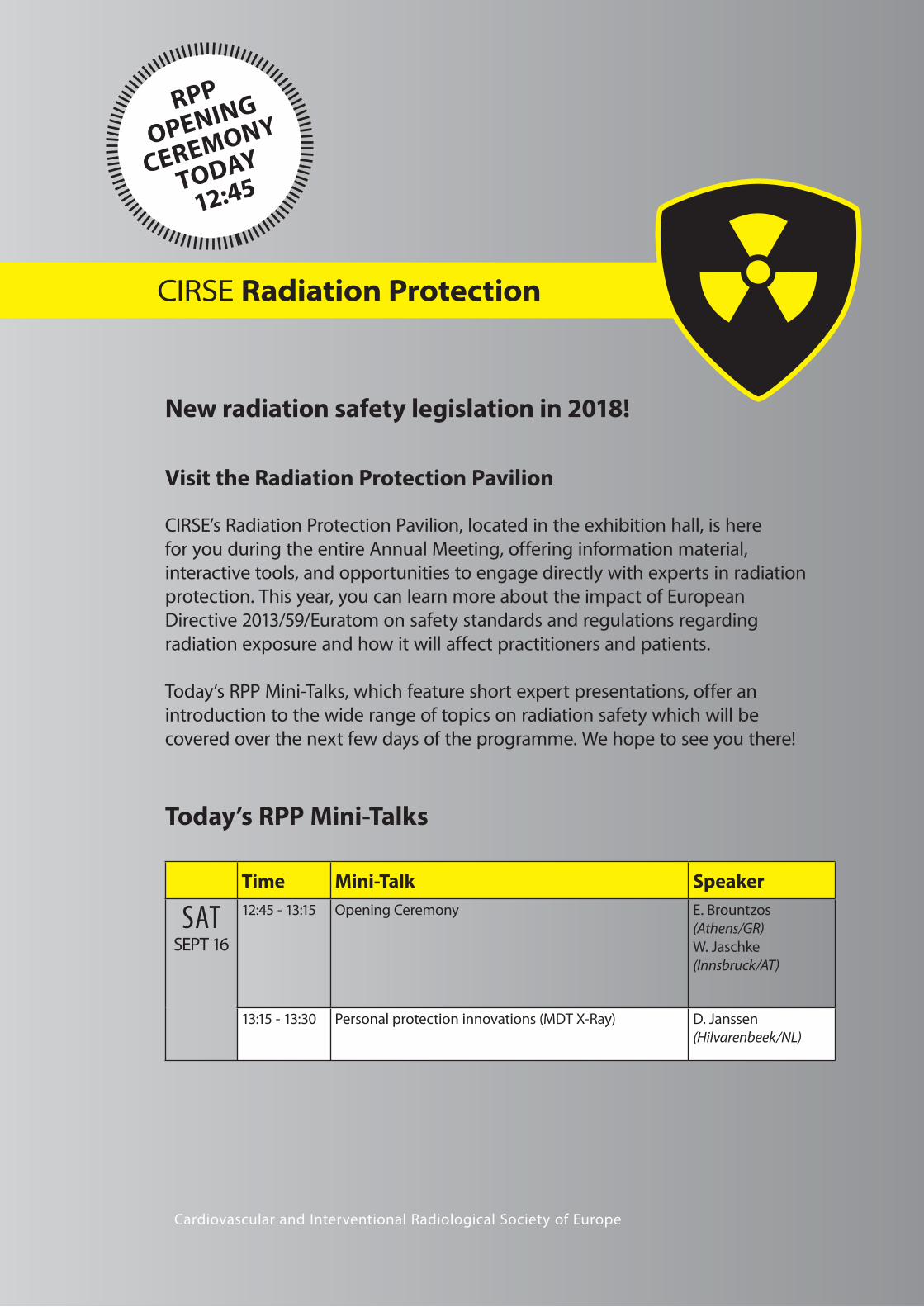
Visit the Radiation Protection Pavilion
CIRSE’s Radiation Protection Pavilion, located in the exhibition hall, is here
for you during the entire Annual Meeting, offering information material,
interactive tools, and opportunities to engage directly with experts in radiation
protection. This year, you can learn more about the impact of European
Directive 2013/59/Euratom on safety standards and regulations regarding
radiation exposure and how it will affect practitioners and patients.
Today’s RPP Mini-Talks, which feature short expert presentations, offer an
introduction to the wide range of topics on radiation safety which will be
covered over the next few days of the programme. We hope to see you there!
Cardiovascular and Interventional Radiological Society of Europe
Time Mini-Talk Speaker
SATSEPT 16
12:45 - 13:15 Opening Ceremony E. Brountzos
(Athens/GR)
W. Jaschke
(Innsbruck/AT)
13:15 - 13:30 Personal protection innovations (MDT X-Ray) D. Janssen
(Hilvarenbeek/NL)
Today’s RPP Mini-Talks
CIRSE Radiation Protection
New radiation safety legislation in 2018!
RPP
OPENING
CEREMONY
TODAY
12:45

Cardiovascular and Interventional Radiological Society of Europe
9IRnewscongress
C RSE
Radiation Protection / ICCIR 2018
Cardiovascular and Interventional Radiological Society of Europe
9IRnewscongress
Initiated in 2014, the Radiation Protection
Pavilion (RPP) has been gaining attention and
popularity at the CIRSE annual congresses over
the last three years. This year marks the fourth
year of its campaign to heighten awareness on
radiation protection and dose management in
interventional radiology.
As radiation protection gains greater awareness,
industry is reacting in tandem with the
European Commission, which has created new
Basic Safety Standards on radiation protection
that must be complied with by February
6, 2018. At this year’s RPP, a carefully selected
programme of mini-talks will supply delegates
with vital information on topics ranging from
research in radiation protection to improving
radiation safety in the IR suite to room design,
workflow and protection verification.
Werner Jaschke, Chairperson of the Radiation
Protection Subcommittee, enthusiastically
agreed to talk with us about the effect the RPP
has had on attending delegates, the potential
ways to improve radiation risks which still exist
and the future plans for the campaign.
CIRSE: Now in its fourth year, how do you
think the RPP has impacted physicians who
attend the congress?
Jaschke: Having the Radiation Protection
Pavilion at CIRSE has increased awareness
significantly. After four years of campaigning,
we believe that we have accomplished a lot,
but there is still a long way to go until radiation
protection is truly an integrated part of our
daily routine work. Each year, more and more
people are attending sessions on radiation
protection. Physicians bring the things they
learn here back to their centres, and the
following year we see the effect of that in
the audience numbers. This year there will
even be a Hot Topic Symposium on radiation
protection, which proves that the interest is
there and the idea is expanding.
CIRSE: What aspects of radiation
protection do you think physicians and
medical professionals still need to keep
improving on?
Jaschke: Physicians still need to improve on
selecting appropriate and effective radiation
protection measures, such as shielding and
equipment settings. It’s easy for people to get
stuck in their bad habits: someone might start
smoking when they’re 20, and then they’re
more likely to still be smoking when they’re
60, and they are also more likely to suffer the
effects of that. We can extend this metaphor
to many things, including radiation risk: if
doctors have gotten into the bad habit of not
wearing the proper garments or glasses, they
are increasing their risk of radiation exposure,
but awareness is increasing and things are
improving. If we look at the anti-smoking
campaign, it took 30 years to get that rolling,
so hopefully we will be able to accomplish this
in less time.
Another point is that, right now, aprons tend
to be "one size fits all." This means that many
people are not getting the proper protection.
A recent article in AJR showed that technicians
attending fluoroscopic procedures have a
higher risk of developing left-sided cancers,
and in female technicians, the study indicated
a higher risk of breast cancer: these effects
may be related to the current design of the
radiation protection garments. As you’ll see
at the Pavilion, industry has reacted to this
by providing additional protection for the
shoulder and upper arms, which also increases
the protection of breast tissue.
It’s also important to keep in mind that if
people are not needed in the angio suite then
they shouldn’t stay in there. In hybrid rooms, it
is common to see 11 people in a room where
only 5 are needed. If it’s not necessary to have
them present, then save them the radiation
exposure and get them out.
CIRSE: Do you think this Pavilion has made a
difference in raising awareness of the risks
of radiation?
Jaschke: It certainly has made a difference,
but to what extent we don’t know. The
Pavilion is only a small part of a worldwide
effort to increase radiation safety for
healthcare professionals and patients. CIRSE
is trying to change attitudes and ignorance
regarding radiation protection issues. It is very
encouraging that interventional radiologists
of all ages and nationalities are attending our
sessions.
CIRSE: The Radiation Protection Pavilion
has worked closely with the European
Society of Radiology over the last four
years, providing a space to showcase ESR’s
Eurosafe Campaign; what’s the importance
of collaborating with other societies when it
comes to radiation protection?
Jaschke: It’s important that we make a
joint effort to avoid risks for patients and
professionals, but also to inform physicians
and the public about the high safety standards
in IR. We need to have everyone reaching for
the same goal: reducing radiation exposure
in patients and physicians. But we also have
to keep in mind that the risk of radiation is
very, very low compared to other risks in
life. Most people we treat are of an older
age and suffering from severe diseases. We
should, therefore, be very sensitive to how
we discuss radiation-associated risks with our
patients. In most cases, benefits outweigh risks
substantially.
CIRSE: What are your hopes with continuing
the Radiation Protection Pavilion?
Jaschke: At CIRSE 2017, an important topic
we are covering is radiation protection in
paediatric patients. Our little patients deserve
our close attention, because they are very
sensitive to radiation. Regarding patient safety
in general, our campaign will be boosted by
the new EU directive which will be part of
national legislation in 2018. This directive states
that EU countries must ensure compliance
of the Basic Safety Standards by February 6,
2018, including placing a greater focus on
awareness of patient protection by taking into
consideration not just the patient’s exposure
during a single procedure but each patient’s
lifetime exposure and documenting this as
well. Therefore, CIRSE 2017 aims to enhance
the awareness for the new legislation in the IR
community.
Make sure to join us at the Radiation Protection Pavilion Opening Ceremony at 12:45!
Focus on Radiation Protection at CIRSE 2017Michelle Weiss, CIRSE Office
ICCIR 2018International Conference on Complications in Interventional Radiology
June 7-9, 2018Poertschach | Austria
C I R S E f o u n d a t i o n

Special Edition / CIRSE 2017 – Copenhagen
Today at 13:15-14:15, in the News on Stage Area
NoS 404 Vascular News on Stage
404.1 Accelerated thrombolysis for post-thrombotic syndrome using the acoustic pulse thrombolysis EkoSonic® Endovascular system (ACCESS PTS) study:
initial results of a multi-centeric study
M.J. Garcia (Wilmington,DE/US)
404.3 Randomized clinical trial to compare ultrasound-enhanced delivery of paclitaxel and DEB in patients with critical limb ischemia and femoral-popliteal
disease: outcome of the PACUS trial after 18 months
C. Del Giudice (Rome/IT)
404.4 Multicenter feasibility study of microwave radiometry thermometry for noninvasive differential diagnosis of critical limb ischemia in diabetic patients
C. Lalenis (Athens/GR)
404.5 Supervised exercise therapy versus percutaneous angioplasty versus combined angioplasty and exercise for intermittent claudication: systematic
review and Bayesian network meta-analysis of randomized controlled trials
M. Pantelidou (London/UK)
404.6 Peripheral endovascular interventions using human Thiel-embalmed cadavers with extracorporeal flow for medical device testing, development,
and training
H.M. McLeod (Dundee/UK)
News on Stage
The aim of this session format is to allow physicians to showcase the latest results from multi-centric trials, ground-breaking techniques and many more
IR hot topics in an informal and open atmosphere. The presentations will be displayed in a dedicated open area next to the exhibition, giving delegates
the opportunity to engage in active, lively discussions.

Cardiovascular and Interventional Radiological Society of Europe
11IRnewscongress
C RSE
Dotter Institute / Members’ Lounge
Join Us in the Members’ Lounge!
As a special service to members, CIRSE is offering a Members‘ Lounge at Copenhagen 2017.
All CIRSE Members are invited to come and relax with colleagues.
The Members’ Lounge is located in the Exhibition, next to the IDEAS Training Village.
Charles Theodore Dotter (1920 –1985) was a
pioneering American vascular radiologist and one
of the founders of interventional radiology. His
vision lives on in the daily work of IRs worldwide
and at the Dotter Interventional Institute, which is
now a fully-fledged IR department at the Oregon
Health and Science University (OHSU).
In many ways, Charles Dotter was ahead of
his time. Towards the end of the 50s, when
invasive approaches using X-rays to enhance
their ability to diagnose diseases were only
just being developed, he was already thinking
beyond the diagnosis and actively working
towards directly treating them. Credited with
the first transluminal angioplasty in 1964, he
fought hard to open colleagues’ minds to
the vast potential of interventional radiology,
recognising the possibilities for both the
patient and the future of medicine in general.
Charles Dotter was a pioneer in every sense of
the word, not only inventing procedures but
sometimes also crafting the tools for them.
His techniques and methods were embraced
by Eberhard Zeitler and Andreas Grüntzig,
whose work was instrumental in kick-starting
the success story that IR has embraced ever
since. Their work also helped open the minds
of the American medical community. This story
also highlights the close cooperation of the
IR communities on both sides of the Atlantic,
which has its roots in the earliest days of IR
and has been a pillar in the development of
the specialty.
Thus, it is exciting to report that at the
OHSU, where Charles Dotter spent most of
his pioneering career and was the chairman
of the School of Medicine Department of
Diagnostic Radiology for 33 years, the Dotter
Interventional Institute has achieved full
departmental status as of July 1, 2017.
"Creating a new department is a big decision.
And in the case of interventional radiology, I
could not be more thrilled to bring the Dotter
Interventional Institute’s new status across the
finish line," Interim Dean John Hunter stated
in a press release, "Its departmental status
recognises not only the legitimacy of the
discipline and its essential function in diagnosis
and cure, but also honours the legacy of OHSU
and Charles Dotter as a birthplace for work that
has transformed medicine."
The Dotter Interventional Institute, a
freestanding division in the OHSU School
of Medicine separate from the Department
of Radiology, was founded a few years
after Dr. Dotter’s passing and has ever
since remained an important centre for
interventional radiology with many influential
minds working there. The late IR pioneer Josef
Rösch, credited with developing TIPS, was also
a colleague of Dotter at OHSU and then went
on to be the first director of the Institute. In
2012, when the American Board of Medical
Specialties recognised interventional radiology
as a primary specialty of medicine, the Oregon
Health and Science University began to take
steps to elevate the Dotter Interventional
Institute to departmental status. Five years
later, the Institute, which proudly bears the
name of one of IRs most influential and
visionary minds, has officially gained the status
of an IR department, with CIRSE Distinguished
Fellow and 2017 Faculty Member, Dr. John
Kaufman serving as inaugural Chair.
The Institute’s new status further reinforces
its position as one of the leading centres of
IR, boasting a unique legacy which is closely
tied to the history of our specialty and some of
its most brilliant minds. Without a doubt, the
Dotter Interventional Institute will also remain
a strong part of the transatlantic ties that have
contributed to the development and success
of IR.
Dr. John Kaufman, the Institute’s inaugural Chair.
Dr. Charles Dotter treating a patient.
The OHSU Does Doctor Dotter Proud
64 65
IDEAS Training Village
Members’ Lounge
Poster Area II

Special Edition / CIRSE 2016 – Barcelona
Communication through open access publication
CIRSE is happy to announce the launch of CVIR Endovascular,
a new online open access journal with a multidisciplinary
approach and open peer review.
Find out more on www.cvirendovascular.org
Join us at the Launch of CVIR EndovascularSaturday, September 16, 11:00-12:00, Room 17
SUBMIT NOW!
RCVENDOVASCULAR
Multidisciplinary and Open Access

C RSECardiovascular and Interventional Radiological Society of Europe
13Venous / Poster AwardsIRnewscongress
Venous disorders of the legs occur frequently
and range in severity from minor asymptomatic
telangiectasia to chronic leg ulceration due
to major incompetence of venous valves.
Venous disease of the legs causes considerable
morbidity and is also costly, with roughly 2% of
national healthcare resources being spent on
treatment.
Varicose veins are a common manifestation
of venous incompetence in the lower
limb, with approximately one third of the
population showing some degree of varicose
veins appearing as dilated, elongated and
tortuous superficial veins. Incompetence of
the superficial and/or perforating vein valves
leads to pathological haemodynamic changes
of the venous pressure in the lower leg. These
haemodynamic changes in the venous pressure
induce inflammatory reaction in the tissue
which can result in skin changes such as
hyper-pigmentation and indurations with
eventual ulceration. Normally, this develops
over a long period of time, and only a
minority of the patients with superficial
vein incompetence seeks initial help in this
advanced stage.
Instead, the typical patients with varicose veins
that contact the healthcare system for the first
time have symptoms such as heaviness, aching,
cramps, swelling and cosmetic issues from the
tortuous superficial veins. Most commonly it is
a female in her mid-forties, even though men
have the same risk of developing superficial
vein incompetence. These patients are often
active people in the middle of their life and
career, well-informed and expecting results.
They wish to get rid of the problem fast and
easily, inside the national healthcare system
or outside through a privately financed
treatment.
During today’s special session, Varicose vein:
time for IR, a number of senior specialists in this
field will try to bring you up to speed on what
strengths and weaknesses the IR community
might have in this area.
You will be guided through how to organise
a venous service unit. Of course there will
be organisational differences between
different countries and healthcare systems,
but the basics are the same. You will learn
about different techniques for truncal
ablation, thermal or non-thermal. What about
phlebectomy when we do truncal ablation?
Is ultrasound-guided sclerotherapy a stand-
alone therapy for truncal ablation or is it a
complementary treatment?
Finally, one of the speakers will deal with
the most complex problem of venous
incompetence: the venous ulcer. There are a
number of issues to consider when treating
these complex patients. What therapy do they
need? Should we organise a multi-specialist
cluster dealing with leg ulcers? What actions
are needed to get all leg ulcer patients referred
to a leg ulcer specialist?
Clinical assessment – a crucial starting point
To get all of the above to fall into position, you
have to start with a well-documented clinical
assessment and work-up. The medical history
can best be taken using a questionnaire. This
saves time, but it is of utmost importance that
the physician can personally clarify the answers
in detail.
Clinical examination remains the foundation
for any medical assessment, even in this time of
advanced technology. It will give the physician
the opportunity to get to know the patient as a
person and allow him/her to take the individual
into consideration when evaluating clinical
findings and offering therapeutic treatment.
Furthermore, the patient’s expectations need
to be considered, and possible outcomes, side
effects and risks of the offered therapy need to
be fully disclosed.
The goal of the clinical exam is to understand
the anatomy and pathophysiology of the
incompetent veins, assess their functional
importance and then develop a therapy plan.
Contraindications to treatment, such as deep
vein thrombosis, lymph oedema, arterial
occlusive disease, immobility and any systemic
disease need to be ruled out.
A preoperative duplex ultrasound investigation
should be performed by the operating
physician to verify venous insufficiency
and to be used as preoperative planning of
what vessels are to be treated. In addition,
ultrasound is important to identify potential
complications and risks. The operator must
have a good knowledge of venous ultrasound
diagnostics and duplex techniques. These skills
are required as the entire treatment procedure
is guided by ultrasound monitoring.
Adding together all the information above, a
classification according to CEAP can be made.
CEAP is the acronym for clinical severity,
aetiology, anatomy and pathophysiology, and
this classification of varicose veins indicates the
degree of symptoms of varicose disease and
is used in many countries for reimbursement
classification.
Join us today and learn more about how
to organise and develop your IR knowledge
to start treating this interesting patient
group!
How to deal with varicose veins Varicose vein: time for IR
Special Session
Saturday, September 16, 11:30-12:30
Auditorium 11
Don’t miss it !
Michael Åkesson
Dr. Åkesson is a senior consultant at the
Scandinavian Venous Centre, and the CEO and
owner of the MedPACS Network AB. He previously
worked as a consultant in interventional radiology
at Skåne University Hospital. Dr. Åkesson has
been actively involved with several CIRSE events,
holding workshops on varicose vein ablation at
CIRSE 2013, and on TEVR at CIRSE 2011. He has
also contributed to various publications, including
articles on endovascular recanalisation of chronic
iliac vein occlusion and sub-intimal angioplasty
of infra-inguinal arterial occlusions for critical
limb ischaemia. Dr. Åkesson is a member of the
Swedish Society of Radiology.
Michael L. Åkesson
Lund University/Scandinavian
Venous Centre
Malmö, Sweden
References
1. McBride K. Changing to endovenous treatment for varicose
veins: How much more evidence is needed?
Surgeon 2011; 9: 150-159
2. National Institute for Health and Clinical Excellence. Varicose
veins in the legs (CG 168)
See http://guidance.nice.org.uk/CG168
3. Butie A. Clinical examination of varicose veins.
Dermatol Surg 1995; 21:52-56
SCIENTIFIC POSTERS
Magna Cum Laude
Idarubicin-loaded DC Bead® for chemoembolization of HCC: interim analysis of IDASPHERE II
(FFCD 1307) multicenter single-arm phase II trial
B. Guiu1, P. Chevallier2, P. Merle3, M.-A. Pierredon Foulongne1, A. Rode3, A. Bouvier4, O. Guillaud3,
P.-J. Valette5, J. Dumortier3, J.-P. Joly6, E. Nguyen Khac6, T. Yzet6, M. Lartournerie7, J.-C. Barbare6,
M. Boulin7, S. Manfredi7; 1Montpellier/FR, 2Nice/FR, 3Lyon/FR, 4Angers/FR, 5Pierre Benite/FR, 6Amiens/FR, 7Dijon/FR
Cum Laude
Randomized clinical trial to compare ultrasound-enhanced delivery of paclitaxel and DEB
in patients with critical limb ischemia and femoral-popliteal disease: outcome of the PACUS
trial after 18 months
R. Gandini1, C. Del Giudice2; 1Rome/IT, 2Paris/FR
Proof of concept of a gene-directed enzyme prodrug therapy with intra-arterial delivery
of mesenchymal stem cells in a rabbit VX2 hepatic tumor model
O. Pellerin, I. Amara, P. Beaune, I. de Waziers, C. Déan, M.R. Sapoval; Paris/FR
Certificate of Merit
Preoperative portal vein embolization and percutaneous intrahepatic split by ablation:
feasibility and safety of radiological stage 1 ALPPS
A. Lunardi, R. Cervelli, C. Lombardo, I. Bargellini, L. Crocetti, U. Boggi, D. Caramella,
R. Cioni; Pisa/IT
Concurrent N-butyl cyanoacrylate embolization and endovascular aneurysm repair (EVAR)
can reduce the risk of endoleak and reintervention compared with conventional EVAR
Y. Watanabe, T. Fukuda, H. Matsuda, A. Kono, K. Kiso, Y. Morita; Suita, Osaka/JP
The origin of the last normal branch from the feeding artery of pulmonary arteriovenous
malformations
M. Maruno, H. Kiyosue, S. Tanoue, N. Hongo, S. Matsumoto, H. Mori; Yufu/JP
Supervised exercise therapy versus percutaneous angioplasty versus combined angioplasty
and exercise for intermittent claudication: systematic review and Bayesian network meta-analysis
of randomized controlled trials
M. Pantelidou1, K.N. Katsanos2, R. Brar1, S. Patel1, A. Diamantopoulos1, H. Zayed1; 1London/UK, 2Patras/GR
Two-year results from the IN.PACT global studies and outcomes in patients with diabetes
M. Brodmann1, G. Ansel2, T. Zeller3, A. Micari4, P. Peeters5, G. Tepe6; 1Graz/AT, 2Columbus, OH/US, 3Bad Krozingen/DE, 4Cotignola/IT, 5Bonheiden/BE, 6Rosenheim/DE
Percutaneous deep venous arterialisation (LimFlow procedure) for end-stage critical limb ischaemia:
early experience from 2 European centres
C. Del Giudice1, D.A. van den Heuvel2, M.R. Sapoval1, S. Kum3; 1Paris/FR, 2Nieuwegein/NL, 3Singapore/SG
EDUCATIONAL POSTERS
Magna Cum Laude
Modified balloon-occluded retrograde transvenous obliteration (BRTO) techniques for the
treatment of gastric varices: balloon-occluded antegrade transvenous obliteration (BATO)/vascular
plug-assisted retrograde transvenous obliteration (PARTO)/coil-assisted retrograde transvenous
obliteration (CARTO)
S.K. Kim1, N.B. Mani1, M.D. Darcy1, A.W. Park2; 1St. Louis, MO/US, 2Charlottesville, VA/US
Cum Laude
Approaches for percutaneous vertebroplasty of the upper cervical spine
I. Genah, B. Hamze, C. Parlier, V. Bousson, J.-D. Laredo; Paris/FR
Certificate of Merit
Transarterial radioembolization of hepatocellular carcinoma: how to interpret post-treatment
imaging
P. Scalise, G. Lorenzoni, I. Bargellini, L. Crocetti, F. Turini, R. Cioni, D. Caramella; Pisa/IT
Poster Awards 2017

SUBMITYOUR ABSTRACTS

C RSECardiovascular and Interventional Radiological Society of Europe
15CIRSE’s Global MembershipIRnewscongress
As one of the largest interventional radiological
societies in the world, CIRSE endeavours to do
all it can to bring together interventionalists
and encourage multidisciplinary collaboration.
The society has, furthermore, always striven
for strong alliances between national societies
to safeguard the interests of interventional
radiology on a global scale.
The Hong Kong Society of Interventional
Radiology (HKSIR) was established in Hong
Kong on January 5, 2001, by a group of
radiologists who had a common interest in
interventional radiology. It is classified as a
public charitable institution under Hong Kong
law. As a society, its objectives are threefold:
1. To engage and support education and
research for interventional radiology;
2. To promote and establish close contacts
among medical professionals who are
interested in working in the field of
interventional radiology;
3. To promote honourable interventional
radiology practice, and to establish an
ethical and healthy environment for the
practice of interventional radiology in
Hong Kong.
Growing Stronger
In October 2017, the HKSIR will be holding
its 17th Annual Scientific Meeting, which
will feature talks from renowned local and
international speakers. Our Annual Scientific
Meeting has a general theme each year.
Last year we focused on peripheral vascular
disease revascularisation. HKSIR also frequently
As a result, more and more IR societies from
around the world are joining CIRSE, making
it an increasingly global network. Among the
35 group members, 15 are from outside
of Europe.
holds other educational activities such as,
inter-hospital educational meetings, and
non-vascular IR and angiogram courses for
trainees. Over the years, we have organised
symposia with live case demonstrations on
vertebroplasty; liver, renal and bone RFA;
balloon-occluded retrograde transvenous
obliteration of varices; and uterine and
prostatic artery embolisation. Every year,
HKSIR sponsors its members to attend
overseas IR conferences and courses, including
CIRSE, APCCVIR and other international IR
meetings.
To date, we have approximately 370 qualified
radiologists in Hong Kong. HKSIR membership
is available to any radiologist with an interest
in IR; we currently have 182 radiologists on our
membership list. Hong Kong has, however,
not developed a formal qualification in
interventional radiology. We estimate that
there are approximately 40 radiologists who
are specialised in IR. We are also working
closely with the Hong Kong College of
Radiologists towards such a qualification.
HKSIR associate membership is also open to
any healthcare professionals who are involved
in IR work; associate members are mostly
radiographers and radiology nurses.
Healthcare in Hong Kong
The population of Hong Kong is over 7 million,
according to our latest census. We have a very
efficient public health care system which takes
care of at least 90% of in-patients. Towards the
end of March 2017, we had performed close
to 60,000 simple to complex IR procedures
on patients in public hospitals. Although it
is not easy for interventional radiologists to
be proficient in all modern IR procedures,
I believe that interventional radiologists in
Hong Kong collectively possess the skills that
enable them to handle most IR procedures in
neurointerventions, vascular & non-vascular IR.
Hong Kong is geographically located in
Southern China, where hepatitis B-related
disease is highly prevalent. To put this in
numbers, there are approximately 1,500
newly diagnosed hepatocellular carcinoma
(HCC) cases per annum. Interventional
radiologists in Hong Kong carry out various
therapies for treating HCC including RFA,
chemoembolisation and Yttrium-90 internal
irradiation. Approximately 80 liver transplants
are performed in Hong Kong each year.
Although our surgical colleagues have been
doing a fabulous job with such a high number
of procedures being carried out, there are
inevitably complications that interventional
radiologists have to deal with.
I think that interventional radiologists in
Hong Kong face the same challenges as our
international counterparts, the main one being
turf battles with other procedural specialists.
Currently, HKSIR is the only representative
of interventional radiologists in Hong Kong.
It is only through continuous learning and
developing new skills that we will stand
a higher chance of becoming stronger
as a specialty. We look forward to future
collaboration with CIRSE.
CIRSE: Your Global Community
The Exciting Future of IR in Hong Kong
Helen Hemblade, CIRSE Office
Dr. Lik-Fai Cheng, President of the Hong Kong Society of Interventional Radiology
Asia
A tenth of all our members
are based in the Asia Pacific
region – and the number is
growing!
Europe
Around three-quarters of
our members are based in
Europe.
Africa
We are proud to have
a number of members
in Africa!
Middle East
Our members in the
Middle East make up an
important part of our
membership.
France
The Société Française de
Radiologie (SFR) finalised its
CIRSE membership in early
2016. CIRSE is honoured to
welcome such a dynamic
member!
North/South America
10% of our members are based
in North America and Latin
America.

Saturday, September 16, 2017Embolisation / EBIR16
Current evidence confirms that uterine fibroid
embolisation improves quality of life and is a
safe, efficacious and cost-effective treatment
for symptomatic fibroids.
However, the current data are insufficient to
provide strong evidence of its place in women
desiring fertility. Uterine fibroids are implicated
as a contributing factor for subfertility even if
the uterine cavity appears hysteroscopically
normal. Fibroids are implicated as the sole
cause of infertility in less than 3% of cases.
The location of the fibroids is important with
submucosal fibroids implicated in decreased
fertility and increased pregnancy loss, possibly
as a result of changes in uterine anatomy,
alteration in uterine function with increased
contractility and local hormone changes
induced by fibroids. Despite this, many women
with large fibroids conceive without difficulty.
There is no recognisable adverse impact on
fertility when uterine artery embolisation has
been used in the management of women
with post-partum haemorrhage [1]. However,
these women are younger and, in general,
do not have symptomatic fibroids. Women
with symptomatic fibroids tend to be older
(late 30s onwards) and many have completed
their families, making comparison with
long-standing surgical procedures, such as
myomectomy, difficult.
There are theoretical reasons why UFE may
affect fertility. These include: ischaemic
injury to the myometrium; the presence of
necrotic tissue in the fibroid left behind,
which may increase the risk of uterine
rupture; residual fibroid mass, which may be
responsible for pregnancy complications; and
impairment of ovarian function, leading to
premature menopause or reducing chances
of achieving pregnancy. For all these reasons,
women actively pursuing fertility have been
discouraged from having this treatment and/or
excluded from UFE trials. This understandable
precautionary measure means that the data
on younger women undergoing invasive
treatment for fibroids are insufficient for
meaningful comparison and interpretation.
If the fibroids are contributing by their mass
effect on the uterine cavity and distortion
of normal structures, then UFE is unlikely to
improve the situation as the bulk of fibroids
remains.
With regard to ovarian reserve, ovarian failure
has mostly been confined to women over 45
years. The evidence to date has shown that
in women <40 years, ovarian insufficiency
recovers after UFE if it is affected at all [2,3].
The technique of embolisation may be
important. It has been proposed that aiming
for stasis in the uterine artery as an end-point
could increase the risk of occlusion of the
ovarian arteries – but there is no objective
evidence to support this theory.
A systematic review of the literature in 2013
compared the cumulative pregnancy rate
following UFE with the age-adjusted rate in the
general population (mean age 36 years) and
concluded that the 68% pregnancy rate was
comparable [4]. A review from 2010, however,
highlighted higher rates of spontaneous
abortion and postpartum haemorrhage in
the UFE population than for non-UFE fibroid
patients [5].
The myomectomy literature reports pregnancy
rates of between 8% and 46%; pregnancy rates
after UFE are also reported to range from 8%
to 47%. Superficially these are similar, but are
there differences in outcome? It is difficult
to interpret the published literature with
confidence. A recent systematic review of the
literature on embolisation for PPH reported
more frequent rates of abnormal placentation.
In theory any treatment that scars the uterus
may lead to increased rates of invasive
placentation with post-partum haemorrhage,
and, logically, both myomectomy and UFE may
cause this. There also seems to be an increased
rate of first-trimester miscarriage after UFE.
Uterine contractility may be affected by fibroid
bulk and it is logical that this is more likely
to occur with UFE, where the fibroids are left
behind, than with myomectomy.
All studies conclude that further research is
needed in this area. The results of another RCT
(the FEMME trial) which will assess ovarian
reserve after UFE and myomectomy are
awaited, but this trial will not inform on fertility
rates, as gynaecologists were reluctant to enter
young women into this trial [6].
UFE has been well investigated and found
to be safe and effective for the treatment
of symptomatic fibroids. However, I am
pessimistic that we will obtain good quality
evidence to directly compare fertility rates and
outcomes with different treatment regimes.
Instead, we are left with a pragmatic approach,
which is to offer myomectomy as a first option
and reserve UFE for those women whose
fibroids recur and in whom repeat surgery is
to be avoided. In a sense, this is a self-fulfilling
prophecy, as these women will be older with
scarred uteruses. Thus we may never learn the
answer to the question of whether there is an
advantage of one therapeutic modality over
another.
Uterine fibroid embolisation and fertilityAnna-Maria Belli, EBIR
Uterine fibroid embolisation
Special Session
Saturday, September 16, 10:00-11:00
Auditorium 3
Don’t miss it !
Former CIRSE President Anna Belli is well-known
for her work in the vascular interventional field,
particularly in uterine artery embolisation.
She is currently Professor of Interventional
Radiology at St George’s Hospital and Medical
School, where she works with a renowned
team of vascular specialists. Alongside her
many contributions to CIRSE over the years,
she has been President of the British Society of
Interventional Radiology (2001-2003), a member
of the Council of the British Institute of Radiology
(2007-2009), and a long-standing advisor for both
NICE and the Medicines & Healthcare Regulatory
Authority. She is the author of more than
130 peer-reviewed publications and more than
20 chapters in IR. She is actively involved in many
meetings, both as an organiser and a lecturer,
including ECR, CIRSE and the BSIR.
Anna-Maria Belli
(EBIR)
St. George’s
London, UK
References:
1. Soro, Denys, de Rham, Baud. Short and long term adverse
outcomes after arterial embolization for the treatment of
postpartum haemorrhage: a systematic review. European
Radiology (2017) 27:749-762
2. Kim, Shim, Jang, Song. The effects of uterine artery
embolizaion on ovarian reserve. Eur J Obstet Gynecol Reprod
Biol (2016) 206:172-176
3. Kaump GR, Spies JB. The impact of uterine artery embolization
on ovarian function. JVIR (2013) 24:459-67
4. Mohan, Hamblin, Vogelzang. Uterine artery embolization and
its effect on fertility. JVIR (2013)24:925-39
5. Homer H, Saridogen E. Uterine artery embolization for
fibroids is associated with an increased risk of miscarriage.
Fertil Steril (2010) 94:324-30
6. McPherson K, Manyonda I, Lumsden MA, Belli AM, Moss J,
Wu O, Middleton L, Daniels J. A randomized trial of treating
fibroids with either embolization or myomectomy to measure
the effect on quality of life among women wishing to avoid
hysterectomy (the FEMME study): study protocol for a
randomized controlled trial. Trials (2014) 15:468
Special Edition / CIRSE 2017 – Copenhagen
European Board of Interventional Radiology
BUILD YOUR CAREER IN IR!
Register now for the upcoming EBIR examinations in Europe:
VIENNA, February 28-March 1, 2018LISBON, September 21-22, 2018
Join a global community of over 500 interventional radiologists!
For more information, please visit www.cirse.org/ebir
or email us at [email protected] RSE C E S M A
U E M S

C RSECardiovascular and Interventional Radiological Society of Europe
Dierk Vorwerk, former CIRSE President and, until
recently, Editor-in-Chief of CVIR, has had a highly
accomplished career and dedicated many years
to the Society. He will be awarded the CIRSE Gold
Medal at this year’s congress.
When I finished medical school I was not
so sure what I wanted to do. I liked surgery
but was afraid of not being able to stand
it physically. Internal medicine was not my
favourite either. I chose radiology because
this was helpful for everything. Quite early in
my training I rotated into IR, and, since there
was a shortage of seniors, I was given plenty
of opportunities and supported by my head
physician, Rolf Günther, to develop my skills.
So, I fell in love with radiology through IR.
It is a pleasure to see CIRSE becoming
the most prolific IR society in the world
and to help organise our large IR conference.
The keystone here is the central office in
Vienna.
The best training for IRs is hands-on to
develop their skills. Nevertheless, continuous
theoretical education by conferences and
reading IR dedicated journals is a must to
stay up-to-date with modern trends and
developments.
Monomania is an attitude which is
frequently found among doctors but is
not helpful for the patient. Modern clinical
management of patients does require an
interdisciplinary approach to serve the patient
best. Shared decision-making is mandatory
and should be required by the regulating
bodies. Radiologists in IR need to be more
prominent to patients by doing clinical rounds;
they should offer office hours and outpatient
treatments. In almost all European countries
these instruments exist for IR and they should
be used.
I loved the challenge of convincing
gynaecologists to use UFE. Unfortunately,
this worked only partially and we still have
a long way to go. Another challenge was to
build-up a neurointerventions service and
training in order to have enough people
to fulfil a 24-7 service to our neurological
partners.
Artificial intelligence will become a
challenge for radiology, but IR seems pretty
robust in this regard as IR is the hands-on
clinical arm of radiology. If IR offers a 24-7
service of excellence, I envision a bright
future for the subspeciality, as it increasingly
becomes a discipline, particularly in emergency
situations and treatments.
IR is important for radiology and radiology
is important for IR. The speciality should stay
together to keep its strength.
Words from a Gold MedallistHelen Hemblade and Michelle Weiss, CIRSE Office
Gold Medallist / Society AppIRnewscongress
The CIRSE 2017 event in the CIRSE society app
Your toolkit for the 2017 Annual Meeting
in Copenhagen:
• browse the programme
• build your personal schedule
• complete the session evaluation
• participate in e-voting polls
• send questions to the moderators
• find your way around using the interactive floor plans
• browse the list of exhibitors
• watch sessions live and on demand
• and much more...
pp
I N N O V A T I O N | E D U C A T I O N | I N T E R V E N T I O N
C RSE
Available for iOS and Android
I N N O V A T I O N | E D U C A T I O N | I N T E R V E N T I O N
17

Special Edition / CIRSE 2017 – Copenhagen
Student Programme Saturday, September 16, 2017
1. The best of IR is at your fingertips…
With around 6,700 participants, 250 hours of
education, and 6,000m2 of technical exhibition,
CIRSE 2017 is rich in opportunities to learn
about interventional radiology. With this
incredible array of activities, it is also a bit
overwhelming for the IR newcomer, so the
CIRSE Student Programme has put together a
recommended sessions guide to help medical
students navigate this exciting event. From
the student-exclusive session Introducing IR,
to workshops, fundamental courses, special
sessions, expert round tables and expert
case discussions, the student-recommended
sessions will be nothing short of inspiring!
2. Networking & finding an IR Mentor
Building and expanding your professional
presence can be heavily influenced by who you
know and who you meet along the way. CIRSE
2017 offers students incredible opportunities
5 Things to Look Forward to at CIRSE 2017
to build their network, find mentors, and
perhaps even meet their future supervisors and
colleagues. Two of these opportunities include
the Student Mentoring Breakfast, where
students and professionals come together in
a relaxed atmosphere to meet and socialise,
and the European Trainee Forum (ETF). The ETF
represents CIRSE’s Junior Members and young
IRs in training, and will hold onsite sessions and
activities in the ETF Pavilion throughout the
congress.
3. Building new relationships
CIRSE’s Student Programme is here not only to
introduce medical students to the fascinating
field of IR, but also to encourage future
generations of interventional radiologists to
build relationships and create a community
with one another at an early stage in their
career. This is why students can find their own
exclusive Students’ Lounge where they can
hang out, eat lunch, plan their day, or simply
use as a meeting place before going to the best
socialising event of the week – the Students’
Evening. The Students’ Evening is the perfect
opportunity to party, drink (each student
gets a free drink voucher), and, in the spirit
of the hosting country, practice their "hygge"
skills with new-found friends in the heart of
Copenhagen.
4. Getting your hands dirty
One of the best ways of learning is by doing,
and CIRSE 2017 is once again offering students
the opportunity to experience IR procedures
first-hand with three different types of
student-exclusive hands-on sessions: hands-
on device training, simulation training and
company learning centres. Each session will
focus on a separate topic and students will
have the chance to use devices and perform
mock procedures. Participation is free, and
session details can be found in the student
recommended sessions. Advance registration
for these sessions is required and requests for
open spots can be made at the registration
desks.
5. Showing off your IR knowledge
Last year, students’ IR knowledge increased
profoundly from the beginning to the end of
the congress (see chart), and 86% of students
stated that, because of this new knowledge,
interventional radiology had become more
attractive as a career choice for them. This
year, to continue the excellence in learning
and information retention, students are
encouraged to take part in the Students’
Quiz – a fun, pub-style, team-based IR quiz.
Students will have the opportunity to show off
everything they learned, and the winning team
will receive prizes. Join the fun on Tuesday in
the Students’ Lounge.
Risha Rose, CIRSE Office
be inspIRed...
How would you judge your knowledge of interventional radiology...?
18
We spoke to students at CIRSE 2016 about how the programme impacted their knowledge of the subspecialty (n=64):
. . . before CIRSE 2016?
good/
very good
11%
poor/
very poor
44%
average
45%
. . . after CIRSE 2016?
good/
very good
47%
poor/
very poor
3%
average
50%
QUESTIONS OF THE DAYSaturday, September 16, 2017
Be in with a chance to win daily prizes
by sending your answers to
[email protected] by 18:00 tonight.
Answers to the below questions can
be found within today’s Congress News
and/or at today’s student-recommended
sessions.
The first two correct responses will win
€20 Amazon vouchers. Ready… set… GO!
1. What does the radiation protection
directive, which is part of the national
legislation in 2018, state EU countries
need to do by February 6, 2018?
2. What are the three short talks that will
be hosted by the European Trainee
Forum in the ETF Pavilion?
3. Approximately what percentage
of IRs in Europe are women?
a. 5-7%
b. 10-12%
c. 25-30%
d. >40%
STUDENT CORNER

Take a selfie
with the new CVIR Editor-in-Chief Prof. Klaus Hausegger
at the CVIR booth and win tickets for the CIRSE 2017
Dinner & Farewell Party!
1. Take a selfie with Prof. Hausegger’s
cutout at the CVIR booth.
2. Post your selfie on our Facebook
page adding #CVIRatCIRSE and
tagging yourself in the picture.
3. Collect the most likes and win one
of our great prizes!
The two entries with the highest number
of likes will win tickets for the CIRSE 2017
Dinner & Farewell Party!
For more information, visit
www.facebook.com/cvirjournal or
www.cvironline.org
The official journal of the Cardiovascular and Interventional Radiological Society of Europecal Society of Europe

Special Edition / CIRSE 2017 – Copenhagen
This year, CIRSE will feature an elaborate on-site
programme for IR trainees, residents and IRs at
the beginning of their career, including two IR
Trainee Sessions, the ETF Pavilion, featuring short
talks on relevant topics, as well as a dedicated ETF
Quiz on Tuesday morning.
Interventional radiology has continuously been
linked with innovation and creative ways of
thinking, and, in order to foster this originality
with an influx of bright young minds, CIRSE
is expanding both its Student Programme as
well as its European Trainee Forum (ETF), an
initiative for trainees, residents and young
IRs. This expansion will be seen at CIRSE 2017
through an extensive on-site programme
solely dedicated to interventional radiologists
in training as well as IRs at the first stages of
their career and will include two IR trainee
sessions offering carefully selected content and
speakers, the first of which will take place today
and will discuss technologies set to shape the
future of IR.
IR Trainee Session: Future IR technologies
During this brand new session at CIRSE,
speakers will give short presentations on
the topics at hand. There will also be plenty
of time for participants to ask questions
and start discussions on these important
themes.
To open the session, Philippe Pereira will
outline the current stance of interventional
oncology and discuss potential future
developments in the field, before passing
the baton to Reto Bale, who will delve into
an emerging area of IO: the application of
stereotaxy and robotics in tumour ablation.
Shifting the focus then to endovascular
interventions, Mohamad Hamady will provide
a comprehensive overview of the advances
in technology that are taking place within
endovascular interventions. With existing
technology rapidly improving and virtual
reality entering the scene, training on
simulators and virtual simulations will be an
increasingly important part of education within
interventional radiology. With this in mind,
Michael Strøm will cover this interesting topic
in his talk and offer insights into the future of
training schemes in IR. The session will then
be rounded off with a contribution by Peter
Kecskemethy, a renowned, international expert
in the role of artificial intelligence and big data
management. He will provide insight into A.I.
and machine learning and its impact on the
future of interventional radiology.
With such a strong line-up, the IR Trainee
Session Future IR technologies is a must for any
trainee, resident and young IR attending CIRSE
2017, and is bound to be a major highlight of
this year’s programme. On Tuesday, the second
IR Trainee Session will cover how to build a
successful career in interventional radiology,
and will feature talks from distinguished
leaders of the European IR community.
The European Trainee Forum Pavilion
In 2016, CIRSE created the European Trainee
Forum in order to develop a framework for
numerous activities dedicated to enhancing
IR training around Europe and to help young
IRs in the first stages of their career. Over the
last year, it has grown rapidly as a network of
young European professionals and, for the first
time at this year’s congress, trainees, residents
and young IRs will get their own designated
area in the exhibition area to socialise and
build their professional network. This space,
called the ETF Pavilion, will be located near the
simulator gallery. The ETF Pavilion will feature a
short talk every day on issues relevant to young
IRs and trainees. The topics for each day are as
follows:
• Saturday Sept 16:
Grants and European mobility as an IR
• Sunday Sept 17:
IR training opportunities in the USA
• Monday Sept 18:
Taking the EBIR Exam – practical advice
On Tuesday morning, there will also be a new
event called the ETF Quiz, taking place in the
ETF Pavilion. This will give trainees the chance
to test their IR knowledge and compete with
each other in a fun, buzz-in quiz. Don’t miss
out on the chance to win tickets to the CIRSE
Dinner Party, Amazon vouchers and other
exciting prizes!
The European Trainee Forum is open to all young
IRs or those still in training and has an inclusive
policy. Do you have ideas on what CIRSE could
do to help improve IR training in your country?
Do you have questions about the work of CIRSE
and the ETF? Come visit us in the ETF Pavilion to
find out more about the activities of the European
Trainee Forum or check out
www.cirse.org/etf.
IR’s Next Generation at CIRSE 2017Ben Raho, CIRSE Office
IR Trainee Session:
Future IR technologies
Saturday, September 16, 17:30-18:30
• The state of the art and future of
interventional oncology
P. Pereira
• Reliability in percutaneous tumour
ablation – fusion, stereotaxy and
robotics
R. Bale
• Future technologies in endovascular
interventions
M. Hamady
• Simulator training: potentials and
limitations
M. Strøm
• Big data, AI and machine learning
P. Kecskemethy
European Trainee Forum Saturday, September 16, 201720

Cardiovascular and Interventional Radiological Society of Europe C RSE
21Interventional OncologyIRnewscongress
Therapies for HCC based on size criteria
Special Session
Saturday, September 16, 10:00-11:00
Auditorium 15
Don’t miss it !
Hepatocellular carcinoma (HCC) represents
one of the few cancers for which locoregional
treatments are recognised as having the
potential to cure and/or prolong survival and,
as such, are included in international guidelines
[1-4]. This is due to the unique nature of HCC,
which, in most cases, occurs in patients with
underlying virus- or alcohol-related cirrhosis.
Therefore, treatment choice in patients with
HCC is driven not only by tumour staging, as
in the great majority of cancers, but also by
careful evaluation of liver function and physical
status. Another specific feature of HCC is that
it is the only tumour that can be cured by
organ transplantation, with the aim to treat
both cancer and underlying liver disease [1-4].
These characteristics create a complex scenario
and prompt the need for close cooperation
among interventional oncologists, surgeons,
hepatologists and anaesthesiologists [4].
Decision-making for the treatment of HCC is
supported by guidelines endorsed by different
societies worldwide, including the European
Association for the Study of the Liver (EASL),
the European Organisation for Research and
Treatment of Cancer (EORTC), the American
Association for the Study of Liver Diseases
(AASLD), the Asian Pacific Association for
the Study of Liver (APASL), the Korean Liver
Cancer Study Group and the Japan Society of
Hepatology (1-3,5,6). Among these guidelines,
differences in recommendations are formed
based on cancer epidemiology, techniques
available and expertise in various countries.
The Barcelona Clinic Liver Cancer (BCLC)
classification has emerged during recent years
as the standard classification that is used for
clinical management of patients with HCC
[7,8]. This classification links stage stratification
with a recommended treatment strategy and
defines the standard of care for each tumour
stage. It has been endorsed by an EASL panel
of experts and the AASLD guidelines [1,2].
According to the BCLC staging system, refined
in 2016, image-guided tumour ablation is
recommended in patients with very early and
early-stage HCC. Very early HCC is defined
as the presence of a single tumour <2 cm in
diameter with good health status (ECOG-0)
and well-preserved liver function (Child-Pugh
class A). Early HCC (BCLC stage A) is defined
in patients presenting single tumours >2 cm
or 3 nodules <3 cm of diameter, ECOG-0 and
Child–Pugh class A or B [2,8].
Even if not specifically categorised by
current guidelines, patients with solitary
intermediate-sized HCC (>3 cm and <5 cm),
can be considered as a specific subset of
early-stage HCC patients. The size of the
tumour makes ablation alone unlikely
to be radical with current techniques,
but transarterial chemoembolisation
(TACE) does not, theoretically, represent
a treatment option at this stage. When
clinically significant portal hypertension and
abnormal bilirubin contraindicate surgical
treatment, a combination of TACE followed
by radiofrequency ablation (RFA) has been
used to minimise heat loss due to perfusion-
mediated tissue cooling and increase the
therapeutic effect of RFA [9]. A number of
randomised control trials (RCTs) and meta-
analyses comparing transarterial therapies
with ablation have been published, but,
unfortunately, the strength and clinical
impact of the results of these meta-analyses
is impaired by the limitations of the included
studies. The third arm of monotherapy (TACE
alone) is often missing, and different TACE
regimens and therapeutical protocols have
been applied. Moreover, in most cases, study
results include combined treatment of HCC
<3 cm or patients who are not stratified
according to tumour size. When this kind of
stratification is done, it has been demonstrated
that combined therapy (TACE+RFA) is
better than ablation alone in patients with
intermediate-sized tumours [10,11]. TACE with
drug-eluting beads has also been performed
after an RFA procedure to increase tumour
necrosis by exposing the peripheral part
of the tumour to high drug concentration,
whereas only sublethal temperatures may be
achieved in a standard RFA treatment [12].
More recently, microwave ablation (MWA)
has been performed as neoadjuvant therapy
before TACE (Fig. 1) [13], but further research
to determine optimal methods of combining
chemotherapeutic regimens (both agent, route
of administration, time interval between TACE
and RFA or vice versa) with RFA is needed.
It has been suggested that a single-step
"combined" approach, with both procedures
performed in the same session, makes it
possible to obtain and amplify the synergistic
effects of RFA and TACE [14]. Initial reports
seem to suggest that with the use of current
MWA devices, percutaneous ablation of HCCs
larger than 3 cm can be performed with high
efficacy [15,16].
Researchers and companies are also
exploring new strategies for increasing the
results of ablation. The HEAT trial, a phase
III, randomised, double-blinded, placebo-
controlled study, has recently been completed
which investigated the efficacy and safety of
lyso-thermosensitive liposomal doxorubicin
in combination with RFA compared to RFA
alone in the treatment of nonresectable HCC
[17]. When heated during an RFA procedure
to ≥40°C, lyso-thermosensitive liposomal
doxorubicin produces high drug concentration
in the surrounding margins of the ablation
zone. RFA + lyso-thermosensitive liposomal
doxorubicin did not improve the efficacy of
normal practice RFA. However, among the 285
patients with a solitary lesion who received at
least 45 minute RFA dwell time, the hazard ratio
for overall survival was 0.63 (95% CI: 0.41–0.96;
p = 0.04) [17]. The ongoing OPTIMA study
is testing the hypothesis that adding lyso-
thermosensitive liposomal doxorubicin to a
standardised RFA lasting ≥45 minutes increases
survival compared with standardised RFA
alone [18].
Patients with solitary large tumours (exceeding
5 cm in size) deserve a special mention. It
has been suggested that patients with large
solitary tumours may benefit from surgery
because surgical mortality has decreased,
and because patients with operable solitary
large tumours may be a self-selected group
with a low tendency for multifocal disease
[19]. Different ablative modalities are not
currently providing a sufficient volume of
ablation to successfully treat these tumours,
and when the tumour size is above 5 cm, the
advantages of combination therapies seem
negligible [9,10]. On the other hand, the results
of TACE as a stand-alone therapy in tumours
larger than 5 cm, which are characterised by
heterogeneous structure and vascularisation,
are suboptimal, with a high rate of incomplete
response and postembolisation syndrome [20].
In this setting, transarterial radioembolisation
has been suggested as a treatment option,
in which the main anti-cancer effect is
given by radiation and the embolic effect
of microparticles containing radionuclide
(Yttrium-90) is negligible [21].
Fig.1. Contrast-enhanced CT shows a single HCC, 43 mm in largest diameter, in segment 7 (a: arterial phase; b: delayed phase). After MWA, angiograms show
peripheral hyperenhacing rim surrounding the avascular ablation zone (c, d) and drug eluting beads TACE (2 ml, 75 mg of Doxorubicin, DC-Beads, BTG) is
performed. 1-month CT (e: arterial phase; f: portal phase) and 1-year CT (g: arterial phase; h: portal phase) confirm complete response.
IO in intermediate-sized tumoursRoberto Cioni
Dr. Roberto Cioni is the Director of the Subdivision
of Interventional Radiology in the Department
of Clinical and Experimental Medicine at the
University of Pisa, where he completed his
specialisation in diagnostic radiology in 1993.
Prior to this, he completed his medical degree at
the University of Florence. Dr. Cioni’s particular
research interests are angioplasty and vascular
stenting, renal and liver diseases, thoracic
biopsies, drainages and thermal implants in
the lung and skeletal areas. He has published
over 100 articles and chapters in national and
international publications and has lectured all
over the world.
Roberto Cioni
Pisa University Hospital
Pisa, Italy
References:
1. Bruix J, Sherman M. Management of hepatocellular carcinoma.
Hepatology 2005;42(5):1208-1236
2. EASL-EORTC clinical practice guidelines: management of
hepatocellular carcinoma. J Hepatol 2012;56(4):908-943
3. Omata M, Lesmana LA, Tateishi R, et al. Asian Pacific
Association for the Study of the Liver consensus
recommendations on hepatocellular carcinoma.
Hepatol Int 2010;4(2):439-474
4. Crocetti L, Bargellini I, Cioni R. Loco-regional treatment
of HCC: current status. Clin Radiol 2017;72(8):626-635
5. Korean Liver Cancer Study Group (KLCSG); National Cancer
Center, Korea (NCC). 2014 Korean Liver Cancer Study Group-
National Cancer Center Korea practice guideline for the
management of hepatocellular carcinoma.
Korean J Radiol 2015;16(3):465-522
6. Kokudo N, Hasegawa K, Akahane M, et al. Evidence-based
clinical practice guidelines for hepatocellular carcinoma:
The Japan Society of Hepatology 2013 update
(3rd JSH-HCC Guidelines). Hepatol Res. 2015;45(2)
7. Llovet JM, Brú C, Bruix J. Prognosis of hepatocellular
carcinoma: the BCLC staging classification.
Semin Liver Dis 1999;19(3):329-338
8. Bruix J, Reig M, Sherman M. Evidence-based diagnosis,
staging, and treatment of patients with hepatocellular
carcinoma. Gastroenterology 2016;150(4):835-853
9. Helmberger T, Dogan S, Straub G, et al. Liver resection or
combined chemoembolization and radiofrequency ablation
improve survival in patients with Hepatocellular carcinoma.
Digestion 2007;75(2-3):104–112
10. Lu Z, Wen F, Guo Q, Liang H, Mao X, Sun H. Radiofrequency
ablation plus chemoembolization versus radiofrequency
ablation alone for hepatocellular carcinoma: a meta-analysis
of randomized-controlled trials. Eur J Gastroenterol
Hepatol 2013;25(2):187-194
11. Wang Y, Deng T, Zeng L, Chen W. Efficacy and safety
of radiofrequency ablation and transcatheter arterial
chemoembolization for treatment of hepatocellular
carcinoma: A meta-analysis. Hepatol Res 2016; 46(1):58-71
12. Lencioni R, Crocetti L, Petruzzi P, et al. Doxorubicin-eluting
bead-enhanced radiofrequency ablation of hepatocellular
carcinoma: a pilot clinical study. J Hepatol 2008;49(2):217-222
13. Sheta E, El-Kalla F, El-Gharib M, et al. Comparison of single-
session transarterial chemoembolization combined with
microwave ablation or radiofrequency ablation in the
treatment of hepatocellular carcinoma: a randomized-
controlled study. Eur J Gastroenterol
Hepatol 2016; 28(10):1198-1203
14. Iezzi R, Pompili M, Posa A, Coppola G, Gasbarrini A,
Bonomo L. Combined locoregional treatment of patients
with hepatocellular carcinoma: State of the art.
World J Gastroenterol 2016;14;22(6):1935-1942
15. Thamtorawat S, Hicks RM, Yu J, et al. Preliminary outcome of
microwave ablation of hepatocellular carcinoma: breaking the
3-cm barrier? J Vasc Interv Radiol 2016;27(5):623-630
16. Zhang NN, Lu W, Cheng XJ, Liu JY, Zhou YH, Li F. High-powered
microwave ablation of larger hepatocellular carcinoma:
evaluation of recurrence rate and factors related to recurrence.
Clin Radiol 2015;70(11):1237-1243
17. Lencioni R, Tak WY, Chen MH, et al. Standardized
radiofrequency ablation (sRFA) ≥ 45 minutes (m) plus
lyso-thermosensitive liposomal doxorubicin (LTLD) for
solitary hepatocellular carcinoma (HCC) lesions 3–7 cm:
a retrospective analysis of Phase III HEAT study.
J Clin Oncol 2014; 32(Suppl.) Abstract e15143
18. https://clinicaltrials.gov/ct2/show/NCT02112656
(Accessed July 2017)
19. Majno PE, Mentha G, Mazzaferro V. Partial hepatectomy
versus radiofrequency ablation for hepatocellular carcinoma:
confirming the trial that will never be, and some comments
on the indications for liver resection.
Hepatology 2010;51(4):1116-1118
20. Kim DY, Ryu HJ, Choi JY, et al. Radiological response predicts
survival following transarteria chemoembolisation in patients
with unresectable hepatocellular carcinoma. Aliment
Pharmacol Ther 2012;35(11):1343-1350
21. Bolondi L, Burroughs A, Dufour JF, et al. Heterogeneity of
patients with intermediate (BCLC B) Hepatocellular Carcinoma:
proposal for a subclassification to facilitate treatment
decisions. Semin Liver Dis 2012;32(4):348-359
a
e
b
f
c
g
d
h

Special Edition / CIRSE 2017 – Copenhagen

Cardiovascular and Interventional Radiological Society of Europe C RSE
IR Congress News is published as an additional source of information for all CIRSE 2017
participants. The articles and advertorials in this newspaper reflect the authors’ opinions.
CIRSE does not accept any responsibility regarding their content.
If you have any questions about this publication, please contact us at [email protected].
Editors-in-Chief: Christoph Binkert and Fabrizio Fanelli
Managing Editor: Helen Hemblade
CIRSE Office Editorial Team: Ciara Madden, Petra Mann and Michelle Weiss
Graphics/Artwork: LO O P. EN T ER P R I SE S media / www.loop-enterprises.com
23IR ManagementIRnewscongress
Importance of R&D in IR
Special Session
Saturday, September 16, 11:30-12:30
Auditorium 10
Don’t miss it !
It has been estimated that at least 50%
of published research studies are poorly
conducted, making them difficult to interpret
and use to inform best practice. Therefore, to
recommend a new approach in daily practice,
and thus assess and improve the quality of
care, it is recommended to select strong
quality indicators (QIs). These are measurable
items referring to structures, processes and
outcomes of care and need to be evidence-
based. For developing QIs in a time-efficient
and resource-saving manner, it is worth
generating them from clinical guidelines
already available or coupling the process of
guideline development with the formulation of
appropriate QIs.
Medical guidelines are documents produced
with the aim of guiding decisions and criteria
regarding diagnosis, management and
treatment in specific areas of healthcare. They
identify, summarise and evaluate the highest
quality evidence (mostly from systematic
reviews) and most current data about
prevention, diagnosis, prognosis, therapy risk/
benefit and cost-effectiveness.
Systematic reviews are a type of literature
review that collect and critically analyse
multiple research studies (mainly randomised
clinical trials) using methods that are selected
before one or more questions are formulated
and then finding and analysing studies that
relate to and answer those questions in a
structured methodology.
Systematic reviews
Systematic reviews (SRs) are considered the
gold standard for healthcare decision-making
as they evaluate the quality and confidence
of all the available evidence. Their power
depends, obviously, on the quality of the
information handled. Although they can be
based on registries, medical records or case-
control studies, randomised clinical trials are
the most rigorous method for generating
evidence of comparative effectiveness.
The methodological quality and completeness
of reporting the systematic reviews is
fundamental for optimal implementation
of evidence-based health care and the
reduction of research waste. An SR must
follow, and maintain constantly, the following
steps: defining a question and agreeing on
an objective method; a search for relevant
data from research that matches certain
criteria; extraction of relevant data; assessing
the quality of the data; and analysing and
combining the data.
In regard to shortcomings within SRs and
meta-analysis (use of statistical techniques to
combine and summarise the results of multiple
studies) which would influence their quality,
recent reports indicate that there is lack of
clear guidance regarding best SR practices to
minimise bias. SRs must be built on a protocol
that describes the rationale, hypothesis and
planned methods of the review. Protocols
should be available and accessible under
their registration in tools such as "Prospective
Register of Ongoing Systematic Reviews"
(PROSPERO). The methodological quality and
the completeness of reporting are evaluated by
the use of protocols/checklists, among others
the "Preferred Reporting Items for Systematic
review and Meta-analysis" (PRISMA). However,
while PRISMA serves as a resource to improve
the quality of reporting of SRs, it is not an
instrument to gauge the quality of a SR. For
achieving the latter, methods like GRADE
(Grading of Recommendations Assessment,
Development and Evaluation) were developed
for grading evidence.
Medical guidelines
Chong et al. assert: "In order for practitioners
to continue to act in the best interest of their
patients, organisations will need to be vigilant
to ensure that the medical guidelines they
produce remain up to date."
Medical guidelines (MG; also called "clinical
practice guidelines") are widely used to
inform decisions on evaluation and treatment;
healthcare providers rely on these documents
to implement evidence-based medicine.
However, many MGs lack quality by failing
to meet widely accepted standards which
undermine their clinical utility. Several
institutions, such as the Institute of Medicine
(IOM) have published a set of standards
for developing rigorous and trustworthy
guidelines. A standard is defined as a process,
action or procedure that is deemed essential to
producing scientifically valid, transparent and
reproducible results.
The process of developing and funding MGs
needs to be transparent because "transparent"
guidelines should give users confidence that
the document is based on the best available
evidence, is largely free from bias, and is
clear about the purpose of recommendations
to individual patients and, therefore,
trustworthy.
One of the more complex issues, when drawing
up MGs, is how to handle the conflict of
interest (COI) defined as a "set of circumstances
that creates a risk that professional judgement
or actions regarding a primary interest will
be unduly influenced by a secondary interest."
The impact of these conflicts may be partially
attenuated by the declaration of all interests
and activities potentially resulting in COI
by the members of the panel. Disclosures
should attend to commercial, non-commercial,
intellectual, institutional and publication
information campaigns.
Ideally, the panel should have 10 to 20
members and should include generalists
and subspecialists of the topic but
also epidemiologists, statisticians,
"guidelineologists" (experts in the
guideline development process) and public
representatives. Multidisciplinarity can lead
to better performance (clarity in creativity in
strategic decisions) and may lessen the impact
of the influence caused by each individual
member of the panel. Multidisciplinarity
increases the likelihood that all relevant
scientific evidence will be critically assessed
and identified and increase the sense of
"ownership" among audiences.
Guideline developers must define outcomes
of interest and the anticipated timing of their
occurrence. Stating that a practice is clinically
effective is insufficient and specification of
the outcomes is required. Guidelines must
process the path between the evidence and
the recommendation with objectivity and
transparency. Recommendations must be
classified in relation to their strength and
direction and should be clearly worded to
avoid any misunderstanding.
Concluding Points
Despite the above points about methodology
and transparency, there are several recent
articles that enhance the lack of quality of
some guidelines and several of them fail to
meet the published standards for trustworthy
recommendations.
Another important aspect to remember is that
the durability of class "I" recommendations
varies across individual guidelines even using
the same literature. The reason for these
discrepancies seems to be related to the low
quality of the SRs used for constructing an MG.
Common shortcomings are: lack of assessment
of publication bias, lack of declaration of
conflicts of interest, and lack of providing an a
priori protocol.
Research data mandatory for guidelinesJosé Ignacio Bilbao, EBIR
Prof. Bilbao heads the University Clinic of
Navarra’s Interventional Radiology Department,
which he co-established. He has been an active
member of CIRSE since its beginnings and has
attended every congress, often presenting award-
winning posters and oral presentations or giving
invited lectures. Prof. Bilbao was awarded a Gold
Medal at CIRSE 2013 and was the Josef Roesch
Lecturer for CIRSE 2010, speaking on TIPS. Earlier
in 2017, he received a Gold Medal at the European
Congress of Radiology. He has served on a
number of CIRSE committees over the years, and
currently leads CIRSE’s Registry for SIR-Spheres
Therapy (CIRT).
José Ignacio Bilbao
(EBIR)
University Clinic of Navarra
Pamplona, Spain
References:
1. "systematic review" (http://getitglossary.org/term/
systematic+review). GET-IT glossary
2. Pussegoda K, Turner L, Garritty C et al. Identifying approaches
for assessing methodological and reporting quality of
systematic reviews: a descripyive study. Syst Rev 2017; 6: 117
3. Moher D, Shammer L, Clarke M et al. Preferred reporting ítems
for systematic review and meta-analysis protocols (PRISMA-P)
2015 statement. Syst Rev 2015; 4: 1
4. Tunis AS, McInnes MD, Hanna R, Esmail K. Association of study
quality with completeness of reporting: have completeness of
reporting and quality of systematic reviews and meta-analyses
in major radiology journals changed since publication of the
PRISMA statement? Radiology.2013; 269:413-426
5. Luce BR, Kramer JM, Goodman SN et al. Rethinking
randomized clinical trials for comparative effectiveness
research: the need for transformational change.
Ann Intern Med. 2009; 151:206-209
6. Current best practices and Proposed Standards for
Development of Trustworthy CPGs: Part 1, Getting Started.
In "Clinical Practice Guidelines We Can Trust" NCBI Bookshelf.
(http://www.ncbi.nlm.nih.gov/books/NBK209537/)
7. Hussain T, Michel G, Shiffman RN. The Yale Guideline
Recommendation Corpus: a representative sample of the
knowledge content of guidelines.
Int J Med Inform. 2009; 78:354-363
8. Kötter T, Blozik E, Scherer M. Methods for the guideline-based
development of quality indicators – a systematic review.
Implement Sci. 2012; 7:21
9. Andrews JC, Schünemann HJ, Oxman AD et al. GRADE
guidelines: 15. Going from evidence to recommendation-
determinants of a recommendation’s direction and strength.
J Clin Epidemiol. 2013; 66:726-735
10. Andrews J, Guyatt G, Oxman AD et al. GRADE guidelines:
14. Going from evidence to recommendations: the
significance and presentation of recommendations.
J Clin Epidemiol. 2013; 66:719-725
11. Neuman MD, Goldstein JN, Cirullo MA, Schwartz JS. Durability
of class I American College of Cardiology/American Heart
Association clinical practice guideline recommendations.
JAMA. 2014; 311:2092-2100
12. Han H, Chao H, Guerra A, et al. Evolution of the American
College of Cardiology/American Heart Association Clinical
Guidelines. J Am Coll Cardiol. 2015; 65:2726-2734
13. Reames BN, Krell RW, Ponto SN, Wong SL. Critical evaluation
of oncology clinical practice guidelines. J Clin Oncol. 2013;
31:2563-2568
14. Chong AB, Taylor M, Schubert G, Vassar M. Interventional
Radiology Clinical Practice Guideline Recommendations
for Neurovascular Disorders Are Not Based on High-Quality
Systematic Reviews. AJNR Am J Neuroradiol. 2017; 38:759-765