Embed Size (px)
Citation preview

Circulatory and Lymphatic System

Contributions of the Circulatory System• *Nutritive: provides cells with food (nutrients)• *Excretory: provides for elimination of wastes• *Protection: provides for defense and resistance
to disease• *Regulatory: provides for internal balance of
heat and fluids• *Respiratory: provides cells with O2 and the
elimination of CO2
**It’s the transportation system of the body!**

Characteristics of Blood• Blood is a fluid connective
tissue• Composition of blood:
• 45% blood cells (formed elements)
• 55% plasma (liquid portion)• Blood contains 3 types of cells:
• Red Blood Cells (RBC/erythrocytes)
• White Blood Cells (WBC/leukocytes)
• Platelets (thrombocytes)• Plasma
• *Amount in an adult• 4-6 quarts

Plasma
• Liquid part of blood• 91.5% water• 7% plasma proteins• Other 1.5 %: salts,
nutrients, electrolytes, hormones, respiratory gases, nitrogenous wastes, antibodies….
• Plasma also carries body heat

Erythrocytes (RBC’s)• Normal range of RBC 4-6
million cells• RBC’s contain protein
hemoglobin: the part of the RBC that carries oxygen
• Function of RBCs: carry oxygen to all body tissues
• Normal range for hemoglobin 12-18 grams per 100cc’s of blood
• Iron = mineral needed for formation of hemoglobin

Leukocytes (white blood cells/WBC)• Normal WBC count = 5,000 to
10,000 cells per mm3• Function of WBCs: defend
against infection and help provide immunity
• Formed in red blood marrow and lymphatic tissue
• Elevation of different WBCs help to diagnose specific problems

Thrombocytes (Platelets)• Platelets are not whole cells;
they are fragments or pieces of cells
• Function of platelets: aid in blood clotting
• Normal platelet count: 250,000 to 400,000 cells per mm3
• Platelets are necessary for homeostasis (prevention of blood loss)

Blood Types• Our blood type is genetic• A type and cross match is
done before a blood transfusion to prevent transfusing incompatible blood
• Type A blood• Has A antigen on RBCs• Has anti-B antibodies in
plasma• Type B blood
• Has B antigen on RBC’s• Has anti-A antibodies in
plasma

Blood Types• Type AB blood
• Has both A&B antigen on RBCs• Has no antibodies in plasma
(neither anti-A or anti-B antibodies)
• ***Universal recipient (no antibodies)***
• Type O blood• Has no antigen on RBCs (no A
or B antigen)• Has anti-A and anti-B
antibodies in plasma• ***Universal donor (no
antigens)***


Rh Factor• Rh factor is another antigen
that may be present on RBCs
• If the Rh factor antigen is present, the person is Rh+
• If the Rh factor antigen is not present, the person is Rh-
• Problem: Rh incompatibility between mother (-) and fetus (+): hemolytic anemia

Hemostasis• Involves 3 events:
• Vascular spasm: a sudden, brief tightening of a blood vessel. Vascular spasms can temporarily reduce blood flow to tissues supplied by that vessel.
• Platelet plugs: platelets clump together at the site of injury, swell, and stick to the injured area, acting as a plug to reduce the bleeding.
• Chemical clotting: Platelets, which come from white blood cell fragments, immediately begin to adhere to the cut edges of the vessel and release chemicals to attract even more platelets. A platelet plug is formed, and the external bleeding stops.

Antigens and Antibodies• Antigen: Substance that
stimulates the body to make antibodies, usually foreign, except proteins present on RBCs
• Antibody: Proteins produced by the body to neutralize antigens

The Heart• Cone shaped, hollow
muscular organ• Beats 100,000 times/day• Located in the mediastinum
• The thoracic cavity
• Primary function: pump blood through arteries, capillaries, and veins
• Animation

Pericardial Membranes• *The heart is enclosed
in the pericardium (or pericardial membranes)

Walls of the Heart• 3 Distinct Layers
• 1. Epicardium• 2. Myocardium- thick
muscular wall of the heart
• 3.Endocardium

Heart Chambers• Upper chambers of the heart –
atria-Right and left atrium• Thin walls• Separated by interatrial septum
• Common wall of myocardium• Atria are receiving chambers for
blood• Lower chambers of heart –
ventricles-Right and left ventricles
Septum

Right Atrium• 2 large veins return blood
from body to right atrium of the heart
• Superior vena cava• Carries blood from upper
body to heart• Inferior vena cava
• Carries blood from lower body to heart
• *Largest vein in the body

Left Atrium
• *4 pulmonary veins return blood from the lungs to the left atrium of the heart

Right Ventricle• Receives blood from the
right atrium• Pulmonary artery
• *Pulmonary artery takes blood from the right ventricle to the lungs. It branches into left and right pulmonary arteries, one for each lung.
*TricuspidValue*Right atrium to right ventricle

Left Ventricle• Receives blood
from left atrium• *Thickest walls• Pumps blood to
body via aorta = largest artery of the body
• Mitral valve• Blood is carried to
the lungs by the pulmonary artery

Heart Valves
• Tricuspid/right atrioventricular (AV) valve • Blood flows from right atrium
through this valve into right ventricle
• Mitral / bicuspid / left AV valve• *Blood flow from left atrium
(auricle) through this valve into left ventricle

Heart Valves
• Pulmonary semilunar valve• Valve at the junction of the
right ventricle and the pulmonary artery
• Aortic semilunar valve• Valve at junction of aorta and
left ventricle

Cardiac Conduction System• Cardiac cycle regulated by electrical
activity of myocardium• Heart generates its own beat and the
electrical impulses follow a specific route
• SA Node in right atrium is the *pacemaker*• Initiates each heartbeat
• *Conduction Pathway: SA Node-> 1.AV Node-> atrial myocardium (atria contract)-> 2.Bundle of HIS-> right and left bundle branches-> 3.Purkinje fibers-> ventricular myocardium (ventricles contract)
• http://www.mhhe.com/biosci/ap/dynamichuman2/content/cardio/VPL-29.MOV

Nervous System Control of the Heart• Cardiac muscle is involuntary in action• Control center is in the brain (medulla)• ANS = Sympathetic and Parasympathetic divisions• *Sympathetic division of the ANS speeds the heart rate
(adrenaline)• *Parasympathetic division of the ANS slows and strengthens
the heart rate

Heart’s Pumping Cycle• Atria contract forcing blood into
ventricles• Ventricles contract forcing blood
out of the heart• Right Ventricle: blood pumped
out of the pulmonary artery to lungs to be oxygenated
• Left Ventricle: blood pumped out of the aorta to the body
• Blood fills both atria• Right atrium: from body• Left atrium: from lungs

Heart Sounds• Each heartbeat produces 2
sounds: lub-dub• Lub-dub sound = closing of heart
valves• *First sound: lub = closing of
valves between atria and ventricles (AV valves) caused by ventricular sound
• *Second sound: dub = closing of aortic and pulmonary semilunar valves
• Heart murmur: an abnormal or extra heart sound caused by a malfunctioning heart valve

Cardiac Cycle• Sequence of events in 1 heartbeat
• Simultaneous contraction of the two atria followed (the “lub” sound) by simultaneous contraction of the two ventricles (the “dub” sound)
• *Systole: contraction (1st sound heard)Lub• *Diastole: relaxation (2nd sound heard) Dub

The Vascular System• Consists of arteries,
capillaries, and veins• Arteries and veins
transport blood between the capillaries and the heart
• Capillaries exchange materials between blood and tissues

Arteries• *Carry blood away from
heart• Blood is high in O2
(oxygenated)• Highest blood pressure• Function as– collectors • Arterioles = small arteries
• *Coronary arteries provide blood supply to the myocardial cells

Main Arteries
Dorsalis Pedis
Popliteal
Temporal

Veins• *Carry blood back to heart• Blood is low in oxygen
(deoxygenated), high in CO2
• Function as– distributors • Venules = small veins• Inner layer of veins is
smooth but at intervals there are valves to prevent the backflow of blood

Capillaries• Carry blood from arterioles to
venules• Site of exchange of materials
between blood and tissue fluids surrounding cells• Gases (O2 and CO2) move by
diffusion• Nutrients move by filtration
• 2 way traffic – nourishment and exchange of wastes
• *Walls are 1 cell thick, extensions of lining of arteries and veins
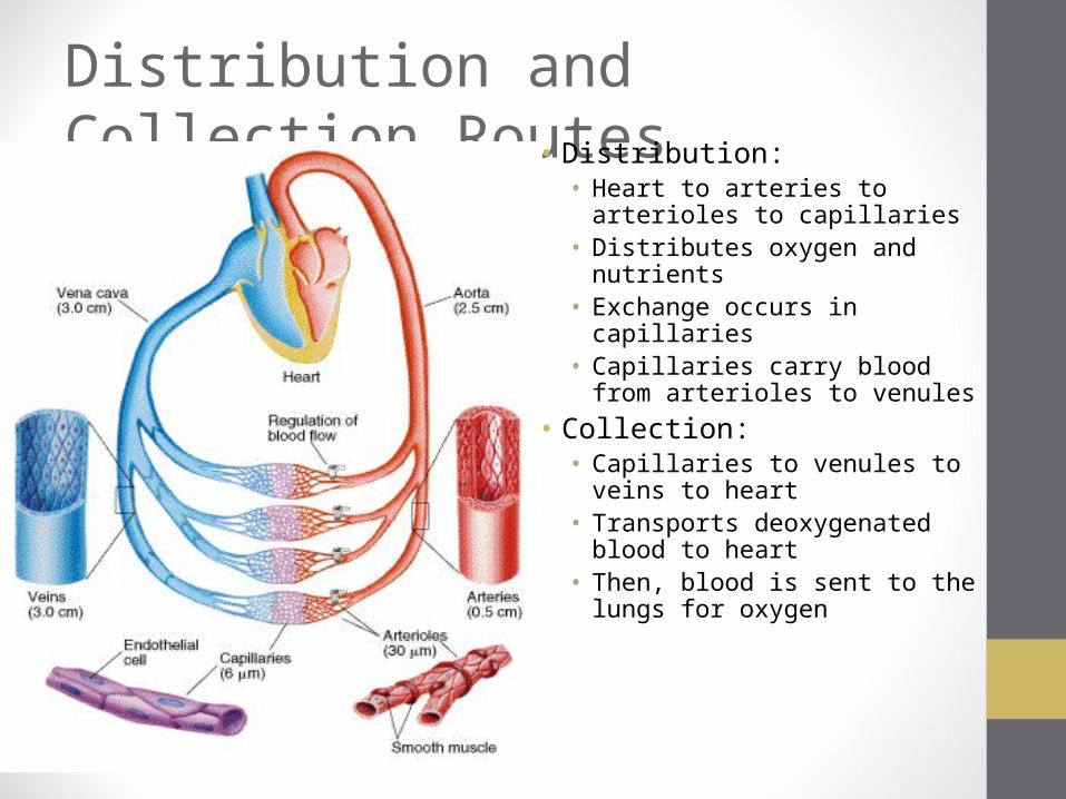
Distribution and Collection Routes• Distribution:
• Heart to arteries to arterioles to capillaries
• Distributes oxygen and nutrients• Exchange occurs in capillaries• Capillaries carry blood from
arterioles to venules• Collection:
• Capillaries to venules to veins to heart
• Transports deoxygenated blood to heart
• Then, blood is sent to the lungs for oxygen

Pathways of Circulation
• 2 major pathways of circulation:• Pulmonary circulation • Systemic circulation

Pulmonary Circulation• Right-sided heart pump• Superior and Inferior Vena Cava return deoxygenated blood back to the heart into the right
atrium• Blood passes from right atrium into the right ventricle going through the tricuspid valve• Right ventricle pumps deoxygenated blood into the pulmonary artery which branches into
the right and left pulmonary arteries, one going to each lung• Pulmonary arteries are low in oxygen, high in carbon dioxide• Carbon dioxide will be exchanged for oxygen in the lungs and returned back to the heart via
the pulmonary veins

Systemic Circulation• Left-sided heart pump• Pulmonary veins return
oxygenated blood back to the heart and into the left atrium
• Blood passes through the mitral valve to the left ventricle
• Left ventricle pumps oxygenated blood into the aorta (passes through the aortic semilunar valve) and out the body

Hepatic Portal Circulation• Subdivision of Systemic
Circulation• Blood from abdominal digestive
organs and spleen circulate through the liver before returning to the heart
• Veins from the spleen, stomach, pancreas, intestines do not drain into the inferior vena cava; they send their blood to the liver via the portal vein. Liver is a manufacturing plant.
• Blood leaves the liver via the hepatic vein and then empties into the inferior vena cava; which goes to the right atrium of the heart

Cerebral Circulation• Part of the Systemic
Circulation• Blood supplied to brain via 2
internal carotid arteries and 2 vertebral arteries. Jugular veins drain back to the superior vena cava.
• Brain requires a constant flow of blood to supply oxygen and remove wastes. Blood flow to the brain doesn’t change with activity.
• Circle of Willis (cerebral arterial circle) provides for a continuous blood supply

Renal Circulation• Part of Systemic
Circulation• Renal circulation
• Renal arteries bring blood to kidneys
• Renal veins drain blood from kidneys

The Lymphatic System
• Functions:• Returns lymph fluid to
blood• Filters injurious agents
and prevents them from entering the bloodstream
• *Forms some WBCs (lymphocytes and monocytes)
• Defends against infection

Lymph
• Name for tissue fluid (watery-like) that enters lymph capillaries
• Filtration in capillaries creates tissue fluid, most is returned to the blood

Lymphatic Ducts• 2 ducts empty
lymph into circulatory system• Thoracic duct
• *Largest duct• *Drains ¾ of body and
empties lymph into left subclavian vein
• Right Lymphatic duct• Drains about ¼ of body
and empties lymph into right subclavian vein
• Flaps in both subclavian veins permit entry of lymph but prevent blood from flowing into lymph vessels

Lymph Nodes and Nodules
• Masses of lymphatic tissue• Found in groups along
pathway of lymph vessels• All located at junctions of
head and extremities with trunk of body
• Functions:• Filtration• Phagocytosis• WBC formation

Lymph Nodes
• *Cervical lymph nodes• *Tonsils = lymphatic
nodules of pharynx• *Axilla = armpit• Iliac=inner hip• Inguinal=groin

Spleen• *Located LUQ of abdominal
cavity, just below diaphragm, behind the stomach
• Functions:• *WBC production• *Defense = contains plasma
cells that produce antibodies• Not considered a vital organ