Embed Size (px)
Citation preview

trations. However, we found no such effect after analyzing theinfluence of fasting insulin concentration on stimulated lipolysis.
Basal glycerol release from adipose tissue was unaffected bythe age of the subjects. This result is in accordance with previousfindings.7,8 Further, we found no relation between basal glyceroloutflow and BMI. By analyzing the female subgroup separately,we found a significant but modest correlation between glycerolrelease and BMI (r 5 0.2062). Thus, basal lipolysis does notappear to be influenced by age or BMI.
The glycerol level in the interstitial fluid of adipose tissuedepends on adipose-tissue blood flow. Therefore, blood-flowchanges might cause variations in glycerol concentration in thedialysate, thereby masking the influence of age and BMI onlipolysis rate. This potential confounding factor was investigatedby the ethanol-escape method. In men only, blood flow wasnegatively correlated with BMI when the lipolysis rate was stim-ulated with norepinephrine. However, the correlation was weak(r 5 0.4335), so we assume that blood flow in human adiposetissue is not influenced by age or BMI.
In conclusion, in vivo basal or stimulated lipolysis, which ishighly variable in subcutaneous adipose tissue, is not influenced byage or BMI in obese subjects.
Marion Flechtner-Mors, PhDAndreas Alt, PhD
Guido Adler, MD, PhDHerwig H. Ditschuneit, MD
University of UlmUlm, Germany
Christopher P. Jenkinson, PhDUniversity of Texas Health Science Center
San Antonio, Texas, USA
REFERENCES
1. Jansson, PA, Larsson A, Smith U, Lo¨nnroth P. Glycerol production in subcuta-neous adipose tissue in lean and obese humans. J Clin Invest 1992;89:1610
2. Coppack SWRD, Evans R, Fisher M, et al. Adipose tissue metabolism in obesity:lipase action in vivo before and after a mixed meal. Metabolism 1992;41:264
3. Arner P. Control of lipolysis and its relevance to development of obesity in man.Diabetes Metab Rev 1988;4:507
4. Arner P. Adrenergic receptor function in fat cells. Am J Clin Nutr 1992;55:228S5. Coppack SW, Jensen MD, Miles JM. In vivo regulation of lipolysis in humans.
J Lipid Res 1994;35:1776. Ostman, J, Efendic S, Arner P. Catecholamine metabolism of human adipose tissue.
I. Comparison between in vitro effects of noradrenaline, adrenaline and theophyllineon lipolysis in omental adipose tissue. Acta Med Scand 1969;186:241
7. Lonnqvist F, Nyberg B, Wahrenberg H, Arner P. Catecholamine-induced lipol-ysis in adipose tissue of the elderly. J Clin Invest 1990;85:1614
8. Imbeault P, Prud’Homme D, Trembley A, Despre´s J-P, Maurie`ge P. Adiposetissue metabolism in young and middle-aged men after control for total bodyfatness. J Clin Endocrinol Metab 2000;85:2455
9. Gerber JG, Detmar-Hanna D, Zahniser NR. Lack of an effect of age on beta-adrenoceptor mediated lipolysis in isolated human adipocytes. J Gerontol A BiolSci Med Sci 1999;54(2):B71
10. Hickner RC, Rosdahl H, Borg I, et al. Ethanol may be used with the microdialysistechnique to monitor blood flow changes in skeletal muscle: dialysate glucoseconcentration is blood flow-dependent. Acta Physiol Scand 1991;143:355
11. Bjorkhelm J, Arner P, Thore A, O¨ stman J. Sensitive kinetic bioluminescent assayof glycerol release from human fat cells. J Lipid Res 1981;22:1142
12. Machata G. U¨ ber die gaschromatische Blutalkoholbestimmung. Analyse derDampfphase. Microchim Acta 1964;262
13. Arner P. Regulation of lipolysis in fat cells. Diabetes Rev 1996;4:450
14. Engfeldt P, Hellme´r J, Wahrenberg H, Arner P. Effects of insulin on adrenoceptor
binding and the rate of catecholamine-induced lipolysis in isolated human fat
cells. J Biol Chem 1988;263:15553
PII S0899-9007(01)00608-6
Circadian Rhythmicity ofWhole-Blood Glutathione inHealthy SubjectsObjectives: Physiologic rhythms of antioxidants have been thesubject of considerable interest in recent years. Physiologically, itis known that 24-h variability in whole-blood glutathione (GSH)could depend on various factors, such as meal composition, proteindietary content, meal-related hormonal modifications, and stress.Experiments were conducted to determine if the circadian varia-tions previously observed in hepatic concentrations of reducedglutathione in animals also occurred in healthy volunteers.Methods: The circadian periodicity of whole-blood glutathionewas explored. All parameters were measured enzymatically at 4-hintervals over a 24-h period.Results:Circadian rhythms were not found. The rhythms peak forglutathione content occurred at 9 h with a small peak at 21 h. Thenadir occurred at 17 h and 1 h.Conclusion:These data show that despite this wide distribution ofglutathione throughout the day we could not find any significantintertime variability.
INTRODUCTION
Glutathione (L-g-glutamyl-L-cysteinyl-glycine; GSH) is the mostabundant antioxidant inside the cells. It performs a variety ofphysiologic and metabolic functions such as thiol-transfer reac-tions, which seem to protect cell membranes and proteins, metab-olism of endogenous compounds, and transport of aminoacids.Lipid peroxidation is an oxidative process leading to the denatur-ation of biological membranes; it is implicated in diverse patho-physiologic conditions such as aging, atherosclerosis, rheumaticdiseases, cancer, cardiac and cerebral ischemias, respiratory-distress syndrome, various liver disorders, sepsis, trauma, burns,and the toxicity to various organs induced by certain metals,solvents, pesticides, and drugs.1,2
Several investigators have suggested that the hepatic, blood andplatelet, content of GSH undergoes marked diurnal fluctuationsand possibly depends on the food intake of animals.3
Physiologic rhythms of amino acids and antioxidants are thesubject of considerable interest. Circadian rhythm alteration mightrepresent an early etiologically determinant stage of disease or a signof illness, and correction of rhythm alteration might prevent or alle-viate certain diseases. The goal of this study was to determine enzy-matically the profile and significance of circadian variations in whole-blood GSH over 24 h, with 4-h intervals, in three healthy subjects whoconsumed a standard diet. We found that GSH in whole blood had adiurnal rhythm but without significant intertime variability.
MATERIALS AND METHODS
Subjects
Three subjects (two male and one female) were classified asclinically healthy on the basis of complete medical check-ups. Themean age was 40 y, mean body weight was 64.6 kg, mean heightwas 166 cm, and mean body mass index was 23.2 kg/m2. The
Correspondence to: Erick Valencia, MD, Pharmaceutical Nutrition, Schoolof Biological and Molecular Sciences, Oxford Brookes University, Head-ington, Oxford OX3 0BP, UK. E-mail: [email protected]
Date accepted: March 10, 2001
Nutrition Volume 17, Number 9, 2001 731Research Letters
purpose of the study was explained fully to each subject who thenprovided informed written consent. Between July and August2000, we investigated the GSH content in whole blood. All sub-jects had followed a regular diurnal rhythm for at least 3 wk beforethe experiment. The experiment started at 1:00PM. The subjectscontinued their usual activities during the study period, apart fromthe sampling periods. They went to sleep before 12:00AM andwere awakened for the 1:00AM and 5:00AM samples.
Diets
Each subject was studied twice with a 1-wk interval diet. Subjectsconsumed the test diet, with known intakes of protein, total GSH,cysteine, and methionine, for 3 d. The meals were prepared indi-vidually to provide 25 kcal of energy per kilogram each day, with16.3% of the energy as protein (1 gz kg21 z d21), 60% ascarbohydrate, and 23.7% as fat (ratio of polyunsaturated to satu-rated fatty acid5 1). The calculated GSH contents of each mealwere 5.2 g for breakfast, 4.6 g for lunch and for dinner, and 0.6 gfor snacks; the cysteine-plus-methionine contents were 0.04 g forbreakfast and snacks and 0.75 g for lunch and dinner. The glu-tamine and glutamate contents of each meal were 6.9 g for break-fast, midmorning and afternoon snacks and 4.98 g for lunch anddinner. Main meals were eaten at 8:00AM, 12:30PM, 7:00PM, andsnacks were eaten at 10:00AM, 3:00 PM, and 9:30PM.
Blood-Sample Collection and GSH Assays
N-ethylmaleimide, reduced GSH, glyoxalase-I, and methylglyoxalwere purchased from Sigma Chemical Company (UK). Approxi-mately 5 mL of whole blood were taken from forearm veins at1:00, 5:00, and 9:00AM and 1:00, 5:00, and 9:00PM. Bloodsamples were obtained by venipuncture and placed into tubescontaining (25 mg) ethylene-diaminetetraacetic for anticoagula-tion. All preparations were performed at room temperature.Whole-blood samples (0.5 mL) were treated immediately with 0.5mL of trichloroacetic acid (25%) containing 40 mM ofN-ethylmaleimide. Within 30 min tubes were centrifuged at 15 000revolutions/min for 10 min, and the acidic supernatants werestored at220°C for 24 to 48 h before analysis.
GSH determinations in whole-blood samples were done byfollowing the glyoxalase-I assay described by Akerboom et al.,4
with minor modifications. Briefly, the reaction between methylg-lyoxal and GSH in 0.1 M of sodium phosphate buffer (pH 7.0) andcatalyzed by glyoxalase-I is followed spectrophotometrically at240 nm. This method is specific for GSH because of the specificityof the glyoxalase-I measurement for GSH.
Statistical Analysis
Data were analyzed by Student’st test (paired, two-tailed) and anal-ysis of variance. Within-day variability was analyzed with analysis ofvariance. Correlation coefficients were computed, and tests were doneto determine whether ther value differed from zero. In addition,individualized time series were analyzed with the single Cosinormethod5 and Student’st test. Rhythmometric data were summarizedby means. For all analytical procedures,P , 0.05 was consideredstatistically significant. All statistical analyses were performed withMicrosoft Excel (2000, Microsoft, Redmond, WA, USA).
RESULTS
We measured reduced GSH in whole blood. The critical step in thedetermination of GSH is the time between blood sampling andanalysis or freezing at220°C or in liquid nitrogen. Analysis ofwhole-blood samples that have been instantly deproteinized andstored at220°C for 48 h produces practically the same value as an
immediate analysis of a fresh sample. We did not find a closecorrelation between each subject’s GSH content and GSH (r 50.86,P 5 0.06), methionine, cysteine (r 5 0.35,P 5 0.55), andprotein (in grams;r 5 0.51,P 5 0.23) concentrations in their diets.
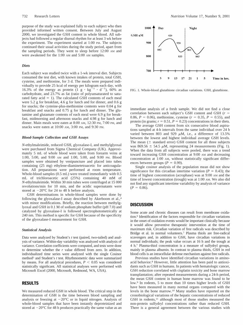
The average GSH content from six consecutive blood aspira-tions sampled at 4-h intervals from the same individual over 24 hvaried between 803 and 929mM, i.e., a difference of 13.5%between the lowest and highest individual average GSH levels.The mean (6 standard error) GSH content for all three subjectswas 869.566 54.5 mM, representing 24 measurements (Fig. 1).When the data from all subjects were pooled, there was a trendtoward increasing GSH concentration at 9:00AM and decreasingconcentration at 1:00AM, without statistically significant differ-ences between groups (P 5 0.99).
Single cosinor analysis of the population mean did not showsignificance for this circadian intertime variation (P 5 0.43); thetime of highest concentration (acrophase) was at 9:00AM and thetime of lowest concentration (trough) was at 1:00AM. We also didnot find any significant intertime variability by analysis of variance(P 5 0.86).
DISCUSSION
Some acute and chronic diseases can result from membrane oxida-tion.6 Identification of the factors responsible for circadian variationsin the onset of oxidation events would be important clinically becauseit would allow preventive therapeutic intervention at the time ofmaximum risk. Circadian variation of free radicals was described byBridge et al. in normal volunteers.7 Plasma thiols are free-radicalscavengers and, in addition to GSH, have circadian variations innormal individuals; the peak value occurs at 16 h and the trough at4 h.7 Plasma-thiol concentration is a measure of sulfydryl groups,mainly in plasma albumin. In contrast to plasma thiols, GSH in redblood cells is an intracellular defense mechanism against free radicals.
Previous studies have identified circadian variations in amino-acid behavior.8 However, little attention has been paid to antioxi-dants such as GSH in humans. In patients with hematologic cancer,GSH reduction correlated with cisplatin toxicity and bone marrowtransplantation; after repeated measurements during a 24-h period,the mean GSH content in human bone marrow was found to below.9 In rodents, 5 to more than 10 times higher levels of GSHhave been measured in many normal organs compared with thelevels in the bone marrow.10 Many studies have reported on thecronobiological variations of the hepatic concentrations of reducedGSH in rodents,11 although most of those studies measured thenon-protein sulfydryl concentrations rather than reduced GSH.There is a general agreement between the various studies with
FIG. 1. Whole-blood glutathione circadian variations. GSH, glutathione.
732 Research Letters Nutrition Volume 17, Number 9, 2001
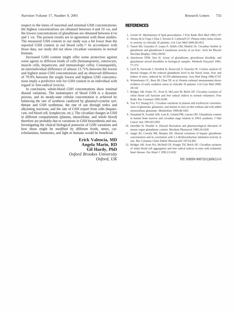
respect to the times of maximal and minimal GSH concentrations:the highest concentrations are obtained between 4 and 10AM, andthe lowest concentrations of glutathione are obtained between 4PM
and 1AM. The present results are in agreement with those studies.The measured GSH content in our study was a bit lower than thereported GSH content in red blood cells.12 In accordance withthose data, our study did not show circadian variations in normalhumans.
Increased GSH content might offer some protection againstsome agents in different kinds of cells (hematopoietic, enterocyte,muscle cells, hepatocyte, and immunologic cells). Consequently,an interindividual difference of almost 13.71% between the lowestand highest mean GSH concentrations and an observed differenceof 79.9% between the single lowest and highest GSH concentra-tions imply a predictive role for GSH content in an individual withregard to free-radical toxicity.
In conclusion, whole-blood GSH concentrations show minimaldiurnal variations. The maintenance of blood GSH is a dynamicprocess, and its steady-state cellular concentration is achieved bybalancing the rate of synthesis catalyzed by glutamyl-cysteine syn-thetase and GSH synthetase, the rate of use through redox andalkylating reactions, and the rate of GSH export from cells (hepato-cyte, red blood cell, lymphocyte, etc.). The circadian changes in GSHin different compartments (plasma, intracellular, and whole blood)therefore are probably due to variations in GSH biosynthesis and use.Investigating the clinical biological potencies of GSH variations andhow those might be modified by different foods, stress, cat-echolamines, hormones, and light in humans would be beneficial.
Erick Valencia, MDAngela Marin, RD
Gil Hardy, PhDOxford Brookes University
Oxford, UK
REFERENCES
1. Girotti W. Mechanisms of lipid peroxidation. J Free Radic Biol Med 1985;1:872. Alonso de la Vega J, Diaz J, Serrano E, Carbonell LF. Plasma redox status relates
to severity in critically ill patients. Crit Care Med 2000;28:18123. Tunon MJ, Gonzalez P, Lopez P, Salido GM, Madrid JA. Circadian rhythm in
glutathione and glutathione-S transferase activity of rat liver. Arch Int PhysiolBiochim Biophys 1992;100:83
4. Akerboom TPM, Sies H. Assay of glutathione, glutathione disulfide, andglutathione mixed disulfides in biological samples. Methods Enzymol 1981;77:373
5. Lach H, Surowiak J, Dziubek K, Krawczyk S, Szaroma W. Cosinor analysis ofdiurnal changes of the reduced glutathione level in the blood, brain, liver andkidney of mice, induced by ACTH administration. Acta Biol Hung 1986;37:93
6. Winterbourn CC, Buss IH, Chan TP, et al. Protein carbonyl measurement showsevidence of early oxidative stress in critically ill patients. Crit Care Med 2000;28:143
7. Bridges AB, Fisher TC, Scott N, McLaren M, Belch JJF. Circadian variation ofwhite blood cell function and free radical indices in normal volunteers. FreeRadic Res Commun 1992;16:89
8. Tsai P-J, Huang P-C. Circadian variations in plasma and erythrocyte concentra-tions of glutamate, glutamine, and alanine in men on diet without and with addedmonosodium glutamate. Metabolism 1999;48:1455
9. Smaaland R, Svardal AM, Lote K, Ueland PM, Laerun OD. Glutathione contentin human bone marrow and circadian stage relation to DNA synthesis. J NatlCancer Inst 1991;83:1092
10. Jaeschke H, Wendel A. Diurnal fluctuation and pharmacological alteration ofmouse organ glutathione content. Biochem Pharmacol 1985;34:1020
11. Jaeger RJ, Conolly RB, Murphy SD. Diurnal variations of hepatic glutathioneconcentration and its correlation with 1,1-dichloroethylene inhalation toxicity inrats. Res Commun Chem Pathol Pharmacolol 1973;6:465
12. Bridges AB, Scott NA, McNeill GP, Pringle TH, Belch JJF. Circadian variationof white blood cell aggregation and free radical indices in men with ischaemicheart disease. Eur Heart J 1992;13:1632
PII S0899-9007(01)00653-0
Nutrition Volume 17, Number 9, 2001 733Research Letters