Embed Size (px)
Citation preview

6
Chytridiomycota
6.1 Introduction
The phylum Chytridiomycota comprises over
900 species in five orders (D. J. S. Barr, 2001; Kirk
et al., 2001). Fungi included here are colloquially
called ‘chytrids’. Most chytrids grow aerobically
in soil, mud or water and reproduce by zoospores
with a single posterior flagellum of the whiplash
type, although the zoospores of some members of
the Neocallimastigales are multiflagellate. Some
species inhabit estuaries and others the sea.
Sparrow (1960) has given an extensive account
of aquatic forms, Karling (1977) a compendium of
illustrations, and Powell (1993) has provided
examples of the importance of the group. Many
members are saprotrophs, utilizing cellulose,
chitin, keratin, etc., from decaying plant and
animal debris in soil and mud, whilst species of
Caulochytrium grow as mycoparasites on the
mycelium and conidia of terrestrial fungi (Voos,
1969). Saprotrophs can be obtained in crude
culture by floating baits such as cellophane,
hair, shrimp exoskeleton, boiled grass leaves
and pollen on the surface of water overlying
samples of soil, mud or pieces of aquatic plant
material (Sparrow, 1960; Stevens, 1974;
Willoughby, 2001). From such crude material,
pure cultures may be prepared by streaking
or pipetting zoospores onto agar containing
suitable nutrients and antibiotics to limit con-
tamination from bacteria. The growth and
appearance of chytrids in pure culture is variable
and often differs significantly from their natural
habit. This has led to problems in classification
systems based on thallus morphology (Barr, 1990,
2001). The availability of cultures has, however,
facilitated studies on chytrid nutrition and
physiology (Gleason, 1976).
Some chytrids are biotrophic parasites of
filamentous algae and diatoms and may severely
deplete the population of freshwater phyto-
plankton (see p. 139). Two-membered axenic
cultures of diatom host and parasite have been
prepared, making possible detailed ultrastruc-
tural studies of comparative morphology, zoo-
spores, infection processes and reproduction.
Other chytrids such as species of Synchytrium
and Olpidium are biotrophic parasites of
vascular plants. Synchytrium endobioticum is the
agent of the potentially serious black wart
disease of potato. Olpidium brassicae, common in
the roots of many plants, is relatively harmless,
but its zoospores are vectors of viruses such
as that causing big vein disease of lettuce.
Coelomomyces spp. are pathogens of freshwater
invertebrates including copepods and the larvae
of mosquitoes. The possibility of using them
in the biological control of mosquitoes has
been explored. The most unusual group are the
Neocallimastigales, which grow in the guts of
herbivorous mammals, are obligately anaerobic
and subsist on ingested herbage.
The cell walls of some chytrids have been
examined microchemically by X-ray diffraction
and other techniques. Chitin has been detected
in many species (Bartnicki-Garcia, 1968, 1987),
and in Gonapodya cellulose is also present
(Fuller & Clay, 1993). The composition of the
wall is of interest because chitin, a polymer of

N-acetylglucosamine, is also found in the walls
of other Eumycota (i.e. Zygomycota, Ascomycota
and Basidiomycota), whilst the cell walls of
members of the Oomycota contain cellulose.
Cellulose and chitin occur together in the walls
of species of Hyphochytrium and Rhizidiomyces,
members of the Hyphochytriomycota (Fuller,
2001; see Section 4.3).
The form of the thallus in the
Chytridiomycota is varied. In biotrophic species
such as Olpidium and Synchytrium, where the
whole thallus is contained within the host cell,
there is no differentiation into a vegetative
and a reproductive part. At maturity the entire
structure, except for the wall which surrounds it,
is converted into reproductive units, i.e. zoo-
spores, gametes or resting sporangia. Such thalli
are termed holocarpic (Fig. 6.1). More usually, the
thallus is differentiated into organs of reproduc-
tion (sporangia and resting sporangia) arising
from a vegetative part which often consists of
rhizoids. These serve in the exploitation of the
substratum and the assimilation of nutrients.
Thalli of this type are eucarpic. Eucarpic thalli
may have one or several sporangia and are then
termed monocentric or polycentric, respectively
(Fig. 6.1). In some species there are both mono-
centric and polycentric thalli, so that these terms
have descriptive rather than taxonomic signifi-
cance. A further distinction has been made,
especially in monocentric forms, between those
in which only the rhizoids are inside the host
cell whilst the sporangium is external (epibiotic),
in contrast with the endobiotic condition in
which the entire thallus is inside the host cell
(Fig. 6.1). In monocentric thalli, the rhizoids
usually radiate from a single position on the
sporangium wall, but in polycentric forms a
more extensive, branched rhizoidal system, the
rhizomycelium, develops.
The zoosporangium is generally a spherical
or pear-shaped sac bearing one or more dis-
charge tubes or exit papillae. The method of
zoospore release has been used in classification.
Fig 6.1 Types of thallus structure in theChytridiales, diagrammatic and not to scale.
128 CHYTRIDIOMYCOTA

In the inoperculate chytrids such as Olpidium,
Diplophlyctis and Cladochytrium, the sporangium
forms a discharge tube which penetrates to the
exterior of the host cell and its tip becomes
gelatinous and dissolves away. In operculate
chytrids such as Chytridium and Nowakowskiella,
the tip of the discharge tube breaks open at a
special line of weakness and is seen as a special
cap or operculum after discharge (see Fig. 6.4b).
6.1.1 The zoosporeThe number of zoospores formed inside zoospo-
rangia of chytrids varies with the size of the
spore and sporangium. Although the zoospore
size is roughly constant for a given species, the
size of the sporangium may be very variable. In
Rhizophlyctis rosea, tiny sporangia containing only
one or two zoospores have been reported from
culture media deficient in carbohydrate, whereas
on cellulose-rich media large sporangia contain-
ing many hundred spores are formed. The release
of zoospores is brought about by internal
pressure which causes the exit papillae to burst
open. In studies of the fine structure of mature
sporangia of R. rosea and Nowakowskiella profusa
(Chambers & Willoughby, 1964; Chambers et al.,
1967), it has been shown that the single
flagellum is coiled round the zoospore like a
watch spring. The zoospores are separated by a
matrix of spongy material which may absorb
water and swell rapidly at the final stages
of sporangial maturation. When the internal
pressure has been relieved by the ejection of
some zoospores, those remaining inside the
sporangium escape by swimming or wriggling
through the exit tube. In some species the spores
are discharged in a mass which later separates
into single zoospores, but in others the zoospores
make their escape individually.
The form of the zoospore is similar in all
chytrids (with the exception of the multiflagel-
late members of the Neocallimastigales). There
is a spherical or ellipsoidal body which in some
forms is capable of plastic changes in shape, and
a long trailing flagellum. When swimming, the
zoospores show characteristic jerky or ‘hopping’
movements; additionally, abrupt changes in
direction are sometimes made. The internal
structure of the zoospore as revealed by light
and electron microscopy is variable, but char-
acteristic of particular genera (Lange & Olson,
1979). In view of the plasticity in morphology of
the thallus under different growth conditions,
zoospore ultrastructure is regarded as a more
satisfactory basis of classification (D. J. S. Barr,
1990, 2001). Two features are of taxonomic
importance, the flagellar apparatus and an assem-
blage of organelles termed the microbody�lipid
globule complex (MLC) (D. J. S. Barr, 2001).
The flagellar apparatus
The whiplash flagellum resembles that of other
eukaryotes, with a smooth membrane enclosing
a cylindrical shaft, the axoneme, made up
internally of nine doublet pairs of microtubules
surrounding two central microtubules. As shown
in Fig. 6.2, the base of the axoneme comprises
three regions, the flagellum proper, the transi-
tional zone and the kinetosome. The function
of the kinetosome is to generate the flagellum.
An interesting feature found in several species
is a second kinetosome or the remainder of
one, the dormant kinetosome. Its presence
has led to the suggestion that the ancestors
of the Chytridiomycota may have had biflagel-
late zoospores, the second flagellum having been
lost in the course of evolution (Olson & Fuller,
1968).
In section, the kinetosome resembles a cart-
wheel (Fig. 6.2f), because to each of the nine
outer microtubule doublets seen in the flagel-
lum proper, a third microtubule is attached.
This is called the C-tubule; in the doublets,
that tubule with extended dynein arms is the
A-tubule, and its partner is labelled B. These
flagellar microtubules radiate as kinetosome
props into the zoospore, perhaps providing
structural support and anchorage of the flagel-
lum (D. J. S. Barr, 2001). Microtubules may also be
attached laterally to the kinetosome, contribut-
ing to the flagellar root system (Figs. 6.2c, 6.19).
In the innermost (proximal) part of the transi-
tional zone, the nine microtubule triplets of
the kinetosome are converted into the doublets
of the flagellum proper; concentric fibres,
possibly arranged helically, surround the nine
doublet pairs. Also within the transitional zone,
129INTRODUCTION

the two central microtubules arise near a
terminal plate. The structure of the flagellum
and kinetosome in transverse section is shown in
Figs. 6.2e and f (Barr & Desaulniers, 1988).
The microbody�lipid complex
The MLC (Fig. 6.3) is made up of a microbody
which is often closely appressed to a large lipid
globule and to simple membrane cisternae or
a tubular membrane system, the rumposome.
This is defined as a cisterna in which there is an
area with hexagonally arranged, honeycomb-like
pores called fenestrae (Fuller, 1976; Powell &
Roychoudhury, 1992). The rumposome may
be involved in signal transduction from the
plasma membrane to the flagellum because it
is known that this organelle sequesters calcium.
Regulation of external calcium concentrations
has an effect on the symmetry of flagellar beat
and hence on the direction of zoospore move-
ment (Powell, 1983).
There are several distinct types of MLC (Powell
& Roychoudhury, 1992) and Fig. 6.3 illustrates
diagrammatically just one of them, that
described for Rhizophlyctis harderi. In this species,
the MLC includes several (3�5) lipid globules.
Fig 6.2 Flagellar apparatus typical of zoospores ofChytridiomycota. (a)Median longitudinal section of the junction of the flagellumwith the body of the zoospore.The labels indicate the flagellumproper (F), transitional zone (TZ), kinetosome (K), electron-denseregion (ED), concentric fibres (CF), transitional fibres (TF), kinetosome props (KP), terminal plate (TP), kinetosome (K) showing acartwheel-like organization (Cw), dormantkinetosome (DK), fibrillarmaterial (Fi) found in some taxa, andmicrotubular roots (Mt)extending from the side or end of the kinetosome into the body of the zoospore. (b) Transverse section near the terminal plateshowing nine kinetosome props extending from doubletmicrotubules to the cellmembrane. (c) Transverse section in the lowerpart of the transition zone showing concentric and transitional fibres. (d) Transverse section of the flagellum proper showing twocentralmicrotubules and nine peripheral doubletmicrotubules enclosed in the flagellar membrane (FM). (e) Schematic drawing ofthe flagellum proper in transverse section.The arrowed line 0��180� shows an imaginary planewhich coincides with the plane ofundulation of the flagellum, passing through doublet pair1and between the centralmicrotubules and doublet pairs 5 and 6.The convention used in labelling the outer doublet pairs ofmicrotubules is shown: themicrotubulewith dynein arms (d) is theAmicrotubule and its partner is the B microtubule. (f) Kinetosome in transverse section showing the triplet arrangement of theperipheralmicrotubules by the addition of a thirdmicrotubule (C).Redrawn from Barr and De¤ saulniers (1988) by copyrightpermission of the National Research Council of Canada, Barr (1992).�TheMycological Society of America, and D. J. S. Barr (2001)with kind permission of Springer Science and Business Media.
130 CHYTRIDIOMYCOTA

Those at the anterior of the cell are embedded
in an aggregation of ribosomes. The surfaces
of lipid globules close to the plasma membrane
are partially covered by one to several simple
cisternae, sometimes with irregularly scattered
pores. Towards the centre of the cell the
lipid bodies are clasped by cup-shaped micro-
bodies. At the posterior of the zoospore near
the kinetosome, 1�3 smaller lipid globules
are partially covered by a rumposome, linked
to the plasma membrane by short bridges and
to the kinetosome by a microtubule root.
Other features
Patches of glycogen are located in the peripheral
cytoplasm of the zoospore and it is likely
that these and the lipid globules represent
sources of energy used in respiration and
propulsion. Mitochondria tend to be concen-
trated in the posterior of the zoospore close
to the kinetosome; in Allomyces and Blastocladiella
(Blastocladiales), the base of the flagellum
passes through the perforation of a single large
mitochondrion (see Fig. 6.19).
Most zoospores are uninucleate. The nucleus
is surrounded in many cases (but not all) by a
nuclear cap of uneven thickness. The nuclear cap
is especially prominent in zoospores of members
of Blastocladiales such as Allomyces and
Blastocladiella (Fig. 6.19). It is rich in RNA and
protein and also contains ribosomes.
6.1.2 Zoospore encystment andgermination
The period of zoospore movement varies. Some
flagellate zoospores seem to be incapable of
active swimming and amoeboid crawling may
take place instead, or swimming may last for
only a few minutes. In other spores, motility
may be prolonged for several hours. Prior to
germination, the zoospore comes to rest and
encysts. The flagellum may contract, it may be
completely withdrawn or it may be cast off,
but the precise details are often difficult to
follow. The subsequent behaviour also differs
in different species. In holocarpic parasites the
zoospore encysts on the host surface and
the cytoplasmic contents of the zoospore are
injected into the host cell. In many monocentric
chytrids rhizoids develop from one point on
the zoospore cyst and the cyst itself enlarges to
form the zoosporangium, but there are variants
of this type of development in which the cyst
enlarges into a prosporangium from which the
zoosporangium later develops. In the polycentric
types, the zoospore on germination may form
a limited rhizomycelium on which a swollen
cell arises, giving off further branches of rhizo-
mycelium. Germination may be from a single
point on the wall of the zoospore cyst (mono-
polar germination) or from two points, enabling
growth to take place in two directions (bipolar
germination). The mode of germination is an
important character in distinguishing, for
example, the Chytridiales (monopolar) from
the Blastocladiales (bipolar).
6.1.3 Life cycles of the ChytridiomycotaMost chytrids have haploid zoospores and thalli
but some Blastocladiales show an alternation
of haploid (gametothallic) and diploid (spor-
othallic) generations. Apart from differences in
Fig 6.3 Schematic diagram of themicrobody�lipid complexof the zoospore of Rhizophlyctis harderi as seen in a longitudinalsection through the base of the zoospore and flagellum.Thefollowing organelles are drawn: mitochondrion (Mc), simplecisterna (C), lipid globule (L), microbody (Mi), flagellum (F) andrumposome (R).Redrawn from Powell and Roychoudhury(1992), by copyright permission of the National ResearchCouncil of Canada.
131INTRODUCTION

the reproductive organs, the morphology of the
two types of thallus is very similar, a phenom-
enon known as isomorphic alternation of
generations.
Sexual reproduction, i.e. a life cycle which
includes nuclear fusion and meiosis, may occur
in several different ways (e.g. Figs. 6.6 and 6.22).
In some chytrids it is by gametogamy, the fusion
of gametes which are posteriorly uniflagellate.
Isogamous conjugation occurs if there is no
morphological distinction between the two
fusing partners, but in some Blastocladiales (e.g.
Allomyces) anisogamy takes place by fusion
between a smaller, more actively motile male
gamete with a larger, sluggish female gamete.
Oogamy, fusion between an actively motile
male gamete and a much larger, non-flagellate,
immobile globose egg, is characteristic of
Monoblepharidales. Somatogamy, the fusion
of undifferentiated hyphae or rhizoids, has
been well documented in cultures of the fresh-
water fungus Chytriomyces hyalinus by Moore
and Miller (1973) and Miller and Dylewski
(1981, 1987). As shown in Fig. 6.4, zoospores of
C. hyalinus are released from the zoosporangium
by the opening of a lid-like operculum. They
germinate to form uninucleate rhizoidal thalli
(contributory thalli) and the tips of the rhizoids
from adjacent thalli, which are apparently not
genetically distinct from each other, may fuse
(Fig. 6.4c). At the point of fusion an incipient
resting body develops (Fig. 6.4d) and swells
while cytoplasm and a nucleus migrate into it
from each contributory thallus. Nuclear fusion
occurs in the resting body to form a diploid
zygote nucleus. The resting body continues to
enlarge and develops a thick wall. This type
of sexual reproduction by somatogamous con-
jugation probably occurs in several genera of
inoperculate and operculate chytrids (Moore &
Miller, 1973).
Fusion of gametangia (gametangio-
gametangiogamy) has been reported by Doggett
and Porter (1996) for Zygorhizidium planktonicum,
a parasite of the diatom Synedra. This species
reproduces asexually by epibiotic zoosporangia.
Germinating zoospores develop either new
zoosporangial thalli or gametangial thalli of
two sizes with globose uninucleate gametangia.
Conjugation occurs when a conjugation tube
grows from the smaller donor to the larger
recipient gametangium (Fig. 6.5a). Following
nuclear fusion, the larger gametangium develops
a thick wall and functions as a diploid rest-
ing spore. After a period of maturation the
resting spore acts as a prosporangium, giving
rise to a thin-walled meiosporangium. Meiosis,
as evidenced by the presence of synaptonemal
complexes, occurs here, followed by mitosis
and cytoplasmic cleavage to form zoospores
(Fig. 6.5b). A variant of this form of sexual
differentiation (gametangio-gametogamy) has
Fig 6.4 Chytriomyces hyalinus somatogamy. (a,b) Epibioticfruiting thallus seated on a pollen grain intowhich rhizoidshave penetrated. In (a) the zoosporangium, containingnumerous zoospores, is seen shortly before dischargewith abulging operculum (o). In (b) the operculum has lifted off andthe zoospores are escaping. (c�e) Stages in somatogamy.(c) Rhizoids from two uninucleate contributory thalli (c) haveundergone anastomosis (arrow). (d) Cytoplasm and a nucleusfrom each contributory thallus havemigrated towards thepoint of anastomosis, where the thallus swells to form aglobose incipient resting body (i) which is binucleate andpackedwith cytoplasm, leaving the contributory thalli empty.(e) The two nuclei in the incipient resting body have fused.After C.E.Miller and Dylewski (1981).
132 CHYTRIDIOMYCOTA

been reported in species of Rhizophydium (Karling,
1977); this involves copulation between the
gametangium of a rhizoid-forming thallus and
a motile gamete that encysts directly on the
gametangium.
Generally the product of sexual reproduction
is a resting spore or resting sporangium with
thick walls, but it is known that thick-walled
sporangia may also develop asexually and in
many chytrids sexual reproduction has not been
described and possibly does not occur. Resting
sporangia of some chytrids may remain viable
for many years.
6.1.4 Classification and evolutionFossil chytrids have been reported from the
400million-year-old Rhynie chert, a site known
for the discovery of fossil remains of the
earliest known vascular land plants. Clusters
of holocarpic, endobiotic thalli resembling the
present day Olpidium have been found inside
cells of a coenobial alga preserved within the
hollow axes of a vascular plant, and epibiotic
sporangia with endobiotic rhizoids have been
seen attached to meiospores of a vascular plant,
much like those of extant chytrids like
Rhizophydium which grow on pollen grains
(Taylor et al., 1992). Chytrid-like fossils have also
been found in strata of the 340million-year-old
Pennsylvanian (Carboniferous) era (Millay &
Taylor, 1978) and from the more recent Eocene
strata (Bradley, 1967).
Formerly thought to have an affinity for
the Oomycota, Hyphochytriomycota or protists,
the Chytridiomycota are now accepted as
members of the true fungi, the Eumycota. They
are probably ancestral to other groups of true
fungi, especially the Zygomycota (Cavalier-Smith,
1987, 2001; D. J. S. Barr, 2001). The inclusion of the
chytrids in the Eumycota is supported by several
DNA-based phylogenetic analyses (e.g. Bowman
et al., 1992; James et al., 2000), but the delimitation
of orders within the Chytridiomycota is still
problematic. Particularly puzzling is the group-
ing of the Blastocladiales with the Zygomycota on
the basis of 18S ribosomal DNA sequences
(see Fig. 1.26).
D. J. S. Barr (2001) and Kirk et al. (2001) have
classified the Chytridiomycota into five orders
(Table 6.1) but the details of their distinguishing
features need not concern us here. We shall
study examples from each order.
Fig 6.5 Sexual reproduction in Zygorhizidiumplanktonicum. (a) Empty donor gametangium to the left connected by aconjugation tube to a mature resting spore. (b) Near-median section of a fully formedmeiosporangiumwhich has developedfrom a germinating resting spore.The donor gametangium is on the right. Scale bar¼ 4mm. After Doggett and Porter (1996).
133INTRODUCTION

6.2 Chytridiales
This is by far the largest order, comprising more
than 50% of the total number of chytrids
described to date. It is difficult to characterize
members of the Chytridiales because they lack
any specific features by which species have been
assigned to the other four orders. The classifica-
tion of the Chytridiales has traditionally been
based on thallus morphology (Sparrow, 1973)
but, as pointed out by D. J. S. Barr (2001), this is
unsatisfactory because of the great variability in
thallus organization shown by the same fungus
growing on its natural substratum and in
culture. Future systems of classification will be
based on zoospore ultrastructure and the
comparison of several different types of DNA
sequences, but too few examples have yet been
studied to provide a definitive framework.
Because of this we shall study genera which
illustrate the range of morphology, life cycles
and ecology of the Chytridiales without attempt-
ing to place them into families.
6.2.1 SynchytriumIn this genus the thallus is endobiotic and
holocarpic, and at reproduction it may become
converted directly into a group (sorus) of
sporangia, or to a prosorus which later gives
rise to a sorus of sporangia. Alternatively the
thallus may turn into a resting spore which
can function either directly as a sporangium
and give rise to zoospores, or as a prosorus. The
zoospores are of the characteristic chytrid type
(Lange & Olson, 1978). Sexual reproduction is
by copulation of isogametes, resulting in the
formation of thalli which develop into thick-
walled resting spores. Synchytrium includes
about 120 species which are biotrophic parasites
of flowering plants. Some species parasitize only
a narrow range of hosts, e.g. S. endobioticum on
Solanaceae, but others, e.g. S. macrosporum, have
a wide host range (Karling, 1964). Most species
are not very destructive to the host plant but
stimulate the formation of galls on leaves,
stems and fruits.
Synchytrium endobioticumThis is the cause of wart disease affecting
cultivated potatoes and some wild species of
Solanum. It is a biotrophic pathogen which has
not yet been successfully cultured outside living
host cells. Wart disease is now distributed
throughout the main potato-growing regions
of the world, especially in mountainous areas
and those with a cool, moist climate. Lange
(1987) has given practical details of techniques
for studying the fungus but in most European
countries handling of living material by
Table 6.1. Orders of Chytridiomycota following D. J. S. Barr (2001) and Kirk et al. (2001).
Order Numberofdescribed taxa
Examples
Chytridiales(see Section 6.2)
80 genera Cladochytrium,Nowakowskiella,Rhizophydium,Synchytrium600 spp.
Spizellomycetales(see Section 6.3)
13 genera Olpidium,Rhizophlyctis86 spp.
Neocallimastigales(see Section 6.4)
5 genera Anaeromyces,Caecomyces,Neocallimastix,Orpinomyces, Piromyces16 spp.
Blastocladiales(see Section 6.5)
14 genera Allomyces,Blastocladiella,Coelomomyces,Physoderma179 spp.
Monoblepharidales(see Section 6.6)
4 genera Gonapodya,Monoblepharella,Monoblepharis19 spp.
134 CHYTRIDIOMYCOTA

unlicensed workers is illegal. Diseased tubers
bear dark brown cauliflower-like excrescences.
Galls may also be formed on the aerial shoots,
and they are then green with convoluted leaf-like
masses of tissue (the leafy gall stage; Plates 3a,b).
Heavily infected tubers may have a considerable
proportion of their tissues converted to warts.
The yield of saleable potatoes from a heavily
infected crop may be less than the actual weight
of the seed potatoes planted. The disease is thus
potentially a serious one, but fortunately vari-
eties of potatoes are available which are immune
from the disease, so that control is practicable.
The possible life cycle of S. endobioticum is
summarized in Fig. 6.6.
The dark warts on the tubers are galls in
which the host cells have been stimulated to
divide by the presence of the fungus. Many
of the host cells contain resting spores which
are more or less spherical cells with thick dark
brown walls folded into plate-like extensions
(see Fig. 6.7a). The resting spores are released
by the decay of the warts and may remain alive
in the soil for over 40 years (Laidlaw, 1985).
The outer wall (exospore) bursts open by an
irregular aperture and the endospore balloons
out to form a vesicle within which a single
sporangium differentiates (Kole, 1965; Sharma
& Cammack, 1976; Hampson et al., 1994). Thus
the resting spore functions as a prosporangium
on germination. Germination of the resting
spore may occur spontaneously but can be stimu-
lated by passage through snails. It is presumed
that abrasion and digestion of the spore wall
Fig 6.6 Schematic outline of the probable life cycle of Synchytrium endobioticum.Haploid and diploid nuclei are represented bysmall empty and larger split circles, respectively.Key events in the life cycle are plasmogamy (P), karyogamy (K) andmeiosis (M).Resting spores within awartedpotato contain a single nucleus which undergoesmeiosis upon germination.Haploid zoosporesare released from a single sporangium. If two zoospores pair up, a zygote is formed and penetration of a potato cell gives rise toa diploid thallus and, ultimately, a resting spore.Diploid infections cause host hyperplasia visible as the potato wart symptoms.If a zoospore infects in thehaploid state, a haploidprosorus (summer spore) is formed, andhypertrophyof the infected and adjacenthost cells ensues. A sorus of several sporangia is ultimately produced, with each sporangium releasing a fresh crop of haploidzoospores. Synchytrium endobioticum appears to be homothallic.
135CHYTRIDIALES

in the snail gut causes breakdown of the thick
wall which contains chitin and branched-chain
wax esters, so overcoming dormancy related
to the impermeability of the wall (Hampson
et al., 1994).
The zoospores are capable of swimming for
about two hours in the soil water. If they alight
on the surface of a potato ‘eye’ or some other
part of the potato shoot such as a stolon or
a young tuber before its epidermis is suberized,
they come to rest and withdraw their flagellum.
During penetration, the contents of the zoospore
cyst are transferred to the host cell whilst the
cyst wall remains attached to the outside. When
a dormant ‘eye’ is infected, dormancy may
be broken and the tuber may begin to sprout. If
the potato variety is susceptible to the disease,
the small fungal thallus inside the host cell
will enlarge. The infected host cell as well as
surrounding cells also enlarge so that a rosette
of hypertrophied cells surrounds a central
infected cell (Fig. 6.7c). The walls of these cells
adjacent to the infected cell are often thickened
and assume a dark brown colour. The infected
cell remains alive for some time but eventually
it dies. The pathogen thallus passes to the
bottom of the host cell, enlarges and becomes
spherical. A double-layered chitinous wall which
Fig 6.7 Synchytrium endobioticum.(a) Resting spores in section of wart.(b) Germinating resting spore showingthe formation of a vesicle containinga single globose sporangium (after Kole,1965). (c) Section of infected host cellcontaining a prosorus.The prosorusis extruding a vesicle.Note the hyper-trophy of the infected cell and adjacentuninfected cells. (d) Cleavage of vesiclecontents to form zoosporangia. (e) Twoextruded zoosporangia. (f) Zoospores.(g) Rosette of hypertrophiedpotato cellsas seen from the surface.The outlineof the infectedhost cell is shown dotted.(h) Young resting sporangium resultingfrom infection by a zygote.Note thatthe infected cell lies beneath theepidermis due to division of thehost cells.
136 CHYTRIDIOMYCOTA

is golden brown in colour is secreted around
the thallus, now termed a prosorus or summer
spore. Further development of the prosorus
involves the protrusion of the inner wall through
a pore in the outer wall, and its expansion as
a vesicle which enlarges upwards and fills the
upper half of the host cell (Fig. 6.7c). The
cytoplasmic contents of the prosorus including
the single nucleus are transferred to the vesicle.
The process is quite rapid and can be completed
in about 4h. During its passage into the vesicle
the nucleus may divide, and mitoses continue
so that the vesicle contains about 32 nuclei.
At this stage the cytoplasmic contents of the
vesicle become cleaved into about 4�9 sporangia
(Fig. 6.7d), forming a sorus. After the deposition
of sporangial walls, further nuclear divisions
occur in each sporangium, and finally each
nucleus with its surrounding mass of cytoplasm
becomes differentiated to form a zoospore. As
the sporangia ripen, they absorb water and
swell, causing the host cell containing them to
burst open. Meanwhile, division of the host
cells underlying the rosette has been taking
place, and enlargement of these cells pushes
the sporangia out onto the surface of the host
tissue (Fig. 6.7e). The sporangia swell if water
is available and burst open by means of a small
slit through which the zoospores escape. There
may be as many as 500�600 zoospores in a single
large sporangium. The zoospores resemble those
derived from resting sporangia and are capable
of initiating further asexual cycles of reproduc-
tion throughout spring and early summer.
Sometimes several zoospores succeed in pene-
trating a single cell so that it contains several
fungal protoplasts.
Alternatively, zoospores may function as
gametes, fusing in pairs (or occasionally in
groups of three or four) to form zygotes which
retain their flagella and swim actively for a time.
Since zoospores acting as gametes do not differ
in size and shape, copulation can be described
as isogamous. However, the gametes may differ
physiologically. Curtis (1921) has suggested that
fusion may not occur between zoospores derived
from a single sporangium, but only between
zoospores from separate sporangia. Kohler (1956)
has claimed that the zoospores are at first
sexually neutral. Later they mature and become
capable of copulation. Maturation may occur
either outside the sporangia or within, so that
in over-ripe sporangia the zoospores are capable
of copulation on release. At first the zoospores
are ‘male’, and swim actively. Later the swarmers
become quiescent (‘female’) and probably secrete
a substance which attracts ‘male’ gametes.
After swimming by means of its two flagella,
the zygote encysts on the surface of the host
epidermis and penetration may then follow by
a process essentially similar to zoospore penetra-
tion. Multiple infections by several zygotes
penetrating a single host cell can also occur.
Nuclear fusion occurs in the young zygote before
penetration.
The results of zygote infections differ from
infection by zoospores. The host cell reacts to
zoospore infection by undergoing hypertrophy,
i.e. increase in cell volume, and adjacent cells
also enlarge to form the characteristic rosette
which surrounds the resulting prosorus. In
contrast, when a zygote infects, the host cell
undergoes hyperplasia, i.e. repeated cell division.
The pathogen lies towards the bottom of the
host cell, adjacent to the host nucleus, and cell
division occurs in such a way that the fungal
protoplast is located in the innermost daughter
cell. As a result of repeated divisions of the
host cells, the typical gall-like potato warts are
formed and fungal protoplasts may be buried
several cell layers deep beneath the epidermis
(see Fig. 6.7h). During these divisions of the
host tissue the zygote enlarges and becomes
surrounded by a two-layered wall, a thick outer
layer which eventually becomes dark brown in
colour and is thrown into folds or ridges which
appear as spines in section, and a thin hyaline
inner wall surrounding the granular cytoplasm
(Lange & Olson, 1981). The host cell eventually
dies and some of its contents are deposited on
the outer wall of the resting sporangium,
forming the characteristic brown ridges. During
its development the resting spore remains uni-
nucleate. Resting spores are released into the
soil and are capable of germination within
about 2months. Before germination, the nucleus
divides repeatedly to form the nuclei of the
zoospores whose further development has
137CHYTRIDIALES

already been described. It has been claimed that
the zygote and the young resting spore
are diploid, and it has been assumed that meiosis
occurs during germination of the resting spo-
rangia prior to the formation of zoospores, so
that these zoospores, the prosori and the soral
zoospores are also believed to be haploid.
These assumptions seem plausible in the light
of knowledge of the life history and cytology
of other species (e.g. Lingappa, 1958b), and an
essentially similar life cycle has been described
for S. lagenariae and S. trichosanthidis, parasitic on
Cucurbitaceae, which differ from S. endobioticum
in that their resting spores function as prosori
instead of prosporangia (Raghavendra Rao &
Pavgi, 1993).
Control of wart disease
Control is based largely on the breeding of
resistant varieties of potato. It was discovered
that certain varieties such as Snowdrop were
immune from the disease and could be planted
on land heavily infected with Synchytrium with-
out developing warts. Following this discovery,
plant breeders have developed a number of
immune varieties such as Maris Piper. However,
some potato varieties that are susceptible to
the disease are still widely grown, including the
popular King Edward. In most countries where
wart disease occurs, legislation has been intro-
duced requiring that only approved immune
varieties be planted on land where wart disease
has been known to occur, and prohibiting the
movement and sale of diseased material. Within
the British Isles, the growing of immune varieties
on infested land has prevented the spread of
the disease, and it is now confined to a small
number of foci in the West Midlands, northwest
England and mid and south Scotland. It has
also persisted in Newfoundland. The majority of
the outbreaks are found in allotments, gardens
and smallholdings.
The reaction of immune varieties to infection
varies (Noble & Glynne, 1970). In some cases
when ‘immune’ varieties are exposed to a heavy
inoculum load of S. endobioticum in the labora-
tory, they may become slightly infected, but
infection is often confined to the superficial
tissues which are soon sloughed off. In the
field such slight infections would probably pass
unnoticed. Occasionally infections of certain
potato varieties may result in the formation
of resting spores, but without the formation
of noticeable galls. Penetration of the parasite
seems to occur in all potato varieties, but when
a cell of an immune variety is penetrated it
may die within a few hours, and since the fungus
is a biotrophic parasite, further development is
checked. In other cases the parasite may persist
in the host cell for up to 2�3 days, apparently
showing normal development, but after this
time the fungal thallus undergoes disorganiza-
tion and disappears from the host cell.
Unfortunately, it has been discovered that
new physiological races (or pathotypes) of
the pathogen have arisen, capable of attack-
ing potato varieties previously thought to be
immune. About 20 pathotypes are now known,
and the implications are obvious. Unless their
spread can be prevented, much of the work of
potato plant breeders over the past century will
have to be started all over again.
Other methods of control are less satisfactory.
Attempts to kill the resting spores of the
fungus in the soil have been made, but this is
a costly and difficult process, requiring large-
scale fungicide applications to the soil. Copper
sulphate or ammonium thiocyanate have been
applied in the past at amounts of up to 1 ton
acre�1, and local treatment with mercuric
chloride or with formaldehyde and steam has
been used to eradicate foci of infection
(Hampson, 1988). Control measures based on
the use of resistant varieties seem more satisfac-
tory. An interesting method of control developed
in Newfoundland is the use of crabshell meal
placed above seed tubers at the time of planting.
This technique has resulted in significant
and sometimes complete control (Hampson &
Coombes, 1991) which may be due to selective
enhancement of chitinolytic soil micro-organ-
isms degrading the chitinous walls of the resting
spores of S. endobioticum.
Other species of SynchytriumNot all species of Synchytrium show the same kind
of life cycle as S. endobioticum. Synchytrium fulgens,
a parasite of Oenothera, resembles S. endobioticum
138 CHYTRIDIOMYCOTA

in that both summer spores and resting spores
are formed (Lingappa, 1958a,b), but in this
species the zoospores from resting sporangia
can also function as gametes and give rise
directly to zygote infections from which further
resting spores arise (Lingappa, 1958b). It has
been suggested that the same phenomenon may
occasionally occur in S. endobioticum. In S. taraxaci
parasitic on Taraxacum (Fig. 6.8; Plate 3c), as
well as a number of other Synchytrium spp., the
mature thallus does not function as a prosorus
but cleaves directly to form a sorus of sporangia,
and the resting spore also gives rise to zoo-
spores directly. In some species, e.g. S. aecidioides,
resting sporangia are unknown, whilst in others,
e.g. S. mercurialis, a common parasite on leaves
and stems of Mercurialis perennis (Fig. 6.9), only
resting sporangia are known and summer spo-
rangial sori do not occur. Mercurialis plants
collected from March to June often show
yellowish blisters on leaves and stems. The
blisters are galls made up of one or two layers
of hypertrophied cells mostly lacking chloro-
phyll, surrounding the Synchytrium thallus
during its maturation to form a resting
sporangium. In this species the resting sporan-
gium functions as a prosorus during the follow-
ing spring. The undivided contents are extruded
into a spherical sac which becomes cleaved into a
sorus containing as many as 120 sporangia from
which zoospores arise. The variations in the life
histories of the various species of Synchytrium
form a useful basis for classifying the genus
(Karling, 1964).
6.2.2 RhizophydiumRhizophydium is a large, cosmopolitan genus of
about 100 species (Sparrow, 1960) which grow
in soil, freshwater and the sea. The thallus is
eucarpic, with a globose epibiotic zoosporan-
gium which develops from the zoospore cyst,
and endobiotic rhizoids which penetrate the
host. Whilst some species are saprotrophic,
others are biotrophic pathogens of algae and
can cause severe epidemics of freshwater phyto-
plankton. Saprotrophic forms such as R. pollinis-
pini and R. sphaerocarpon colonize pollen grains
and are easily isolated by sprinkling pollen onto
the surface of water overlying soil (Fig. 6.10).
Within 3days, sporangia with exit papillae are
Fig 6.8 Synchytriumtaraxaci.(a) Undivided thallus in epidermal cell ofscape of Taraxacum.Outline of host cellshown dotted. (b) Section of Taraxacumscape showing thallus divided into a sorusof sporangia. (c) A sorus ofsporangia seen from above.Two sporangia are releasing zoospores.(d) A ripe sporangium. (e) Sporangiumreleasing zoospores. (f) Zoospores andzygotes.The triflagellate zoosporeprobably arose by incompleteseparation of zoospore initials.(g) Section of host leaf showing a restingsporangium. (a�e) and (g) to same scale.
139CHYTRIDIALES

found in crude cultures on pine pollen. The
zoospores are at first released into a hyaline
vesicle which soon dissolves, allowing them to
swim away. Gauriloff and Fuller (1987) have
outlined techniques for growing R. sphaerocarpon
in pure culture. This species can also grow
parasitically on filaments of the green alga
Spirogyra.
Douglas Lake (Michigan, USA) is surrounded
by conifers shedding pollen which floats on
the lake and becomes colonized by Rhizophydium
spp. Using the MPN (most probable number)
technique, Ulken and Sparrow (1968) have
estimated that the number of chytrid propagules
in the surface waters (epilimnion) can rise to
over 900 l�1 by late June. Some infected pollen
grains sink through the hypolimnion to the mud
at the floor of the lake. It is thought likely that
these develop resting sporangia which survive
the winter and provide inoculum to start off
colonization of new pollen deposits in the
following season.
Rhizophydium planktonicumThis species is the best-studied chytrid phyto-
plankton parasite. It is a biotrophic pathogen of
Fig 6.9 Synchytriummercurialis.(a) Section of stem ofMercurialisperennis showing hypertrophied cellssurrounding a resting sporangium.(b) Germination of a restingsporangium to release a sorus ofzoosporangia.Thus in S. mercurialisthe resting sporangium functions asa prosorus (after Fischer,1892).
Fig 6.10 Pine pollen grainscolonizedby Rhizophydium sp.(a) The rhizoid system attachingthe epibiotic sporangium to thecolonizedpollen grain. (b) Maturesporangium; the cytoplasm hasbecome cleaved into numerouszoospores.
140 CHYTRIDIOMYCOTA
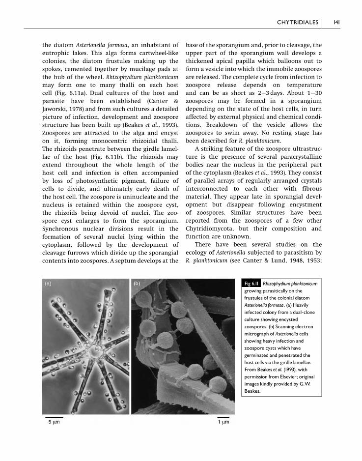
the diatom Asterionella formosa, an inhabitant of
eutrophic lakes. This alga forms cartwheel-like
colonies, the diatom frustules making up the
spokes, cemented together by mucilage pads at
the hub of the wheel. Rhizophydium planktonicum
may form one to many thalli on each host
cell (Fig. 6.11a). Dual cultures of the host and
parasite have been established (Canter &
Jaworski, 1978) and from such cultures a detailed
picture of infection, development and zoospore
structure has been built up (Beakes et al., 1993).
Zoospores are attracted to the alga and encyst
on it, forming monocentric rhizoidal thalli.
The rhizoids penetrate between the girdle lamel-
lae of the host (Fig. 6.11b). The rhizoids may
extend throughout the whole length of the
host cell and infection is often accompanied
by loss of photosynthetic pigment, failure of
cells to divide, and ultimately early death of
the host cell. The zoospore is uninucleate and the
nucleus is retained within the zoospore cyst,
the rhizoids being devoid of nuclei. The zoo-
spore cyst enlarges to form the sporangium.
Synchronous nuclear divisions result in the
formation of several nuclei lying within the
cytoplasm, followed by the development of
cleavage furrows which divide up the sporangial
contents into zoospores. A septum develops at the
base of the sporangium and, prior to cleavage, the
upper part of the sporangium wall develops a
thickened apical papilla which balloons out to
form a vesicle into which the immobile zoospores
are released. The complete cycle from infection to
zoospore release depends on temperature
and can be as short as 2�3 days. About 1�30
zoospores may be formed in a sporangium
depending on the state of the host cells, in turn
affected by external physical and chemical condi-
tions. Breakdown of the vesicle allows the
zoospores to swim away. No resting stage has
been described for R. planktonicum.
A striking feature of the zoospore ultrastruc-
ture is the presence of several paracrystalline
bodies near the nucleus in the peripheral part
of the cytoplasm (Beakes et al., 1993). They consist
of parallel arrays of regularly arranged crystals
interconnected to each other with fibrous
material. They appear late in sporangial devel-
opment but disappear following encystment
of zoospores. Similar structures have been
reported from the zoospores of a few other
Chytridiomycota, but their composition and
function are unknown.
There have been several studies on the
ecology of Asterionella subjected to parasitism by
R. planktonicum (see Canter & Lund, 1948, 1953;
Fig 6.11 Rhizophydiumplanktonicumgrowing parasitically on thefrustules of the colonial diatomAsterionella formosa. (a) Heavilyinfected colony from a dual-cloneculture showing encystedzoospores. (b) Scanning electronmicrograph of Asterionella cellsshowing heavy infection andzoospore cysts which havegerminated and penetrated thehost cells via the girdle lamellae.From Beakes et al. (1993), withpermission from Elsevier; originalimages kindly provided by G.W.Beakes.
141CHYTRIDIALES

Canter & Jaworski, 1981; van Donk & Bruning,
1992). Asterionella is also parasitized by two
other chytrids, Zygorhizidium planktonicum and
Z. affluens, and some of the early studies in fresh-
water lakes may well have included a mixture
of species.
Studies on the epidemiology of infection of
Asterionella by R. planktonicum in lakes have
shown that there are peak periods of Asterionella
population density both in spring and in autumn,
related to the availability of dissolved nutrients,
water temperature, thermal stratification and its
breakdown, daylength and light intensity.
Asterionella cells infected with Rhizophydium can
occur throughout the year, but epidemics in
which a high proportion of cells are infected only
occur at concentrations of around 10host cells
ml�1 (Holfeld, 1998). Interpretation of the condi-
tions conducive to the occurrence of epidemics
has been aided by experiments using dual
cultures of pathogen and host in which effects
such as light intensity, temperature and phos-
phorus concentration have been varied (van Donk
& Bruning, 1992). The effects of light are complex.
Although Rhizophydium zoospores are not photo-
tropic, they are quiescent and incapable of
infection in the dark or at low light intensity.
Experiments by Canter and Jaworski (1981) have
indicated that a light intensity below 200 lx is
inadequate for zoospore settlement on host cells.
In light-limited cultures of Asterionella, the spo-
rangia of the pathogen and hence the number of
zoospores produced are smaller than when light
is not limiting (Bruning, 1991a). Similarly, zoo-
spore production is also reduced when the
concentration of phosphorus limits growth of
the host (Bruning, 1991b). Temperature affects
the rate of sporangium development and the
size of sporangia, with maximum dimensions
at 2�C at fairly high light intensities (Bruning,
1991a). It also affects the duration of swimming of
zoospores and therefore their infective lifetime
which can vary from about 10 days at 3�C to only
2 days at 20�C. Epidemic development may result
from a combination of factors and there is a
remarkable interaction between the effects of
light intensity and temperature (Bruning, 1991c).
At higher temperatures, optimal conditions for
epidemic development occur at high light
intensities, but at temperatures below 5�6�Cepidemic development is encouraged by lower
light intensities. This may explain why, in nature,
epidemics can occur both in summer (high light
intensity, high temperature) and winter (low
light intensity, low temperature).
Rhizophydium planktonicum is a specialized
parasite infecting only Asterionella. It is more
compatible with certain clones of host cells
than others, and cells from incompatible clones
show hypersensitivity, undergoing rapid death
following infection (Canter & Jaworski, 1979).
6.2.3 CladochytriumThere are about a dozen species of Cladochytrium
(Sparrow, 1960) which are widespread sapro-
trophs, mostly of aquatic plant debris. The
thallus is eucarpic and polycentric and the
vegetative system may bear intercalary swellings
and septate turbinate cells (sometimes termed
spindle organs). The sporangia are inoperculate.
Cladochytrium replicatum is a common representa-
tive in decaying pieces of aquatic vegetation
and can be distinguished from other chytrids
by the bright orange lipid droplets found in the
sporangia. It is frequently isolated if moribund
aquatic vegetation is placed in a dish of water
and baited with boiled grass leaves or cellulosic
materials such as dialysis tubing. Lucarotti (1987)
has given details of its isolation and growth
in culture. The bright orange sporangia which
are visible under a dissecting microscope
appear on baits within about 5 days, arising
from an extensively branched hyaline rhizomy-
celium bearing two-celled intercalary swellings.
Sporangium development is encouraged by
exposure to light. On release from the sporan-
gium, the zoospores each contain a single orange
lipid droplet and bear a single posterior flagel-
lum. Lucarotti (1981) has described the fine
structure of the zoospore. After swimming for
a short time, the zoospore attaches itself to
the surface of the substratum and puts out
usually a single germ tube which can penetrate
the tissues of the host plant. The germ tube
expands to form an elliptical or cylindrical
turbinate cell which is often later divided into
two by a transverse septum (Fig. 6.12d). The
142 CHYTRIDIOMYCOTA

zoospore is uninucleate and during germina-
tion the single nucleus is transferred to the
swollen turbinate cell which becomes a vegeta-
tive centre from which rhizoids are put out
which in turn produce further turbinate cells
(see Figs. 6.12b,d). Nuclear division is apparently
confined to the turbinate cells, and although
nuclei are transported through the rhizoidal
system they are not resident there. The thallus
so established branches profusely, and at cer-
tain points spherical zoosporangia form,
either terminally or in intercalary positions.
Sometimes one of the cells of a pair of turbinate
cells swells and becomes transformed into a
sporangium. In culture, both cells may
be modified in this way. The spherical to pear-
shaped zoosporangium undergoes progressive
nuclear division, and the contents of the spor-
angium acquire a bright orange colour due
to accumulation of lipid droplets containing
the carotenoid lycopene. These lipid reserves are
later found in the zoospores. Cleavage of the
cytoplasm to form uninucleate zoospore initials
follows. The zoospores escape through a narrow
Fig 6.12 Cladochytriumreplicatum.(a) Rhizomyceliumwithin the epidermisof an aquatic plant bearing thetwo-celled hyaline turbinate cells andglobose orange zoosporangia. (b)Rhizomycelium and turbinate cells froma culture. (c) Zoosporangia from atwo-week-old culture.Onezoosporangium has released zoospores,each of which contains a bright orange-coloured globule. (d) Germinatingzoospores on boiled wheat leaves.The empty zoospore cysts are spherical.The germ tubes have expanded to formturbinate cells. (e) A zoosporangiumwhich has proliferated internally to forma second sporangium. (f) Rhizomyceliumwithin a boiledwheat leaf bearing athick-walled, spiny resting sporangium.
143CHYTRIDIALES

exit tube which penetrates to the exterior of
the substratum and becomes mucilaginous at
the tip. There is no operculum. Sometimes
zoosporangia may proliferate internally, a new
zoosporangium being formed inside the wall of
an empty one. Resting sporangia with thicker
walls and a more hyaline cytoplasm are also
formed either terminally or in an intercalary
position on the rhizomycelium. In some cases
the wall of the resting sporangium is reported
to be smooth and in others spiny, and it has
been suggested (Sparrow, 1960) that the two
kinds of resting sporangia may belong to dif-
ferent species. However, studies by Willoughby
(1962) of a number of single-spore isolates have
shown that the presence or absence of spines is
a variable character. The contents of the resting
sporangia divide to form zoospores which also
have a conspicuous orange droplet, and escape
by means of an exit tube as in the thin-walled
zoosporangia. Whether the resting sporangia are
formed as a result of a sexual process is not
known. Pure cultures of C. replicatum have been
studied by Willoughby (1962), Goldstein (1960)
and Lucarotti (1981). The fungus is heterotrophic
for thiamine. Biotin, while not absolutely
required, stimulates growth. Nitrate and sul-
phate are utilized, as are a number of different
carbohydrates; a limited amount of growth takes
place on cellulose.
6.2.4 NowakowskiellaSpecies of Nowakowskiella are widespread sapro-
trophs in soil and on decaying aquatic plant
debris, and can be obtained by baiting aquatic
plant remains in water with boiled grass
leaves, cellophane, dialysis tubing and the like.
Nowakowskiella elegans is often encountered in
such material, and pure cultures can be obtained
and grown on cellulosic materials overlying
agar, or directly in liquid culture media
(Emerson, 1958; Johnson, 1977; Lucarotti, 1981;
Lucarotti & Wilson, 1987). In culture, consider-
able variation in growth habit and morphology
can result from changing the concentration of
nutrients and the availability of water (Johnson,
1977). In boiled grass leaves the fungus forms an
extensive rhizomycelium with turbinate cells
(Fig. 6.13c). Zoosporangia are formed terminally
or in an intercalary position (Fig. 6.13c) and
are globose or pear-shaped with a subsporangial
swelling (apophysis), and granular or refractile
hyaline contents. At maturity some sporangia
develop a prominent beak, but in others this
is not present. When an operculum becomes
detached, zoospores escape and initially remain
clumped together at the mouth of the sporan-
gium (Figs. 6.13b,c). The fine structure of the
zoospore is very similar to that of Rhizophydium
but paracrystalline bodies have not been observed
(Lucarotti, 1981). It also has close resemblance
to the zoospore ultrastructure of the inopercu-
late, polycentric Cladochytrium replicatum.
Yellowish resting sporangia (Fig. 6.13e) have
been described (Emerson, 1958; Johnson, 1977;
Lucarotti & Wilson, 1987). They develop as
spherical to fusiform swellings in the rhizomy-
celium which become delimited by septa, dev-
elop thick walls and a large central vacuole
surrounded by dense cytoplasm with small
spherical lipid droplets. The resting sporangium
is at first binucleate. After nuclear fusion the
diploid nucleus divides meiotically. Further
nuclear divisions are mitotic and the contents
of the resting sporangium cleave into zoospores
which may be released through a papilla in
the sporangium wall. Alternatively, the resting
sporangium may give rise to a thin-walled zoo-
sporangium from which the zoospores are
released, i.e. the resting sporangium may func-
tion as a prosporangium as in some other chytrids
( Johnson, 1977).
In N. profusa, which is probably synonymous
with N. elegans (Johnson, 1977), three kinds of
sporangial dehiscence have been described:
exo-operculate, in which the operculum breaks
away to the outside of the sporangium; endo-
operculate, in which the operculum remains
within the sporangium; and inoperculate,
where the exit papilla opens without any clearly
defined operculum (Chambers et al., 1967;
Johnson, 1973). Such variations within a single
chytrid strain add emphasis to criticisms of
the value of dehiscence as a primary criterion in
classification.
Goldstein (1961) has reported that N. elegans
requires thiamine and can utilize nitrate,
144 CHYTRIDIOMYCOTA

sulphate and a number of carbohydrates includ-
ing cellulose, but cannot utilize starch.
6.3 Spizellomycetales
Members of this order differ from the
Chytridiales in possessing zoospores which
contain more than one lipid droplet and are
capable of limited amoeboid movement. Thalli
are generally monocentric. The order takes its
name from the genus Spizellomyces which in turn
was named in honour of the chytrid pioneer
F. K. Sparrow after Spizella, a genus of North
American sparrows (Barr, 1980). Some 86 species
of Spizellomycetales are currently recognized.
6.3.1 OlpidiumAbout 30 species of Olpidium are known, but
the genus is in need of revision and possibly
Fig 6.13 Nowakowskiella elegans.(a) Polycentric mycelium bearingzoosporangia. (b) Empty zoosporangiashowing opercula. (c) Mycelium showingturbinate cells and zoosporangia.(d) Zoospores from culture. (e) Restingsporangium from culture.
145SPIZELLOMYCETALES

some of the species should be classified else-
where. Typical species are holocarpic. Some are
parasitic on fungi and aquatic plants or algae,
or saprotrophic on pollen (Sparrow, 1960).
Others parasitize rotifers (Glockling, 1998),
nematodes and their eggs (Tribe, 1977; Barron
& Szijarto, 1986), moss protonemata or leaves
and roots of higher plants (Macfarlane, 1968;
Johnson, 1969). Olpidium bornovanus (¼ O. radicale)
develops on various monocotyledonous and
dicotyledonous plant roots following inoculation
(Lange & Insunza, 1977). Olpidium brassicae is
common on the roots of cabbages, especially
when growing in wet soils, and is also found on
a wide range of unrelated hosts, but some
host specialization has been reported. Both
O. bornovanus and O. brassicae are vectors of a
number of plant viruses (Barr, 1988; Adams,
1991; Hiruki, 1994; Campbell, 1996) and this
topic is discussed more fully below. Weber and
Webster (2000a) have given practical details of
how to grow O. brassicae for observation on
Brassica seedlings. A film featuring O. brassicae is
also available (Webster, 2006a).
Epidermal cells and root hairs of infected
cabbage roots contain one or more spherical or
cylindrical thalli, sometimes filling the whole
cell (Fig. 6.14a). The cytoplasm of the thallus
is granular and the entire contents divide into
numerous posteriorly uniflagellate zoospores
that escape through one or more discharge
tubes which penetrate the outer wall of the
host cell (Temmink & Campbell, 1968). Release of
the zoospores takes place within a few minutes
of washing the roots free from soil. The tip of
the discharge tube breaks down and zoospores
rush out and swim actively in the water. The
zoospores are very small, tadpole-like, with
Fig 6.14 Olpidiumbrassicae in cabbageroots. (a) Two ripe sporangia and oneempty sporangium in an epidermal cell.Each sporangium has a single exit tube.(b) Empty sporangium showing three exittubes. (c) Zoospores. (d) Zoospore cystson a root hair.Note that some cysts areuninucleate and some are binucleate.(e) Resting sporangia. (a,b,d,e) to samescale.
146 CHYTRIDIOMYCOTA
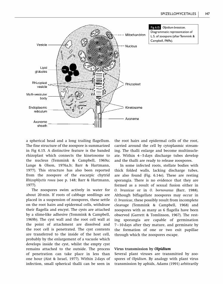
a spherical head and a long trailing flagellum.
The fine structure of the zoospore is summarized
in Fig 6.15. A distinctive feature is the banded
rhizoplast which connects the kinetosome to
the nucleus (Temmink & Campbell, 1969a;
Lange & Olson, 1976a,b; Barr & Hartmann,
1977). This structure has also been reported
from the zoospore of the eucarpic chytrid
Rhizophlyctis rosea (see p. 148; Barr & Hartmann,
1977).
The zoospores swim actively in water for
about 20min. If roots of cabbage seedlings are
placed in a suspension of zoospores, these settle
on the root hairs and epidermal cells, withdraw
their flagella and encyst. The cysts are attached
by a slime-like adhesive (Temmink & Campbell,
1969b). The cyst wall and the root cell wall at
the point of attachment are dissolved and
the root cell is penetrated. The cyst contents
are transferred to the inside of the host cell,
probably by the enlargement of a vacuole which
develops inside the cyst, whilst the empty cyst
remains attached to the outside. The process
of penetration can take place in less than
one hour (Aist & Israel, 1977). Within 2days of
infection, small spherical thalli can be seen in
the root hairs and epidermal cells of the root,
carried around the cell by cytoplasmic stream-
ing. The thalli enlarge and become multinucle-
ate. Within 4�5days discharge tubes develop
and the thalli are ready to release zoospores.
In some infected roots, stellate bodies with
thick folded walls, lacking discharge tubes,
are also found (Fig. 6.14e). These are resting
sporangia. There is no evidence that they are
formed as a result of sexual fusion either in
O. brassicae or in O. bornovanus (Barr, 1988).
Although biflagellate zoospores may occur in
O. brassicae, these possibly result from incomplete
cleavage (Temmink & Campbell, 1968) and
zoospores with as many as 6 flagella have been
observed (Garrett & Tomlinson, 1967). The rest-
ing sporangia are capable of germination
7�10 days after they mature, and germinate by
the formation of one or two exit papillae
through which the zoospores escape.
Virus transmission by OlpidiumSeveral plant viruses are transmitted by zoo-
spores of Olpidium. By analogy with plant virus
transmission by aphids, Adams (1991) arbitrarily
Fig 6.15 Olpidiumbrassicae.Diagrammatic representation ofL.S. of zoospore (afterTemmink &Campbell,1969a).
147SPIZELLOMYCETALES

distinguished viruses with non-persistent and
persistent transmission by fungi, although
Campbell (1996) objected to the use of these
terms, distinguishing instead between viruses
which can be acquired in vitro (i.e. outside the
plant) and those that can only be acquired in vivo
(within the host cell).
Tobacco necrosis virus (TNV) and cucumber
necrosis virus (CNV) are non-persistent viruses
which can be acquired in vitro by zoospores
of O. brassicae or O. bornovanus (respectively).
Virus particles (virions) are adsorbed onto the
plasmalemma of the zoospore and onto the
flagellar axonemal sheath which is continuous
with it (Temmink et al., 1970). Binding seems to
occur between the virus coat and specific mole-
cules at the zoospore surface, possibly oligosac-
charide side chains of proteins (Kakani et al.,
2003; Rochon et al., 2004). When the flagellum is
withdrawn into the body of the zoospore at
encystment, virus particles are introduced into
the fungal cytoplasm and are then transmitted
into the plant upon infection. Air-dried roots
containing TNV virus and O. brassicae resting
sporangia, or living virus-infected roots with
resting sporangia treated with 5NHCl, were
incapable of transmitting virus even though
the resting sporangia survived these treatments,
indicating that TNV is not carried inside the
resting sporangia (Campbell & Fry, 1966).
Lettuce big vein virus, LBVV, in contrast, is
an example of the persistent type (Grogan
et al., 1958). In this case it has been shown that
the virus can persist in air-dried resting spor-
angia for 18�20 years (Campbell, 1985). Here
the virions are acquired in vivo and they are
present inside the zoospores which emerge from
sporangia and resting sporangia (Campbell,
1996).
Classification of OlpidiumAlthough previously classified within the
family Olpidiaceae in the order Chytridiales,
D. J. S. Barr (2001) has placed Olpidium in the
order Spizellomycetales along with Rhizophlyctis
on the basis of similarities in zoospore structure.
Ribosomal DNA sequence comparisons are
inconclusive in that they do not show any close
similarity between Olpidium and either Chytridium
or Spizellomyces (Ward & Adams, 1998).
6.3.2 RhizophlyctisThere are about 10 known species of Rhizophlyctis
with monocentric eucarpic thalli, growing as
saprotrophs on a variety of substrata in soil,
freshwater and the sea. Rhizophlyctis rosea grows
on cellulose-rich substrata in soil, and it probably
plays an active but currently underestimated
role in cellulose decay (Powell, 1993). It can
survive for prolonged periods in dry soil, even
when this is heated to 90�C for two days (Gleason
et al., 2004) and, in fact, the recovery of R. rosea
is greatly enhanced if soil samples are air-dried
prior to isolation experiments (Willoughby,
2001). Willoughby (1998b) has estimated that
over 1000 thallus-forming units could be recov-
ered per gram of air-dry soil or leaf humus
fragments from Provence, France. These numbers
may arise from one or a few sporangia, since
a single sporangium about 100mm in diameter
may discharge up to 30 000 zoospores. Mitchell
and Deacon (1986) have shown that zoospores
of R. rosea accumulate preferentially on cellulosic
materials.
The fungus is readily isolated and grown
in culture, and details of techniques have
been provided by Stanier (1942), Barr (1987),
Willoughby (1998b) and Weber and Webster
(2000a). The placing of a small crumb of soil
onto moist tissue paper or cellophane overlying
agar containing mineral salts, or the floating
of squares of cellophane on water containing
a soil sample, are followed within a few days
by the development of thalli with bright pink
sporangia. The sporangia are attached to coarse
rhizoids which arise at several points on the
sporangial wall and extend throughout the
cellulosic substratum, tapering to fine points.
Extensive corrosion of the substrate underneath
the thallus and rhizoids points at the secretion
of powerful cellulases (Fig. 6.17).
Although the fungus is usually monocentric,
there are also records of some polycentric
isolates. When ripe, the sporangia have pink
granular contents which differentiate into
numerous uninucleate posteriorly uniflagellate
148 CHYTRIDIOMYCOTA

zoospores (Fig. 6.16a). One to several discharge
tubes are formed, and the tip of each tube
contains a clear mucilaginous plug which, prior
to discharge, is exuded in a mass from the tip of
the tube (Fig. 6.16c). While the plug of mucilage
dissolves, the zoospores within the sporangium
show active movement and then escape by
swimming through the tube. In some specimens
of R. rosea it has been found that a membrane
may form over the cytoplasm at the base of
the discharge tubes. If the sporangia do not
discharge their spores immediately, the mem-
brane may thicken. When spore discharge
occurs, these thickened membranes can be seen
floating free within the sporangia, and the term
endo-operculum has been applied to them. The
genus Karlingia was erected for forms possessing
such endo-opercula, including R. rosea, which
is therefore sometimes referred to as Karlingia
rosea, but the validity of this separation is
questionable because the presence or absence
of endo-opercula is a variable character
(Blackwell & Powell, 1999).
Zoospores of R. rosea are capable of swimming
for several hours. The head of the zoospore is
often globose, but can become pear-shaped or
show amoeboid changes in shape. It contains a
prominent lipid body, several bright refringent
globules, and bears a single trailing flagellum.
Ultrastructural details resemble those of
Olpidium brassicae in the presence of a striated
rhizoplast connecting kinetosome and nucleus
(Barr & Hartmann, 1977). On coming to rest on
a suitable substratum, the flagellum is with-
drawn and the body of the zoospore enlarges
to form the rudiment of the sporangium, whilst
rhizoids appear at various points on its surface.
Within the sporangium, the flagella are tightly
wrapped around the zoospores (Chambers
& Willoughby, 1964).
Resting sporangia are also found. They are
brown, globose or angular and have a thickened
Fig 6.16 Rhizophlyctis rosea. (a) Zoospores. (b) Young thallus formed on germination of zoospore.The zoospore cyst has enlargedandwill form the sporangium. (c) Older sporangium showing three discharge tubes. (d) Sporangium showingmucilage plugs at thetips of the discharge tubes and thickenings of the cellmembrane at the bases of the tubes. Such thickenings are termedendo-opercula. (e) Globose sporangium and seven visible papillae. (f) Resting sporangium formed inside an empty zoosporangium.(a,b) to same scale; (c�f) to same scale.
149SPIZELLOMYCETALES

wall (Fig. 6.16f ). Whether they are formed
sexually in R. rosea is not known. Couch (1939)
has, however, put forward evidence that the
fungus is heterothallic because single isolates
grown in culture failed to produce resting
sporangia whereas these structures did form
when certain cultures were paired. Stanier (1942)
has reported the occurrence of biflagellate zoo-
spores, but whether these represented zygotes
seemed doubtful. In the homothallic chitino-
philic fungus Rhizophlyctis oceanis, Karling (1969)
has described frequent fusions between zoos-
pores. These fusions are possibly sexual, but
unfortunately Karling was unable to cultivate
the resulting thalli to the stage of resting spore
development.
On germination, the resting sporangium of
R. rosea functions as a prosporangium, although
it is uncertain whether resting sporangia are
important for survival in nature. Willoughby
(2001) has shown that R. rosea could be recovered
from cellophane baits in as little as 5�6h after
placing air-dried soil samples in water, and it
was concluded that these zoospores were derived
from sporangia instead of resting spores which
need a longer time to produce zoospores.
The nutritional requirements of R. rosea are
simple. It shows vigorous growth on cellulose
as the sole carbon source but it can utilize
a range of carbohydrates such as glucose,
cellobiose and starch. The pink colour of the
sporangia is due to the presence of carotenoid
pigments such as g-carotene, lycopene and a
xanthophyll.
6.4 Neocallimastigales(rumen fungi)
A very interesting and unusual group of zoo-
sporic fungi inhabits the rumens (foreguts) of
ruminants (herbivorous mammals which regur-
gitate and masticate previously ingested food)
like cows, sheep and deer. They have also been
found in some non-ruminants such as horses and
elephants and probably occur in the guts of
many large herbivores. These fungi are obligate
anaerobes which can flourish in the rumen
because oxygen is depleted there by the intense
respiratory activity of a dense population of
protozoa and bacteria, some of which are
facultative anaerobes capable of scavenging free
oxygen. Their zoospores were at first thought to
be protozoa and were not recognized as belong-
ing to fungi because obligately anaerobic fungi
Fig 6.17 Scanning electronmicrograph of two thalli ofRhizophlyctis rosea on a cellophanemembrane. Pit corrosion is visiblewhere a thallus has been liftedfrom the substratum (arrows).
150 CHYTRIDIOMYCOTA
were not believed to exist. Further, microbiolo-
gists working on microbes from the ruminant
gut studied only strained rumen fluid and
therefore failed to see the thalli of fungi attached
to herbage fragments. The view that the motile
cells swimming in rumen fluid belonged to
flagellates was challenged by Orpin (1974), who
observed that there was an enormous increase in
the concentration of ‘flagellates’ in the rumen of
sheep within a short time of feeding. The ratio of
minimum (pre-feeding) to maximum concentra-
tion of motile cells could vary between 1:15 and
1:296 (average 1:47), and if these were organisms
reproducing by binary fission it would be
necessary for them to undergo six successive
cell divisions in 15min. The explanation for the
rapid increase in motile cells is that sedentary
fungal thalli, anchored by rhizoids to partially
digested food fragments floating in the rumen,
are stimulated to release zoospores by soluble
substances such as haems released from the
newly ingested food material. The zoospores
attach themselves in large numbers to the
herbage fragments, and germinate to form
rhizoidal or rhizomycelial thalli with sporangia
capable of releasing further zoospores within
about 30h.
Some 5 genera and 15 species have now been
distinguished (Theodorou et al., 1992, 1996;
Trinci et al., 1994). They include Caecomyces
which has mono- and polycentric thalli,
Anaeromyces and Orpinomyces with polycentric
thalli, and Piromyces and Neocallimastix which
are monocentric. The zoospores of Anaeromyces,
Caecomyces and Piromyces are uniflagellate
whilst those of Neocallimastix and Orpinomyces
are multiflagellate (see Fig. 6.18). They were
classified within the order Spizellomycetales,
family Callimasticaceae by Heath et al. (1983)
and Barr et al. (1989) but are now placed in a
separate order Neocallimastigales (Li et al., 1993;
D. J. S. Barr, 2001). Special techniques and media
are needed for isolating and handling anaerobic
fungi, but the life cycle details of several have
now been followed in pure culture. One of the
best known is N. hurleyensis, isolated from sheep
(Fig. 6.18). Minutes after the arrival of fresh
food, globose ripe zoosporangia on previously
colonized grass fragments release zoospores
through an apical pore and these attach them-
selves to herbage fragments and germinate to
produce rhizoids which penetrate and digest the
ingested plant material. The walls of the thallus
contain chitin. A single zoosporangium develops
and is cut off from the rhizoidal system by
a septum. The rhizoidal part is devoid of nuclei,
but within the zoosporangium repeated nuclear
divisions occur before the cytoplasm cleaves
to form 64�128 zoospores. The life cycle of
N. hurleyensis from zoospore germination to the
release of a fresh crop of zoospores lasts about
29�31h at 39�C (Lowe et al., 1987a). The
zoospores bear 8�16 whiplash flagella inserted
posteriorly in two rows.
The ultrastructure of zoospores has been
described for several species of Neocallimastix,
including N. patriciarum (Orpin & Munn, 1986),
N. frontalis (Munn et al., 1981; Heath et al., 1983)
and N. hurleyensis (Webb & Theodorou, 1988,
1991). There are differences in detail. For
example, the zoospore of N. frontalis has a waist-
like constriction, with the majority of the cyto-
plasmic organelles concentrated in the posterior
portion near the insertion of the flagella.
Characteristic organelles known from zoospores
of aerobic chytridiomycetes such as mitochon-
dria, Golgi bodies, lipid droplets or gamma
particles (seen in zoospores of Blastocladiella
emersonii; Fig. 6.19) are absent. In the posterior
portion of the zoospore of N. hurleyensis near the
point of insertion of the flagella, an irregularly
shaped complex structure interpreted as a
hydrogenosome has been reported in place of a
mitochondrion. In zoospores of N. patriciarum
there are many presumed hydrogenosomes
concentrated around the region of flagellar
insertion. Hydrogenosomes are organelles
capable of the anaerobic metabolism of hexoses
to acetic and formic acids. Protons (Hþ) act as
electron acceptors, so that gaseous H2 is released
by the activity of the enzyme hydrogenase
(Muller, 1993; Boxma et al., 2004). The hydrogen,
in turn, is used by anaerobic methanogenic
bacteria to reduce CO2 to CH4 (methane) which
escapes in profusion through the front and hind
exits of the ruminant digestive tracts.
Hydrogenosomes are found in several anaerobic
lower eukaryotes and are believed to be derived
151NEOCALLIMASTIGALES (RUMENFUNGI)

from mitochondria (Embley et al., 2002). Whereas
mitochondria of most fungi contain a limited
amount of DNA, hydrogenosomes of rumen
chytrids seem to have lost their genome alto-
gether (Bullerwell & Lang, 2005).
Granular inclusion bodies which contain
aggregates of ribosome-like particles and also
free ribosome-like arrays are found anterior to
the nucleus. Rosettes of glycogen represent the
energy reserve of the zoospore. The shafts of
the flagella contain the familiar eukaryotic 9 þ 2
arrangement of microtubules, but in N. frontalis
the microtubules do not extend into the tips
of the flagella which are narrower than the
proximal part.
Ecologically, these anaerobic fungi play an
important role in the early colonization of
ingested herbage and have a wide range of
enzymes which enable them to utilize mono-
saccharides, disaccharides and polysaccharides
such as xylan, cellulose, starch and glycogen
(Theodorou et al., 1992). They may play an active
role in fibre breakdown. It is likely that coloniza-
tion of straw particles by these fungi aids further
attack by bacteria. The survival of anaerobic
fungi outside the unusual and protective envir-
onment of the herbivore gut occurs in dried
faeces in the form of cysts or as melanized
thick-walled thalli whilst transmission to young
animals takes place in saliva during licking
Fig 6.18 Neocallimastixhurleyensis. (a) Rhizoidalthallus with zoosporangium. (b) Release ofzoospores. (c) Tracing of T.E.M. of zoosporewith14 flagella in longitudinal section.Diagrams based onWebb andTheodorou (1991).
152 CHYTRIDIOMYCOTA

and grooming (Lowe et al., 1987b; Wubah et al.,
1991; Theodorou et al., 1992).
6.5 Blastocladiales
6.5.1 IntroductionSpecies belonging to the Blastocladiales are
mostly saprotrophs in soil, water, mud or aquatic
plant and animal debris, and some are pathogens
of plants, invertebrate animals or fungi. Most
are obligate aerobes, but Blastocladia spp. are
facultatively anaerobic, requiring a fermentable
substrate and growing on submerged fleshy
fruits, twigs or other plant materials rich in
soluble carbohydrates (Emerson & Robertson,
1974). The life cycles of Blastocladiales show
great variations and in some forms there is an
alternation of distinct haploid gametothallic
and diploid sporothallic generations. These
terms are used in preference to the botanical
terms gametophytic and sporophytic. Species
of Physoderma, previously grouped with the
Chytridiales (Lange & Olson, 1980), are biotrophic
parasites of higher plants (Karling, 1950). They
include P. maydis, the cause of brown spot of
maize, and P. alfalfae (Lange et al., 1987). One
genus, Coelomomyces, consists of obligate parasites
of insects, usually mosquito larvae (Couch &
Bland, 1985). This genus is unusual in that
the vegetative thallus is a wall-less plasmodium-
like structure lacking rhizoids. The life cycle
is completed in unrelated alternate animal
hosts, sporothalli occurring in mosquito larvae
(Insecta) and gametothalli in a copepod
(Crustacea) (Whisler et al., 1975; Federici, 1977).
Attempts are being made to use Coelomomyces
in the biological control of mosquitoes. Catenaria
anguillulae, a facultative parasite of nematodes
and their eggs, liver fluke eggs and some other
invertebrates, can be grown in culture (Couch,
1945; Barron, 1977; Barstow, 1987), whilst
Catenaria allomycis is a biotrophic parasite of
Allomyces (Couch, 1945; Sykes & Porter, 1980).
With the exception of Coelomomyces, the
thallus of members of the Blastocladiales is
eucarpic. The morphologically simpler forms
such as Blastocladiella (Fig. 6.22) are monocentric,
with a spherical or sac-like zoosporangium or
resting sporangium arising directly or on
a short one-celled stalk from a tuft of radiating
rhizoids. These simpler types show considerable
similarity to monocentric Chytridiales of
other orders such as Rhizophlyctis rosea (Figs. 6.16
and 6.17), and in the vegetative state they
may be difficult to distinguish. The more
complex organisms such as Allomyces are poly-
centric, and the thallus is differentiated into
a trunk-like portion which has rhizoids below
whilst branching above, often dichotomously,
and bearing sporangia of various kinds at the
tips of the branches. Chitin has been demon-
strated in the walls of Allomyces and Blastocladiella
(Porter & Jaworski, 1966; Youatt, 1977; Maia,
1994).
The zoospore of Blastocladiales
The zoospore of Blastocladiales has a single
posterior flagellum of the whiplash type.
Details of the fine structure of this kind of
zoospore have been reviewed by Fuller (1976)
and Lange and Olson (1979). The best known
are Blastocladiella emersonii (Cantino et al., 1963;
Reichle & Fuller, 1967) and Allomyces macrogynus
(Fuller & Olson, 1971). The structure of the
zoospore of B. emersonii is summarized diagram-
matically in Fig. 6.19. The zoospore is tadpole-
like with a pear-shaped head about 7 � 9mm and
a single, trailing flagellum about 20 mm long.
Under the light microscope, the most conspic-
uous internal structure is the dense crescent-
shaped nuclear cap which surrounds the more
transparent nucleus. The nuclear cap is rich in
RNA and protein, and is filled with ribosomes.
The zoospore of B. emersonii is unusual in that
it contains only a single large mitochondrion,
situated near the flagellar kinetosome.
The organization of the flagellum is essen-
tially as described on p. 129. The nine triplet
microtubules extend in a funnel-shaped manner
from the proximal end of the kinetosome
towards the nucleus and nuclear cap, maintain-
ing its conical shape. Extending into the mito-
chondrion and linking up the kinetosome
153BLASTOCLADIALES

with it are three striated bodies variously
referred to as flagellar rootlets, striated rootlets
or banded rootlets. They are contained within
separate channels, and each is surrounded by
a unit membrane. Since the energy for propul-
sion is generated within the mitochondrion, it is
possible that the banded rootlets are, in some
way, responsible for transmitting energy to the
base of the axoneme. It is also possible that the
banded rootlets serve to anchor the flagellum
within the body of the zoospore.
There are two other obvious kinds of organelle
within the body of the Blastocladiella zoospore.
The lipid sac attached to the mitochondrion
contains a group of lipid droplets which is
surrounded by a unit membrane. It is not
known whether lipid forms the energy reserve
used in swimming, cytoplasmic glycogen deposits
being a more plausible alternative (Cantino
et al., 1968). In the anterior of the zoospore
between the nuclear cap and the plasma
membrane, there is a group of granules about
0.5 mm in diameter, called gamma particles.
They consist of an inner core, shaped like
an elongated cup and bearing two unequal
openings at opposite sides of the cup. This cup-
shaped structure is enveloped in a unit mem-
brane (Myers & Cantino, 1974). Gamma particles
are only present in developing and motile
zoospores but disappear as the zoospore encysts.
Formerly thought to represent the chytrid
equivalent of the chitosome found in higher
fungi (see p. 6), this notion has now been
discarded (Hohn et al., 1984).
Fig 6.19 Blastocladiella emersonii zoospore,fine structure, diagrammatic and not to scale.(a) L.S. of zoospore along the axis of theflagellum. (b) T.S. of kinetosome showing ninetriplets ofmicrotubules. (c) T.S. of kinetosomeat a slightly lower level showing the origin oftwo of the banded rootlets which extendinto themitochondrion.The cristae of themitochondrion are close to themembranewhich surrounds the banded rootlets.(d) T.S. of axoneme showing the nine pairedperipheralmicrotubules and the two centralmicrotubules.
154 CHYTRIDIOMYCOTA

The zoospore of Allomyces macrogynus broadly
resembles that of B. emersonii (Fuller & Olson,
1971). Gamma particles are present in the
zoospore and, during encystment, these form
vesicles which fuse with the plasma membrane.
Fusion coincides with the appearance of
wall material around the cyst (Barstow &
Pommerville, 1980). The zoospore of Allomyces
differs from that of Blastocladiella in some other
ways. Although there is a large basal mitochon-
drion, many smaller mitochondria are also
present, generally located along the membrane
of the nuclear cap in the anterior part of the
cell. A complex structure situated laterally at
the base of the body of the zoospore, between
the nucleus and the zoospore membrane, has
been termed the side body complex by Fuller
and Olson (1971). It consists of two closely
appressed membranes separated by an electron-
opaque material. These membranes subtend
numerous electron-opaque, membrane-bound
bodies, lipid bodies and a portion of the basal
mitochondrion. In addition, there are mem-
brane-bound non-lipid bodies termed Stuben
bodies by Fuller and Olson (1971), whose func-
tion and composition are uncertain.
The zoospore is propelled forward by rhyth-
mic lashing of the flagellum, and it can swim
for a period even under anaerobic conditions.
It is also capable of amoeboid changes of shape.
On coming to rest, the flagellum is retracted
into the body of the zoospore. There are different
interpretations of the manner in which flagellar
retraction is achieved. Cantino et al. (1968)
have suggested that the flagellum is retracted
by a revolving action of the nucleus, whereas
in the ‘lash-around’ mechanism the flagellum
coils around the body of the spore, the flagellar
membrane fuses with the plasmalemma of
the spore and the axoneme enters the spore
cytoplasm (Olson, 1984). In Allomyces, the zoo-
spore cyst produces, at one point, a narrow germ-
tube which branches to form the rhizoidal
system. At the opposite pole, the zoospore cyst
forms a wider germ tube which gives rise to
hyphae which branch and later bear sporangia.
This bipolar germination pattern is a point
of difference between the Blastocladiales and
the Chytridiales, in which germination is
typically unipolar. The rhizoids are strongly
chemotropic and specialize in nutrient uptake
and transport. An inwardly directed electrical
current has been detected around the rhizoids,
and an outwardly directed current around
the hyphae and hyphal tips. The inward current
at the rhizoids may be the consequence of
localized proton-driven solute transport
(de Silva et al., 1992).
Life cycles of Blastocladiales
A number of distinct life history patterns are
found. In Allomyces arbuscula, for example, iso-
morphic alternation of haploid gametothallic
and diploid sporothallic generations has been
demonstrated. In A. neo-moniliformis (¼ A. cysto-
genes) there is no free-living sexual generation,
but this stage is represented by a cyst (see
below). In A. anomalus, only the asexual stage
has been found in normal cultures, but experi-
mental treatments may result in the develop-
ment of sexual thalli. Similar variations in life
cycles have been found in other genera such
as Blastocladiella. A characteristic feature of
the asexual thalli of the Blastocladiales is the
presence of resting sporangia with chitinous,
pitted walls impregnated with a dark brown,
melanin-type pigment. The pits are inwardly
directed conical pores in the wall. The inner
ends of the pores abut against a smooth, colour-
less inner layer of wall material surrounding
the cytoplasm (Skucas, 1967, 1968). The resting
sporangia of Allomyces can remain viable for up
to 30 years in dried soil. The ease with which
certain members of the group can be grown
in culture has facilitated extensive studies of
their nutrition and physiology, and the results
of some of these investigations are discussed
below.
Four families have been recognized �Coelomomycetaceae, Catenariaceae, Physoderm-
ataceae and Blastocladiaceae � but of these we
shall study only Allomyces and Blastocladiella,
both representatives of the Blastocladiaceae.
6.5.2 AllomycesSpecies of Allomyces are found in mud or soil of
the tropics or subtropics, including desert soil,
155BLASTOCLADIALES

and if dried samples of soil are placed in water
and ‘baited’ with boiled hemp seeds, the baits
may become colonized by zoospores. From such
material, it is possible to obtain pure cultures by
streaking or pipetting zoospores onto suitable
agar media and to follow the complete life
history of these fungi in the laboratory. Olson
(1984) has given a full account of the taxonomy,
life cycles, morphogenesis and genetics of
different species of Allomyces, with practical
details of how to grow and handle them. Good
growth occurs on a medium containing yeast
extract, peptone and soluble starch (YPsS), but
chemically defined media have also been used.
There is a requirement for thiamine and organic
nitrogen in the form of amino acids.
Emerson (1941) isolated species of Allomyces
from soil samples from all over the world. He
distinguished three types of life history, repre-
sented by three subgenera.
Sub-genus Eu-AllomycesThe Eu-Allomyces type of life history is exemplified
by A. arbuscula and A. macrogynus (Fig. 6.21; for
a film, see Webster & Hard, 1998a). Resting
sporangia are formed on asexual diploid thalli.
They contain about 12 nuclei which undergo
meiosis during the early stages of germination
(Olson, 1974). The cytoplasm cleaves around the
48 haploid nuclei to form the zoospores. Since
meiosis occurs in the resting sporangia, these
have been termed meiosporangia, and the
haploid zoospores meiospores. The meiospores
are released when the outer wall of the brown
pitted resting sporangium cracks open by a slit
and the inner wall balloons outwards and
eventually opens by one or more pores. The
meiospores swim by movement of the trailing
flagellum and, on coming to rest, encyst and
germinate as described above to form a rhizoidal
system and a trunk-like region which bears
dichotomous branches.
The tips of the branches have been claimed
to resemble the Spitzenkorper of higher fungi
in being actin-rich, although secretory vesicles
and/or microvesicles (chitosomes) have not been
clearly shown (Srinivasan et al., 1996). Repeated
nuclear division occurs to form a coenocytic
structure, and finger-like ingrowths from the
walls of the trunk-region and branches form
incomplete septa, sometimes termed pseudo-
septa, with a pore in the centre through which
cytoplasmic connections can be seen (Fig. 6.20d;
Meyer & Fuller, 1985). The haploid thalli which
develop from the meiospores are gametothallic,
i.e. sexual. They are monoecious, and the tips
of their branches swell to form paired sacs � the
male and female gametangia. The male gam-
etangia can be identified by the presence of
a bright orange pigment, g-carotene, whilst the
female gametangia are colourless. In A. arbuscula
the male gametangium is subterminal or
hypogynous, i.e. beneath the terminal female
gametangium, but in A. macrogynus the positions
are reversed and the male gametangium is
terminal or epigynous (Figs. 6.20e,i). The gam-
etangia bear a number of colourless papillae on
their walls, blocked by pulley-shaped plugs
which eventually dissolve.
The contents of the gametangia differentiate
into uninucleate gametes which differ in size
and pigmentation. The female gametangium
forms larger, colourless motile gametes (swarm-
ers) whilst the male gametangium releases
smaller, more active, orange-coloured swarmers.
After escaping through the papillae in the walls
of the gametangia, the gametes swim for a time
and then pair off. A female gamete which fails
to pair can function as a zoospore by germinat-
ing to form a new sexual thallus. A hormone,
sirenin, is secreted by female gametangia dur-
ing gametogenesis and by the released female
gametes, and this stimulates a chemotactic
response in male gametes at the extremely low
concentration of 8 � 10�11M (Machlis, 1972;
Carlile, 1996a). The chemical structure of sirenin
has been determined (Fig. 6.22), and both d- and
l-forms have been synthesized. Only l-sirenin is
active. It is a bicyclic sesquiterpene, probably
derived from the parent hydrocarbon sesqui-
carene (Nutting et al., 1968; Plattner & Rapoport,
1971). A second hormone, parisin, which attracts
female gametes, is secreted by male gametes.
Its structure has not been determined, although
it may well be related to sirenin (Pommerville
& Olson, 1987).
The biflagellate zygote resulting from the
fusion of two gametes may swim for a while
156 CHYTRIDIOMYCOTA

before it encysts and casts off the flagella.
Nuclear fusion then follows (Pommerville &
Fuller, 1976). The zygote develops immediately
into a diploid asexual thallus which differs
from gametothalli in bearing two types of
zoosporangia instead of gametangia. The first
formed are thin-walled papillate zoosporangia
formed singly or in rows at the tips of the
Fig 6.20 (a�h) Allomycesarbuscula. (a) Zoospores (haploidmeiospores). (b) Young gametothalli, 24h old. (c) Young sporothalli,18hold. (d) Sporothallus, 30h old. Perforations are visible in some of the septa. (e) Gametangia at the tips of the branches of thegametothallus.Note the disparity in the size of the gametes (anisogamy).The smaller male gametes are orange in colour whilstthe larger female gametes are colourless.Compare the hypogynous arrangement of themale gametangiawith the epigynousarrangement in A. macrogynus shown at (i). (f) Meiosporangia (resting sporangia, R.S.) andmitosporangia (zoosporangia, Z.S.) on asporothallus. (g) Release ofmitospores from zoosporangia (¼mitosporangia) on sporothallus. (h) Rupture ofmeiosporangium(¼ resting sporangium). (i) Allomycesmacrogynus.Branch tip from gametothallus showing the arrangement of gametangiawithterminal, epigynousmale gametangia and anisogamous gametes.
157BLASTOCLADIALES

branches (Fig. 6.20g). Within these thin-walled
sporangia the nuclei undergo mitosis. Initially
the nuclei are arranged in the cortical region
of the cytoplasm, but later they migrate and
become uniformly spaced apart. Movement of
the nuclei is controlled by forces generated by
actin microfilaments whilst their spacing and
positioning is controlled by microtubules (Lowry
et al., 1998). Cleavage of the cytoplasm around
the nuclei to form diploid colourless zoospores
is initiated by the formation of membranes seen
first at the plasmalemma, then extending into
the cortex to form a complex membranous
network (Fisher et al., 2000). The process of
cytokinesis, i.e. the extension and fusion of
membranes, seems to be mediated principally
by the actin component of the cytoskeleton
(Lowry et al., 2004). According to Barron and
Hill (1974), the development of the cleavage
Fig 6.21 Life cycle diagram of Eu-Allomyces as exemplified by A. macrogynus. A diploid sporothallusmay produce diploidmitosporesfrom a colourless, thin-walled papillatemitosporangium, and haploidmeiospores from a thick-walled pittedmeiosporangium inwhichmeiosis occurs.Meiospores germinate to form a haploid gamethothallus which produces two different gametangia andreleases haploid gametes of two kinds, small carotenoid-rich (shaded) ‘male’gametes and larger colourless ‘female’ones.Uponcopulation, a diploid zygote gives rise to a sporothallus. Alternatively, if failing to pair up, the female gametesmay function aszoospores, inwhich case they give rise to a new gametothallus. Small open circles represent haploid nuclei whereas diploid nucleiare drawn larger and split. It should be noted thatmany field strains of A. macrogynus have a higher ploidy level, e.g. alternatingbetween diploid (small circles) and tetraploid (large split circles) conditions.Key events in the life cycle are plasmogamy (P),karyogamy (K) andmeiosis (M).
Fig 6.22 Chemical structure of the hormone l-sireninwhichattractsmale gametes of Allomycesmacrogynus.The structureofparisin, attractive to female gametes, does not seem tohavebeen elucidated as yet.
158 CHYTRIDIOMYCOTA

membranes is induced by the availability of free
water. Zoospores are released from the sporangia
after dissolution of the plugs blocking the exit
papillae. Since nuclear division in the thin-
walled sporangia is mitotic, these are termed
mitosporangia, and the diploid swarmers they
release are mitospores. The mitospores, after a
swimming phase, encyst and are capable of
immediate germination, developing into a
further diploid asexual thallus.
The second type of zoosporangium is the dark
brown, thick-walled, pitted resting sporangium
(meiosporangium), formed at the tips of the
branches. Meiotic divisions within these sporan-
gia result in the formation of the haploid
meiospores, which develop into sexual thalli.
The life cycle of a member of the subgenus
Eu-Allomyces is thus an isomorphic alternation
of gametothallic and sporothallic generations
(Fig. 6.21). Comparisons of the nutrition and
physiology of the two generations show no
essential distinction between them up to the
point of production of gametangia or sporangia.
Emerson and Wilson (1954) have made cyto-
logical and genetic studies of a number of
collections of Allomyces. Interspecific hybrids
between A. arbuscula and A. macrogynus have
been produced in the laboratory, and it has
been shown that the fungus earlier described
as A. javanicus is a naturally occurring hybrid
between these two species. Cytological examina-
tion of the two parent species and of artificial
and natural hybrids showed a great variation
in chromosome number. In A. arbuscula the
basic haploid chromosome number is 8, but
strains with 16, 24 and 32 chromosomes have
been found. In A. macrogynus the lowest haploid
number encountered is 14, but strains with
28 and 56 chromosomes are also known. The
demonstration that these two species each
represent a polyploid series was the first to
be made in fungi. The wild-type strain of
A. macrogynus appears to be an autotetraploid
which, after meiosis, produces diploid gameto-
thalli (Olson & Reichle, 1978).
The behaviour of the hybrid strains is of
considerable interest. As seen above, the parent
species differ in the arrangement of the primary
pairs of gametangia, A. arbuscula being
hypogynous whilst A. macrogynus is epigynous.
Following fusion of gametes derived from differ-
ent parents, zygotes formed, germinated and
gave rise to sporothalli. The meiospores from the
hybrid sporothalli had a low viability (0.1�3.2%),
as compared with a viability of about 63% for
A. arbuscula meiospores, but some germinated
to form gametothalli. The arrangement of the
gametangia on these F1 gametothalli showed
a complete range from 100% epigyny to 100%
hypogyny. Also, in certain gametothalli the ratio
of male to female gametangia (normally about
1:1) was very high, with less than one female
per 1000 male gametangia. It was concluded
from these experiments that, since intermediate
gametangial arrangements are found in hybrid
haploids, this arrangement is not under the
control of a single pair of non-duplicated allelic
genes, but that a fairly large number of genes
must be involved. Hybridization in some way
upsets the mechanism which controls the
arrangement of gametangia in the parental
species. By treating meiospores of A. macrogynus
with DNA extracted from gametothallic cultures
of A. arbuscula, Ojha and Turian (1971) have
demonstrated an inversion of the normal gamet-
angial arrangement, i.e. a proportion of the
DNA-treated meiospores developed colonies
with hypogynous antheridia instead of the
normal epigynous arrangement. Similar inver-
sions were also obtained in converse experi-
ments. In an isolate of the naturally occurring
hybrid A. javanicus, Ji and Dayal (1971) have
shown that although copulation between anisog-
amous gametes results in the formation of
sporothalli bearing thin-walled and thick-walled
sporangia, the swarmers from the thick-walled
sporangia rarely develop into gametothalli, but
into sporothalli. This is not surprising for a
hybrid, and is possibly due to a failure of meiosis
in the thick-walled sporangia.
Sub-genus CystogenesA life cycle different from Eu-Allomyces is found
in Allomyces moniliformis and A. neo-moniliformis.
There is no independent gametothallic genera-
tion, but this stage is probably represented by
a cyst (C.M. Wilson, 1952). The asexual thalli
resemble those of subgenus Eu-Allomyces, bearing
159BLASTOCLADIALES

both thin-walled mitosporangia and brown,
thick-walled, pitted meiosporangia. The mito-
spores encyst and germinate to form a further
crop of asexual thalli. In the meiosporangium,
meiosis takes place, but before cytoplasmic
cleavage occurs, the haploid nuclei pair, the
paired nuclei being united by a common nuclear
cap. When cleavage does occur it therefore
results in the formation of some 30 binucleate
cells. When the meiosporangial wall cracks open,
the binucleate cells are released as amoeboid
bodies which may or may not bear flagella, and
it is these cells which form the cysts. A mitotic
division in each cyst results in four haploid
nuclei, and cytoplasmic cleavage gives rise to
four colourless uniflagellate isogametes. These
copulate to form biflagellate zygotes, each of
which can develop into an asexual sporothallus.
In the Cystogenes life cycle there is thus a free-
living diploid asexual sporothallic generation,
whereas the haploid generation is reduced to the
cysts and gametes.
Sub-genus Brachy-AllomycesIn certain isolates of Allomyces which have been
placed in a ‘form species’ A. anomalus, there are
neither sexual thalli nor cysts. Asexual thalli
bear mitosporangia and brown resting sporan-
gia. The spores from the resting sporangia
develop directly to give asexual thalli again.
The cytological explanation proposed by C.M.
Wilson (1952) for this unusual behaviour is that,
due to complete or partial failure of chromo-
some pairing in the resting sporangia, meiosis
does not occur and nuclear divisions are mitotic.
Consequently the zoospores produced from rest-
ing sporangia are diploid, like their parent thalli
and, on germination, give rise to diploid asexual
thalli again. Similar failures in chromosome
pairing were also encountered in the hybrids
between A. arbuscula and A. macrogynus leading to
very low meiospore viability from certain crosses.
In view of this it seemed possible that some of
the forms of A. anomalus might have arisen
through natural hybridization. In a later study,
Wilson and Flanagan (1968) showed that there
is a second way in which the life cycle of
this fungus is maintained without a sexual
phase. In certain isolates, meiosis does occur in
the resting sporangia, followed by apomixis,
i.e. the fusion of two meiosis-derived nuclei in
the same thallus. Propagules from the resting
sporangia are therefore diploid and the cysts
develop into sporothalli. By germinating resting
sporangia in dilute K2HPO4, a small percentage
of zoospores were produced which developed
into gametothalli, some of which were identified
as A. macrogynus and some as A. arbuscula. No
hybrids were found. Thus A. anomalus is not
a single species, but represents sporothalli of
these two species in which the normal alterna-
tion of generations has been upset by cytological
deviations.
6.5.3 BlastocladiellaAbout a dozen species of Blastocladiella have been
isolated from soil or water, and one is parasitic
on the cyanobacterium Anabaena (Canter &
Willoughby, 1964). The form of the thallus is
comparatively simple, resembling that of some
monocentric chytrids. There is an extensive
branched rhizoidal system which is attached
either to a sac-like sporangium or to a cylindrical
trunk-like region bearing a single sporangium
at the tip. In B. emersonii it has been shown that
the rhizoids are chemotropic and function
not only in attachment, but in absorption and
selective translocation of nutrients (Kropf &
Harold, 1982).
Different species of Blastocladiella have life
cycles resembling those of the three subgenera
of Allomyces, and Karling (1973) has proposed
that Blastocladiella should similarly be divided
into three subgenera, i.e. Eucladiella correspond-
ing to Eu-Allomyces, Cystocladiella corresponding
to Cystogenes, and Blastocladiella corresponding
to Brachy-Allomyces. In some species there is an
isomorphic alternation of generations, probably
matching in essential features the Eu-Allomyces
pattern, but cytological details are needed to
confirm this. For example, in Blastocladiella
variabilis two kinds of asexual thallus are found.
One bears thin-walled zoosporangia which
release posteriorly uniflagellate swarmers.
These swarmers may develop to form thalli
resembling their parents or may give rise to
the second type of asexual thallus bearing
160 CHYTRIDIOMYCOTA

a thick-walled dark-brown sculptured resting
sporangium within the terminal sac. The resting
sporangium releases posteriorly uniflagellate
swarmers which, after swimming, germinate
to form sexual thalli of two kinds. About half
of the sexual thalli are colourless (‘female’),
and about half are orange-coloured (‘male’).
However, in contrast to the anisogamy of
Eu-Allomyces, in Blastocladiella there is no distinc-
tion in size between the gametes. The orange
and colourless gametes pair to produce zygotes,
which germinate directly to produce asexual
thalli. In other species (e.g. B. cystogena) the life
cycle is of the Cystogenes type, i.e. there are no
gametothalli.
In yet other species there is no clear evidence
of sexual fusion. In B. emersonii (Fig. 6.23), the
resting sporangial thallus contains a single
globose, dark reddish brown resting sporangium
with a dimpled wall. Meiosis occurs during
development of the resting sporangium (Olson
& Reichle, 1978). After a resting period, the wall
cracks open and one to four papillae protrude
from which swarmers are released. The swarmers
germinate to form two types of thallus bearing
thin-walled zoosporangia. About 98% of the
swarmers give rise to thalli bearing colourless
sporangia (Fig. 6.23a), and about 2% to thalli
with sporangia coloured orange due to the
presence of g-carotene. The colourless thalli
develop rapidly and are ready to discharge
zoospores within 24h. These have about twice
the DNA content as the swarmers released from
resting sporangia (Horgen et al., 1985). Thus
young colourless thalli are at first haploid, but
release diploid zoospores. The manner in which
the diploid state of the colourless thalli or of the
resting sporangia is brought about is not known.
The life cycle of B. emersonii thus corresponds
to that of the sub-genus Brachyallomyces.
Blastocladiella emersonii has a number of other
unusual features. If zoospore suspensions are
pipetted onto yeast�peptone�glucose (YPG)
agar, the majority of thalli which develop will
be of the thin-walled colourless type. On the
same medium containing 10mM bicarbonate,
resting sporangial thalli develop. The addition
of 40�80mM KCl, NaCl or NH4Cl, or exposure
of cultures to ultra-violet light, will similarly
induce the formation of resting sporangia
(Horgen & Griffin, 1969). Thus, by means of
simple manipulation of the environment it is
possible to switch the metabolic activities of
the fungus into one of two morphogenetic
Fig 6.23 Blastocladiella emersonii. (a) Thin-walled thallus releasing zoospores. (b) Three-day-old thallus with immature restingsporangium. (c) Thallus with germinating resting sporangium showing the crackedwall and four exit tubes. (d) Zoospores fromthin-walled thallus.
161BLASTOCLADIALES
pathways. There are important differences in the
activities of certain enzymes (Cantino et al., 1968;
Lovett, 1975). In the absence of bicarbonate,
there is evidence for the operation of a tricar-
boxylic acid cycle, whereas in the presence of
bicarbonate, part of this cycle is reversed, leading
to alternative pathways of primary carbon
metabolism. In addition, a polyphenol oxidase,
absent in the thin-walled thallus, replaces the
normal cytochrome oxidase. There is also
increased synthesis of melanin and of chitin in
the presence of bicarbonate. The effect of
bicarbonate can be brought about by increased
levels of CO2.
Another unusual feature is that B. emersonii
fixes CO2 more rapidly in the light than in the
dark. In the presence of CO2, light-grown thalli
show a number of differences when compared
with dark-grown controls. Illuminated thalli take
about three hours longer to mature, and are
larger than dark-grown thalli. They also have an
increased rate of nuclear division and a higher
nucleic acid content. The most effective wave-
lengths for this increased CO2 fixation (or
lumisynthesis) lie between 400 and 500nm, i.e.
at the blue end of the spectrum. This suggests
that the photoreceptor should be a yellowish
substance. Attempts to identify the photorecep-
tor have as yet been unsuccessful, but it is known
not to be a carotenoid.
6.6 Monoblepharidales
This group includes about 20 species and is
represented by 5 genera, namely Monoblepharis,
Monoblepharella, Gonapodya, Oedogoniomyces and
Harpochytrium. Fungi belonging to this order
can be isolated from soil samples or from twigs
or fruits submerged in freshwater, sometimes
under anoxic conditions (Karling, 1977; Fuller
& Clay, 1993). Whisler (1987) has given details
of isolation techniques. Most species are sapro-
trophs and several are available in culture. In all
genera the thallus is eucarpic either with
rhizoids or a holdfast, and with branched or
unbranched filaments. The walls contain
microfibrils of chitin (Bartnicki-Garcia, 1968),
but the walls of G. prolifera also contain cellulose
(Fuller & Clay, 1993). A characteristic feature is
the frothy or alveolate appearance of the
cytoplasm caused by the presence of numerous
vacuoles often arranged in a regular pattern.
Asexual reproduction is by posteriorly uniflagel-
late zoospores which are borne in terminal,
cylindrical or flask-shaped sporangia. Sexual
reproduction, where known, is unique for fungi
in being oogamous with a large egg and a
smaller, posteriorly flagellate spermatozoid. The
egg may be retained within the oogonium or
may move to its mouth by amoeboid movement
in some species of Monoblepharis, or propelled by
the lashing of the flagellum of the spermatozoid
in Monoblepharella and Gonapodya.
6.6.1 The zoosporeThe fine structure of zoospores is similar in
representatives of all five genera (Fig. 6.24; see
Mollicone & Longcore, 1994, 1999). In all cases
the body of the zoospore is oval, the narrow
part facing forward and with a long whiplash
flagellum trailing from the wider posterior.
Amoeboid changes of shape may occur and
swimming zoospores may develop pseudopodia
anteriorly. The body of the zoospore is differ-
entiated into three regions: an anterior region
which is often devoid of organelles apart from
lipid globules, a few vacuoles and tubular
cisternae; a central region which contains the
nucleus, surrounded by ribosomal aggregations
(sometimes termed the nuclear cap), micro-
bodies and spherical mitochondria with flat-
tened cristae; and a posterior ‘foamy’ region at
the base of which are the functional kinetosome,
a non-functional kinetosome and a rumposomal
complex. The functional kinetosome is sur-
rounded by a striated disc, apparently anchored
to annular cisternae. From an electron-dense
region of the striated disc, about 31�34 micro-
tubules extend outwards into the body of the
zoospore. Water expulsion vacuoles have been
identified in the anterior part of the zoospore
of G. prolifera. Another distinctive feature in this
fungus is the presence of a pair of paraxonemal
structures, solid cylindrical fibres which are
162 CHYTRIDIOMYCOTA