Embed Size (px)
Citation preview

CASE REPORT Open Access
Chronic eosinophilic pneumonia presentingwith ipsilateral pleural effusion: a casereportNarin Sriratanaviriyakul1,2,3,4*, Hanh H. La1,3,5 and Timothy E. Albertson1,2,3
Abstract
Background: Chronic eosinophilic pneumonia is a rare idiopathic interstitial lung disease. The nearly pathognomonicradiographic finding is the peripheral distribution of alveolar opacities. Pleural effusions are rarely seen. We report acase of chronic eosinophilic pneumonia with transudative eosinophilic pleural effusion.
Case presentation: A 57-year-old Hispanic woman, a nonsmoker with a history of controlled asthma, presented to thehospital with unresolving pneumonia despite three rounds of antibiotics over a 2-month period. She was laterdiagnosed with chronic eosinophilic pneumonia based on the presence of peripheral blood eosinophilia, theperipheral distribution of alveolar infiltrates on chest radiograph, and a lung parenchymal biopsy with infiltrates ofeosinophils. Upon presentation, our patient had a right-sided moderate-sized pleural effusion. The pleural fluid profilewas consistent with a transudative effusion with eosinophil predominance. Our patient responded promptly to oralcorticosteroid treatment in a few days. The pulmonary infiltrates and pleural effusion subsided on a 1-month follow-upchest radiograph after starting corticosteroid treatment.
Conclusions: We report the first case of chronic eosinophilic pneumonia presenting with pneumonia with ipsilateraltransudative eosinophilic pleural effusion. Like other cases of chronic eosinophilic pneumonia, early recognition anddiagnosis is essential and prompt treatment with corticosteroids is the mainstay of therapy. Pleural effusion resolvedwithout the further need for therapeutic thoracentesis.
Keywords: Chronic eosinophilic pneumonia, Pleural effusion
BackgroundChronic eosinophilic pneumonia (CEP) is a rare dis-order, accounting for approximately 2.5 % of interstitiallung disease [1]. It is idiopathic and can occur in any agegroup but is rarely seen in childhood [2]. Up to half ofCEP cases have a history of asthma preceding CEP. Clin-ical manifestations are nonspecific with subacute tochronic respiratory symptoms being the common pres-entation. The presence of peripheral blood eosinophiliaand characteristic radiographic findings in a patient withpneumonia that fails to resolve with antibiotic treatmentshould raise the suspicion of CEP and other pulmonaryinfiltrates with eosinophilia syndromes such as CEP,
allergic bronchopulmonary aspergillosis, fungal andparasitic infections, eosinophilic granulomatosis with po-lyangiitis, hypereosinophilic syndrome.CEP has a distinctive radiographic feature, which in-
cludes peripheral parenchymal opacities involving theupper lobes [3]. The presence of pleural effusion is a veryrare finding. Here we report a rare case of CEP presentingwith ipsilateral transudative eosinophilic pleural effusion.
Case presentationA 57-year-old Hispanic woman, a never smoker with a20-year past medical history of well-controlled asthma,presented with fever, productive cough, and fatigue for 3months. Her symptoms did not improve with threecourses of antibiotics that included levofloxacin anddoxycycline over these 3 months. She also reportedsymptoms of night sweats and a 10-lb weight loss duringthis period. She denied sick contacts and any significant
* Correspondence: [email protected] of California, Davis, USA2Department of Internal Medicine, Division of Pulmonary, Critical Care andSleep Medicine, 4150 V Street, Suite 3100, Sacramento, CA 95817, USAFull list of author information is available at the end of the article
© 2016 The Author(s). Open Access This article is distributed under the terms of the Creative Commons Attribution 4.0International License (http://creativecommons.org/licenses/by/4.0/), which permits unrestricted use, distribution, andreproduction in any medium, provided you give appropriate credit to the original author(s) and the source, provide a link tothe Creative Commons license, and indicate if changes were made. The Creative Commons Public Domain Dedication waiver(http://creativecommons.org/publicdomain/zero/1.0/) applies to the data made available in this article, unless otherwise stated.
Sriratanaviriyakul et al. Journal of Medical Case Reports (2016) 10:227 DOI 10.1186/s13256-016-1005-5

environmental exposure history. She had a full-time jobas an office worker. Two weeks prior to the presentationher activities of daily living (ADL) were limited to short-distance ambulation within her house. On the day of ad-mission, our patient was not able to get out of bed due toshortness of breath. Her vital signs included a temperatureof 38.9 °C, a blood pressure of 99/69 mmHg, a heart rateof 91 beats/min, respiratory rate of 18 breaths/min, andan oxygen saturation of 94 % on 3 L nasal cannula. Aphysical examination revealed an ill-appearing woman. Apulmonary examination was notable for increased breathsounds and crackles at right middle chest and decreasedbreath sounds at the right lower chest.Laboratory studies revealed leukocytosis with a white
blood cell count of 20.2 × 10⁹/L with a markedly ele-vated 16 % eosinophils (3.3 × 10⁹/L), 66 % neutrophils,and 12 % lymphocytes. Her hemoglobin level was 10.9g/dL and platelet count was 392 × 109/L. Her blood bio-chemical profiles as well as serum immunoglobulinswere all unremarkable. Infectious disease etiologiesworkup including serologies for human immunodefi-ciency virus (HIV), coccidioides, blastomyces, cryptococ-cus, strongyloides, and toxocara were all negative. Herblood cultures, urine culture and sputum cultureyielded no growth. Stool examinations for ova and par-asites were negative. Vasculititides and connective tis-sue diseases workup including anti-nuclear antibody,anti-myeloperoxidase, anti-serine protease, anti-double-stranded DNA, rheumatoid factor, and cyclic citrulli-nated peptide were unremarkable. A chest X-rayshowed multifocal consolidation predominantly in herright lung with a moderate-sized pleural effusion(Fig. 1). A chest computed tomography scan confirmedthe findings seen on chest X-ray (Fig. 2 and 3). Echo-cardiographic findings were normal.Our patient did not improve despite empirical antibiotic
treatment with vancomycin and cefepime. She underwentthoracentesis of her right chest and bronchoscopy. Pleural
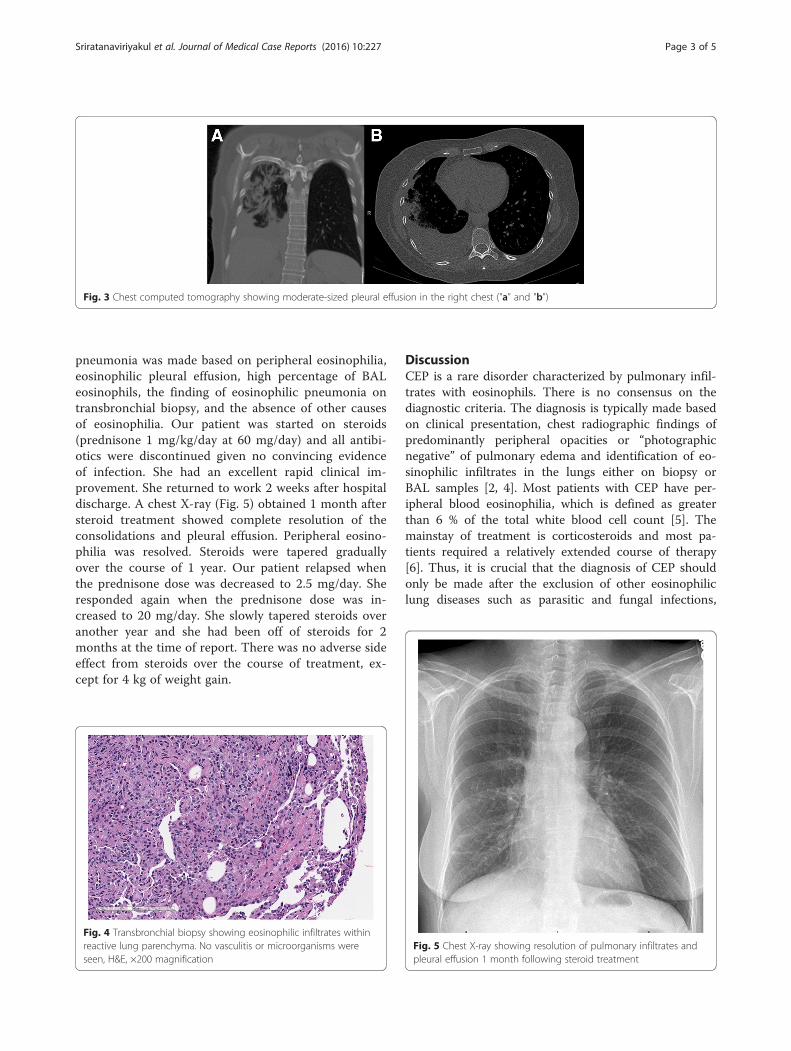
fluid demonstrated a white blood cell count of 41,000cells per mL with 38 % eosinophils, 32 % lymphocytesand 27 % neutrophils. Pleural fluid chemistries wereconsistent with a transudative profile with lactate dehy-drogenous (LDH) of 70 U/L (serum LDH 184 U/L),protein 2.9 g/dL (serum protein 7 g/dL) and glucose 86mg/dL. Bronchoalveolar lavage (BAL) fluid demon-strated a white blood cell count of 1985 cells per mLwith 74 % eosinophils, 13 % lymphocytes and 7 % neu-trophils. A transbronchial biopsy performed from theright lower lobe was suggestive of eosinophilic pneu-monia with marked increased eosinophils without evi-dence of vasculitis, malignancy or fungal organisms(Fig. 4). The diagnosis of chronic eosinophilic
Fig. 1 Chest X-ray showing peripheral pulmonary infiltratespredominantly in the right lung with pleural effusion a posterioranteriorfilm, b lateral film
Fig. 2 Chest computed tomography showing peripheral pulmonaryopacities predominantly in the right lung a apical cut, b mid cut, clower cut
Sriratanaviriyakul et al. Journal of Medical Case Reports (2016) 10:227 Page 2 of 5
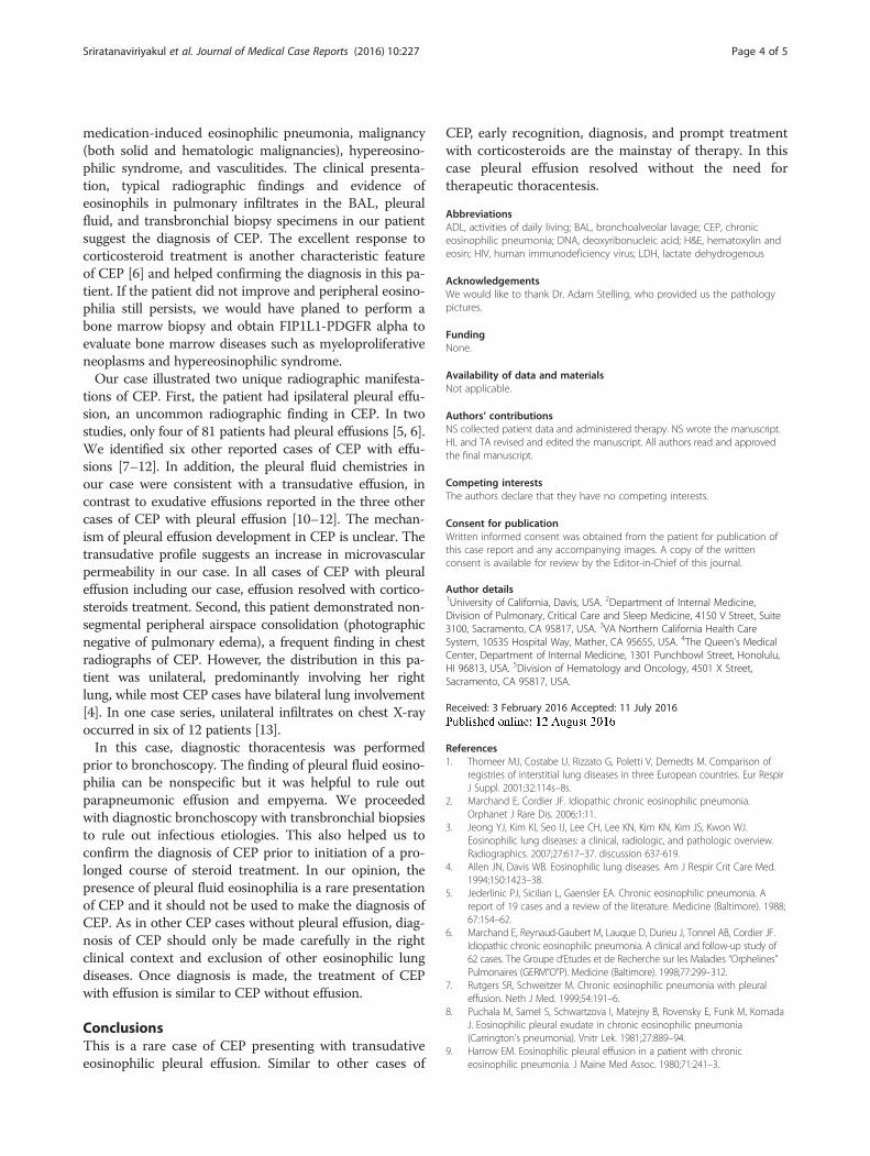
pneumonia was made based on peripheral eosinophilia,eosinophilic pleural effusion, high percentage of BALeosinophils, the finding of eosinophilic pneumonia ontransbronchial biopsy, and the absence of other causesof eosinophilia. Our patient was started on steroids(prednisone 1 mg/kg/day at 60 mg/day) and all antibi-otics were discontinued given no convincing evidenceof infection. She had an excellent rapid clinical im-provement. She returned to work 2 weeks after hospitaldischarge. A chest X-ray (Fig. 5) obtained 1 month aftersteroid treatment showed complete resolution of theconsolidations and pleural effusion. Peripheral eosino-philia was resolved. Steroids were tapered graduallyover the course of 1 year. Our patient relapsed whenthe prednisone dose was decreased to 2.5 mg/day. Sheresponded again when the prednisone dose was in-creased to 20 mg/day. She slowly tapered steroids overanother year and she had been off of steroids for 2months at the time of report. There was no adverse sideeffect from steroids over the course of treatment, ex-cept for 4 kg of weight gain.
DiscussionCEP is a rare disorder characterized by pulmonary infil-trates with eosinophils. There is no consensus on thediagnostic criteria. The diagnosis is typically made basedon clinical presentation, chest radiographic findings ofpredominantly peripheral opacities or “photographicnegative” of pulmonary edema and identification of eo-sinophilic infiltrates in the lungs either on biopsy orBAL samples [2, 4]. Most patients with CEP have per-ipheral blood eosinophilia, which is defined as greaterthan 6 % of the total white blood cell count [5]. Themainstay of treatment is corticosteroids and most pa-tients required a relatively extended course of therapy[6]. Thus, it is crucial that the diagnosis of CEP shouldonly be made after the exclusion of other eosinophiliclung diseases such as parasitic and fungal infections,
Fig. 3 Chest computed tomography showing moderate-sized pleural effusion in the right chest ("a" and "b")
Fig. 4 Transbronchial biopsy showing eosinophilic infiltrates withinreactive lung parenchyma. No vasculitis or microorganisms wereseen, H&E, ×200 magnification
Fig. 5 Chest X-ray showing resolution of pulmonary infiltrates andpleural effusion 1 month following steroid treatment
Sriratanaviriyakul et al. Journal of Medical Case Reports (2016) 10:227 Page 3 of 5
medication-induced eosinophilic pneumonia, malignancy(both solid and hematologic malignancies), hypereosino-philic syndrome, and vasculitides. The clinical presenta-tion, typical radiographic findings and evidence ofeosinophils in pulmonary infiltrates in the BAL, pleuralfluid, and transbronchial biopsy specimens in our patientsuggest the diagnosis of CEP. The excellent response tocorticosteroid treatment is another characteristic featureof CEP [6] and helped confirming the diagnosis in this pa-tient. If the patient did not improve and peripheral eosino-philia still persists, we would have planed to perform abone marrow biopsy and obtain FIP1L1-PDGFR alpha toevaluate bone marrow diseases such as myeloproliferativeneoplasms and hypereosinophilic syndrome.Our case illustrated two unique radiographic manifesta-
tions of CEP. First, the patient had ipsilateral pleural effu-sion, an uncommon radiographic finding in CEP. In twostudies, only four of 81 patients had pleural effusions [5, 6].We identified six other reported cases of CEP with effu-sions [7–12]. In addition, the pleural fluid chemistries inour case were consistent with a transudative effusion, incontrast to exudative effusions reported in the three othercases of CEP with pleural effusion [10–12]. The mechan-ism of pleural effusion development in CEP is unclear. Thetransudative profile suggests an increase in microvascularpermeability in our case. In all cases of CEP with pleuraleffusion including our case, effusion resolved with cortico-steroids treatment. Second, this patient demonstrated non-segmental peripheral airspace consolidation (photographicnegative of pulmonary edema), a frequent finding in chestradiographs of CEP. However, the distribution in this pa-tient was unilateral, predominantly involving her rightlung, while most CEP cases have bilateral lung involvement[4]. In one case series, unilateral infiltrates on chest X-rayoccurred in six of 12 patients [13].In this case, diagnostic thoracentesis was performed
prior to bronchoscopy. The finding of pleural fluid eosino-philia can be nonspecific but it was helpful to rule outparapneumonic effusion and empyema. We proceededwith diagnostic bronchoscopy with transbronchial biopsiesto rule out infectious etiologies. This also helped us toconfirm the diagnosis of CEP prior to initiation of a pro-longed course of steroid treatment. In our opinion, thepresence of pleural fluid eosinophilia is a rare presentationof CEP and it should not be used to make the diagnosis ofCEP. As in other CEP cases without pleural effusion, diag-nosis of CEP should only be made carefully in the rightclinical context and exclusion of other eosinophilic lungdiseases. Once diagnosis is made, the treatment of CEPwith effusion is similar to CEP without effusion.
ConclusionsThis is a rare case of CEP presenting with transudativeeosinophilic pleural effusion. Similar to other cases of
CEP, early recognition, diagnosis, and prompt treatmentwith corticosteroids are the mainstay of therapy. In thiscase pleural effusion resolved without the need fortherapeutic thoracentesis.
AbbreviationsADL, activities of daily living; BAL, bronchoalveolar lavage; CEP, chroniceosinophilic pneumonia; DNA, deoxyribonucleic acid; H&E, hematoxylin andeosin; HIV, human immunodeficiency virus; LDH, lactate dehydrogenous
AcknowledgementsWe would like to thank Dr. Adam Stelling, who provided us the pathologypictures.
FundingNone.
Availability of data and materialsNot applicable.
Authors’ contributionsNS collected patient data and administered therapy. NS wrote the manuscript.HL and TA revised and edited the manuscript. All authors read and approvedthe final manuscript.
Competing interestsThe authors declare that they have no competing interests.
Consent for publicationWritten informed consent was obtained from the patient for publication ofthis case report and any accompanying images. A copy of the writtenconsent is available for review by the Editor-in-Chief of this journal.
Author details1University of California, Davis, USA. 2Department of Internal Medicine,Division of Pulmonary, Critical Care and Sleep Medicine, 4150 V Street, Suite3100, Sacramento, CA 95817, USA. 3VA Northern California Health CareSystem, 10535 Hospital Way, Mather, CA 95655, USA. 4The Queen’s MedicalCenter, Department of Internal Medicine, 1301 Punchbowl Street, Honolulu,HI 96813, USA. 5Division of Hematology and Oncology, 4501 X Street,Sacramento, CA 95817, USA.
Received: 3 February 2016 Accepted: 11 July 2016
References1. Thomeer MJ, Costabe U, Rizzato G, Poletti V, Demedts M. Comparison of
registries of interstitial lung diseases in three European countries. Eur RespirJ Suppl. 2001;32:114s–8s.
2. Marchand E, Cordier JF. Idiopathic chronic eosinophilic pneumonia.Orphanet J Rare Dis. 2006;1:11.
3. Jeong YJ, Kim KI, Seo IJ, Lee CH, Lee KN, Kim KN, Kim JS, Kwon WJ.Eosinophilic lung diseases: a clinical, radiologic, and pathologic overview.Radiographics. 2007;27:617–37. discussion 637-619.
4. Allen JN, Davis WB. Eosinophilic lung diseases. Am J Respir Crit Care Med.1994;150:1423–38.
5. Jederlinic PJ, Sicilian L, Gaensler EA. Chronic eosinophilic pneumonia. Areport of 19 cases and a review of the literature. Medicine (Baltimore). 1988;67:154–62.
6. Marchand E, Reynaud-Gaubert M, Lauque D, Durieu J, Tonnel AB, Cordier JF.Idiopathic chronic eosinophilic pneumonia. A clinical and follow-up study of62 cases. The Groupe d’Etudes et de Recherche sur les Maladies “Orphelines”Pulmonaires (GERM“O”P). Medicine (Baltimore). 1998;77:299–312.
7. Rutgers SR, Schweitzer M. Chronic eosinophilic pneumonia with pleuraleffusion. Neth J Med. 1999;54:191–6.
8. Puchala M, Samel S, Schwartzova I, Matejny B, Rovensky E, Funk M, KomadaJ. Eosinophilic pleural exudate in chronic eosinophilic pneumonia(Carrington’s pneumonia). Vnitr Lek. 1981;27:889–94.
9. Harrow EM. Eosinophilic pleural effusion in a patient with chroniceosinophilic pneumonia. J Maine Med Assoc. 1980;71:241–3.
Sriratanaviriyakul et al. Journal of Medical Case Reports (2016) 10:227 Page 4 of 5
10. d’Amours P, Leblanc P, Boulet LP. Chronic eosinophilic pneumoniaassociated with thrombocytosis and pleural effusion. CMAJ. 1990;142:837–9.
11. Jee YG, Ra SH, Park YM, Cha JW, Kang YS, Park JH, Kang TY. A case ofrheumatoid arthritis with chronic eosinophilic pneumonia associated witheosinophilic pleural effusion. J Rheum Dis. 2013;20:328–31.
12. Samman YS, Wali SO, Abdelaal MA, Gangi MT, Krayem AB. Chroniceosinophilic pneumonia presenting with recurrent massive bilateral pleuraleffusion: case report. Chest. 2001;119:968–70.
13. Naughton M, Fahy J, FitzGerald MX. Chronic eosinophilic pneumonia. Along-term follow-up of 12 patients. Chest. 1993;103:162–5.
• We accept pre-submission inquiries
• Our selector tool helps you to find the most relevant journal
• We provide round the clock customer support
• Convenient online submission
• Thorough peer review
• Inclusion in PubMed and all major indexing services
• Maximum visibility for your research
Submit your manuscript atwww.biomedcentral.com/submit
Submit your next manuscript to BioMed Central and we will help you at every step:
Sriratanaviriyakul et al. Journal of Medical Case Reports (2016) 10:227 Page 5 of 5