Embed Size (px)
Citation preview


Chromosomal Mutations:Variation in Chromosome Number
Henry County Professional Learning High School Session II

Chromosomal Mutations
• Deletion – part of the chromosome is missing• Starts with breaks in the chromosome• Radiation, heat, viruses, chemicals, etc.• May cause an unpaired loop• May give rise to pseudodominance• Cri-du-chat syndrome (Chrom. #5)

Chromosomal Mutations

Chromosomal Mutations
• To "read" a set of chromosomes, scientists use three key features to identify their similarities and differences:• Size. This is the easiest way to
tell chromosomes apart.• Banding pattern. The size and
location of Giemsa bands make each chromosome unique.
• Centromere position. Centromeres appear as a constriction. They have a role in the separation of chromosomes into daughter cells during cell division (mitosis and meiosis).

Chromosomal Mutations
• Genetic disorders resulting from chromosome deletions include:• Cri-du-Chat Syndrome (Chromosome 5)• Williams Syndrome (Chromosome 7)

What is Cri-Du-Chat syndrome?
Cri-du-chat is a French term for “cry of the cat”
Also known as 5p- syndrome
Cri-du-chat syndrome occurs in an estimated 1 in 20,000 to 50,000 newborns

Cri-du-chat syndrome occurs in an estimated 1 in 20,000 to 50,000 newbornsThis condition is found in people of all ethnic backgrounds
HOW COMMON IS CRI-DU-CHAT
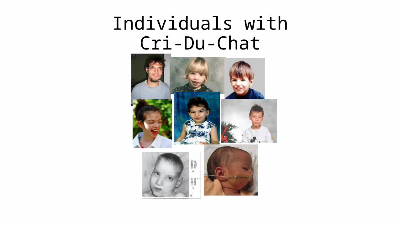
Individuals with Cri-Du-Chat

Centromere Position
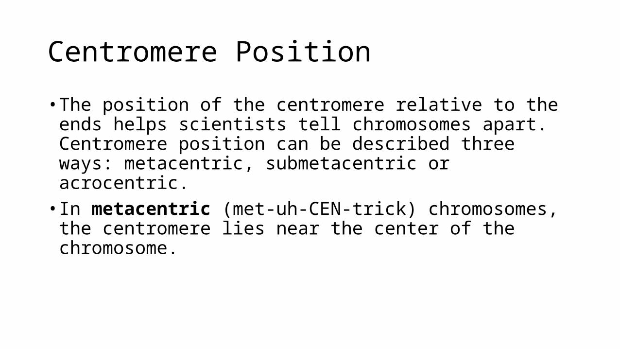
Centromere Position
• The position of the centromere relative to the ends helps scientists tell chromosomes apart. Centromere position can be described three ways: metacentric, submetacentric or acrocentric.• In metacentric (met-uh-CEN-trick) chromosomes, the centromere lies
near the center of the chromosome.

Centromere Position
• Submetacentric (SUB-met-uh-CEN-trick) chromosomes have a centromere that is off- center, so that one chromosome arm is longer than the other. The short arm is designated "p" (for petite), and the long arm is designated "q" (because it follows the letter "p").• In acrocentric (ACK-ro-CEN-trick) chromosomes, the centromere is
very near one end.


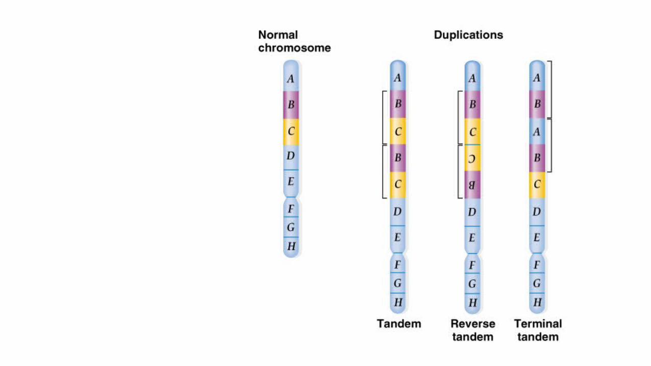
Chromosomal Mutations
• Duplication – doubling of a segment of a chromosome• Tandem• Reverse tandem• Terminal tandem• Position Effect of barring eyes in Drosophila

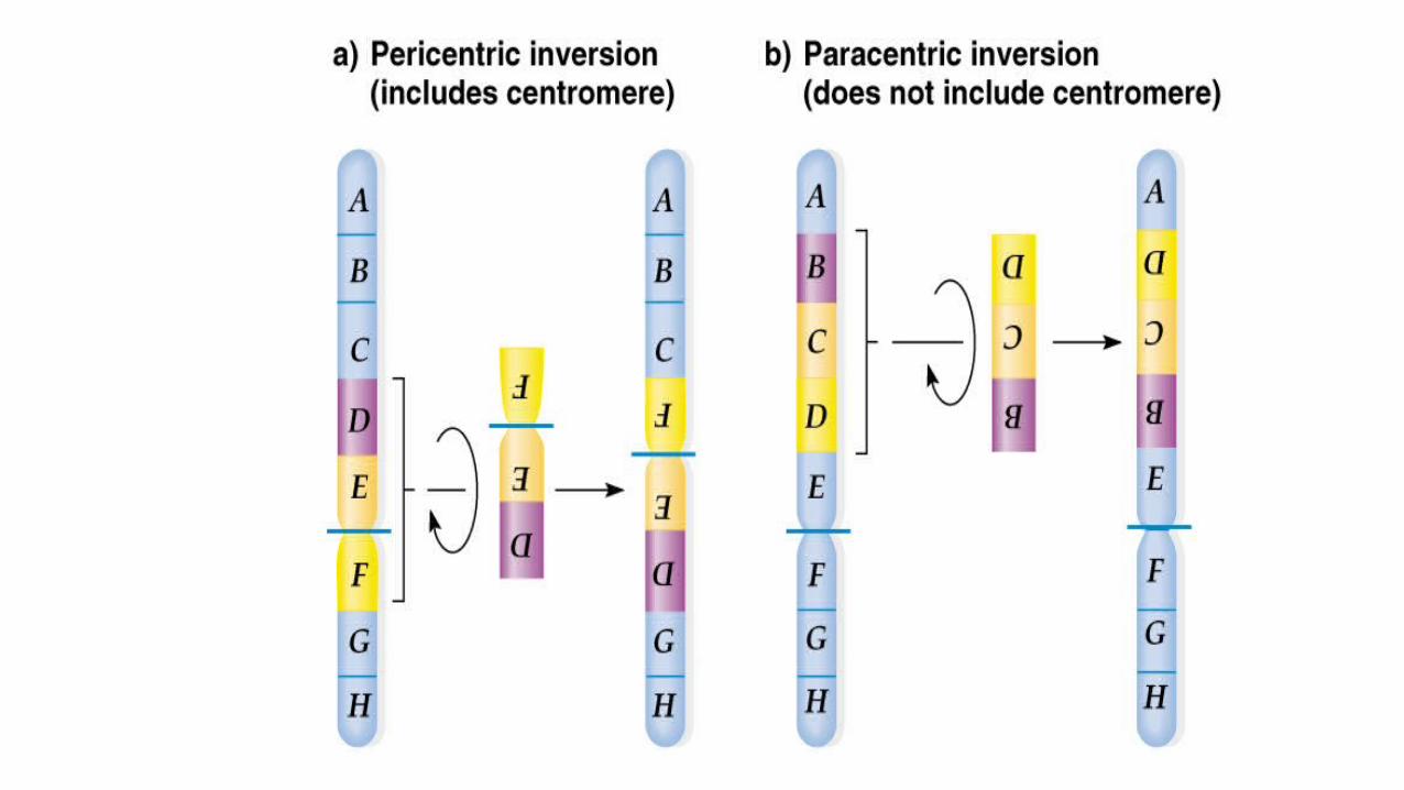
Chromosomal Mutations
• Inversion – results when a segment of a chromosome is excised and then reintegrated in an orientation 180 degrees from the original orientation.• Pericentric inversion• Paracentric inversion• Resulting from a dicentric bridge
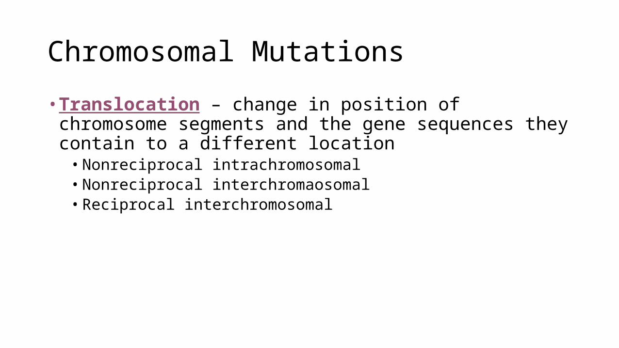
Chromosomal Mutations
• Translocation – change in position of chromosome segments and the gene sequences they contain to a different location• Nonreciprocal intrachromosomal • Nonreciprocal interchromaosomal• Reciprocal interchromosomal


A Robertsonian translocation occurs when the long arms of two acrocentric chromosomes fuse at a centromere. The two short arms are lost, leaving a total of 45 chromosomes. But because the short arms carry very little genetic information, individuals with Robertsonian translocations are usually healthy.

Chromosomal Mutations
• Fragile Sites and Fragile X Syndrome• Narrowings or unstained areas (gaps)• Recessive X-linked traits (predominately males)• Normal transmitting males carrying a premutation will have grandsons with
mental retardations and some granddaughters (up to 33%) with mild retardation


Variation in Chromosomal Number
• Euploidy – the correct number of sets of chromosomes in an organism• Monoploidy – only one set of chromosomes when there should be
more• Male wasps, ants, and bees are monoploids because they develop from
unfertilized eggs• Used in plant-breeding experiments/colchicine (inhibits mitotic spindle)
Variation in Chromosomal Number
Polyploidy – having three or more sets of chromosomes• Usually occurs due to a breakdown of the mitotic spindle• Usually occurs in plants (self-fertilization)• Even numbered polyploids have a better chance of being fertile than odd
numbered polyploids• In humans, triploidy is usually lethal.
Variation in Chromosomal Number
Polyploidy (continued) • Autopolyploidy• All sets of chromosomes originate in the same species/defect in meiosis• Example – a diploid gamete fuses with a haploid gamete to produce a triploid
zygote (bananas and seedless fruit)

Variation in Chromosomal Number
Polyploidy (continued)• Allopolyploidy • Sets of chromosomes originate from different species though usually related• Because of differences between chromosomes, the hybrid, no crossing over
occurs and no viable gametes produced making hybrids sterile• Occasionally, tow sets of different chromosomes will double, producing
tissues of 2N + 2N2 / produces fully fertile allotetraploid, 2N + 2N2, plants


Variation in Chromosomal Number
• Aneuploidy – variation in the number of chromosomes• Nullisomy - loss of one homologous chromosome pair; 2N – 2 • Monosomy – loss of a single chromosome; 2N – 1 • Trisomy – single extra chromosome; 2N + 1• Tetrasomy – extra chromosome pair; 2N + 2

Nondisjunction

Variation in Chromosomal Number
• In humans, Down Syndrome is the only disorder where an extra autosomal chromosome exists and the individuals lives past three months. All other embryos will self-abort or the infant will die within 6 months. However, extra sex chromosomes will not cause an embryo to necessarily abort; but the individual may have other distinguishable characteristics.

Variation in Chromosomal Number
• Trisomy-21 – Down Syndrome• Trisomy-13 (Patau syndrome) – cleft lip and palate, small eyes,
polydactyly and mental retardation. Dies within 3 months• Trisomy-18 (Edwards syndrome) – developmental retardation,
congenital malformations, and die within 6 months

• Dr. Harry Klinefelter
• Massachusetts General Hospital in Boston
• 1942 symptoms first identified
• 1950’s syndrome was discovered
• Early 1970’s screening of infants began
Klinefelter

• Sex Chromosomes are XXY
• May display all or none of the symptoms
• Some live their entire lives without any suspicion that anything is
wrong
• Is not passed through genetics
About The Disorder

Male with Klinefelter’s Syndrome
Common Symptoms

• Hormone Testing
• Semen analysis
• Usually not diagnosed before puberty
• Can be detected prenatally by amniocentesis or chorionic villus
sampling
Diagnosis
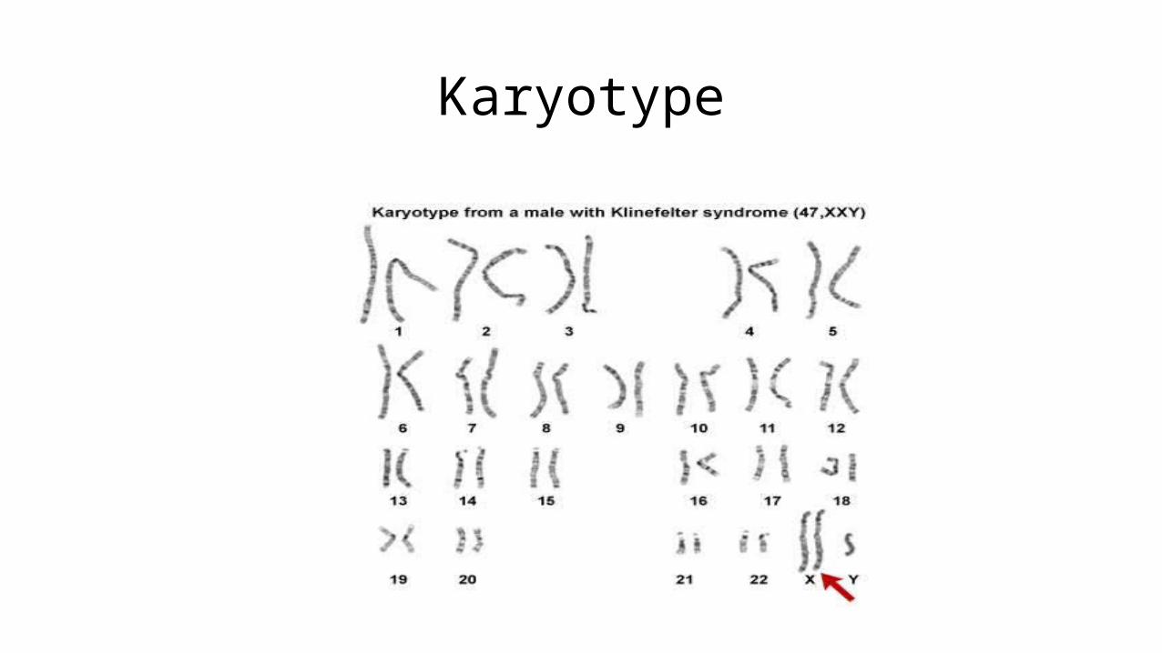
Karyotype
• Testosterone can be injected or administered through as skin patch or
gel
• Usually continues throughout the lifespan
• Does not help with reproduction
Treatment

Marfan Syndrome
• French Physician Antoine Bernard- Jean Marfan• Discovered in 1896• He described a five year old girl with
abnormally long and frail limbs and fingers and lots of other skeletal abnormalities.

Cause of Disorder
• Marfan Syndrome is caused by a defect in a gene• It is a disorder that affects connective tissue in the
body• Connective tissue is what provides our body strength
and flexibility to structures of out body such as:• Bones• Ligaments• Muscles• Blood Vessels• Heart Valves

Chromosomes Involved
• This disorder is located on chromosome 15• Chromosome 15 controls the production of the protein fibrillin• Fibrillin is responsible for elasticity and strength of connective
tissue• The mutations that cause Marfan Syndrome change a single
amino acid in the fibrillin-1 protein. • Researchers found more than 1,300 Fibrillin-1 gene mutations
that cause this disorder

Complications
• Aortic Aneurysm • Aortic Dissection• Valve Malformation• Lens Dislocation• Retinal Problems• Early-onset Glaucoma or Cataracts
Treatments
• Special medicines called beta blockers and ARBs (angiotensin-receptor blockers) which work to lower the blood pressure and reduce wear and tear on the blood vessels.• Teens with Marfan syndrome that develop scoliosis have to
wear a special back brace. • Calcium channel blockers are recommended if the patient can
not take beta blockers.• Regular follow up appointments.• Routine cardiovascular, eye, and orthopedic exams.

Famous People Affected By
Marfan
Abraham Lincoln
Michael Phelps
Jonathan Larson

Jacobs syndrome (47, XYY)
• In 1961 Dr. Avery Sandberg and associates at the Roswell Park Memorial Institute observed for the first time ever a man who had a karyotype of XYY.• The subject was a 44 year old
man who had sought the help of a geneticist after having a daughter who suffered from down syndrome.
• XYY syndrome is a condition when a male patient acquires an extra Y chromosome in addition to the normal one X and one Y chromosomes. It is often argued whether the term ‘syndrome' should be used in connection to the XYY condition since the patients' phenotypes are normal and they do not have knowledge of their karyotype.

XYY Occurences• 47 XYY syndrome is not an inherited
condition. This is just the result of an accidental event during sperm cell formation. The ‘accident' happens either during metaphase I or metaphase II.
• An error in cell division called nondisjunction can result in sperm cells with an extra copy of the Y chromosome. If one of these atypical reproductive cells contributes to the genetic makeup of a child, the child will have an extra Y chromosome in each of the body's cells.
• This condition cannot be corrected but to prevent the occurrence of this syndrome on the patient's posterity, it is best that genetic testing be completed.
• ~1 in every 1,000 males acquire the extra Y chromosome.

The truth about XYY

Normal v. mutation

Normal v. mutation
• People normally have 46 chromosomes in each cell. Two of the 46 chromosomes, known as X and Y, are called sex chromosomes because they help determine whether a person will develop male or female sex characteristics. Females typically have two X chromosomes (46,XX), and males have one X chromosome and one Y chromosome (46,XY).
• 47,XYY syndrome is caused by the presence of an extra copy of the Y chromosome in each of a male's cells. As a result of the extra Y chromosome, each cell has a total of 47 chromosomes instead of the usual 46. It is unclear why an extra copy of the Y chromosome is associated with tall stature, learning problems, and other features in some boys and men.

Mutation v. mutation
XYY• Jacobs Syndrome is usually associated with
early growth development in young adolescents, which results in taller stature.
• Increased presence of acne has also been associated with male individuals who have the presence of an extra Y chromosome.
• Decreased muscle tone and general atrophy has been linked to Jacobs Syndrome.
• No true indication of increased masculinity with the exception of height.
XXY• Klinefelter Syndrome is usually
associated with increased femininity in males who have an extra X chromosome.
• As infants, males have decreased muscularity and strength; as adults, reduced coordination and muscle control are associated with this chromosomal mutation.
• During puberty, decreased levels of testosterone result in increased femaleness.

Medications to ease symptoms
• How do treat Jacob’s Syndrome?
• Currently there is no cure for Jacob’s Syndrome, however scientists have developed therapy treatments to help the affected person tolerate the disorder. Males with Jacob’s Syndrome can have explosive tempers.

There is often pain associated with Jacob’s Syndrome
• Males with Jacob’s Syndrome often have joint pain and painful bone growth due to growing at an accelerated rate. • Pain killers and anti-
inflammatory medicines are prescribed to ease the pain. • In some cases support braces
are used to relieve joint pain.
Learning difficulties are associated with Jacob’s Syndrome • Males with Jacob’s Syndrome sometimes have
developmental learning problems. They may require speech therapy, specialized learning techniques and sometimes require special needs
consideration.

Dystrophic Epidermolysis
Bullosa

Background
-Discovered by Heinrich Koebner in 1886-Broken down into subgroups in 1960s-Specific causes discovered in 1980s/1990s-Epidermis is the outer layer of skin
- “lysis” means breakdown- “bullosa” means blister

About DEB
-Three subgroups, all genetic mutations-Dominant and recessive-Outlook depends on severity
What does it look like?
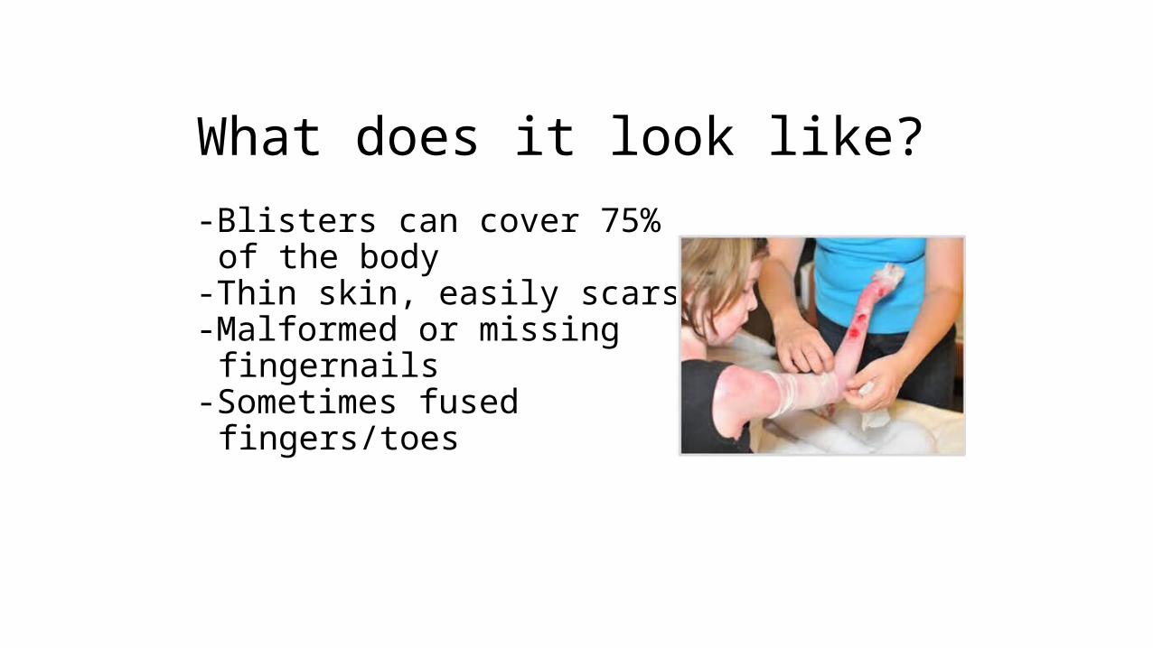
-Blisters can cover 75% of the body-Thin skin, easily scars-Malformed or missing fingernails-Sometimes fused fingers/toes

What’s the Problem?
-Collagen VII-protein that holds layers of skin together-misshapen, reduced number, completely missing-coding gene on chromosome 3p21.1


Living with DEB
-Draining blisters-Bandages-Liquids and soft foods-Loose fitting clothing-Oral problems-“Butterfly Children”

Discovery• Turner Syndrome was discovered in 1938 by Dr. Henry Turner.
• In 1938, he also described and came up with the first treatment for Turner Syndrome.
• He observed a set of common physical features in his patients, however it wasn’t until 1960 when the chromosomal abnormality was found.

• Turner Syndrome occurs in 1 out of 2,500 newborn girls worldwide.
• Usually the fetus does not survive to term.• Turner Syndrome affects 60,000 girls in
the United States.
STAT
ISTI
CS

• Most cases of Turner Syndrome are not inherited.• Most females affected by Turner Syndrome suffer from monosomy X which means that the female is missing the second X chromosome.
• Nondisjunction, which causes abnormal numbers of chromosomes in reproductive cells, is usually the cause of Turner Syndrome.
←Karyotype
CAUSES

Symptoms of Turner Syndrome
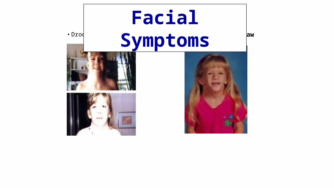
• Drooping eye lids Receding or small lower jaw
Facial Symptoms

Body
cha
ract
eris
tics