Embed Size (px)
Citation preview
Copyright © 2010 Pearson Education, Inc.
Figure 4.8g Connective tissues.
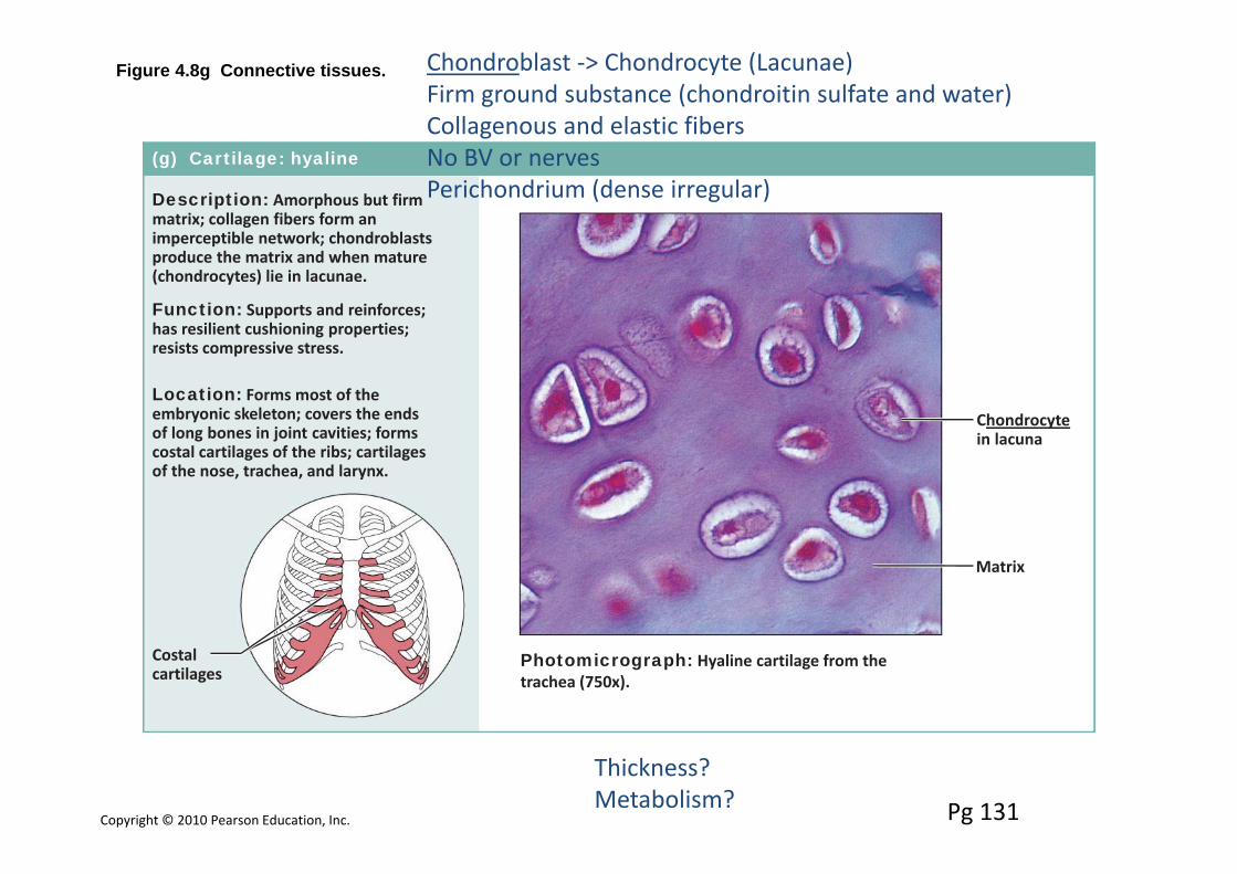
(g) Cartilage: hyaline
Description: Amorphous but firmmatrix; collagen fibers form animperceptible network; chondroblastsproduce the matrix and when mature(chondrocytes) lie in lacunae.
Function: Supports and reinforces;has resilient cushioning properties;resists compressive stress.
Location: Forms most of theembryonic skeleton; covers the endsof long bones in joint cavities; formscostal cartilages of the ribs; cartilagesof the nose, trachea, and larynx.
Photomicrograph: Hyaline cartilage from thetrachea (750x).
Costalcartilages
Chondrocytein lacuna
Matrix
Pg 131
Chondroblast ‐> Chondrocyte (Lacunae)Firm ground substance (chondroitin sulfate and water)Collagenous and elastic fibersNo BV or nervesPerichondrium (dense irregular)
Thickness?Metabolism?
Copyright © 2010 Pearson Education, Inc.
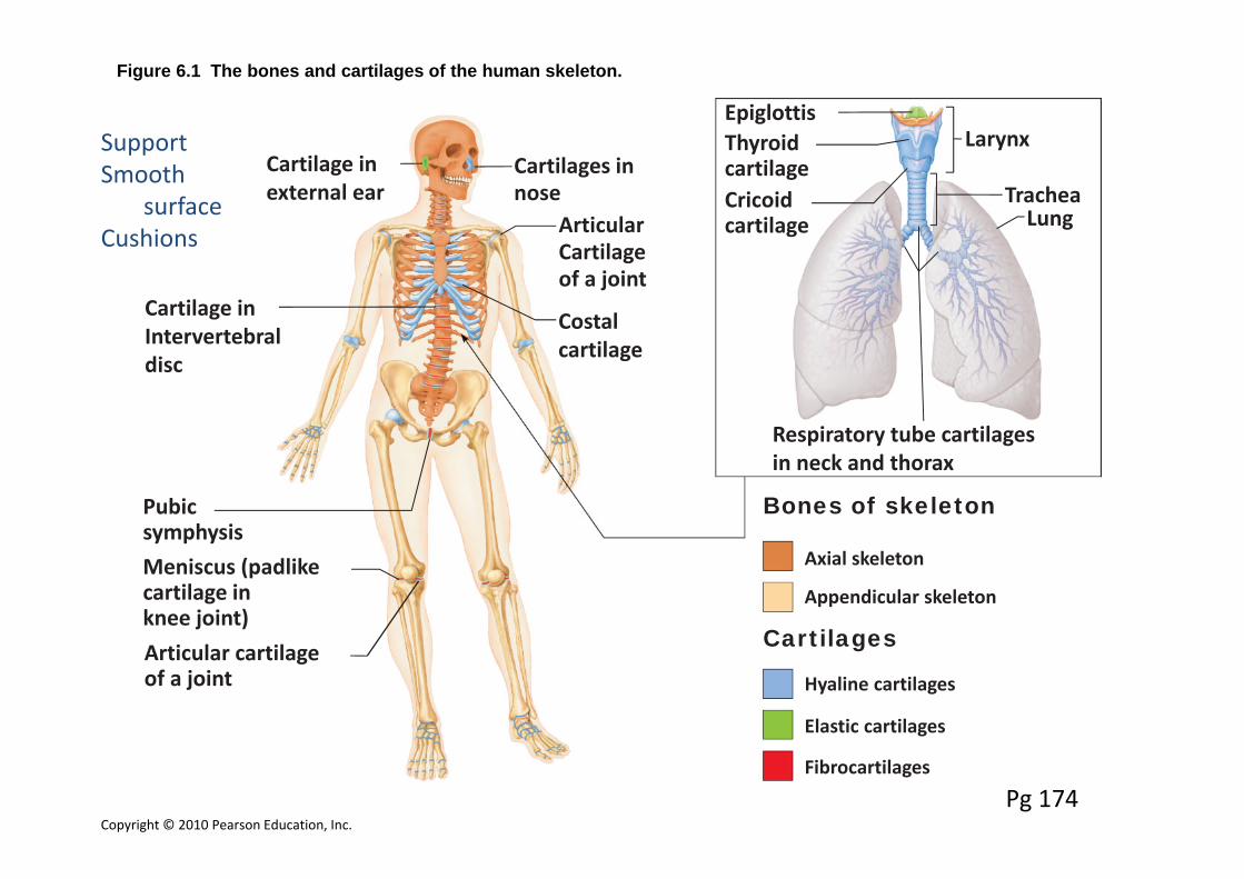
Figure 6.1 The bones and cartilages of the human skeleton.
Axial skeleton
Appendicular skeleton
Hyaline cartilages
Elastic cartilages
Fibrocartilages
Cartilages
Bones of skeleton
EpiglottisLarynx
TracheaCricoidcartilage Lung
Respiratory tube cartilagesin neck and thorax
ThyroidcartilageCartilage in
external earCartilages innose
ArticularCartilageof a joint
Costalcartilage
Cartilage inIntervertebraldisc
Pubicsymphysis
Articular cartilageof a joint
Meniscus (padlikecartilage inknee joint)
Pg 174
SupportSmooth
surfaceCushions
Copyright © 2010 Pearson Education, Inc.
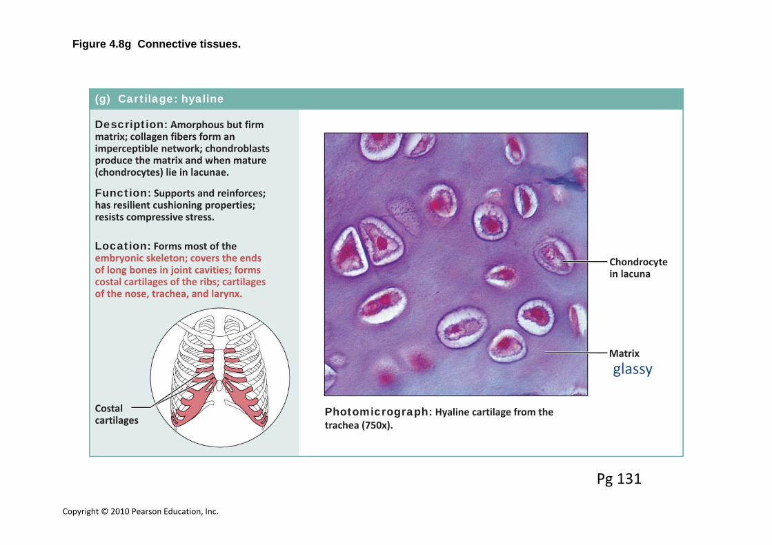
Figure 4.8g Connective tissues.
(g) Cartilage: hyaline
Description: Amorphous but firmmatrix; collagen fibers form animperceptible network; chondroblastsproduce the matrix and when mature(chondrocytes) lie in lacunae.
Function: Supports and reinforces;has resilient cushioning properties;resists compressive stress.
Location: Forms most of theembryonic skeleton; covers the endsof long bones in joint cavities; formscostal cartilages of the ribs; cartilagesof the nose, trachea, and larynx.
Photomicrograph: Hyaline cartilage from thetrachea (750x).
Costalcartilages
Chondrocytein lacuna
Matrixglassy
Pg 131
Copyright © 2010 Pearson Education, Inc.
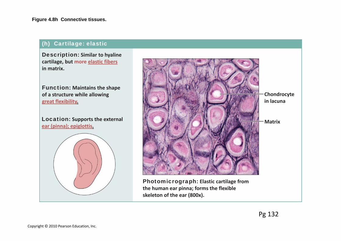
Figure 4.8h Connective tissues.
(h) Cartilage: elastic
Description: Similar to hyalinecartilage, but more elastic fibersin matrix.
Function: Maintains the shapeof a structure while allowinggreat flexibility.
Location: Supports the externalear (pinna); epiglottis.
Photomicrograph: Elastic cartilage fromthe human ear pinna; forms the flexibleskeleton of the ear (800x).
Chondrocytein lacuna
Matrix
Pg 132
Copyright © 2010 Pearson Education, Inc.
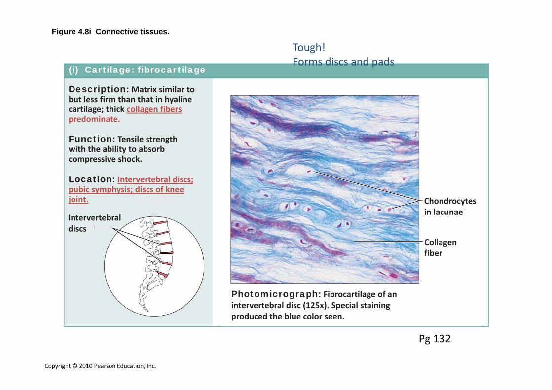
Figure 4.8i Connective tissues.
(i) Cartilage: fibrocartilage
Description: Matrix similar tobut less firm than that in hyalinecartilage; thick collagen fiberspredominate.
Function: Tensile strengthwith the ability to absorbcompressive shock.
Location: Intervertebral discs;pubic symphysis; discs of kneejoint.
Photomicrograph: Fibrocartilage of anintervertebral disc (125x). Special stainingproduced the blue color seen.
Intervertebraldiscs
Chondrocytesin lacunae
Collagenfiber
Pg 132
Tough!Forms discs and pads
Copyright © 2010 Pearson Education, Inc.
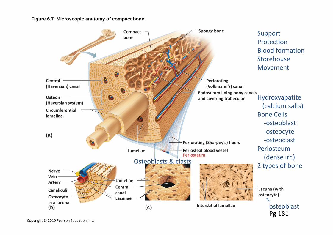
Figure 6.7 Microscopic anatomy of compact bone.
Endosteum lining bony canalsand covering trabeculae
Perforating (Volkmann’s) canal
Perforating (Sharpey’s) fibers
Periosteal blood vesselPeriosteum
Lacuna (withosteocyte)
(a)
(b) (c)
Lacunae
Lamellae
NerveVeinArtery
CanaliculiOsteocytein a lacuna
Circumferentiallamellae
Osteon(Haversian system)
Central(Haversian) canal
Centralcanal
Interstitial lamellae
Lamellae
Compactbone
Spongy bone
Pg 181
SupportProtectionBlood formationStorehouseMovement
Hydroxyapatite(calcium salts)
Bone Cells‐osteoblast‐osteocyte‐osteoclast
Periosteum(dense irr.)
2 types of bone
osteoblast
Osteoblasts & clasts

Copyright © 2010 Pearson Education, Inc.
Proximalepiphysis
(b)
(c)(a)
Yellowbone marrow
Endosteum
Epiphysealline
Articularcartilage
Periosteum
Spongy bone
Compact boneMedullarycavity (linedby endosteum)
Compact bone
Compact bone
Periosteum
Perforating(Sharpey’s)fibersNutrientarteries
Diaphysis
Distalepiphysis
Figure 6.3 The structure of a long bone (humerus of arm).
Pg 176
Spongy = Cancellous
Spaces contain marrowTrabeculae
‐Ends of long bones‐Shaft of young bones‐In flat, short and irregular bones
Copyright © 2010 Pearson Education, Inc.
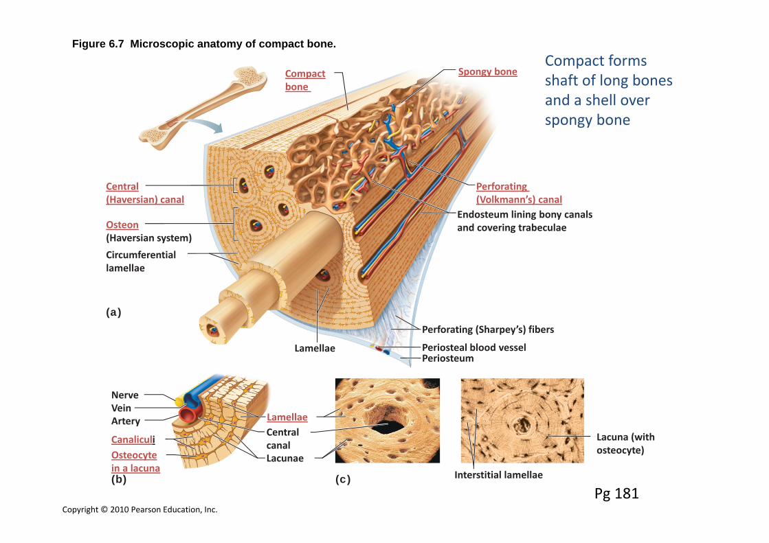
Figure 6.7 Microscopic anatomy of compact bone.
Endosteum lining bony canalsand covering trabeculae
Perforating (Volkmann’s) canal
Perforating (Sharpey’s) fibers
Periosteal blood vesselPeriosteum
Lacuna (withosteocyte)
(a)
(b) (c)
Lacunae
Lamellae
NerveVeinArtery
CanaliculiOsteocytein a lacuna
Circumferentiallamellae
Osteon(Haversian system)
Central(Haversian) canal
Centralcanal
Interstitial lamellae
Lamellae
Compactbone
Spongy bone
Pg 181
Compact forms shaft of long bones and a shell over spongy bone

Copyright © 2010 Pearson Education, Inc.
Figure 4.8j Connective tissues.
(j) Others: bone (osseous tissue)
Description: Hard, calcifiedmatrix containing many collagenfibers; osteocytes lie in lacunae.Very well vascularized.
Function: Bone supports andprotects (by enclosing);provides levers for the musclesto act on; stores calcium andother minerals and fat; marrowinside bones is the site for bloodcell formation (hematopoiesis).
Location: Bones
Photomicrograph: Cross‐sectional viewof bone (125x).
Lacunae
Lamella
Centralcanal
Pg 133
Copyright © 2010 Pearson Education, Inc.
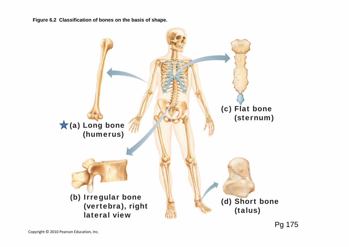
Figure 6.2 Classification of bones on the basis of shape.
(a) Long bone(humerus)
(b) Irregular bone(vertebra), rightlateral view
(d) Short bone(talus)
(c) Flat bone(sternum)
Pg 175
Copyright © 2010 Pearson Education, Inc.
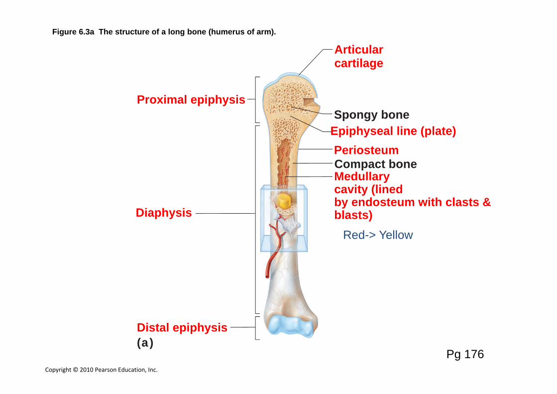
Figure 6.3a The structure of a long bone (humerus of arm).
Proximal epiphysis
(a)
Epiphyseal line (plate)
Articularcartilage
Periosteum
Spongy bone
Compact boneMedullarycavity (linedby endosteum with clasts & blasts)Diaphysis
Distal epiphysis
Pg 176
Red-> Yellow
Formation of bony skeleton
Intramembranous: skull bones, mandible, part of clavicle
Endochondral: Most bones of the body

Bone is only deposited in an area with less highly specialized connective tissue.
Intramembranousmesenchyme ‐> Dense Irr. ‐> Spongy ‐> Compact
C.T. Bone Bone(inside)
Copyright © 2010 Pearson Education, Inc.
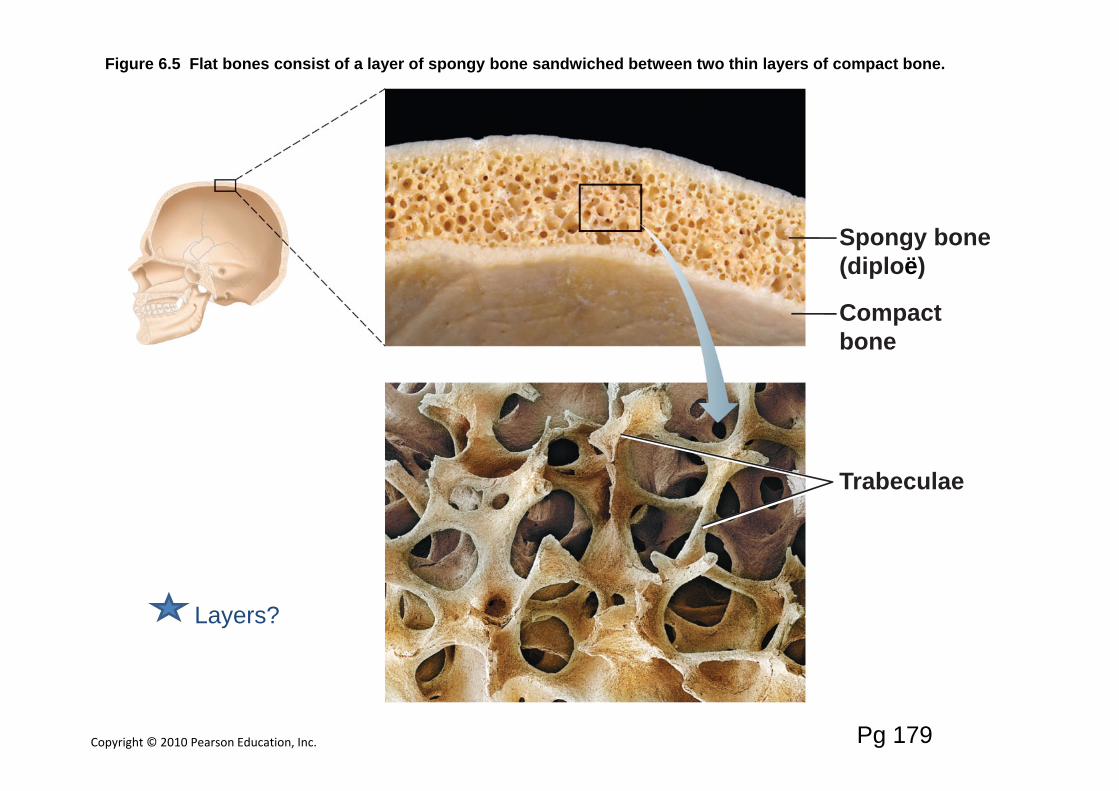
Figure 6.5 Flat bones consist of a layer of spongy bone sandwiched between two thin layers of compact bone.
Compactbone
Trabeculae
Spongy bone(diploë)
Layers?
Pg 179
Bone is only deposited in an area with less highly specialized connective tissue.
Intramembranousmesenchyme ‐> Dense Irr. ‐> Spongy ‐> Compact
C.T. Bone Bone(inside)
Endochondralmesenchyme ‐> Hyaline ‐> Spongy ‐> Compact
Cartilage Bone Bone
Copyright © 2010 Pearson Education, Inc.
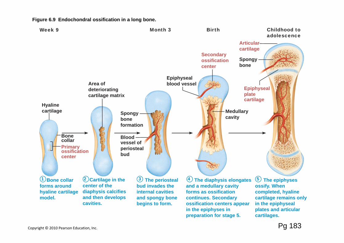
Figure 6.9 Endochondral ossification in a long bone.
1 2 3 4 5Bone collarforms aroundhyaline cartilagemodel.
Cartilage in thecenter of thediaphysis calcifiesand then developscavities.
The periostealbud invades theinternal cavitiesand spongy bonebegins to form.
The diaphysis elongatesand a medullary cavityforms as ossificationcontinues. Secondaryossification centers appearin the epiphyses inpreparation for stage 5.
The epiphysesossify. Whencompleted, hyalinecartilage remains onlyin the epiphysealplates and articularcartilages.
Hyalinecartilage
Area ofdeterioratingcartilage matrix
Epiphysealblood vessel
Spongyboneformation
Epiphysealplatecartilage
Secondaryossificationcenter
Bloodvessel ofperiostealbud
Medullarycavity
Articularcartilage
Childhood toadolescence
BirthWeek 9 Month 3
Spongybone
Bonecollar Primaryossificationcenter
Pg 183
Copyright © 2010 Pearson Education, Inc.
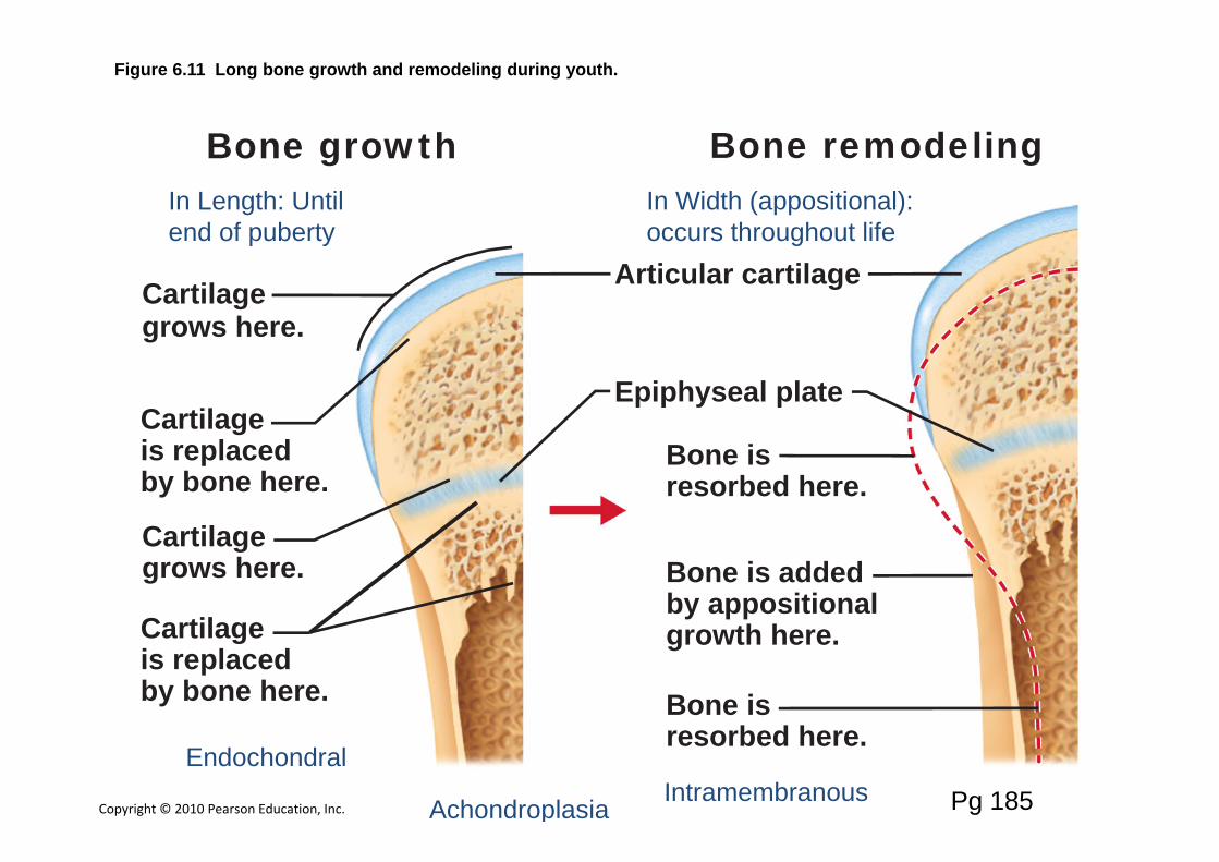
Figure 6.11 Long bone growth and remodeling during youth.
Bone growth Bone remodeling
Articular cartilage
Epiphyseal plate
Cartilagegrows here.
Cartilageis replacedby bone here.
Cartilagegrows here.
Bone isresorbed here.
Bone isresorbed here.
Bone is addedby appositionalgrowth here. Cartilage
is replacedby bone here.
Pg 185
In Length: Until end of puberty
Endochondral
In Width (appositional): occurs throughout life
IntramembranousAchondroplasia
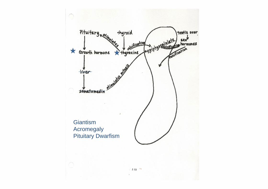
GiantismAcromegalyPituitary Dwarfism
Copyright © 2010 Pearson Education, Inc.
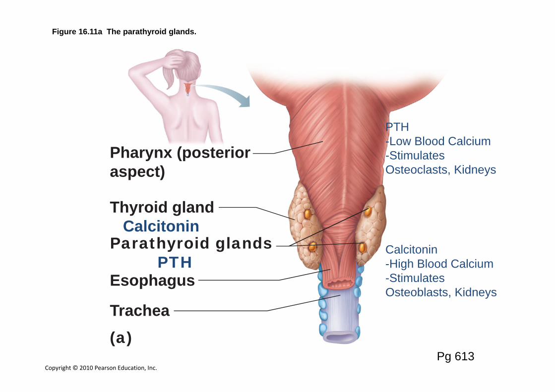
Figure 16.11a The parathyroid glands.
Pharynx (posterioraspect)
Thyroid glandCalcitonin
Parathyroid glandsPTH
Trachea
Esophagus
(a)Pg 613
PTH-Low Blood Calcium-Stimulates Osteoclasts, Kidneys
Calcitonin-High Blood Calcium-Stimulates Osteoblasts, Kidneys
Osteoporosis
Estrogen‐Reduced with menopause‐Loss of bone massCalciumVitamin DExercise
Copyright © 2010 Pearson Education, Inc.
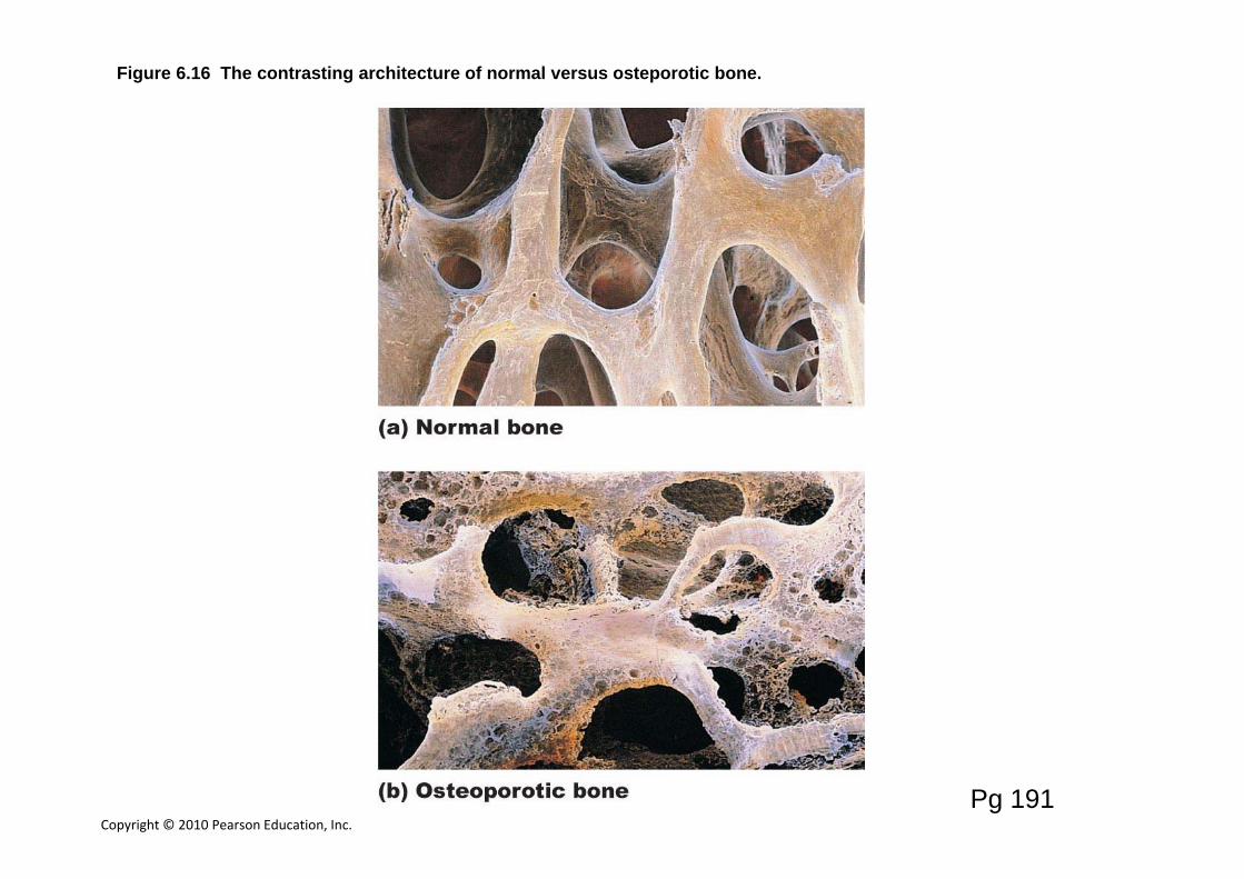
Figure 6.16 The contrasting architecture of normal versus osteporotic bone.
Pg 191
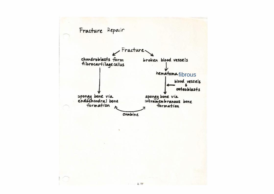
fibrous
Copyright © 2010 Pearson Education, Inc.
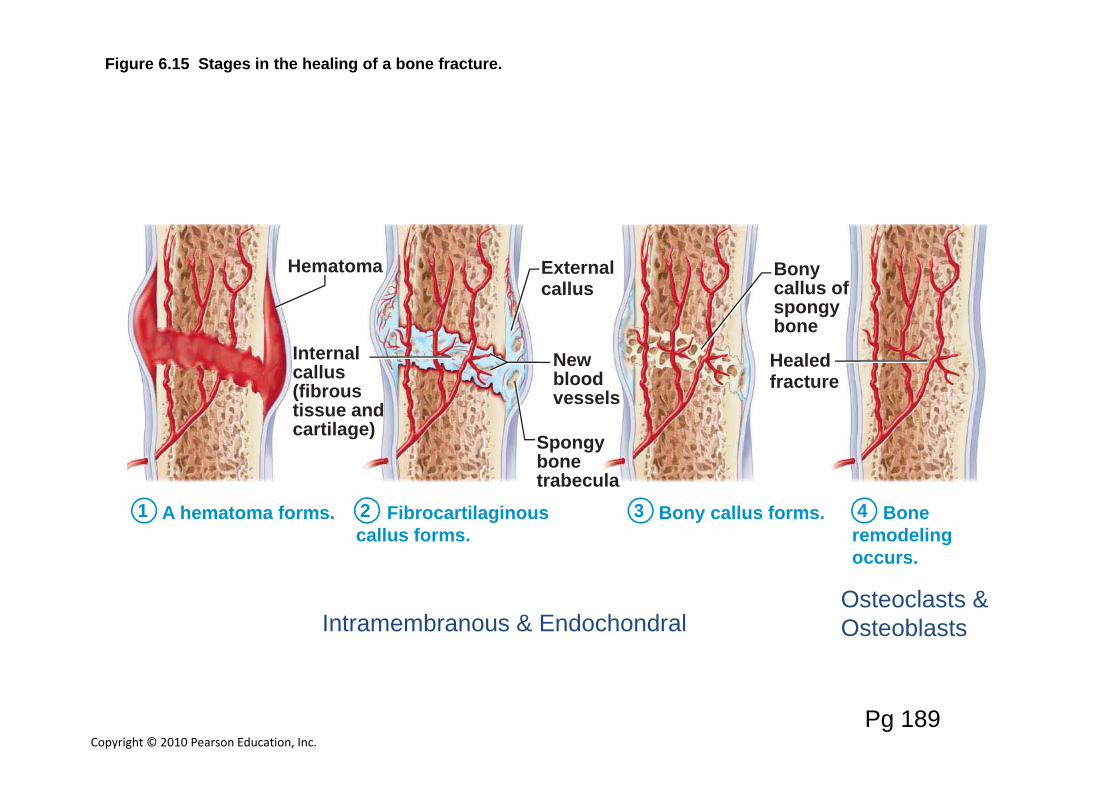
Figure 6.15 Stages in the healing of a bone fracture.
Hematoma Externalcallus
Bonycallus ofspongybone
Healedfracture
Newbloodvessels
Spongybonetrabecula
Internalcallus(fibroustissue andcartilage)
1 A hematoma forms. 2 Fibrocartilaginouscallus forms.
3 Bony callus forms. 4 Boneremodelingoccurs.
Pg 189
Intramembranous & EndochondralOsteoclasts & Osteoblasts


![TRIPLE TALAQ BILL: LACUNAE AND RECOMMENDATIONSnlujodhpur.ac.in/uploads/5 (2) NLUJ Law Review 49 (2018).pdfWinter, 2018] Triple Talaq Bill: Lacunae and Recommendations 53 between the](https://img.dokumen.tips/doc/110x75/5e758c0dbe319a7ca27f7695/triple-talaq-bill-lacunae-and-recom-2-nluj-law-review-49-2018pdf-winter-2018.jpg)