Embed Size (px)
Citation preview
JourmU of Chromatography, 325 (1985) 333-335 Elsevier Science Publishers B.V., Amsterdam - Printed in The Netherlands
CHROM. 17 697
Note
Chloral hydrate is compatible with isoelectric focusing
DIANE FQYNTER and MICHAEL LANDDN*
Department of Biochembtry, University of Nottingham Medical School, Queen’s Medical Centre. Notting- ham NG7 2UH (U.K.)
(Received March 4th, 1985)
Chloral hydrate has been found to be a potent non-ionic protein solvent and dissociating agent when used at high concentration (lOO%, w/v; 100 g chloral hydrate
I 4 8 12 16
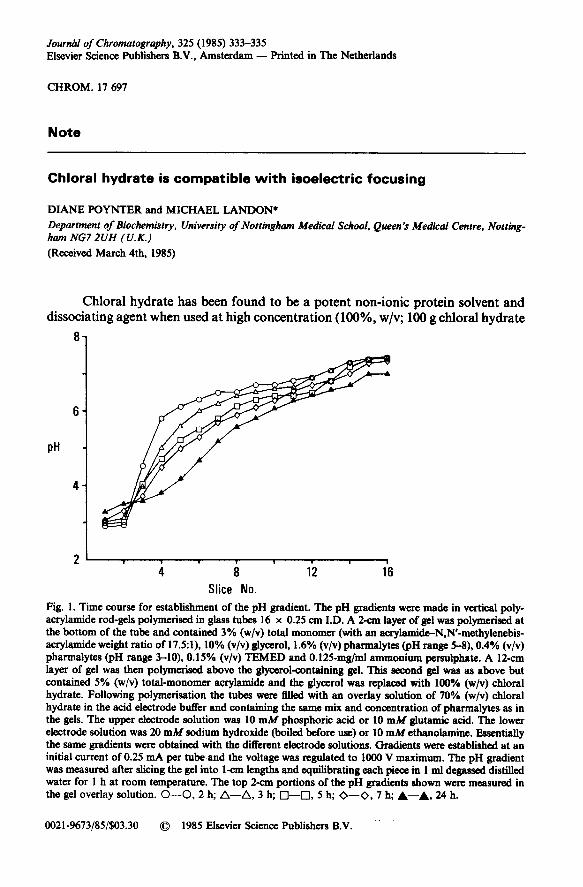
Slice No. Fig. 1. Time course for establishment of the pH gradient. The pH gradients were made in vertical poly- acrylamide rod-gels polymerised in glass tubes 16 x 0.25 cm I.D. A 2-cm layer of gel was polymer&d at the bottom of the tube and contained 3% (w/v) total monomer (with an acrylamide-N.N’-methylenebis- acrylamide weight ratio of 17.5:1), 10% (v/v) glycerol, 1.6% (v/v) pharmalytes @H range 5-8), 0.4% (v/v) pharmalytes (pH range 3-IO), 0.15% (v/v) TEMED and 0.125-mg/ml ammonium persulphate. A 12-cm layer of gel was then polymerised above the glycerol-containing gel. This second gel was as above but contained 5% (w/v) total-monomer acrylamide and the glycerol was replaced with 100% (w/v) chloral hydrate. Following polymer&ion the tubes were 6lled with an overlay solution of 70% (w/v) chloral hydrate in the acid electrode buffer and containing the same mix and concentration of pharmalytes as in the gels. The upper electrode solution was 10 mM phosphoric acid or 10 mM glutamic acid. The lower electrode solution was 20 mM sodium hydroxide (boiled before use) or 10 mM ethanolamine. Essentially the same gradients were obtained with the different electrode solutions. Gradients were established at an initial current of 0.25 mA per tubeand the voltage was regulated to 1000 V maximum. The pH gradient was measured after slicing the gel into l-cm lengths and equilibrating each piece in 1 ml &gassed distilled water for 1 h at room temperature. The top 2-cm portions of the pH gradients shown were measured in the gel overlay solution. O-O, 2 h; A-A, 3 h; q -_O, 5 h; O-O, 7 h; &--A, 24 h.
0021-9673/85/$03.30 0 1985 Elsevier Science Publishers B.V.
334 NOTES
made UP to 100 ml with water) in aqueous solution, and it has also been demonstrated to be compatible with polyacrylamide gel electrophoresisl+ and with ion-exchange chromatography5. However, in an investigation of its use in protein isoelectric fo- cusing (IEF) it has been suggested that it is unsuitable since it appears to bring about modification of ampholytes and protein@.
Here we show that 100% (w/v) aqueous choral hydrate can be used in IEF, albeit with certain constraints and limitations. Chloral hydrate at this high concen- tration yields a viscous solution which will reduce ion mobilities and experimental times must be extended to take this into account. Chloral hydrate should be recrys- tallised from chloroform prior to use in order to remove decomposition products. An aqueous solution of 100% (w/v) recrystallised chloral hydrate has a pH of about 4. Treatment with alkali will cause decomposition to chloroform and formic acid and should be avoided, as should mixing with urea, with which it forms a complex7.
. . cm
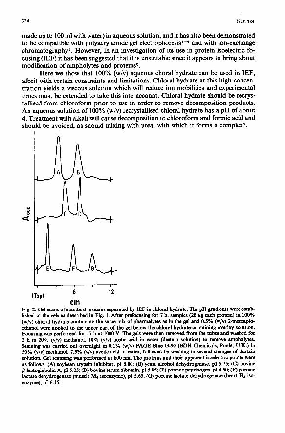
Fig. 2. Gel scans of standard proteins separated by IEF in chloral hydrate. The pH gradients were estab lished in the gels as described in Fig. 1. After prefocusing for 7 h, samples (20 pg each protein) in 100% (w/v) chloral hydrate containing the same mix of pharmalytes as in the gel and 0.5% (w/v) 2-mercapto- ethanol were applied to the upper part of the gel below the chloral hydrate-containing overlay solution. Focusing was performed for 17 h at 1000 V. The gels were then removed from the tubes and washed for 2 h in 20% (v/v) methanol, 10% (v/v) acetic acid in water (destain solution) to remove antpholyks. Staining was carried out overnight in 0.1% (w/v) PAGE Blue G-90 (BDH Chemicals, Poole, U.K.) in 50% (v/v) methanol, 7.5% (v/v) acetic acid in water, followed by washing in several changes of destain solution. Gel scarming was performed at 600 nm. The proteins and their apparent isoelectric points were as follows: (A) soybean trypsin inhibitor, p1 5.00, (B) yeast alcohol dehydrogenase, p1 5.75; (C) bovine /I-lactoglobulin A, pI 5.25; (D) bovine serum albumin, ~15.85; (E) porcine pepsinogen, ~14.50; (F) porcine lactate dehydrogenase (muscle Mq isoenzyme), ~15.65; (G) porcine lactate dehydrogenase (heart Hd iso- enzyme), p1 6.15.
NOT& 335
We have carried out IEF in polyacrylamide rod gels in the presence of 100% (w/v) chloral hydrate with mixtures of ampholytes (Pharmacia, Hounslow, U.K.) chosen to generate pH gradients that extend no higher than pH 7. Chloral hydrate is protected from contact with the alkaline electrolyte by the incorporation of a 2-cm length at the bottom of the gel with 10% (v/v) glycerol replacing chloral hydrate. An overlay (2 cm in depth) of 70% (w/v) chloral hydrate protects the top of the gel from the acidic electrolyte and prevents swelling in the upper part of the gel. In this way stable pH gradients can be generated in gels that can be conveniently handled. It is noteworthy that even at a high voltage the pH gradient is established slowly and several hours are required before linearity is achieved (Fig. 1). Once established the gradient shows reasonable s$bility for at least 24 h (Fig. 1). These long time periods are necessary because of the low mobilities of ions in solutions containing high con- centrations of chloral hydrate. The restricted gradient we obtained after 2 h is similar to that observed in the earlier work on IEF in chloral hydrate6 where experimental times were not extended beyond this period. Our attempts to extend pH gradients at the alkaline end by including higher pH-range ampholytes were not successful. The ability to achieve stable pH gradients in chloral hydrate using conventional am- pholytes suggests that no modification of these compounds is occurring.
We have established the suitability of our system of IEF by applying it to a number of proteins. For the same reasons as gradient formation is slow, proteins must be focused for long periods before an equilibrium position is obtained. The proteins we have used give sharply focused bands (Fig. 2).
The protein band patterns show no evidence of the partial chemical modifi- cation proposed in the previous study6. Storage of protein samples in chloral hydrate over a ‘period of several weeks did not affect the band pattern or location in the pH gradient. Our results fit with the compatibility of acidic solutions of chloral hydrate with proteins that has been established in earlier studies1*4.
In view of the known membrane solubilising’ and membrane protein disso- ciating power of chloral hydrate4 it would appear that IEF in chloral hydrate should be of value in the analysis of membrane protein complexes.
ACKNOWLEDGEMENT
D.P. wishes to thank the Science and Engineering Research Council for a Research Studentship.
REFERENCES
1 B. Ballou, G. Sundharadas and M. L. Bach, Science, 185 (1974) 531-533. 2 B. Ballou and 0. Smithies, Anal. Biochem., 80 (1977) 616-623. 3 A. G. Booth, Eiochem. J., 163 (1977) 165-168. 4 D. C. Griffin and M. Landon, Biochem. J., 197 (1981) 333-344. 5 D. C. Griffin and M. Landon, Biochem. J., 201 (1982) 227-231. 6 0. Brenna, E. Gianazza and P. G. Righetti, J. Chromatogr., 237 (1982) 293-296. 7 J. E. Fairbrother, in K. Florey (Editor), Analytical Profiles of Drug Substances, Vol. 2, Academic Press,
New York, 1973, 85-143. pp.