Embed Size (px)
Citation preview

439
1. Exposure Data
1.1 Chemical and physical data
1.1.1 Nomenclature
(a) Chloral
Chem. Abstr. Serv. Reg. No.: 75-87-6
Chem. Abstr. Serv. Name: Trichloroacetaldehyde
IUPAC Systematic Name: Chloral
Synonyms: Anhydrous chloral; 2,2,2-trichloro acetaldehyde; trichloro-ethanal; 2,2,2-tri-chloroethanal
(b) Chloral hydrate
Chem. Abstr. Serv. Reg. No.: 302-17-0
Deleted Chem. Abstr. Serv. Reg. No.: 109128-19-0
Chem. Abstr. Serv. Name: 2,2,2-Trichloro-1,1-ethanediol
IUPAC Systematic Name: Chloral hydrate
Synonyms: Chloral monohydrate; trichloroacetaldehyde hydrate; trichloroacetaldehyde monohydrate; 1,1,1-trichloro-2,2-dihydroxyethane
1.1.2 Structural and molecular formulae and relative molecular mass
(a) Chloral
C C H
O
Cl
Cl
Cl
C2HCl3O
Relative molecular mass: 147.39
(b) Chloral hydrate
C C H
OH
Cl
Cl
Cl
OH
C2H3Cl3O2
Relative molecular mass: 165.40
CHLORAL AND CHLORAL HYDRATEChloral and chloral hydrate were considered by previous IARC Working Groups in 1995 and 2004 (IARC, 1995, 2004). New data have since become available and these have been taken into consideration in the present evaluation. Chloral and chloral hydrate are considered together since the two substances exist in equilibrium in aqueous solution.

IARC MONOGRAPHS – 106
440
1.1.3 Chemical and physical properties of the pure substances
(a) Chloral
Description: Oily liquid. Pungent, irritating odour (O’Neil et al., 2006)
Boiling-point: 97.8 °C (O’Neil et al., 2006)
Melting-point: –57.5 °C (O’Neil et al., 2006)
Density: 1.510 at 20 °C/relative to H2O at 4 °C (O’Neil et al., 2006)
Spectroscopy data: Infrared (prism [4626, 4426]), ultraviolet [5–3], nuclear magnetic resonance [8241] and mass [814] spectral data have been reported (Weast & Astle, 1985; Sadtler Research Laboratories, 1991)
Solubility: Freely soluble in water, in which it is converted to chloral hydrate; soluble in diethyl ether and ethanol (O’Neil et al., 2006)
Volatility: Vapour pressure, 10 kPa at 33.8 °C (Haynes, 2012)
Stability: Polymerizes under the influence of light and in presence of sulfuric acid to form a white solid trimer called metachloral (O’Neil et al., 2006)
Conversion factor: mg/m3 = 6.03 × ppm, calculated from: mg/m3 = (relative molec-ular mass/24.45) × ppm, assuming normal temperature (25 °C) and pressure (101 kPa).
(b) Chloral hydrate
Description: Colourless, transparent, or white crystals with aromatic, penetrating and slightly acrid odour, slightly bitter, caustic taste. (O’Neil et al., 2006)
Boiling-point: 96 °C, decomposes into chloral and water (Haynes, 2012)
Melting-point: 57 °C (O’Neil et al., 2006)
Density: 1.9081 at 20 °C/4 °C (Haynes, 2012)
Spectroscopy data: Infrared (prism [5423]), nuclear magnetic resonance [10 362] and mass [1054] spectral data have been reported (Weast & Astle, 1985; Sadtler Research Laboratories, 1991)
Solubility: Very soluble in water, olive oil. Freely soluble in acetone, methyl ethyl ketone (O’Neil et al., 2006)
Volatility: Vapour pressure, 4.7 kPa at 20 °C; slowly evaporates on exposure to air (Jira et al., 1986; O’Neil et al., 2006)
Octanol/water partition coefficient (P): Log P, 0.99 (Hansch et al., 1995)
Conversion factor: mg/m3 = 6.76 × ppm, calculated from: mg/m3 = (relative molec-ular mass/24.45) × ppm, assuming normal temperature (25 °C) and pressure (101 kPa)
1.1.4 Technical products and impurities
Technical-grade chloral ranges in purity from 94% to 99% by weight, with water being the main impurity. Other impurities can include chloro-form, hydrogen chloride, dichloroacetaldehyde and phosgene (Jira et al., 1986).
Trade names for chloral include Grasex and Sporotal 100.
Trade names for chloral hydrate include: Ansopal, Aquachloral, Chloradorm, Chloraldurat, Chloralix, Dormel, Elix-nocte, Escre, Hydral, Lanchloral, Lorinal, Medianox, Nervifene, Noctec, Novochlorhydrate, Nycton, Phaldrone, Rectules, Somnos, Suppojuvent Sedante, Tosyl, Trawotox and Welldorm.
The United States Pharmacopeia (USP) specifies that USP-grade chloral hydrate must contain not less than 99.5% chloral hydrate (US Pharmacopeial Convention, 2012). Chloral hydrate is available as a liquid-filled capsule containing 500 mg of chloral hydrate and as a syrup containing 500 mg/5 mL (PDR Network, 2012).

Chloral and chloral hydrate
441
1.1.5 Analysis
Methods for the analysis of chloral hydrate have been reviewed by Delinsky et al. (2005) and Demeestere et al. (2007). Selected methods for the analysis of chloral hydrate in water are identified in Table 1.1. A biomonitoring method using gas chromatography-electron capture detection has been developed for measuring chloral hydrate in blood (Schmitt, 2002) and in urine (Garrett & Lambert, 1966).
1.2 Production and use
1.2.1 Production process
(a) Manufacturing processes
Chloral was first synthesized by J. von Liebig in 1832 by chlorination of ethanol (Jira et al., 2007).
Chloral is produced by chlorinating acetal-dehyde or ethanol in acidic solution by gradually increasing the temperature from 0 °C to 90 °C (Jira et al., 2007). Antimony trichloride is some-times used as a catalyst. Chloral is distilled from the reaction mixture as the hydrate. The hydrate is then mixed with concentrated sulfuric acid, the heavier acid layer is drawn off, and chloral is distilled through a fractionating column of moderate height.
(b) Production volume
Estimated production and use of chloral in the Member States of the European Union in 1984 was 2500 tonnes (Environmental Chemicals Data and Information Network, 1993).
Chloral (anhydrous) is known to have been produced by 14 companies in China, seven companies in India and one company each in Brazil, France, Japan, Mexico, the Russian Federation and the USA. Chloral hydrate was produced by four companies in China, three companies in Germany, two companies in Japan and one company each in Mexico, the Russian Federation and Spain (Chemical Information Services, 2002).
Chloral hydrate has been produced for use as a hypnotic drug in relatively low and gradually declining volume for many years. As an indica-tion of the scale, production in the USA for this purpose was about 135 tonnes in 1978 (Jira et al., 1986).
1.2.2 Use
(a) Chloral
The principal historical use of chloral has been in the production of the insecticide dichloro-diphenyltrichloroethane (DDT) and, to a lesser extent, other insecticides such as methoxychlor, naled, trichlorfon, and dichlorvos and the herbi-cide trichloroacetic acid (Jira et al., 2007). In the USA in 1975, about 40% of chloral was used in the manufacture of DDT, about 10% in the manufac-ture of other pesticides and about 50% in other applications (IARC, 1995). After the banning of DDT in many countries, demand for chloral for this use has declined dramatically.
Chloral has also been used in the production of rigid polyurethane foam (IARC 1995; Boĭtsov et al., 1970) and to induce swelling of starch
Table 1.1 Methods for the analysis of chloral hydrate in water
Sample preparation Assay procedure Limit of detection Reference
Extract with methyl-t-butyl ether or pentane
GC/ECD 0.002 µg/L EPA (1995)
Extract with ethyl acetate at pH 3.2 GC/MS 0.006 µg/L Serrano et al. (2011)ECD, electron capture detection; GC, gas chromatography; MS, mass spectrometry

IARC MONOGRAPHS – 106
442
granules at room temperature (Whistler & Zysk, 1978).
(b) Chloral hydrate
Chloral hydrate has been used as a hypnotic drug since the 1870s, principally for the short-term treatment of insomnia. It was also used to allay anxiety and to induce sedation and/or sleep post-operatively, before electroencephalo-gram evaluations, and to treat the symptoms of withdrawal of alcohol and other drugs such as opiates and barbiturates. For many years chloral hydrate was widely used for the sedation of chil-dren before diagnostic, dental or medical proce-dures, but although still in use, it has largely been replaced by newer drugs with a lower risk of overdose (Pershad et al., 1999).
After oral administration, chloral hydrate is converted rapidly to trichloroethanol, which is largely responsible for its hypnotic action. Externally, chloral hydrate has a rubefacient action (producing redness of the skin) and has been used as a counter-irritant. It is administered by mouth as a liquid or as gelatin capsules. It has also been dissolved in a bland fixed oil and given by enema or as suppositories (Gennaro, 2000; Royal Pharmaceutical Society of Great Britain, 2002).
Chloral hydrate is also an ingredient in Hoyer’s solution, which is used in microscopy to mount organisms such as bryophytes, ferns, seeds, and arthropods (Anderson, 1954).
1.3 Occurrence and exposure
1.3.1 Natural occurrence
Chloral and chloral hydrate are not known to occur as natural products.
1.3.2 Environmental occurrence
(a) Air
No data are available on human exposure to chloral or chloral hydrate in air. The low vola-tility of chloral hydrate from a water solution precludes significant exposure by inhalation (EPA, 2000).
(b) Water
When raw water, containing natural organic material such as humic, tannic or amino acids, is treated by chlorination for use as drinking-water or in swimming pools, by-products resulting from disinfection can be formed, such as chloral, which is rapidly transformed into chloral hydrate (Miller & Uden, 1983; Sato et al., 1985; Trehy et al., 1986; Italia & Uden, 1988). Chloral hydrate is primarily produced by the treatment of water with chlorine or chloramine, but the use of pre-ozonation before chlorination or chlora-mination can favour its formation (Richardson et al., 2007).
Table 1.2 summarizes recent measurements of chloral hydrate in drinking-water and swim-ming pool water in several countries.
1.3.3 Occupational exposure
The National Occupational Exposure Survey conducted between 1981 and 1983 indicated that 11 278 employees in the USA were potentially exposed to chloral hydrate (NIOSH, 1994). The estimate is based on a survey of companies and did not involve measurement of actual exposures (IARC, 2004).
Chloral has been detected in the work envi-ronment during spraying and casting of poly-urethane foam (Boĭtsov et al., 1970). It has also been identified as an autoxidation product of trichloroethylene during extraction of vegetable oil (McKinney et al., 1955), and detected in the output of etching chambers in semiconductor processing (Ohlson, 1986).

Chloral and chloral hydrate
443
In spite of the use of chloral as an intermediate in the synthesis of insecticides and herbicides, no specific measurement data from workers exposed during synthesis or formulation were available to the Working Group. Given the use of chloral hydrate as a sedative and hypnotic drug, workers could be exposed in the pharmaceutical industry during production. However no data on exposure measurement were identified by the Working Group.
1.3.4 Exposure in the general population
Chloral is an intermediate metabolite of trichloroethylene in humans, and chloral hydrate has been found in the plasma of people who have undergone anaesthesia with trichloroethylene (Cole et al., 1975; Davidson & Beliles, 1991).
1.4 Regulations and guidelines
Chloral hydrate is a controlled substance in Canada and the USA, available by prescription only. Chloral hydrate is banned for marketing
in India. There are no occupational limits for chloral or chloral hydrate.
2. Cancer in Humans
Chloral hydrate is a chemical that occurs in drinking-water and swimming pools as part of a mixture of by-products resulting from disin-fection of drinking-water by chlorination. The chemicals in water-disinfection by-products do not occur in an isolated manner and there is no epidemiological evidence on risk of cancer associated specifically with these by-products. A detailed description of water-disinfection by-products and cancer risk is given in IARC Monograph Volume 101 (IARC, 2012a).
Only one epidemiological study has exam-ined risk of cancer in humans exposed to chloral hydrate. Haselkorn et al. reported cancer morbidity among 2290 users of chloral hydrate within a cohort of 143 574 patients at Kaiser Permanente, USA, who had prescriptions filled
Table 1.2 Concentrations of chloral hydrate in drinking-water
Country Location Concentration (µg/L) Reference
Mean Range
Drinking-water Australia Seven cities NR 0.2–19 Simpson & Hayes (1998)Canada Country-wide
(summer 1993)6.1 < 0.1–18.9 Koudjonou et al. (2008)3.6 0.3–13.68.4 0.2–23.4
China Beijing 0.93 NR–10.44 Wei et al. (2010)Greece Athens NR 0.2–12.5 Golfinopoulos & Nikolaou (2005) Mytilene NR NR–0.5 Leivadara et al. (2008)Spain Eleven provinces < 1a < 1–12.1 Villanueva et al. (2012) Cordoba NR 1.2–38 Serrano et al. (2011)Swimming-pool water Republic of Korea Seoul 16.9 5.1–34.9 Lee et al. (2010)
3.6 ND–10.410.2 ND–23.4
Spain Cordoba NR 53–340 Serrano et al. (2011)a MedianND, not detected; NR, not reported
IARC MONOGRAPHS – 106
444
for the 215 most commonly used drugs between 1969 and 1973 (Haselkorn et al., 2006). Study subjects in this cohort comprised an ethnically and socioeconomically diverse population, who had received both inpatient and outpatient care within the prepaid system. Cohort members were linked to the pharmacy records by each patient’s unique medical record number. Cancer occurrence (1976–1998) for each study subject was ascertained via the local tumour registry and hospital records of all northern California hospitals in the Kaiser Permanente programme. Cancers diagnosed were verified by the review of medical records by a trained medical analyst. Expected numbers of overall and specific cancer were calculated based on age and sex standard-ized rates from the entire Kaiser Permanente cohort. By 1998, there were 285 cancer cases observed versus 258 expected [standardized inci-dence ratio, SIR, 1.1; 95% CI, 0.98–1.24]. Further, observed and expected numbers for cancers of mouth floor [SIR, 5.0; 95% CI, 1.03–14.6], stomach [SIR, 2.1; 95% CI, 1.2–3.3], lung [SIR, 1.3; 95% CI, 0.9–1.7], skin melanoma [SIR, 1.4; 95% CI, 0.7–2.4], prostate [SIR, 1.0; 95% CI, 0.7–1.4] and kidney [SIR, 0.2; 95% CI, 0.05–1.1] are reported. Relations between numbers of prescriptions of chloral hydrate and overall cancer, cancer of lung, stomach, prostate and melanomas were examined in a series of nested case–control studies, adjusted for potential confounders. No significant trends were observed. [The Working Group noted the relatively low power of this study to detect statistically significant deviations from unity for specific cancers].
3. Cancer in Experimental Animals
See Table 3.1
3.1 Mouse
Oral administration
Three groups of 20–35 male C57BL × C3HF1 mice (age, 15 days) were given chloral hydrate in water intragastrically as a single dose at 0, 5, or 10 mg/kg body weight (bw). The mice were killed at various time intervals up to 92 weeks. Liver nodules were examined by histopathology and included hyperplastic nodules, hepatocel-lular adenoma and hepatocellular carcinoma [termed trabecular]. In mice killed between 48 and 92 weeks after dosing, a statistically signif-icant increase in the incidence of hepatocellular adenoma or carcinoma (combined) was observed in mice receiving chloral hydrate at a dose of 10 mg/kg bw (6 out of 8 versus 2 out of19 controls) (Rijhsinghani et al. (1986)). [The Working Group noted the small number of mice evaluated in this study and the low doses administered.]
The hepatocarcinogenicity of chloral hydrate was studied in two groups of 33–40 male B6C3F1 mice given drinking-water containing chloral hydrate at a dose of 0 or 1 g/L (ingested dose, 166 mg/kg bw per day) for 104 weeks (Daniel et al., 1992). Five animals per group were killed after 30 and 60 weeks of exposure. Microscopic examina-tions were performed on all gross lesions, liver, kidney, testis and spleen of mice surviving 104 weeks. Statistically significant increases in the incidence of hepatocellular adenoma, hepatocel-lular carcinoma, and hepatocellular adenoma or carcinoma (combined) were observed in treated mice surviving 104 weeks. Results are shown in Table 3.1. [The Working Group noted that a single dose was used, the group size was small, histopathological evaluation was limited to mice surviving at least 104 weeks, and the histopatho-logical examinations were limited to the liver.]
The initial study by Daniel et al. (1992) was followed up with a study in groups of 72 male B6C3F1 mice given repeated doses of chloral hydrate for 104 weeks (George et al., 2000).

Chloral and chloral hydrate
445
Tabl
e 3.
1 St
udie
s of
car
cino
geni
city
in e
xper
imen
tal a
nim
als
give
n ch
lora
l hyd
rate
by
oral
adm
inis
trat
ion
Spec
ies,
stra
in (s
ex)
Dur
atio
n R
efer
ence
Dos
ing
regi
men
A
nim
als/
grou
p at
star
tIn
cide
nce
of tu
mou
rsSi
gnifi
canc
eC
omm
ents
Mou
se, C
57BL
×C3H
F 1 (M
) U
p to
92
wk
Rijh
sing
hani
et a
l. (1
986)
Sing
le d
ose,
by
gava
ge a
t 0, 5
, 10
mg/
kg b
w, to
mic
e ag
ed 1
5 da
ys
35, 2
5, a
nd 2
0/gr
oup
Hep
atic
nod
ules
[hep
atoc
ellu
lar a
deno
ma
or c
arci
nom
a (c
ombi
ned)
]: 2
/19,
2/9
, 6/8
*
*P <
0.0
5La
bora
tory
gra
de
Smal
l num
bers
of m
ice
and
low
do
se
Mou
se, B
6C3F
1 (M
) 10
4 w
k D
anie
l et a
l. (1
992)
0, 1
g/L
, in
drin
king
-wat
er
33, 4
0/gr
oup
Inte
rim
kill
s of 1
0/gr
oup
Fi
sher
’s ex
act t
est
Puri
ty, >
95%
H
isto
path
olog
ical
dat
a on
ly fo
r th
e liv
erH
epat
ocel
lula
r ade
nom
a:
1/20
, 7/2
4**P
≤ 0
.05
Hep
atoc
ellu
lar c
arci
nom
a:
2/20
, 11/
24**
**P
≤ 0.
03
Hep
atoc
ellu
lar a
deno
ma
or c
arci
nom
a (c
ombi
ned)
: 3/
20, 1
7/24
***
***P
≤ 0
.01
Mou
se, B
6C3F
1 (M
) 10
4 w
k G
eorg
e et
al.
(200
0)
0, 0
.12,
0.5
8, 1
.28
g/L,
in
drin
king
-wat
er
72/g
roup
Prev
alen
ce in
mic
e su
rviv
ing
> 78
wk:
H
epat
ocel
lula
r ade
nom
a:
21.4
(n =
42)
, 43.
5 (n
= 4
6)*,
51.3
(n =
39)
*, 50
.0%
(n =
32)
* H
epat
ocel
lula
r car
cino
ma:
54
.8 (n
= 4
2), 5
4.3
(n =
46)
, 59.
0 (n
= 3
9),
84.4
% (n
= 3
2)*
Hep
atoc
ellu
lar a
deno
ma
or c
arci
nom
a (c
ombi
ned)
: 64
.3 (n
= 4
2), 7
8.3
(n =
46)
, 79.
5 (n
= 3
9)*,
90.6
% (n
= 3
2)*
*P ≤
0.0
5Pu
rity
, > 9
9%
Mea
sure
d co
ncen
trat
ions
; hi
stop
atho
logi
cal d
ata
only
for
the
liver
; neo
plas
ms o
bser
ved
in th
e ki
dney
, spl
een
and
test
is
repo
rted
not
to e
xcee
d th
e in
cide
nces
in c
ontr
ol g
roup
or
hist
oric
al c
ontr
ols.
Mou
se, B
6C3F
1 (M
) 12
or 2
0 m
onth
s Vo
n Tu
ngel
n et
al.
(200
2)
12 m
onth
s Tw
o i.p
. dos
es a
t age
8 a
nd
15 d
ays;
tota
l dos
e, 0
(DM
SO
cont
rol)
or 2
000
nmol
per
m
ouse
24
, 24/
grou
p
12 m
onth
s H
epat
ocel
lula
r ade
nom
a:
1/24
, 5/2
4
NS
Puri
ty, N
R Sm
all n
umbe
rs o
f mic
e; lo
w to
tal
dose
; his
topa
thol
ogy
rest
rict
ed
to th
e liv
er. F
emal
es w
ere
also
in
ject
ed w
ith c
hlor
al h
ydra
te a
nd
no h
epat
ocel
lula
r tum
ours
wer
e ob
serv
ed.
20 m
onth
s Tw
o i.p
. dos
es a
t age
8 a
nd
15 d
ays;
tota
l dos
e, 0
(DM
SO
cont
rol)
or 1
000
nmol
per
m
ouse
23
, 23/
grou
p
20 m
onth
s H
epat
ocel
lula
r ade
nom
a:
6/23
, 9/2
3 H
epat
ocel
lula
r car
cino
ma:
2/
23, 2
/23
Hep
atoc
ellu
lar a
deno
ma
or c
arci
nom
a (c
ombi
ned)
: 7/
23, 1
0/23
NS

IARC MONOGRAPHS – 106
446
Spec
ies,
stra
in (s
ex)
Dur
atio
n R
efer
ence
Dos
ing
regi
men
A
nim
als/
grou
p at
star
tIn
cide
nce
of tu
mou
rsSi
gnifi
canc
eC
omm
ents
Mou
se, B
6C3F
1 (M
) 10
4 w
k N
TP (2
002a
),
Leak
ey et
al.
(200
3)
Gav
age
0, 2
5, 5
0, 1
00 m
g/kg
bw
5 d
ays/
wk
by g
avag
e to
ad
libitu
m-fe
d or
die
tary
con
trol
led
mic
e.
48/g
roup
Fed
ad li
bitu
mPo
ly-3
test
Puri
ty, >
99.
5%
No
trea
tmen
t-rel
ated
redu
ctio
n in
surv
ival
Hep
atoc
ellu
lar c
arci
nom
a:
4/48
, 10/
48, 1
0/47
, 7/4
8 H
epat
ocel
lula
r ade
nom
a or
car
cino
ma
(com
bine
d):
16/4
8, 2
5/48
*, 23
/47,
22/4
8
*P =
0.0
437
**P
= 0.
0371
(tr
end)
**
*P =
0.0
422
****
P =
0.04
5 (tr
end)
Die
tary
cont
rolle
d H
epat
ocel
lula
r car
cino
ma:
2/
48**
, 5/4
8, 4
/48,
8/4
8***
H
epat
ocel
lula
r ade
nom
a or
car
cino
ma
(com
bine
d):
11/4
8***
*, 11
/48,
14/
48, 1
8/48
Mou
se, B
6C3F
1 (F)
10
4 w
k N
TP (2
002b
)
Regi
men
A
Gav
age;
0, 2
5, 5
0, 1
00 m
g/kg
bw
, 5 d
ays/
wk
48/g
roup
Pitu
itary
gla
nd (p
ars d
ista
lis) a
deno
ma:
0/
45*,
2/44
, 0/4
7, 5/
41**
M
alig
nant
lym
phom
a:
9/48
***,
7/48
, 8/4
8, 1
5/48
Poly
-3 te
st
*P =
0.0
73 (t
rend
) **
P =
0.02
4 **
*P =
0.0
455
(tren
d)
Puri
ty, 9
9%
No
expo
sure
-rel
ated
redu
ctio
n in
su
rviv
al.
Rat,
F344
/N (M
) 10
4 w
k G
eorg
e et
al.
(200
0)
0, 0
.12,
0.5
8, 2
.51
g/L,
in
drin
king
-wat
er
78/g
roup
Prev
alen
ce in
rats
surv
ivin
g >
78 w
k:
Hep
atoc
ellu
lar a
deno
ma:
0
(n =
42)
, 7.1
(n =
44)
, 2.3
(n =
44)
, 4.5
%
(n =
42)
H
epat
ocel
lula
r car
cino
ma:
2.
4 (n
= 4
2), 7
.1 (n
= 4
4), 0
(n =
44)
, 2.3
%
(n =
42)
H
epat
ocel
lula
r ade
nom
a or
car
cino
ma
(com
bine
d):
2.4
(n =
42)
, 14.
3 (n
= 4
4), 2
.3 (n
= 4
4),
6.8%
(n =
42)
NS
Puri
ty, >
99%
M
easu
red
conc
entr
atio
ns;
hist
opat
holo
gica
l dat
a on
ly fo
r th
e liv
er; n
eopl
asm
s obs
erve
d in
the
kidn
ey, s
plee
n an
d te
stes
re
port
ed n
ot to
exc
eed
the
inci
denc
es in
con
trol
gro
up o
r hi
stor
ical
con
trol
s.
bw, b
ody
wei
ght;
F, fe
mal
e; i.
p., i
ntra
peri
tone
al; M
, mal
e; m
o, m
onth
; NR
, not
repo
rted
; NS,
not
sign
ifica
nt; w
k, w
eek
Tabl
e 3.
1 (
cont
inue
d)
Chloral and chloral hydrate
447
Measured concentrations of chloral hydrate in the drinking-water were 0, 0.12, 0.58, and 1.28 g/L, corresponding to mean daily doses of 0, 13.5, 65, and 146.6 mg/kg bw. Six mice per group were killed after 26, 52, and 78 weeks. Histopathological examinations of the liver, kidney, spleen and testis were performed on mice surviving more than 78 weeks. Hepatocellular adenomas and carcinomas were observed as early as 52 weeks and incidence increased progressively with duration of treatment. In mice surviving more than 78 weeks, the incidence of hepatocellular adenoma was increased (P ≤ 0.05) in all dose groups, the incidence of hepatocellular carcinoma was increased in the group at 1.28 g/L, and the incidence of hepatocellular adenoma or carcinoma (combined) was increased in the groups at 0.58 and 1.28 g/L. The incidences of neoplasms observed in the kidney, spleen, and testes were reported not to exceed those in the control group or in historical controls (data not shown). [The Working Group noted that the evaluations of tumour incidence were limited to the four organs examined microscopically.]
Groups of 24 male and 21 female neonatal B6C3F1 mice were given intraperitoneal injec-tions of chloral hydrate in dimethyl sulfoxide (DMSO) on postnatal day 8 (three sevenths of the total dose) and day 15 (four sevenths of the total dose) (Von Tungeln et al., 2002). The total dose administered was 2000 nmol [0.33 mg] per mouse, and the mice were observed for 12 months. In a second experiment, groups of 23 males and 22 females were given intraperitoneal injections of chloral hydrate in DMSO on post-natal day 8 (one third of the total dose) and day 15 (the remaining two thirds of the total dose). In this experiment, the total dose administered was 1000 nmol [0.165 mg] per mouse and the groups were observed for 20 months. Control groups of 23–24 mice were injected with the vehicle (DMSO) only. Histopathological examination was restricted to the liver. In males, a non-sta-tistically significant increase in the incidence of
hepatocellular adenoma was observed after 12 months and 20 months (5 out of 24 versus 1 out of 24, and 9 out of 23 versus 6 out of 23, respec-tively). No hepatocellular tumours were observed in the control or treated groups of female mice. [The Working Group noted that only the liver was examined microscopically and that the total doses administered were much lower than those used in other long-term studies of this chemical.]
The National Toxicology Program performed two studies of the carcinogenic effects of chloral hydrate in B6C3F1 mice (NTP, 2002a, b). The first study (NTP, 2002a; Leakey et al., 2003) was an investigation of the influence of dietary restriction on the development of hepatocellular adenoma and hepatocellular carcinoma. Groups of 48 male B6C3F1 mice were given chloral hydrate (dissolved in distilled water) by gavage at a dose of 0, 25, 50, or 100 mg/kg bw, 5 days per week for 104 weeks. The incidences of hepatocellular adenoma and hepatocellular carcinoma were much lower in mice with a controlled diet than in mice fed ad libitum. There was a statistically significant increase in the incidence of hepato-cellular adenoma or carcinoma (combined) in mice fed ad libitum at 25 mg/kg bw, but not at 50 or 100 mg/kg bw. There was a statistically significant increase in the incidence of hepa-tocellular carcinoma in mice with a controlled diet containing chloral hydrate at 100 mg/kg bw (Table 3.1). [The Working Group noted that the mice in this study were dosed for 5 days per week, while the doses in studies by Daniel et al. (1992) and George et al. (2000) were higher and were administered 7 days per week.]
In the second study (NTP, 2002b), female B6C3F1 mice were given chloral hydrate (dissolved in distilled water) by gavage in two different regi-mens. In regimen A, groups of 48 mice were given chloral hydrate at a dose of 0, 25, 50, or 100 mg/kg bw, 5 days per week for 104 weeks (24 months). In regimen B, groups of 48 mice were given chloral hydrate at a dose of 0 (n = 24) or 100 mg/kg bw for 3, 6 or 12 months. Eight mice each from the
IARC MONOGRAPHS – 106
448
groups at 0 and 100 mg/kg bw were killed at 3, 6, or 12 months. The remaining mice were held until termination of the study at 24 months. For regimen A, an increasing trend in incidence of adenoma of the pituitary gland (pars distalis) in the group at the highest dose was observed in mice treated for 24 months. A statistically signif-icant increasing trend in incidence of malignant lymphoma was also observed, but the incidences of hepatocellular adenoma, hepatocellular carci-noma, or hepatocellular adenoma or carcinoma (combined) were not increased. For regimen B, tumour incidences were not increased in female mice exposed to chloral hydrate for 3, 6, or 12 months. [The Working Group noted that mice in this study were dosed for 5 days per week, while the studies by Daniel et al. (1992) and George et al. (2000) used higher doses that were admin-istered 7 days per week.]
3.2 Rat
Oral administration
Groups of 50 male and 50 female Sprague-Dawley rats were given drinking-water containing chloral hydrate at nominal doses of 0, 0, 15, 45, and 135 mg/kg bw (Leuschner & Beuscher, 1998). The rats were examined after 124 weeks (males) or 128 weeks (females) of exposure. A statistically significantly increase in hepatic hypertrophy was reported in the group of male rats at the highest dose. It was reported that the incidence of neoplastic lesions was not increased in treated males or females compared with rats in the control groups. [The Working Group noted that the lack of incidence data in this paper hampered evaluation of the study.]
Groups of 78 male F344/N rats were given drinking-water containing chloral hydrate at measured concentrations of 0, 0.12, 0.58, or 2.51 g/L (George et al., 2000). These treat-ments provided mean daily doses of 0, 7.4, 37.4, and 162.6 mg/kg bw. Six rats per group were
killed after 13, 26, 52, and 78 weeks. Survivors were maintained on treatment for 104 weeks. Histopathological examination of the liver, kidney, spleen and testis was performed on rats surviving more than 78 weeks. There were no treatment-related increases in the incidence of hepatocellular adenoma, hepatocellular carci-noma, or hepatocellular adenoma or carcinoma (combined). However, 3 out of 44 (7%) of rats receiving the lowest dose developed hepatocel-lular carcinomas, while the incidence of hepa-tocellular carcinomas for historical controls in this strain of rats was only 19 out of 2255 (0.8%) (Haseman et al., 1998). The incidences of neoplasms of the kidney, spleen, and testis were reported not to exceed those observed in the control group or historical controls (data not shown). [The Working Group noted that the evaluations of tumour incidence were limited to the four organs examined microscopically.]
4. Mechanistic and Other Relevant Data
4.1 Toxicokinetic data
4.1.1 Absorption
(a) Humans
Studies in humans have shown that chloral hydrate is rapidly absorbed after oral admin-istration, with peak concentrations in blood occurring within 1 hour after dosing (Merdink et al., 2008). The extent of oral absorption has not been measured precisely in humans, but is likely to be high due to the rapidity of absorp-tion. Recovery of metabolites in urine, which represents a lower boundary on absorption, has been reported to be between 47% and 60% for subjects followed for 1 week after administration of a single oral dose (Müller et al., 1974; Merdink et al., 2008). It should be noted that 1 week is
Chloral and chloral hydrate
449
insufficient for complete urinary excretion of the metabolites of chloral hydrate. With repeated oral dosing, Owens & Marshall (1955) reported high inter-individual variation in recovery, with average daily excretion ranging from 7% to 94% of the daily dose of chloral hydrate.
No data were available to the Working Group on absorption in humans via other routes of exposure.
(b) Experimental systems
Orally administered chloral hydrate is rapidly absorbed in rats and mice, with peak concentra-tions reported at 15 minutes after administration (Beland et al., 1998). The extent of oral absorption has not been measured precisely in experimental systems, but is likely to be high due to the rapidity of absorption. The percentage absorption was not estimated since no studies on urinary excretion or mass balance for orally administered chloral hydrate were available to the Working Group.
No experimental data on absorption via other routes of exposure were available to the Working Group.
4.1.2 Distribution
(a) Humans
In humans, orally administered chloral hydrate enters the liver where it undergoes exten-sive metabolism (see Section 4.1.3); only a limited amount enters the systemic circulation (Merdink et al., 2008). Chloral hydrate in the blood is rapidly eliminated by metabolism, as shown by Zimmermann et al. (1998), who reported a half-life of less than 1 hour, and Merdink et al. (2008), who reported a rapid initial decline with a terminal half-life of about 10 hours. [Although no data on distribution in human tissues were available, the Working Group noted that due to the extensive first-pass effect, it was likely that concentrations of chloral hydrate were much lower in extra-hepatic tissues than in the liver of humans exposed orally.]
No data on distribution via other routes of administration were available to the Working Group.
(b) Experimental systems
In rats and mice, orally administered chloral hydrate enters the liver where it undergoes exten-sive metabolism (see Section 4.1.3); only a limited amount enters the systemic circulation (Beland et al., 1998). Chloral hydrate in the blood is rapidly eliminated, probably by metabolism, with levels reported to be below the limit of detection within 3 hours after oral administration (Beland et al., 1998). [Although no data on tissue distribution were available, the Working Group noted that due to the extensive first-pass effect, it was likely that concentrations of chloral hydrate in extra-hepatic tissues were much lower than those in the liver of rodents exposed orally.]
The half-life of chloral hydrate after intra-venous administration has been reported to be about 5–20 minutes in mice and rats (Abbas et al., 1996; Merdink et al., 1999), indicating rapid elimination from the systemic circulation. Abbas et al. (1996) reported concentrations of chloral hydrate in the liver to be about one quarter to one half of concentrations in the blood. No data on distribution to other tissues after intravenous administration were available to the Working Group.
No data on distribution via other routes of administration were available to the Working Group.
4.1.3 Metabolism
(a) Humans
The metabolic pathways of chloral hydrate and its metabolites in humans are depicted in Fig. 4.1.
Multiple studies in humans have reported that the metabolites of chloral hydrate include trichloroethanol, its glucuronide, and trichlo-roacetic acid. For instance, Owens & Marshall

IARC MONOGRAPHS – 106
450
(1955), Breimer et al. (1974), and Merdink et al. (2008) measured free trichloroethanol, total trichloroethanol (free plus glucuronidated), and trichloroacetic acid in blood and/or urine after administration of chloral hydrate to human volunteers. The terminal half-lives of trichlo-roethanol and trichloroacetic acid after oral exposure to chloral hydrate have been measured to be about 8–13 hours for trichloroethanol and 4–5 days for trichloroacetic acid (Breimer, 1977; Zimmermann et al., 1998; Merdink et al., 2008).
Merdink et al. (2008) also examined the importance of enterohepatic recirculation, by which trichloroethanol-glucuronide is excreted with bile into the intestine, where trichloroeth-anol is regenerated and reabsorbed in the gut. In this study, the subjects consumed a high-fat
meal 4 hours after dosing with chloral hydrate, to “synchronize” the secretion of bile and subse-quent excretion of trichloroethanol-glucuronide into the intestine. The resulting cyclic variation in the amount of trichloroethanol-glucuronide and the complex kinetic behaviour of plasma trichloroacetic acid (e.g. two distinct peak concentrations) are consistent with enterohe-patic recirculation.
Considerable amounts of dichloroacetic acid were reported as a urinary metabolite of chloral hydrate in children (Henderson et al., 1997a). However, only trace amounts were detected after administration of chloral hydrate to adults (Merdink et al., 2008). [The Working Group noted that due to uncertainties as to artefac-tual (ex vivo) formation of dichloroacetic acid
Fig. 4.1 Metabolism of chloral hydrate
C HCl3C
OH
OH
C HCl3C
H
OH
C OHCl3C
O
ADH
Chloral hydrate (CH)
ALDHCYP450
UDP-glucuronosyl-transferase
Trichloroethanol (TCOH)
Trichloroacetic acid (TCA) Trichloroethanol-glucuronide (TCOG)
Cl2C C
H O
OH
Dichloroacetic acid (DCA)
CYP450
CH2O gluCl3C
ADH, alcohol dehydrogenase; ALDH, aldehyde dehydrogenase; CYP450, enzyme of the cytochrome P450 family

Chloral and chloral hydrate
451
in biological samples (Ketcha et al., 1996), it was unclear whether this difference was due to life stage or to the analytical methodologies used.]
Bronley-DeLancey et al. (2006) used cryogen-ically preserved human hepatocytes to simulta-neously evaluate the kinetics of the metabolism of chloral hydrate and alcohol dehydrogenase/alde-hyde dehydrogenase (ADH/ALDH) genotype. Thirteen samples of human hepatocytes were examined, and large inter-individual variation in the Vmax values for formation of trichloroethanol and trichloroacetic acid was reported. In this sample of limited size, no correlation with ADH/ALDH genotype was apparent. Furthermore, despite the large variation in Vmax values between individuals, disposition of chloral hydrate into downstream metabolites was found to be rela-tively constant.
(b) Experimental systems
A similar spectrum of metabolites has been reported in experimental studies. Studies in rats, mice, and dogs have all identified trichloroacetic acid, trichloroethanol-glucuronide, and trichlo-roethanol as the major metabolites of chloral hydrate (Breimer et al., 1974; Abbas et al., 1996; Beland et al., 1998; Merdink et al., 1998, 1999).
The importance of enterohepatic recirculation in the kinetics of chloral hydrate metabolites has been examined by Merdink et al. (1999) through comparison between intact and bile-cannulated rats given chloral hydrate, trichloroethanol, and trichloroacetic acid via the jugular vein. A statis-tically significant difference in the kinetics of trichloroethanol and its glucuronide was reported for between intact and bile-cannulated rats given trichloroethanol. Kinetic differences for chloral hydrate, trichloroethanol and its glucuronide, or trichloroacetic acid after administration of chloral hydrate were evident only at the highest dose of chloral hydrate (192 mg/kg bw, compared with 12 and 48 mg/kg bw). Moreover, chloral hydrate, trichloroethanol and its glucuronide, and trichloroacetic acid were all detected in bile,
with trichloroethanol-glucuronide exhibiting the highest peak concentrations. Therefore, while the data are consistent with enterohepatic recir-culation of trichloroethanol and its glucuronide occurring in rats, this process appears to have little impact on the kinetics of chloral hydrate or its metabolites in rats, except at higher doses.
Abbas et al. (1996) reported detecting dichlo-roacetic acid in mice given chloral hydrate by oral administration, but it was later determined that these data were probably confounded by arte-factual formation of dichloroacetic acid during sample preparation (Ketcha et al., 1996; Abbas & Fisher, 1997; Merdink et al., 1998). Later studies (Beland et al., 1998; Merdink et al., 1998, 1999) reported only trace or undetectable amounts of dichloroacetic acid in mice or rats given chloral hydrate. Merdink et al. (1998) have suggested that it is likely that some dichloroacetic acid is being formed as a short-lived intermediate, but that the extremely rapid elimination kinetics of dichloroacetic acid relative to its formation do not allow for accumulation (and detection) of dichloroacetic acid in the blood.
Chloral hydrate was shown to be an inhib-itor of ALDH (Wang et al., 1999), suggesting that production of trichloroacetic acid from chloral hydrate may not increase in a linear fashion with dose. An inhibitory effect of chloral hydrate on liver ADH was also reported in studies in mice (Sharkawi et al., 1983). In a short-term study in rats, Poon et al. (Poon et al., 2002) showed that exposure to drinking-water containing chloral hydrate led to statistically significant reduction in activity of liver ALDH, while the activity of liver aniline hydroxylase (a marker for CYP2E1) was significantly elevated in males and females receiving chloral hydrate at 200 ppm. In the same study, the findings of Wang et al. (1999) were confirmed, showing that chloral hydrate is a potent inhibitor of liver ALDH in vitro, with an IC50 of 8 μM, while trichloroacetic acid was weakly inhibitory, and trichloroethanol was without effect.

IARC MONOGRAPHS – 106
452
4.1.4 Excretion
(a) Humans
The primary known excretion route for chloral hydrate in humans is as the metabolites trichloroethanol-glucuronide and trichloro-acetic acid in urine (Owens & Marshall, 1955; Merdink et al., 2008). Owens & Marshall (1955) found that recovery of urinary metabolites was not complete, with average daily excretion ranging from 7% to 94% of daily doses of chloral hydrate, according to individual. Therefore, it is possible that other excretion routes exist that have not been well characterized. For instance, low concentrations of chloral hydrate have been found in breast milk. Although breastfeeding infants may be sedated by chloral hydrate in breast milk, the highest concentration measured in milk (about 15 μg/mL) is considerably lower than that measured in blood after administra-tion of chloral hydrate at a clinically active dose (100 μg/mL) (Bernstine et al., 1956; Wilson, 1981).
(b) Experimental systems
In mice and rats, chloral hydrate appears to be excreted primarily in urine as the metabolites trichloroethanol-glucuronide and trichloroacetic acid (Merdink et al., 1998, 1999); however, there were no comprehensive studies of mass balance available to the Working Group. Therefore, the extent of recovery represented by urinary excre-tion is unknown, although it is often assumed to be 100% (Beland et al., 1998).
Few notable differences have been found in the excretion of chloral hydrate in rats and mice. While Beland et al. (1998) noted statisti-cally significant differences in the half-lives of trichloroethanol and its glucuronide in rats and mice, all estimated half-lives were very short (< 1 hour). Beland et al. (1998) also reported that with the same regime of repeated doses (12 doses of chloral hydrate at 50 or 200 mg/kg bw over 16 days), the area under the concentration–time
curve (AUC) of trichloroacetic acid in rats was greater than that in mice.
4.2 Genotoxicity and related effects
4.2.1 Humans
Studies on the genotoxic effects of chloral hydrate in humans in vivo and in vitro are presented in Table 4.1.
Ikbal et al. (2004) assessed the frequencies of micronucleus formation and sister-chromatid exchange in cultured peripheral blood lympho-cytes of 18 infants (age range, 31–55 days) before and after administration of a single dose (50 mg/kg bw) of chloral hydrate in breast milk or formula administered for the purposes of sedation before a hearing test. There was a statistically signifi-cant increase in the mean frequency of micro-nucleus formation (2.57 ± 0.20/1000 cells before treatment versus 3.56 ± 0.17/cell after treatment; P = 0.004), as well as in the mean frequency of sister-chromatid exchange (7.03 ± 0.18/cell before treatment versus 7.90 ± 0.19/cell after treatment; P < 0.001). On an individual level, 15 out of 18 individuals showed an increase in the frequency of micronucleus formation with treatment, and 18 out of 18 individuals showed an increase in the frequency of sister-chromatid exchange with treatment. Sister-chromatid exchange was also assessed by Gu et al. (1981) in human lympho-cytes exposed in vitro, with inconclusive results.
The ability of chloral hydrate to induce aneuploidy and polyploidy was tested in human lymphocyte cultures established from blood samples obtained from two healthy nonsmokers (Sbrana et al., 1993). Cells were exposed to chloral hydrate at doses of 50–250 µg/mL for 72 hours or 96 hours. No increases in the percentage of cells with hyperdiploidy, tetraploidy, or endoredupli-cation were observed when cells were exposed for 72 hours at any dose tested. Although no dose–response relationships was observed after 96 hours of exposure, there was a statistically

Chloral and chloral hydrate
453
Tabl
e 4.
1 St
udie
s of
gen
oxic
ity
wit
h ch
lora
l hyd
rate
in h
uman
s in
vit
ro a
nd in
viv
o
Test
syst
em/e
nd-p
oint
Dos
ea (L
ED o
r HID
)R
esul
tsR
efer
ence
Wit
h ex
ogen
ous
met
abol
ic
acti
vati
on
Wit
hout
exo
geno
us
met
abol
ic
acti
vati
on
In v
itro
DN
A d
amag
e (c
omet
ass
ay),
TK6
cells
16.5
NT
+Li
viac
et a
l. (2
010)
DN
A d
amag
e (c
omet
ass
ay),
Hep
G2
cells
3.3
NT
+Zh
ang
et a
l. (2
012)
DN
A S
SB, l
ymph
obla
stoi
d ce
lls16
50N
T–
Cha
ng et
al.
(199
2)G
ene
mut
atio
n, T
K a
nd H
PRT
locu
s, ly
mph
obla
stoi
d ce
lls10
00N
T+
Bela
nd (1
999)
SCE,
lym
phoc
ytes
54N
T(+
)G
u et
al.
(198
1)M
icro
nucl
eus f
orm
atio
n, ly
mph
ocyt
es10
0–
+Va
n H
umm
elen
& K
irsc
h-Vo
lder
s (19
92)
Mic
ronu
cleu
s for
mat
ion,
lym
phob
last
oid
AH
H-1
cel
l lin
e10
0N
T+
Parr
y et
al.
(199
6)M
icro
nucl
eus f
orm
atio
n, ly
mph
obla
stoi
d m
axim
um c
onta
min
ant
leve
l-5 c
ell l
ine
500
NT
–Pa
rry
et a
l. (1
996)
Mic
ronu
cleu
s for
mat
ion
(kin
etoc
hore
-pos
itive
), di
ploi
d LE
O fi
brob
last
s12
0N
T+
Bona
tti e
t al.
(199
2)M
icro
nucl
eus f
orm
atio
n, T
K6
cells
413
NT
–Li
viac
et a
l. (2
010)
Ane
uplo
idy
(dou
ble
Y in
duct
ion)
, lym
phoc
ytes
250
NT
+Va
gnar
elli
et a
l. (1
990)
Ane
uplo
idy
(hyp
erdi
ploi
dy a
nd h
ypod
iplo
idy)
, lym
phoc
ytes
50N
T+
Sbra
na et
al.
(199
3)Po
lypl
oidy
, lym
phoc
ytes
137
NT
+Sb
rana
et a
l. (1
993)
C-M
itosi
s, ly
mph
ocyt
es75
NT
+Sb
rana
et a
l. (1
993)
In v
ivo
Mic
ronu
cleu
s for
mat
ion,
infa
nts,
peri
pher
al ly
mph
ocyt
es50
, ora
lN
T+
Ikba
l et a
l. (2
004)
a D
oses
are
in µ
g/m
L fo
r tes
ts in
vitr
o, a
nd m
g/kg
bw
for t
ests
in v
ivo.
+, p
ositi
ve; (
+), w
eakl
y po
sitiv
e; –
, neg
ativ
e; H
ID, h
ighe
st in
effec
tive
dose
; LED
, low
est e
ffect
ive
dose
; NT,
not
test
ed; S
CE,
sist
er-c
hrom
atid
exc
hang
e; S
SBs,
sing
le st
rand
bre
aks.

IARC MONOGRAPHS – 106
454
significant increase in the percentage of hyper-diploid cells at 150 μg/mL, and in the percentage of tetraploid cells at 137 μg/mL.
4.2.2 Experimental systems
Chloral hydrate has been evaluated for geno-toxic potential in a variety of assays in experi-mental systems (see Table 4.2, 4.3 and 4.4).
(a) DNA binding and damage
There has been limited analysis of the DNA-binding potential of chloral hydrate (Keller & Heck, 1988; Ni et al., 1995; Von Tungeln et al., 2002). Keller & Heck (1988) conducted experi-ments in B6C3F1 mice in vitro and in vivo. The mice were pretreated by gavage with trichloro-ethylene at a dose of 1500 mg/kg bw per day for 10 days, and then given [14C]-labelled chloral intraperitoneally at a dose of 800 mg/kg bw. No detectable covalent binding of the radiolabel to DNA in the liver was observed.
Keller & Heck (1988) investigated the poten-tial of chloral hydrate to form DNA–protein crosslinks in rat liver nuclei. No statistically significant increase in the frequency of DNA–protein crosslinks was observed with chloral hydrate at concentrations of 25, 100, or 250 mM [3.7, 14.7, 36.8 mg/mL]. DNA and RNA isolated from the nuclei treated with [C14]-labelled chloral did not have any detectable bound radiolabel; however, concentration-dependent binding of the radiolabel to proteins from choral-treated nuclei was observed.
Incubation of chloral hydrate with liver microsomes from male B6C3F1 mice resulted in increases in the amounts of lipid-peroxidation products (malondialdehyde and formaldehyde). This effect was inhibited by free radical scav-engers, α-tocopherol or menadione (Ni et al., 1994). Ni et al. (1995) subsequently observed malondialdehyde adducts in calf thymus DNA in the presence of chloral hydrate and liver microsomes from male B6C3F1 mice. In another
study in B6C3F1 mice, exposure in vivo to nonra-diolabelled chloral hydrate at a concentration of 2000 nmol [330 μg] resulted in an increase in malondialdehyde-derived adducts and 8-oxo-2′-deoxyguanosine adducts in liver DNA, indirect indicators of oxidative DNA damage (Von Tungeln et al., 2002).
Kiffe et al. (2003) carried out the single-cell gel electrophoresis assay (comet assay) in Chinese hamster ovary K5 cells under standard assay conditions or with modifications involving collection of all cells, concurrent treatment with ethyl methanesulfonate (for detecting crosslinking properties) and/or analysis of subcel-lular DNA breakage. Chloral hydrate gave nega-tive results except at the highest concentration (5000 μg/mL), at which cell viability was about 30%. Zhang et al. (2012) conducted the single-cell gel electrophoresis assay in HepG2 cells to assess the DNA-damaging potential of chloral hydrate. A statistically significant increase (P < 0.01) in DNA damage was reported after treatment with chloral hydrate at 20 μM [3.3 μg/mL] for 4 hours, with cell viability exceeding 75%. Cell viability decreased below 75% at concentrations of 80 μM [13.2 μg/mL] and higher. Liviac et al. (2010) carried out the single-cell gel electrophoresis assay in TK6 cells and reported statistically significant increases in DNA damage at concentrations of 100 μM [16.5 μg/mL] and higher, with no change in cell viability up to the highest concentration tested (10 mM) [1654 μg/mL]. Liviac et al. (2010) also examined DNA-repair kinetics, reporting that induced DNA damage was repaired after 45 minutes. Therefore, the degree of DNA damage appears to depend heavily on the type of cell used in the experimental system.
(b) Mutations
Chloral hydrate induced gene mutation in Salmonella typhimurium TA100 and TA104 strains, but not in most other strains assayed. Four out of six studies of reverse mutation in S. typhimurium TA100 and two out of two

Chloral and chloral hydrate
455
Tabl
e 4.
2 G
enot
oxic
ity
of c
hlor
al h
ydra
te in
bac
teri
al, y
east
, and
fung
al s
yste
ms
Test
syst
em/e
nd-p
oint
Dos
esa
(LED
or H
ID)
Res
ults
Ref
eren
ce
Wit
h ex
ogen
ous
met
abol
ic a
ctiv
atio
nW
itho
ut e
xoge
nous
m
etab
olic
act
ivat
ion
SOS
chro
mot
est,
Esch
eric
hia
coli
PQ37
10 0
00–
–G
iller
et a
l. (1
995)
Salm
onel
la ty
phim
uriu
m T
A15
35, T
A98
, rev
erse
mut
atio
n10
000
––
Was
kell
(197
8)S.
typh
imur
ium
TA
100,
reve
rse
mut
atio
n20
00 µ
g/pl
ate
++
Ni e
t al.
(199
4)S.
typh
imur
ium
TA
100,
reve
rse
mut
atio
n, li
quid
med
ium
300
+–
Gill
er et
al.
(199
5)S.
typh
imur
ium
TA
100,
TA
104,
reve
rse
mut
atio
n10
00 µ
g/pl
ate
++
Bela
nd (1
999)
S. ty
phim
uriu
m T
A10
4, re
vers
e m
utat
ion
1000
µg/
plat
e+
+N
i et a
l. (1
994)
S. ty
phim
uriu
m T
A15
35, r
ever
se m
utat
ion
1850
––
Leus
chne
r & L
eusc
hner
(199
1)S.
typh
imur
ium
TA
1535
, TA
1537
reve
rse
mut
atio
n66
67–
–H
awor
th et
al.
(198
3)S.
typh
imur
ium
TA
1535
, rev
erse
mut
atio
n10
000
––
Bela
nd (1
999)
S. ty
phim
uriu
m T
A98
, rev
erse
mut
atio
n75
00–
–H
awor
th et
al.
(198
3)S.
typh
imur
ium
TA
98, r
ever
se m
utat
ion
10 0
00 µ
g/pl
ate
–+
Bela
nd (1
999)
A.n
idul
ans,
dipl
oid
stra
in 3
5X17
, mito
tic c
ross
over
1650
NT
–C
rebe
lli et
al.
(198
5)A
. nid
ulan
s, di
ploi
d st
rain
30,
mito
tic c
ross
over
6600
NT
–K
äfer
(198
6)A
. nid
ulan
s, di
ploi
d st
rain
NH
, mito
tic c
ross
over
1000
NT
–K
appa
s (19
89)
A. n
idul
ans,
dipl
oid
stra
in P
1, m
itotic
cro
ssov
er99
0N
T–
Cre
belli
et a
l. (1
991)
A. n
idul
ans,
dipl
oid
stra
in 3
5X17
, non
disju
nctio
n82
5N
T+
Cre
belli
et a
l. (1
985)
A. n
idul
ans,
dipl
oid
stra
in 3
0, a
neup
loid
y82
5N
T+
Käf
er (1
986)
A. n
idul
ans,
hapl
oid
coni
dia,
ane
uplo
idy,
poly
ploi
dy16
50N
T+
Käf
er (1
986)
A. n
idul
ans,
dipl
oid
stra
in N
H, n
ondi
sjunc
tion
450
NT
+K
appa
s (19
89)
A. n
idul
ans,
dipl
oid
stra
in P
1, n
ondi
sjunc
tion
660
NT
+C
rebe
lli et
al.
(199
1)A
. nid
ulan
s, ha
ploi
d st
rain
35,
hyp
erpl
oidy
2640
NT
+C
rebe
lli et
al.
(199
1)Sa
ccha
rom
yces
cere
visia
e, m
eiot
ic re
com
bina
tion
3300
NT
Inco
nclu
sive
Sora
& A
gost
ini C
arbo
ne (1
987)
S. ce
revi
siae,
dis
omy
in m
eios
is25
00N
T+
Sora
& A
gost
ini C
arbo
ne (1
987)
S. ce
revi
siae,
dis
omy
in m
eios
is33
00N
T+
Sora
& A
gost
ini C
arbo
ne (1
987)
S. ce
revi
siae,
D61
.M, m
itotic
chr
omos
omal
mal
segr
egat
ion
1000
NT
+A
lber
tini (
1990
)D
roso
phila
mel
anog
aste
r, so
mat
ic m
utat
ion
win
g sp
ot te
st82
5N
T+
Zord
an et
al.
(199
4)D
. mel
anog
aste
r, in
duct
ion
of se
x-lin
ked
leth
al m
utat
ion
37.2
(fee
d)N
TIn
conc
lusiv
eBe
land
(199
9)D
. mel
anog
aste
r, in
duct
ion
of se
x-lin
ked
leth
al m
utat
ion
67.5
( inj
ecte
d)N
T–
Bela
nd (1
999)
a D
oses
are
in µ
g/m
L fo
r tes
ts in
vitr
o un
less
oth
erw
ise
spec
ified
.+,
pos
itive
; –, n
egat
ive;
HID
, hig
hest
ineff
ectiv
e do
se; L
ED, l
owes
t effe
ctiv
e do
se; N
T, n
ot te
sted

IARC MONOGRAPHS – 106
456
Tabl
e 4.
3 G
enot
oxic
ity
of c
hlor
al h
ydra
te in
mam
mal
ian
syst
ems
in v
itro
Test
syst
em/e
nd-p
oint
Dos
e a
(LED
or
HID
)
Res
ults
Ref
eren
ce
Wit
h ex
ogen
ous
met
abol
ic a
ctiv
atio
nW
itho
ut e
xoge
nous
m
etab
olic
act
ivat
ion
DN
A–p
rote
in c
ross
links
, rat
nuc
lei
41 2
50N
T–
Kel
ler &
Hec
k (1
988)
DN
A S
SB, r
at p
rim
ary
hepa
tocy
tes
1650
NT
–C
hang
et a
l. (1
992)
DN
A d
amag
e (s
ever
al v
aria
nts o
f com
et a
ssay
), C
hine
se h
amst
er o
vary
(C
HO
K5)
cel
ls50
00N
T–/
+K
iffe
et a
l. (2
003)
Gen
e m
utat
ion,
mou
se ly
mph
oma
L517
8Y/T
k+/– c
ells
1000
NT
(+)
Har
ring
ton-
Broc
k et
al.
(199
8)G
ene
mut
atio
n, m
ouse
lym
phom
a L5
178Y
/Tk+/
– cel
ls16
5N
T–
Livi
ac et
al.
(201
1)G
ene
mut
atio
n, m
ouse
lym
phom
a L5
178Y
/Tk+/
– cel
ls56
2N
T(+
)Fe
llow
s et a
l. (2
011)
SCE,
Chi
nese
ham
ster
ova
ry c
ells
100
++
Bela
nd (1
999)
Mic
ronu
cleu
s for
mat
ion
(kin
etoc
hore
-pos
itive
), C
hine
se h
amst
er C
1 ce
lls16
5N
T+
Deg
rass
i & T
anza
rella
(1
988)
Mic
ronu
cleu
s for
mat
ion
(kin
etoc
hore
-neg
ativ
e), C
hine
se h
amst
er C
1 ce
lls25
0N
T–
Deg
rass
i & T
anza
rella
(1
988)
Mic
ronu
cleu
s for
mat
ion
(kin
etoc
hore
-pos
itive
), C
hine
se h
amst
er
LUC
2 ce
lls40
0N
T+
Parr
y et
al.
(199
0)
Mic
ronu
cleu
s for
mat
ion
(kin
etoc
hore
-pos
itive
), C
hine
se h
amst
er
LUC
2 ce
lls40
0N
T+
Lync
h &
Par
ry (1
993)
Mic
ronu
cleu
s for
mat
ion,
Chi
nese
ham
ster
V79
cel
ls31
6N
T+
Seel
bach
et a
l. (1
993)
Mic
ronu
cleu
s for
mat
ion,
mou
se ly
mph
oma
L517
8Y/T
k+/– c
ells
1300
NT
–H
arri
ngto
n-Br
ock
et a
l. (1
998)
Mic
ronu
cleu
s for
mat
ion,
mou
se ly
mph
oma
L517
8Y/T
k+/– c
ells
500
NT
+N
essla
ny &
Mar
zin
(199
9)C
hrom
osom
al a
berr
atio
n, C
hine
se h
amst
er C
HED
cel
ls20
NT
+Fu
rnus
et a
l. (1
990)
Chr
omos
omal
abe
rrat
ion,
Chi
nese
ham
ster
ova
ry c
ells
1000
++
Bela
nd (1
999)
Chr
omos
omal
abe
rrat
ion,
mou
se ly
mph
oma
L517
8Y/T
k+/– c
ells
1250
NT
(+)
Har
ring
ton-
Broc
k et
al.
(199
8)A
neup
loid
y, C
hine
se h
amst
er C
HED
cel
ls10
NT
+Fu
rnus
et a
l. (1
990)

Chloral and chloral hydrate
457
Test
syst
em/e
nd-p
oint
Dos
e a
(LED
or
HID
)
Res
ults
Ref
eren
ce
Wit
h ex
ogen
ous
met
abol
ic a
ctiv
atio
nW
itho
ut e
xoge
nous
m
etab
olic
act
ivat
ion
Ane
uplo
idy,
prim
ary
Chi
nese
ham
ster
em
bryo
nic
cells
250
NT
+N
atar
ajan
et a
l. (1
993)
Ane
uplo
idy,
Chi
nese
ham
ster
LU
C2p
4 ce
lls25
0N
T+
War
r et a
l. (1
993)
Ane
uplo
idy,
mou
se ly
mph
oma
L517
8Y/T
k+/–
1300
NT
–H
arri
ngto
n-Br
ock
et a
l. (1
998)
Tetr
aplo
idy
and
endo
redu
plia
tion,
Chi
nese
ham
ster
LU
C2p
4cel
ls50
0N
T+
War
r et a
l. (1
993)
Cel
l tra
nsfo
rmat
ion,
Syr
ian
ham
ster
em
bryo
cel
ls (2
4-ho
ur tr
eatm
ent)
350
NT
+G
ibso
n et
al.
(199
5)C
ell t
rans
form
atio
n, S
yria
n ha
mst
er e
mbr
yo c
ells
(7-d
ay tr
eatm
ent,
cond
ition
ed m
edia
with
out X
-ray
irra
diat
ed fe
eder
laye
r)5
NT
+Pa
nt et
al.
(200
8)
Cel
l tra
nsfo
rmat
ion,
Syr
ian
ham
ster
der
mal
cel
l lin
e (2
4-ho
ur
trea
tmen
t)50
NT
+Pa
rry
et a
l. (1
996)
a D
oses
are
in µ
g/m
L fo
r tes
ts in
vitr
o.+,
pos
itive
; (+)
, wea
kly
posit
ive;
–, n
egat
ive;
–/+
, som
e va
rian
ts o
f tes
t gav
e ne
gativ
e re
sults
, som
e ga
ve p
ositi
ve re
sults
; HID
, hig
hest
ineff
ectiv
e do
se; L
ED, l
owes
t effe
ctiv
e do
se; N
T, n
ot
test
ed; S
CE,
sist
er-c
hrom
atid
exc
hang
e; S
SB, s
ingl
e-st
rand
bre
ak
Tabl
e 4.
3 (
cont
inue
d)

IARC MONOGRAPHS – 106
458
Tabl
e 4.
4 G
enot
oxic
ity
of c
hlor
al h
ydra
te in
mam
mal
ian
syst
ems
in v
ivo
Test
syst
em/e
nd-p
oint
Dos
esa
(LED
or H
ID)
Res
ults
Ref
eren
ce
DN
A S
SB, m
ale
Spra
gue-
Daw
ley
rat l
iver
300,
ora
l+
Nel
son
& B
ull (
1988
)D
NA
SSB
, mal
e F3
44 ra
t liv
er16
50, o
ral
–C
hang
et a
l. (1
992)
DN
A S
SB, m
ale
B6C
3F1 m
ouse
live
r10
0, o
ral
+N
elso
n &
Bul
l (19
88)
DN
A S
SB, m
ale
B6C
3F1 m
ouse
live
r82
5, o
ral
–C
hang
et a
l. (1
992)
Mic
ronu
cleu
s for
mat
ion,
Ple
urod
eles
wal
tl ne
wt l
arva
e pe
riph
eral
er
ythr
ocyt
es (r
aise
d in
wat
er c
onta
inin
g ch
lora
l hyd
rate
)20
0+
Gill
er et
al.
(199
5)
Mic
ronu
cleu
s for
mat
ion,
Car
assiu
s aur
atus
gib
elio
(cru
cian
car
p) b
lood
, gi
ll, a
nd fi
n ce
lls
(rai
sed
in w
ater
con
tain
ing
chlo
ral h
ydra
te)
400
+A
rkhi
pchu
k &
Gar
anko
(200
5)
Mic
ronu
cleu
s for
mat
ion,
mal
e an
d fe
mal
e N
MR
I mic
e, b
one-
mar
row
er
ythr
ocyt
es50
0, i.
p.–
Leus
chne
r & L
eusc
hner
(199
1)
Mic
ronu
cleu
s for
mat
ion,
BA
LB/c
mou
se sp
erm
atid
s83
, i.p
.–
Russ
o &
Lev
is (1
992a
)M
icro
nucl
eus f
orm
atio
n, m
ale
BALB
/c m
ouse
bon
e-m
arro
w e
ryth
rocy
tes
and
early
sper
mat
ids
83, i
.p.
+Ru
sso
& L
evis
(199
2b)
Mic
ronu
cleu
s for
mat
ion,
mal
e BA
LB/c
mou
se b
one-
mar
row
ery
thro
cyte
s20
0, i.
p.+
Russ
o et
al.
(199
2)M
icro
nucl
eus f
orm
atio
n, m
ale
F1 m
ouse
bon
e-m
arro
w e
ryth
rocy
tes
400,
i.p.
–Le
opar
di et
al.
(199
3)M
icro
nucl
eus f
orm
atio
n, C
57B1
mou
se sp
erm
atid
s41
, i.p
.+
Alle
n et
al.
1994
Mic
ronu
cleu
s for
mat
ion,
mal
e Sw
iss C
D-1
mou
se b
one-
mar
row
er
ythr
ocyt
es20
0, i.
p.+
Mar
razz
ini e
t al.
(199
4)
Mic
ronu
cleu
s for
mat
ion,
B6C
3F1 m
ouse
sper
mat
ids a
fter s
perm
atog
onia
l st
em-c
ell t
reat
men
t16
5, i.
p.+
Nut
ley
et a
l. (1
996)
Mic
ronu
cleu
s for
mat
ion,
B6C
3F1 m
ouse
sper
mat
ids a
fter m
eiot
ic c
ell
trea
tmen
t41
3, i.
p.–
Nut
ley
et a
l. (1
996)
Mic
ronu
cleu
s for
mat
ion,
mal
e F1
, BA
LB/c
mou
se p
erip
hera
l-blo
od
eryt
hroc
ytes
200,
i.p.
–G
raw
é et
al.
(199
7)
Mic
ronu
cleu
s for
mat
ion,
mal
e B6
C3F
1 mou
se b
one-
mar
row
ery
thro
cyte
s50
0, i.
p., ×
3+
Bela
nd (1
999)
Chr
omos
omal
abe
rrat
ion,
mal
e an
d fe
mal
e F1
mou
se b
one-
mar
row
cel
ls60
0, i.
p.–
Xu
& A
dler
(199
0)C
hrom
osom
al a
berr
atio
n, m
ale
and
fem
ale
Spra
gue-
Daw
ley
rat b
one-
mar
row
cel
ls10
00, o
ral
–Le
usch
ner &
Leu
schn
er (1
991)
Chr
omos
omal
abe
rrat
ion,
BA
LB/c
mou
se sp
erm
atog
onia
trea
ted
83, i
.p.
–Ru
sso
& L
evis
(199
2b)

Chloral and chloral hydrate
459
Test
syst
em/e
nd-p
oint
Dos
esa
(LED
or H
ID)
Res
ults
Ref
eren
ce
Chr
omos
omal
abe
rrat
ion,
F1
mou
se se
cond
ary
sper
mat
ocyt
es82
.7, i.
p.+
Russ
o et
al.
(198
4)C
hrom
osom
al a
berr
atio
n, m
ale
Swis
s CD
-1 m
ouse
bon
e-m
arro
w
eryt
hroc
ytes
400,
i.p.
–M
arra
zzin
i et a
l. (1
994)
Chr
omos
omal
abe
rrat
ion,
ICR
mou
se o
ocyt
es60
0, i.
p.–
Mai
lhes
et a
l. (1
993)
Poly
ploi
dy, m
ale
and
fem
ale
F1, m
ouse
bon
e-m
arro
w c
ells
600,
i.p.
–X
u &
Adl
er (1
990)
Ane
uplo
idy
F1 m
ouse
seco
ndar
y sp
erm
atoc
ytes
200,
i.p.
+M
iller
& A
dler
(199
2)A
neup
loid
y, m
ale
F1 m
ouse
seco
ndar
y sp
erm
atoc
ytes
400,
i.p.
–Le
opar
di et
al.
(199
3)H
yper
ploi
dy, m
ale
Swis
s CD
-1 m
ouse
bon
e-m
arro
w e
ryth
rocy
tes
200,
i.p.
+M
arra
zzin
i et a
l. (1
994)
a D
oses
are
in m
g/kg
bw
for t
ests
in v
ivo.
+, p
ositi
ve; –
, neg
ativ
e; H
ID, h
ighe
st in
effec
tive
dose
; i.p
., in
trap
erito
neal
; LED
, low
est e
ffect
ive
dose
; SSB
, sin
gle-
stra
nd b
reak
s
Tabl
e 4.
4 (
cont
inue
d)

IARC MONOGRAPHS – 106
460
studies in S. typhimurium TA104 gave positive results (Haworth et al., 1983; Ni et al., 1994; Giller et al., 1995; Beland, 1999). Waskell (1978) studied the effect of chloral hydrate (dose range, 1.0–13 mg/plate) on gene mutation in different S. typhimurium strains (TA98, TA100, TA1535) in the Ames assay. No revertant colonies were observed in strains TA98 or TA1535 either in the presence and absence of metabolic activation by S9 (9000 × g rat liver supernatant). Similar results were obtained by Leuschner & Leuschner (1991); however, in TA100, a dose-dependent statistically significant increase in the frequency of revertant colonies was obtained in the presence and absence of metabolic activation. It should be noted that the chloral hydrate used (obtained from Sigma), recrystallized one to six times from chloroform, was described as “crude.” However, this positive result was consistent with those of other studies in this strain, as noted above (Waskell, 1978). Giller et al. (1995) studied the genotoxicity of chloral hydrate in three short-term tests. Chloral induced mutations in strain TA100 of S. typhi-murium (fluctuation test). Similar results were obtained by Haworth et al. (1983). [The Working Group noted that these results were consistent with those of several studies with trichloro-ethylene, in which low, but positive, responses were observed in the TA100 strain in the pres-ence of metabolic activation from S9, even when genotoxic stabilizers were not present.]
A statistically significant increase in the frequency of mitotic segregation was observed in Aspergillus nidulans treated with chloral hydrate at a concentration of 5 [827 μg/mL] or 10 mM [1654 μg/mL] (Crebelli et al., 1985). Studies of mitotic crossing-over in A. nidulans gave nega-tive results, while these same studies gave posi-tive results for aneuploidy (Crebelli et al., 1985, 1991; Käfer, 1986; Kappas, 1989).
Two studies in Saccharomyces cerevisiae investigated chromosomal malsegregation after exposure to chloral hydrate (Sora & Agostini Carbone, 1987; Albertini, 1990). Chloral hydrate
(1–25 mM) [165–4135 μg/mL] was dissolved in the sporulation medium and the frequencies of various meiotic events such as recombination and disomy were analysed. Chloral hydrate inhibited sporulation as a function of dose and increased the frequency of diploid and disomic clones. Chloral hydrate was also tested for mitotic chro-mosome malsegregation in S. cerevisiae D61.M (Albertini, 1990). The test strain was exposed at a dose range of 1–8 mg/mL. An increase in the frequency of chromosomal malsegregation was observed after exposure to chloral hydrate.
Two studies of mutagenicity with chloral hydrate were performed in Drosophila (Zordan et al., 1994; Beland, 1999). In these two studies, chloral hydrate gave positive results in the wing spot test for somatic mutation (Zordan et al., 1994), while the results of a test for induction of sex-linked lethal mutation were equivocal when chloral hydrate was administered in the feed, but negative when chloral hydrate was administered by injection (Beland, 1999).
In a mammalian system, Harrington-Brock et al. (1998) noted that increases in mutant frequency observed in Tk +/– mouse lymphoma cell lines treated with chloral hydrate were not statistically significant. The mutants were primarily small-colony Tk mutants, indicating that most mutants induced by chloral hydrate resulted from chromosomal mutation rather than point mutation. It should be noted that cytotoxicity was observed at most concen-trations tested (350–1600 μg/mL). Percentage cell survival ranged from 96% to 4%. Fellows et al. (2011) and Liviac et al. (2011) also tested for mutagenicity with chloral hydrate in Tk +/– mouse lymphoma cell lines without metabolic activation from S9. Fellows et al. (2011) reported a statistically significant increase in mutant frequency at the highest tested concentration of 3.4 mM [562 μg/mL], at which a high degree of cytotoxicity was observed. Liviac et al. (2011) tested chloral hydrate at lower concentrations of 1 μM to 1 mM [0.16–165 μg/mL], at which

Chloral and chloral hydrate
461
relative total growth was at least 66%, and did not observe any statistically significant increases in mutant frequency. [The Working Group noted that reported increases in mutant frequencies in mammalian systems were not statistically signif-icant, except at highly cytotoxic concentrations.]
(c) Chromosomal effects
Exposure to chloral hydrate induces the formation of micronuclei in most test systems, including assays in vitro and in vivo in mamma-lian species (Degrassi & Tanzarella, 1988; Leuschner & Leuschner, 1991; Bonatti et al., 1992; Russo et al., 1992; Russo & Levis, 1992a, b; Van Hummelen & Kirsch-Volders, 1992; Leopardi et al., 1993; Lynch & Parry, 1993; Seelbach et al., 1993; Allen et al., 1994; Marrazzini et al., 1994; Giller et al., 1995; Nutley et al., 1996; Parry et al., 1996; Grawé et al., 1997; Harrington-Brock et al., 1998; Beland, 1999; Nesslany & Marzin, 1999; Ikbal et al., 2004; Arkhipchuk & Garanko, 2005; Liviac et al., 2010). For instance, chloral hydrate has been shown to induce micronucleus forma-tion, but not structural chromosomal aberra-tions in mouse bone-marrow cells. Micronuclei induced by non-clastogenic agents are generally believed to represent intact chromosomes that have failed to segregate into either daughter-cell nucleus at cell division (Xu & Adler, 1990; Russo et al., 1992). Furthermore, micronuclei induced by chloral hydrate in mouse bone-marrow cells (Russo et al., 1992) and in cultured mammalian cells (Degrassi & Tanzarella, 1988; Bonatti et al., 1992) have been shown to be predominantly kinetochore-positive in composition upon anal-ysis with immunofluorescence methods. The presence of a kinetochore in a micronucleus is considered evidence that the micronucleus contains a whole chromosome lost at cell divi-sion (Degrassi & Tanzarella, 1988; Hennig et al., 1988; Eastmond & Tucker, 1989). In contrast, Allen et al. (1994) harvested spermatids from male C57B1/6J mice given chloral hydrate by intraperitoneal administration 49 days before
harvesting, and found statistically significantly increased frequencies of kinetochore-negative micronuclei; however, no dose–response rela-tionship was observed. The results of this study contrast with those of studies described above (Degrassi & Tanzarella, 1988; Bonatti et al., 1992) which demonstrated predominantly kineto-chore-positive micronuclei.
Chloral hydrate induced aneuploidy in vitro in multiple Chinese hamster cell lines (Furnus et al., 1990; Natarajan et al., 1993; Warr et al., 1993) and in human lymphocytes (Vagnarelli et al., 1990; Sbrana et al., 1993), but not in mouse lymphoma cells (Harrington-Brock et al., 1998). Studies performed in vivo in various mouse strains led to increased aneuploidy in spermat-ocytes (Russo et al., 1984; Liang & Pacchierotti, 1988; Miller & Adler, 1992), but not in oocytes (Mailhes et al., 1993) or bone-marrow cells (Xu & Adler, 1990; Leopardi et al., 1993).
The potential of chloral hydrate to induce aneuploidy in mammalian germ cells has been of particular interest since Russo et al. (1984) first demonstrated that treatment of male mice with chloral hydrate results in statistically significant increases in the frequencies of hyperploidy in metaphase II cells. This hyperploidy was thought to have arisen from chromosomal nondisjunc-tion in premeiotic/meiotic cell division, and may be a consequence of chloral hydrate interfering with spindle formation [reviewed by Russo et al. (1984) and Liang & Brinkley (1985)]. Chloral hydrate also causes meiotic delay, which may be associated with aneuploidy (Miller & Adler, 1992).
Several studies have included analysis of chromosomal aberration in vitro and in vivo after exposure to chloral hydrate; there have been some positive results in vitro (Furnus et al., 1990; Harrington-Brock et al., 1998; Beland, 1999). In mouse lymphoma cell lines and Chinese hamster embryo cells treated with chloral hydrate, there was no significant increase in chromosomal aber-ration (Harrington-Brock et al., 1998; Furnus
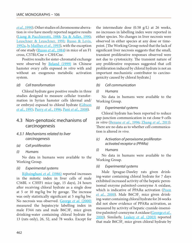
IARC MONOGRAPHS – 106
462
et al., 1990). Other studies of chromosome aberra-tion in vivo have mostly reported negative results (Liang & Pacchierotti, 1988; Xu & Adler, 1990; Leuschner & Leuschner, 1991; Russo & Levis, 1992a, b; Mailhes et al., 1993), with the exception of one study (Russo et al., 1984) in mice of an F1 cross, C57B1/Cne × C3H/Cne.
Positive results for sister-chromatid exchange were observed by Beland (1999) in Chinese hamster ovary cells exposed in vitro with and without an exogenous metabolic activation system.
(d) Cell transformation
Chloral hydrate gave positive results in three studies designed to measure cellular transfor-mation in Syrian hamster cells (dermal and/or embryo) exposed to chloral hydrate (Gibson et al., 1995; Parry et al., 1996; Pant et al., 2008).
4.3 Non-genotoxic mechanisms of carcinogenesis
4.3.1 Mechanisms related to liver carcinogenesis
(a) Cell proliferation
(i) HumansNo data in humans were available to the
Working Group.
(ii) Experimental systemsRijhsinghani et al. (1986) reported increases
in the mitotic index in liver cells of male C56BL × C3HF1 mice (age, 15 days), 24 hours after receiving chloral hydrate as a single dose at 5 or 10 mg/kg bw by gavage. The increase was only statistically significant at 5 mg/kg bw. No necrosis was observed. George et al. (2000) measured the hepatocyte labelling index in male F344 rats and male B6C3F1 mice given drinking-water containing chloral hydrate for 13 (rats only), 26, 52, and 78 weeks. Except for
the intermediate dose (0.58 g/L) at 26 weeks, no increases in labelling index were reported in either species. No changes in liver necrosis were observed in either species at any dose or time-point. [The Working Group noted that the lack of significant liver necrosis suggests that the small, transient proliferative responses observed were not due to cytotoxicity. The transient nature of any proliferative responses suggested that cell proliferation induced by chloral hydrate is not an important mechanistic contributor to carcino-genicity caused by chloral hydrate.]
(b) Cell communication
(i) HumansNo data in humans were available to the
Working Group.
(ii) Experimental systemsChloral hydrate has been reported to reduce
gap-junction communication in rat clone 9 cells in vitro (Benane et al., 1996; Zhang et al., 2011). There are no data as to whether cell communica-tion is altered in vivo.
(c) Activation of peroxisome proliferator-activated receptor α (PPARα)
(i) HumansNo data in humans were available to the
Working Group.
(ii) Experimental systemsMale Sprague-Dawley rats given drink-
ing-water containing chloral hydrate for 7 days exhibited increased activity of the hepatic perox-isomal enzyme palmitoyl-coenzyme A oxidase, which is indicative of PPARα activation (Poon et al., 2000). Male B6C3F1 mice given drink-ing-water containing chloral hydrate for 26 weeks did not show evidence of PPARα activation, as measured by activity of hepatic cyanide-insensi-tive palmitoyl-coenzyme A oxidase (George et al., 2000). Similarly, Leakey et al. (2003) reported that male B6C3F1 mice given chloral hydrate by

Chloral and chloral hydrate
463
gavage for 15 months showed no induction of the markers of PPARα activation lauric acid ω-hy-droxylase activity and CYP4A immunoreactive protein when fed ad libitum, but increases in these markers were reported at the highest dose, (100 mg/kg bw) in dietary-controlled mice.
There were increases in the incidence of liver tumours in mice fed ad libitum and dietary-con-trolled mice (George et al., 2000; Leakey et al., 2003). Sprague-Dawley rats have not been tested for carcinogenicity, but a long-term bioassay in male F344 rats did not report increased inci-dences of liver tumours (George et al., 2000). The lack of association between PPARα activa-tion and liver tumorigenicity across experiments suggests that PPARα activation is not an impor-tant mechanistic contributor to carcinogenicity attributable to chloral hydrate.
4.3.2 Mechanisms related to lung carcinogenesis
(a) Cytotoxicity
(i) HumansNo data on mechanisms for lung carcinogen-
esis of chloral hydrate in humans were available to the Working Group.
(ii) Experimental systemsNo data on mechanisms of lung carcinogen-
esis of chloral hydrate in animals were avail-able to the Working Group. Odum et al. (1992) reported that exposure of mice to chloral hydrate at a concentration of 100 ppm [100 μg/mL] by inhalation for 6 hours led to pulmonary cytotox-icity in the form of bronchiolar lesions, alveolar necrosis, desquamation of the epithelium, and alveolar oedema. These effects appeared to be a direct effect of chloral hydrate, rather than a result of metabolism, since exposures to trichloroeth-anol and trichloroacetic acid did not result in any pulmonary effects. Odum et al. (1992) reported that the bronchiolar lesions appeared identical to
those caused by exposure to trichloroethylene by inhalation.
4.4 Susceptibility data
4.4.1 Inter-individual variability
Early studies of sedative effects of chloral hydrate have demonstrated that its effects on the central nervous system are strain-dependent in rats (Riley et al., 1979) and mice (McIntyre & Alpern, 1985), similar to that reported for ethyl alcohol. These observations suggested simi-larities in pathways of metabolism for chloral hydrate and ethyl alcohol. Indeed, Lipscomb et al. (Lipscomb et al., 1997) showed that CYP2E1 is a key pathway for oxidative metabolism of trichloroethylene to chloral hydrate, that there exists up to 10-fold difference in CYP2E1 activity among humans, and that human inter-indi-vidual variability in microsomal formation of chloral hydrate correlates with CYP2E1 activity and protein levels. This study did not address the potential for these inter-individual differences in formation of chloral hydrate to affect its toxicity or carcinogenicity.
Chloral hydrate is further metabolized to trichloroacetic acid by ALDH, and to trichlo-roethanol by ADH. ALDH and ADH are known to be polymorphic in humans, and these poly-morphisms are well known to have a major impact on cancer susceptibility in people who consume beverages containing ethanol, espe-cially in Asian countries (IARC, 2010, 2012b, 2012c; Chang et al., 2012). It has therefore been suggested that subpopulations with certain poly-morphisms in the ALDH and ADH metabolic pathways may have greater than expected forma-tion of trichloroacetic acid and thus an enhanced risk of adverse health effects after exposure to chloral hydrate or other chlorinated solvents.
The effects of chloral hydrate on the metab-olism of alcohol and acetaldehyde have been suggested as a mechanism for the dramatic

IARC MONOGRAPHS – 106
464
effects of coexposure to chloral hydrate (as well as other chlorinated solvents) and ethyl alcohol. Firstly, such coexposures lead to more than addi-tive sedative effects in rodents (Sharkawi et al., 1983) and humans (Sellers et al., 1972). Secondly, adverse health effects indicative of elevated blood levels of acetaldehyde have been described as “degreaser’s flush” (Stewart et al., 1974). Thus, an additional factor for individual susceptibility to adverse health effects of chloral hydrate may be coexposure to ethyl alcohol.
4.4.2 Life-stage susceptibility
Several clinical reports document adverse health effects in neonates or infants who under-went anaesthesia by chloral hydrate (Reimche et al., 1989). It has been suggested that accumu-lation of trichloroacetic acid and trichloroeth-anol in the tissues of infants may be the reason for the enhanced frequency of toxic effects. Because of the immaturity of hepatic metabo-lism, particularly the glucuronidation pathway, and decreased glomerular filtration, the half-life of trichloroethanol is longer in infants (pre-term and full-term) than in adults. There is indirect evidence of competition for hepatic glucuro-nidation for bilirubin with trichloroethanol in pre-term infants. In addition, it has been shown that enzymes participating in alcohol metabo-lism are expressed at lower levels in infants than in adults (Tran et al., 2007).
Neuroapoptosis has been detected in the infant mouse brain after exposure to chloral hydrate (Cattano et al., 2008).
4.4.3 Sex differences
With regard to differences in tumour inci-dence between males and females in studies of long-term exposure to chloral hydrate, some studies reported differential susceptibility in males and females (see Section 3). However, major inconsistencies exist between studies for
target tissue and tumour incidence, which makes it difficult to conclude with certainty that either males or females may be more susceptible to carcinogenesis induced by chloral hydrate.
[The Working Group noted that although male laboratory rodents seem to be more sensi-tive than females to the hepatic effects of chloral hydrate, there was no evidence for differences between men and women in their sensitivity to the sedative or adverse health effects of chloral hydrate at the recommended clinical doses.]
4.5 Toxic non-cancer effects
4.5.1 Liver toxicity, including hepatomegaly, necrosis, and enzyme changes
(a) Humans
One study has shown that sedative doses of chloral hydrate to newborns increase the like-lihood of hyperbilirubinaemia (Lambert et al., 1990).
(b) Experimental systems
Long-term (104 weeks) exposure of male mice to chloral hydrate at a dose equivalent to 166 mg/kg bw per day resulted in no changes in organ weights except in the liver, which showed an approximate 40% increase in absolute liver weight and in the liver-to-body weight ratio (Daniel et al., 1992a). The same type of study was conducted in male and female rats with the only non-cancer effect reported as focal hepato-cellular necrosis in 2 out of 10 male rats (Daniel et al., 1992b).
Similar studies in mice and rats by George et al. (2000) also showed no effects in rats and only proliferative lesions in the liver of mice at doses up to approximately 150 mg/kg bw per day. The National Toxicology Program (NTP, 2002a) conducted studies of long-term exposure in male and female mice given chloral hydrate at doses of up to 71 mg/kg bw per day, and

Chloral and chloral hydrate
465
observed no non-neoplastic effects. A long-term (approximately 125 weeks) study by Leuschner & Beuscher (1998) in rats given chloral hydrate at doses of up to 135 mg/kg bw per day showed no increase in non-neoplastic lesions.
The short-term toxicity of chloral hydrate has been studied in CD1 mice and Sprague-Dawley rats. In mice, administration of chloral hydrate at daily doses of 14.4 or 144 mg/kg bw by gavage for 14 consecutive days resulted in an increase in relative liver weight and a decrease in spleen size. No other changes were seen. Mice given drinking-water containing chloral hydrate at a concentration of 0.07 or 0.7 mg/mL for 90 days showed dose-related hepatomegaly in males only and significant changes in hepatic microsomal enzymes in both males and females, indicative of hepatic toxicity (Sanders et al., 1982).
Male Sprague-Dawley rats were given drink-ing-water containing chloral hydrate at a concen-tration of 0.13, 1.35, or 13.5 mg/L for 7 days (Poon et al., 2000). No changes were observed in body or organ weights.
4.5.2 Central nervous system
(a) Humans
Choral hydrate has long been used in both human and veterinary medicine as a sedative and hypnotic drug. The effects on the central nervous system are due to the metabolite, trichloroeth-anol (Shapiro et al., 1969; Miller & Greenblatt, 1979).
(b) Experimental systems
Exposure of female CD1 mice to chloral at a dose of 100 ppm [603 μg/L] for 6 hours induced deep anaesthesia, which was fully reversible on cessation of exposure (Odum et al., 1992).
4.5.3 Kidney toxicity
(a) Humans
No other data in humans were available to the Working Group.
(b) Experimental systems
The National Toxicity Program (NTP, 2002a) reported an increase in the incidence of glomer-ulosclerosis in mice fed ad libitum with diets containing chloral hydrate at a concentration of 25 or 100 mg/kg. Other long-term studies did not report any lesions in the kidney (Daniel et al., 1992a; Leuschner & Beuscher, 1998).
4.6 Synthesis of mechanistic considerations
A comprehensive database exists to charac-terize the absorption, distribution, metabolism and excretion of chloral hydrate in humans and experimental animals; this database supports the conclusion that major similarities are evident between humans and rodents. Chloral hydrate is rapidly metabolized to trichloroethanol and trichloroacetic acid, which have been detected in blood and urine. Trichloroacetic acid that is formed from the metabolism of chloral hydrate is slowly metabolized and has a longer plasma half-life in humans (2–4 days) than in rodents (5–6 hours). The major excretion pathway for metabolites of chloral hydrate is in the urine. The major enzymes responsible for the metabo-lism of chloral hydrate are ADH and ALDH. This metabolic pathway is very similar in humans and rodents.
Strong evidence suggests that chloral hydrate is genotoxic, both in vivo and in vitro, in mammalian and other test systems (including the standard genotoxicity battery, with and without metabolic activation). The types of geno-toxic damage detected encompassed mutations, chromosomal aberrations, and micronucleus
IARC MONOGRAPHS – 106
466
formation. One study found that in infants exposed orally to chloral hydrate, a significant increase in micronucleus formation in peripheral blood lymphocytes was observed.
Adverse health outcomes of chloral hydrate have been reported for the liver, kidney, and central nervous system, which suggests that these organs are potential targets for this chemical.
The strength of evidence for liver as a target tissue is strong; in addition to cancer in mice, some evidence of liver toxicity (hyperbilirubi-naemia) has been reported in infants exposed to chloral hydrate. Available data on non-genotoxic mechanisms in the liver are almost exclusively from studies in animals. Multiple mechanisms have been suggested to operate, including a transient increase in cell proliferation, induc-tion of the peroxisome proliferation response (inconclusive evidence for activation of PPARα in both studies in rodents, with poor correlation with tumours), and disruption of gap-junction intercellular communications (limited evidence from one study in vitro). The strength of evidence for non-genotoxic mechanisms in liver cancer is weak.
The strength of evidence for the central nervous system as a target tissue is strong. Chloral hydrate is a sedative in humans and in animals. The relevance of neurotoxicity to cancer is unknown.
The strength of evidence for kidney as a target tissue is weak. Some evidence of kidney toxicity (glomerular sclerosis) has been reported in studies in animals.
There is strong potential for inter-individual variability in the adverse effects of chloral hydrate. ADH and ALDH are the major metabolic enzymes for chloral hydrate, and common poly-morphisms that result in differences in alcohol metabolism have been reported in humans. Consumption of alcoholic beverages may also be a susceptibility factor because of effects on alcohol metabolism enzymes that are important for the biotransformation of chloral hydrate.
5. Summary of Data Reported
5.1 Exposure data
Chloral is a chlorinated aldehyde that is readily converted to chloral hydrate in the presence of water. It can be formed as a disin-fection by-product as a result of chlorine-based disinfection of water. Chloral has been used historically in the production of dichlorodiphe-nyltrichloroethane (DDT) and, to a lesser extent, other insecticides and pharmaceuticals. The use of chloral has declined steadily since the use of DDT has been restricted. Chloral hydrate is used as a sedative during medical procedures and to reduce anxiety related to withdrawal from drugs. Exposure to chloral hydrate may occur at micro-gram-per-litre levels through consumption of chlorinated drinking-water and ingestion of swimming-pool water.
5.2 Human carcinogenicity data
Only one epidemiological study has exam-ined risk of cancer in humans after exposure to chloral hydrate. Relationships between the number of prescriptions for chloral hydrate and morbidity for all cancers, and cancers of the lung, stomach, prostate, and melanoma were examined in nested case–control analyses based on records from a health plan in California, USA, and no significant associations were observed. The single available study of carcinogenicity in humans provides no support for relationship between exposure to chloral hydrate and risk of cancer.
5.3 Animal carcinogenicity data
Chloral hydrate was evaluated for carcino-genicity in male mice in two studies with drink-ing-water and in two gavage studies. In the studies with drinking-water, chloral hydrate

Chloral and chloral hydrate
467
induced a significant increase in the incidence of hepatocellular adenoma and hepatocellular carcinoma. In the gavage studies, an increased incidence in hepatocellular adenoma or carci-noma (combined) was observed.
In another study in male dietary-controlled mice treated by gavage, chloral hydrate increased the incidence and the trend in incidence of hepa-tocellular carcinoma.
In the single study in female mice treated by gavage, exposure to chloral hydrate was asso-ciated with increases in the trend and in the incidence of malignant lymphoma and in the incidence of adenoma of the pituitary gland.
Chloral hydrate in drinking-water was evalu-ated for carcinogenicity in one study in male and female rats, and in a second study in male rats. No dose-related increases in tumour incidence were reported. In one of the studies, however, the incidence of hepatocellular carcinoma in one treatment group of male rats was higher than that in historical controls. Several design and reporting deficiencies limited the power of both studies to adequately evaluate the carcinogenic potential of chloral hydrate in rats.
5.4 Mechanistic and other relevant data
Major similarities exist between humans and experimental animals with regard to the absorption, distribution and metabolism of chloral hydrate. In all species studied, rapid metabolism of chloral hydrate by alcohol and aldehyde dehydrogenases results in formation of trichloroacetic acid and trichloroethanol). However, trichloroacetic acid has a much longer plasma half-life in humans (2–4 days) than in rodents (5–6 hours), which is indicative of much slower excretion or metabolism of this metabo-lite in humans. Strong evidence is available from studies in humans in vivo, animals in vivo and in vitro, and from bacterial and other test systems,
to support the conclusion that chloral hydrate is a genotoxic agent. Cancer findings in animals and toxicity findings in humans and animals designate liver as a major target organ for chloral hydrate. In the liver, non-genotoxic mechanisms may participate in carcinogenesis. Some data on kidney toxicity in rodents suggest that kidney may also be a target organ. The central nervous system is also a potential target organ because chloral hydate has a sedative effect in humans and animals. The relevance of the apparent effects on the kidney and central nervous system to the potential cancer hazard of chloral hydrate in these organs is unknown. There is potential for inter-individual variability in the adverse effects of chloral hydrate because it is metabolized through alcohol and aldehyde dehydrogenases; polymorphisms in these enzymes have been shown to have a major impact on their function. Consumption of beverages containing alcohol may also be a susceptibility factor due effects on these same enzymes.
6. Evaluation
6.1 Cancer in humans
There is inadequate evidence in humans for the carcinogenicity of chloral and chloral hydrate.
6.2 Cancer in experimental animals
There is sufficient evidence in experimental animals for the carcinogenicity of chloral and chloral hydrate.
6.3 Overall evaluation
Chloral and chloral hydrate are probably carcinogenic to humans (Group 2A).
IARC MONOGRAPHS – 106
468
6.4 Rationale
In reaching the evaluation, the Working Group considered the following:
• Chloral hydrate is absorbed, distributed and metabolized similarly in humans and in rodents;
• Chloral hydrate is genotoxic in numerous assays for genotoxicity;
• Evidence exists that chloral hydrate is geno-toxic in exposed humans, supporting the conclusion that a genotoxic mechanism also operates in humans.
References
Abbas R & Fisher JW (1997). A physiologically based pharmacokinetic model for trichloroethylene and its metabolites, chloral hydrate, trichloroacetate, dichlo-roacetate, trichloroethanol, and trichloroethanol glucuronide in B6C3F1 mice. Toxicol Appl Pharmacol, 147: 15–30. doi:10.1006/taap.1997.8190 PMID:9356303
Abbas RR, Seckel CS, Kidney JK, Fisher JW (1996). Pharmacokinetic analysis of chloral hydrate and its metabolism in B6C3F1 mice. [Erratum in: Drug Metab. Dispos., 25, 1449]Drug Metab Dispos, 24: 1340–1346. PMID:8971140
Albertini S (1990). Analysis of nine known or suspected spindle poisons for mitotic chromosome malseg-regation using Saccharomyces cerevisiae D61.M. Mutagenesis, 5: 453–459. doi:10.1093/mutage/5.5.453 PMID:2263203
Allen JW, Collins BW, Evansky PA (1994). Spermatid micronucleus analyses of trichloroethylene and chloral hydrate effects in mice. Mutat Res, 323: 81–88. doi:10.1016/0165-7992(94)90049-3 PMID:7508572
Anderson LE (1954). Hoyer’s Solution as a rapid perma-nent mounting medium for bryophytes. Bryologist, 57: 242–244.
Arkhipchuk VV & Garanko NN (2005). Using the nucle-olar biomarker and the micronucleus test on in vivo fish fin cells. Ecotoxicol Environ Saf, 62: 42–52. doi:10.1016/j.ecoenv.2005.01.001 PMID:15978290
Beland FA (1999). NTP technical report on the toxicity and metabolism studies of chloral hydrate (CAS No. 302–17–0). Administered by gavage to F344/N rats and B6C3F1 mice. Toxic Rep Ser, 59: 1–66, A1–E7. PMID:11803702
Beland FA, Schmitt TC, Fullerton NF, Young JF (1998). Metabolism of chloral hydrate in mice and rats after single and multiple doses. J Toxicol Environ Health A, 54: 209–226. doi:10.1080/009841098158917 PMID:9643873
Benane SG, Blackman CF, House DE (1996). Effect of perchloroethylene and its metabolites on intercellular communication in clone 9 rat liver cells. J Toxicol Environ Health, 48: 427–437. doi:10.1080/009841096161168 PMID:8751833
Bernstine JB, Meyer AE, Bernstine RL (1956). Maternal blood and breast milk estimation following the administration of chloral hydrate during the puer-perium. J Obstet Gynaecol Br Emp, 63: 228–231. doi:10.1111/j.1471-0528.1956.tb05467.x PMID:13320217
Boĭtsov AN, Rotenberg IuS, Mulenkova VG (1970). [Toxicologic assessment of chloral in the process of its liberation during filling and pouring of foam polyu-rethanes] Gig Tr Prof Zabol, 14: 26–29. PMID:5433673
Bonatti S, Cavalieri Z, Viaggi S, Abbondandolo A (1992). The analysis of 10 potential spindle poisons for their ability to induce CREST-positive micronuclei in human diploid fibroblasts. Mutagenesis, 7: 111–114. doi:10.1093/mutage/7.2.111 PMID:1579065
Breimer DD (1977). Clinical pharmacokinetics of hypnotics. Clin Pharmacokinet, 2: 93–109. doi:10.2165/00003088-197702020-00002 PMID:16715
Breimer DD, Ketelaars HCJ, Van Rossum JM (1974). Gas chromatographic determination of chloral hydrate, trichloroethanol, and trichloroacetic acid in blood and urine employing head-space analysis. J Chromatogr A, 88: 55–63. doi:10.1016/S0021-9673(01)91772-8
Bronley-DeLancey A, McMillan DC, McMillan JM et al. (2006). Application of cryopreserved human hepat-ocytes in trichloroethylene risk assessment: relative disposition of chloral hydrate to trichloroacetate and trichloroethanol. Environ Health Perspect, 114: 1237–1242. doi:10.1289/ehp.9047 PMID:16882532
Cattano D, Straiko MM, Olney JW (2008). Chloral hydrate induces and lithium prevents neuroapoptosis in the infant mouse brain. Anesthesiology, 109: A315
Chang JS, Straif K, Guha N (2012). The role of alcohol dehy-drogenase genes in head and neck cancers: a system-atic review and meta-analysis of ADH1B and ADH1C. Mutagenesis, 27: 275–286. doi:10.1093/mutage/ger073 PMID:22042713
Chang LW, Daniel FB, DeAngelo AB (1992). Analysis of DNA strand breaks induced in rodent liver in vivo, hepatocytes in primary culture, and a human cell line by chlorinated acetic acids and chlorinated acetalde-hydes. Environ Mol Mutagen, 20: 277–288. doi:10.1002/em.2850200406 PMID:1330547
Chemical Information Services (2002). Directory of World Chemical Producers (Version 2002.1). Dallas, TX, USA.
Cole WJ, Mitchell RG, Salamonsen RF (1975). Isolation, characterization and quantitation of chloral hydrate

Chloral and chloral hydrate
469
as a transient metabolite of trichloroethylene in man using electron capture gas chromatography and mass fragmentography. J Pharm Pharmacol, 27: 167–171. doi:10.1111/j.2042-7158.1975.tb09431.x PMID:237998
Crebelli R, Conti G, Conti L, Carere A (1985). Mutagenicity of trichloroethylene, trichloroethanol and chloral hydrate inaspergillus nidulans. Mutat Res Genet Toxicol Environ Mutagen, 155: 105–111. doi:10.1016/0165-1218(85)90126-0
Crebelli R, Conti G, Conti L, Carere A (1991). In vitro studies with nine known or suspected spindle poisons: results in tests for chromosome mal segregation in Aspergillus nidulans. Mutagenesis, 6: 131–136. doi:10.1093/mutage/6.2.131 PMID:2056914
Daniel FB, DeAngelo AB, Stober JA et al. (1992). Hepatocarcinogenicity of chloral hydrate, 2-chlo-roacetaldehyde, and dichloroacetic acid in the male B6C3F1 mouse. Fundam Appl Toxicol, 19: 159–168. doi:10.1016/0272-0590(92)90147-A PMID:1516771
Daniel FB, DeAngelo AB, Stober JA et al. (1992a). Hepatocarcinogenicity of chloral hydrate, 2-chlo-roacetaldehyde, and dichloroacetic acid in the male B6C3F1 mouse. Fundam Appl Toxicol, 19: 159–168. doi:10.1016/0272-0590(92)90147-A PMID:1516771
Daniel FB, Robinson M, Stober JA et al. (1992b). Ninety-day toxicity study of chloral hydrate in the Sprague-Dawley rat. Drug Chem Toxicol, 15: 217–232. doi:10.3109/01480549209014152 PMID:1425361
Davidson IW & Beliles RP (1991). Consideration of the target organ toxicity of trichloroethylene in terms of metabolite toxicity and pharmacokinetics. Drug Metab Rev, 23: 493–599. doi:10.3109/03602539109029772 PMID:1802654
Degrassi F & Tanzarella C (1988). Immunofluorescentstaining of kinetochores in micro-nuclei: A new assay for the detection ofaneuploidy. Mutat Res Genet Toxicol Environ Mutagen, 203: 339–345. doi:10.1016/0165-1161(88)90030-1
Delinsky AD, Bruckner JV, Bartlett MG (2005). A review of analytical methods for the determination of trichloroethylene and its major metabolites chloral hydrate, trichloroacetic acid and dichloroacetic acid. Biomed Chromatogr, 19: 617–639. doi:10.1002/bmc.488 PMID:15828053
Demeestere K, Dewulf J, De Witte B, Van Langenhove H (2007). Sample preparation for the analysis of vola-tile organic compounds in air and water matrices. J Chromatogr A, 1153: 130–144. doi:10.1016/j.chroma.2007.01.012 PMID:17258752
Eastmond DA & Tucker JD (1989). Identification of aneu-ploidy-inducing agents using cytokinesis-blocked human lymphocytes and an antikinetochore anti-body. Environ Mol Mutagen, 13: 34–43. doi:10.1002/em.2850130104 PMID:2783409
Environmental Chemicals Data and Information Network (1993). Chloral. Ispra, JRC-CEC.
EPA (1995). Determination of Chlorination Disinfection Byproducts, Chlorinated Solvents, and Halogenated Pesticides/Herbicides in Drinking Water by Liquid-liquid Extraction and Gas Chromatography with Electron Capture Detection. (Method 551.1, Rev 1.0) Cincinnati, OH: Environmental Monitoring Systems Laboratory, pp. 1–61.
EPA (2000). Toxicological Review of Chloral Hydrate. In Support of Summary Information on the Integrated Risk Information System (IRIS). (EPA/635/R-00/006) Washington, DC: Environmental Protection Agency.
Fellows MD, Doherty AT, Priestley CC et al. (2011). The ability of the mouse lymphoma TK assay to detect aneugens. Mutagenesis, 26: 771–781. doi:10.1093/mutage/ger045 PMID:21775299
Furnus CC, Ulrich MA, Terreros MC, Dulout FN (1990). The induction of aneuploidy in cultured Chinese hamster cells by propionaldehyde and chloral hydrate. Mutagenesis, 5: 323–326. doi:10.1093/mutage/5.4.323 PMID:2398816
Garrett ER & Lambert HJ (1966). Gas chromatographic analysis of trichloroethanol, chloral hydrate, trichlo-roacetic acid, and trichloroethanol glucuronide. J Pharm Sci, 55: 812–817. doi:10.1002/jps.2600550812 PMID:5975292
Gennaro AR (2000). Remington: The Science and Practice of Pharmacy. 20th Ed. Baltimore, MD: Lippincott Williams & Wilkins.
George MH, Moore T, Kilburn S et al. (2000). Carcinogenicity of chloral hydrate administered in drinking water to the male F344/N rat and male B6C3F1 mouse. Toxicol Pathol, 28: 610–618. doi:10.1177/019262330002800415 PMID:10930049
Gibson DP, Aardema MJ, Kerckaert GA et al. (1995). Detection of aneuploidy-inducing carcinogens in the Syrian hamster embryo (SHE) cell transforma-tion assay. Mutat Res, 343: 7–24. doi:10.1016/0165-1218(95)90058-6 PMID:7753109
Giller S, Le Curieux F, Gauthier L et al. (1995). Genotoxicity assay of chloral hydrate and chloropicrine. Mutat Res, 348: 147–152. doi:10.1016/0165-7992(95)90002-0 PMID:8544866
Golfinopoulos SK & Nikolaou AD (2005). Survey of disinfection by-products in drinking water in Athens, Greece. Desalination, 176: 13–24. doi:10.1016/j.desal.2004.10.029
Grawé J, Nüsse M, Adler I-D (1997). Quantitative and qualitative studies of micronucleus induction in mouse erythrocytes using flow cytometry. I. Measurement of micronucleus induction in peripheral blood poly-chromatic erythrocytes by chemicals with known and suspected genotoxicity. Mutagenesis, 12: 1–8. doi:10.1093/mutage/12.1.1 PMID:9025090
Gu ZW, Sele B, Chmara D et al. (1981). [Effects of trichlo-rethylene and its metabolites on the rate of sister-chro-matid exchange. In vivo and in vitro study on the
IARC MONOGRAPHS – 106
470
human lymphocytes. (author’s transl)] Ann Genet, 24: 105–106. PMID:6977287
Hansch C, Leo A, Hoekman D (1995). Exploring QSAR: Hydrophobic, Electronic, and Steric Constants. Washington, DC: American Chemical Society, p. 4.
Harrington-Brock K, Doerr C, Moore M (1998). Mutagenicity of three disinfection by-products: Di- and trichloroacetic acidand chloral hydrate in l5178y(+/−) — 3.7.2c mouse lymphoma cells. Mutat Res, 413: 265–276. doi:10.1016/S1383-5718(98)00026-6
Haselkorn T, Whittemore AS, Udaltsova N, Friedman GD (2006). Short-term chloral hydrate admin-istration and cancer in humans. Drug Saf, 29: 67–77. doi:10.2165/00002018-200629010-00005 PMID:16454535
Haseman JK, Hailey JR, Morris RW (1998). Spontaneous neoplasm incidences in Fischer 344 rats and B6C3F1 mice in two-year carcinogenicity studies: a National Toxicology Program update. Toxicol Pathol, 26: 428–441. doi:10.1177/019262339802600318 PMID:9608650
Haworth S, Lawlor T, Mortelmans K et al. (1983). Salmonella mutagenicity test results for 250 chemi-cals. Environ Mutagen, 5: Suppl 11–142. doi:10.1002/em.2860050703 PMID:6365529
Haynes WM (2012). CRC Handbook of Chemistry and Physics, (Internet Version 2012). No. EPA/625/R-96/010b, 92nd Edition. Boca Raton, FL: CRC Press/Taylor and Francis.
Henderson GN, Yan Z, James MO et al. (1997a). Kinetics and metabolism of chloral hydrate in children: iden-tification of dichloroacetate as a metabolite. Biochem Biophys Res Commun, 235: 695–698. doi:10.1006/bbrc.1997.6868 PMID:9207222
Hennig UG, Rudd NL, Hoar DI (1988). Kinetochore immunofluorescence in micronuclei: a rapid method for the in situ detection of aneuploidy and chromosome breakage in human fibroblasts. Mutat Res, 203: 405–414. doi:10.1016/0165-1161(88)90013-1 PMID:3057373
IARC (1995). Dry cleaning, some chlorinated solvents and other industrial chemicals. IARC Monogr Eval Carcinog Risks Hum, 63: 33–477. PMID:9139128
IARC (2004). Some drinking-water disinfectants and contaminants, including arsenic. IARC Monogr Eval Carcinog Risks Hum, 84: 1–477. PMID:15645577
IARC (2010). Alcohol consumption and ethyl carbamate. IARC Monogr Eval Carcinog Risks Hum, 96: 1–1428.PMID:21735939
IARC (2012a). Some chemicals present in industrial and consumer products, food and drinking-water. IARC Monogr Eval Carcinog Risks Hum, 101: 1–586.Available on-line at http://monographs.iarc.fr/ENG/Monographs/vol101/index.php
IARC (2012b). Chemical agents and related occupations. IARC Monogr Eval Carcinog Risks Hum, 100F: 1–599.PMID:23189753
IARC (2012c). Personal habits and indoor combustions. IARC Monogr Eval Carcinog Risks Hum, 100E: 1–575.PMID:23193840
Ikbal M, Tastekin A, Dogan H et al. (2004). The assessment of genotoxic effects in lymphocyte cultures of infants treated with chloral hydrate. Mutat Res, 564: 159–164. doi:10.1016/j.mrgentox.2004.08.007 PMID:15507380
Italia MP & Uden PC (1988). Multiple element emission spectral detection gas chromatographic profiles of halogenated products from chlorination of humic acid and drinking water. J Chromatogr A, 438: 35–43. doi:10.1016/S0021-9673(00)90230-9
Jira R, Kopp E, McKusick BC (1986). Chloroacetaldehydes. In: Ullmann’s Encyclopedia of Industrial Chemistry. Gerhartz W, Yamamoto YS, Campbell FT et al., editors. New York: VCH Publishers, pp. 527–536.
Jira R, Kopp E, McKusick BC et al. (2007). Chloroacetaldehydes. In: Ullmann’s Encyclopedia of Industrial Chemistry. Available at: http://onlineli-brary.wiley.com/doi/10.1002/14356007.a06_527.pub2/abstract
Käfer E (1986). Tests which distinguish induced cross-ing-over and aneuploidy from secondary segregation in Aspergillus treated with chloral hydrate or gamma-rays. Mutat Res, 164: 145–166. doi:10.1016/0165-1161(86)90006-3 PMID:3520302
Kappas A (1989). On the mechanisms of inducedaneu-ploidy in aspergillus nidulans and validation of tests for genomicmutations. In: Mechanisms of Chromosome Distribution and Aneuploidy, Vol. 318. Resnick MA, Vig BK, editors. New York, NY: Wiley, pp. 377–384.
Keller DA & Heck HD (1988). Mechanistic studies on chloral toxicity: relationship to trichloro-ethylene carcinogenesis. Toxicol Lett, 42: 183–191. doi:10.1016/0378-4274(88)90076-8 PMID:3406960
Ketcha MM, Stevens DK, Warren DA et al. (1996). Conversion of trichloroacetic acid to dichloroacetic acid in biological samples. J Anal Toxicol, 20: 236–241. doi:10.1093/jat/20.4.236 PMID:8835661
Kiffe M, Christen P, Arni P (2003). Characterization of cytotoxic and genotoxic effects of different compounds in CHO K5 cells with the comet assay (single-cell gel electrophoresis assay). Mutat Res, 537: 151–168. doi:10.1016/S1383-5718(03)00079-2 PMID:12787820
Koudjonou B, Lebel GL, Dabeka L (2008). Formation of halogenated acetaldehydes, and occurrence in Canadian drinking water. Chemosphere, 72: 875–881. doi:10.1016/j.chemosphere.2008.03.062 PMID:18499222
Lambert GH, Muraskas J, Anderson CL, Myers TF (1990). Direct hyperbilirubinemia associated with chloral hydrate administration in the newborn. Pediatrics, 86: 277–281. PMID:2371103
Leakey JEA, Seng JE, Latendresse JR et al. (2003). Dietary controlled carcinogenicity study of chloral hydrate in male B6C3F1 mice. Toxicol Appl Pharmacol,

Chloral and chloral hydrate
471
193: 266–280. doi:10.1016/j.taap.2003.07.007 PMID:14644627
Lee J, Jun MJ, Lee MH et al. (2010). Production of various disinfection byproducts in indoor swimming pool waters treated with different disinfection methods. Int J Hyg Environ Health, 213: 465–474. doi:10.1016/j.ijheh.2010.09.005 PMID:20961810
Leivadara SV, Nikolaou AD, Lekkas TD (2008). Determination of organic compounds in bottled waters. Food Chem, 108: 277–286. doi:10.1016/j.foodchem.2007.10.031
Leopardi P, Zijno A, Bassani B, Pacchierotti F (1993). In vivo studies on chemically induced aneuploidy in mouse somatic and germinal cells. Mutat Res, 287: 119–130. doi:10.1016/0027-5107(93)90151-5 PMID:7683378
Leuschner J & Beuscher N (1998). Studies on the muta-genic and carcinogenic potential of chloral hydrate. Arzneimittelforschung, 48: 961–968. PMID:9825112
Leuschner J & Leuschner F (1991). Evaluation of the mutagenicity of chloral hydrate in vitro and in vivo. Arzneimittelforschung, 41: 1101–1103. PMID:1799393
Liang J & Pacchierotti F (1988). Cytogeneticinvestigation of chemically-induced aneuploidy in mouse spermatocytes. Mutat Res, 201: 325–335. doi:10.1016/0027-5107(88)90021-8
Liang JC & Brinkley BR (1985). Chemical probes and possible targets for the induction of aneuploidy. Basic Life Sci, 36: 491–505. PMID:4096703
Lipscomb JC, Garrett CM, Snawder JE (1997). Cytochrome P450-dependent metabolism of trichloroethylene: interindividual differences in humans. Toxicol Appl Pharmacol, 142: 311–318. doi:10.1006/taap.1996.8040 PMID:9070354
Liviac D, Creus A, Marcos R (2010). DNA damage induc-tion by two halogenated acetaldehydes, byproducts of water disinfection. Water Res, 44: 2638–2646. doi:10.1016/j.watres.2010.01.026 PMID:20189624
Liviac D, Creus A, Marcos R (2011). Mutagenic analysis of six disinfection by-products in the Tk gene of mouse lymphoma cells. J Hazard Mater, 190: 1045–1052. doi:10.1016/j.jhazmat.2011.04.062 PMID:21561708
Lynch AM & Parry JM (1993). The cytochalasin-B micronucleus/kinetochore assay in vitro: studies with 10 suspected aneugens. Mutat Res, 287: 71–86. doi:10.1016/0027-5107(93)90146-7 PMID:7683386
Mailhes JB, Aardema MJ, Marchetti F (1993). Investigation of aneuploidy induction in mouse oocytes following exposure to vinblastine-sulfate, pyrimethamine, diethylstilbestrol diphosphate, or chloral hydrate. Environ Mol Mutagen, 22: 107–114. doi:10.1002/em.2850220209 PMID:8359152
Marrazzini A, Betti C, Bernacchi F et al. (1994). Micronucleus test and metaphase analyses in mice exposed to known and suspected spindle poisons. Mutagenesis, 9: 505–515. doi:10.1093/mutage/9.6.505 PMID:7854141
McIntyre TD & Alpern HP (1985). Reinterpretation of the literature indicates differential sensitivities of long-sleep and short-sleep mice are not specific to alcohol. Psychopharmacology (Berl), 87: 379–389. doi:10.1007/BF00432499 PMID:2867574
McKinney LL, Uhing EH, White JL, Picken JC Jr (1955). Vegetable oil extraction Autoxidation products of trichloroethylene. Agric. Food Chem, 3: 413–419. doi:10.1021/jf60051a004
Merdink JL, Gonzalez-Leon A, Bull RJ, Schultz IR (1998). The extent of dichloroacetate formation from trichloroethylene, chloral hydrate, trichloroacetate, and trichloroethanol in B6C3F1 mice. Toxicol Sci, 45: 33–41. doi:10.1093/toxsci/45.1.33 PMID:9848108
Merdink JL, Robison LM, Stevens DK et al. (2008). Kinetics of chloral hydrate and its metabolites in male human volunteers. Toxicology, 245: 130–140. doi:10.1016/j.tox.2007.12.018 PMID:18243465
Merdink JL, Stenner RD, Stevens DK et al. (1999). Effect of enterohepatic circulation on the pharmacokinetics of chloral hydrate and its metabolites in F344 rats J Toxicol Environ health, Part A57: 357–368.PMID:10405189
Miller BM & Adler I-D (1992). Aneuploidy induction in mouse spermatocytes. Mutagenesis, 7: 69–76. doi:10.1093/mutage/7.1.69 PMID:1635458
Miller JW & Uden PC (1983). Characterization of nonvolatile aqueous chlorination products of humic substances. Environ Sci Technol, 17: 150–157. doi:10.1021/es00109a006 PMID:22263679
Miller RR & Greenblatt DJ (1979). Clinical effects of chloral hydrate in hospitalized medical patients. J Clin Pharmacol, 19: 669–674. PMID:512063
Müller G, Spassovski M, Henschler D (1974). Metabolism of trichloroethylene in man. II. Pharmacokinetics of metabolites. Arch Toxicol, 32: 283–295. doi:10.1007/BF00330110 PMID:4480028
Natarajan AT, Duivenvoorden WC, Meijers M, Zwanenburg TS (1993). Induction of mitotic aneu-ploidy using Chinese hamster primary embryonic cells. Test results of 10 chemicals. Mutat Res, 287: 47–56. doi:10.1016/0027-5107(93)90144-5 PMID:7683384
Nelson MA & Bull RJ (1988). Induction of strand breaks in DNA by trichloroethylene and metabolites in rat and mouse liver in vivo. Toxicol Appl Pharmacol, 94: 45–54. doi:10.1016/0041-008X(88)90335-3 PMID:3376113
Nesslany F & Marzin D (1999). A micromethod for the in vitro micronucleus assay. Mutagenesis, 14: 403–410. doi:10.1093/mutage/14.4.403 PMID:10390508
Ni Y-C, Kadlubar FF, Fu PP (1995). Formation of malon-dialdehyde-modified 2′-deoxyguanosinyl adduct from metabolism of chloral hydrate by mouse liver micro-somes. Biochem Biophys Res Commun, 216: 1110–1117. doi:10.1006/bbrc.1995.2735 PMID:7488187
Ni YC, Wong TY, Kadlubar FF, Fu PP (1994). Hepatic metabolism of chloral hydrate to free radical(s) and induction of lipid peroxidation. Biochem Biophys Res

IARC MONOGRAPHS – 106
472
Commun, 204: 937–943. doi:10.1006/bbrc.1994.2550 PMID:7980564
NIOSH (1994). National Occupational Exposure Survey (1981-1983). Cincinnati, OH: National Institute for Occupational Safety and Health. Available at: http://www.cdc.gov/noes/noes2/81560occ.html
NTP (2002a). Toxicology and carcinogenesis study of chloral hydrate (ad libitum and dietary controlled) (CAS no. 302-17-0) in male B6C3F1 mice (gavage study). Natl Toxicol Program Tech Rep Ser, 503: 1–218. PMID:12533745
NTP (2002b). NTP Technical Report on the Toxicology and Carcinogenesis Studies of Chloral Hydrate (Cas No. 302–17–0) in B6C3F1 mice (Gavage Studies). Natl Toxicol Program Tech Rep Ser, 502: 1–197. PMID:11965861
Nutley EV, Tcheong AC, Allen JW et al. (1996). Micronuclei induced in round spermatids of mice after stem-cell treatment with chloral hydrate: evaluations with centromeric DNA probes and kinetochore anti-bodies. Environ Mol Mutagen, 28: 80–89. doi:10.1002/(SICI)1098-2280(1996)28:2<80::AID-EM3>3.0.CO;2-I PMID:8844988
O’Neil MJ, Heckelman PE, Roman CB (2006). The Merck Index, 14th Ed. Monograph Number 09639. Whitehouse Station, NJ: Merck & Co.
Odum J, Foster JR, Green T (1992). A mechanism for the development of Clara cell lesions in the mouse lung after exposure to trichloroethylene. Chem Biol Interact, 83: 135–153. doi:10.1016/0009-2797(92)90042-J PMID:1505057
Ohlson J. (1986). Dry etch chemical safety Solid State Technol, July69–73..
Owens AH Jr & Marshall EK Jr (1955). Further studies on the metabolic fate of chloral hydrate and trichlo-roethanol. Bull Johns Hopkins Hosp, 97: 320–326. PMID:13260821
Pant K, Sly JE, Bruce SW et al. (2008). Syrian hamster embryo (SHE) cell transformation assay with condi-tioned media (without X-ray irradiated feeder layer) using 2,4-diaminotoluene, 2,6-diaminotoluene and chloral hydrate. Mutat Res, 654: 108–113. doi:10.1016/j.mrgentox.2008.05.006 PMID:18606566
Parry EM, Hague A, Parry JM (1990). A study of mitotic division fidelity and numerical chromosome changes in ageing Syrian hamster dermal cells. Mutat Res, 237: 83–93. doi:10.1016/0921-8734(90)90014-I PMID:2366802
Parry JM, Parry EM, Bourner R et al. (1996). The detec-tion and evaluation of aneugenic chemicals. Mutat Res, 353: 11–46. doi:10.1016/0027-5107(95)00242-1 PMID:8692188
PDR Network (2012). Physicians’ Desk Reference. 66th Ed. Montvale, NJ, USA. Available at: http://isbndb.com/book/physicians_desk_reference_66th_edition
Pershad J, Palmisano P, Nichols M (1999). Chloral hydrate: the good and the bad. Pediatr Emerg Care, 15: 432–435. doi:10.1097/00006565-199912000-00018 PMID:10608336
Poon R, Nadeau B, Chu I (2000). Biochemical effects of chloral hydrate on male rats following 7-day drinking water exposure. J Appl Toxicol, 20: 455–461. doi:10.1002/1099-1263(200011/12)20:6<455::AID-JAT714>3.0.CO;2-A PMID:11180267
Poon R, Nakai J, Yagminas A et al. (2002). Subchronic toxicity of chloral hydrate on rats: a drinking water study. J Appl Toxicol, 22: 227–236. doi:10.1002/jat.843 PMID:12210539
Reimche LD, Sankaran K, Hindmarsh KW et al. (1989). Chloral hydrate sedation in neonates and infants–clin-ical and pharmacologic considerations. Dev Pharmacol Ther, 12: 57–64. PMID:2714158
Richardson SD, Plewa MJ, Wagner ED et al. (2007). Occurrence, genotoxicity, and carcinogenicity of regu-lated and emerging disinfection by-products in drinking water: a review and roadmap for research. Mutat Res, 636: 178–242. doi:10.1016/j.mrrev.2007.09.001 PMID:17980649
Rijhsinghani KS, Abrahams C, Swerdlow MA et al. (1986). Induction of neoplastic lesions in the livers of C57BL x C3HF1 mice by chloral hydrate. Cancer Detect Prev, 9: 279–288. PMID:3527415
Riley EP, Shapiro NR, Lochry EA (1979). Hypnotic suscep-tibility to various depressants in rats selected for differ-ential ethanol sensitivity. Psychopharmacology (Berl), 60: 311–312. doi:10.1007/BF00426674 PMID:108710
Royal Pharmaceutical Society of Great Britain (2002). Martindale, The Complete Drug Reference, 33rd Ed. London, United Kingdom: The Pharmaceutical Press.
Russo A & Levis AG (1992a). Detection of aneuploidy in male germ cells of mice by means of a meiotic micronu-cleus assay. Mutat Res, 281: 187–191. doi:10.1016/0165-7992(92)90007-5 PMID:1371841
Russo A & Levis AG (1992b). Further evidence for the aneuploidogenic properties of chelating agents: induc-tion of micronuclei in mouse male germ cells by EDTA. Environ Mol Mutagen, 19: 125–131. doi:10.1002/em.2850190206 PMID:1541253
Russo A, Pacchierotti F, Metalli P (1984). Nondisjunction induced in mouse spermatogenesis by chloral hydrate, a metabolite of trichloroethylene. Environ Mutagen, 6: 695–703. doi:10.1002/em.2860060507 PMID:6479114
Russo A, Stocco A, Majone F (1992). Identification of kinetochore-containing (CREST+) micronuclei in mouse bone marrow erythrocytes. Mutagenesis, 7: 195–197. doi:10.1093/mutage/7.3.195 PMID:1602974
Sadtler Research Laboratories (1991). Sadtler Standard Spectra, 1981–1991 Supplementary Index. Philadelphia, PA, USA.
Sanders VM, Kauffmann BM, White KL Jr et al. (1982). Toxicology of chloral hydrate in the mouse. Environ
Chloral and chloral hydrate
473
Health Perspect, 44: 137–146. doi:10.1289/ehp.8244137 PMID:7084146
Sato T, Mukaida M, Ose Y et al. (1985). Chlorinated products from structural compounds of soil humic substances. Sci Total Environ, 43: 127–140. doi:10.1016/0048-9697(85)90035-X
Sbrana I, Di Sibio A, Lomi A, Scarcelli V (1993). C-mitosis and numerical chromosome aberration analyses in human lymphocytes: 10 known or suspected spindle poisons. Mutat Res, 287: 57–70. doi:10.1016/0027-5107(93)90145-6 PMID:7683385
Schmitt TC (2002). Determination of chloral hydrate and its metabolites in blood plasma by capillary gas chromatography with electron capture detec-tion. J Chromatogr B Analyt Technol Biomed Life Sci, 780: 217–224. doi:10.1016/S1570-0232(02)00371-9 PMID:12401346
Seelbach A, Fissler B, Madle S (1993). Further evaluation of a modified micronucleus assay with V79 cells for detection of aneugenic effects. Mutat Res, 303: 163–169. doi:10.1016/0165-7992(93)90018-Q PMID:7694134
Sellers EM, Lang M, Koch-Weser J et al. (1972). Interaction of chloral hydrate and ethanol in man. I. Metabolism. Clin Pharmacol Ther, 13: 37–49. PMID:4550322
Serrano M, Silva M, Gallego M (2011). Micro liquid-liquid extraction combined with large-volume injec-tion gas chromatography-mass spectrometry for the determination of haloacetaldehydes in treated water. J Chromatogr A, 1218: 8295–8302. doi:10.1016/j.chroma.2011.09.048 PMID:21982994
Shapiro S, Slone D, Lewis GP, Jick H (1969). Clinical effects of hypnotics. II. An epidemiologic study. JAMA, 209: 2016–2020. doi:10.1001/jama.1969.03160260020005 PMID:5820263
Sharkawi M, De Saint Blanquat G, Elfassy B (1983). Inhibition of alcohol dehydrogenase by chloral hydrate and trichloroethanol: possible role in the chloral hydrate-ethanol interaction. Toxicol Lett, 17: 321–328. doi:10.1016/0378-4274(83)90245-X PMID:6353674
Simpson KL & Hayes KP (1998). Drinking water disin-fection by-products: An Australian perspective. Water Res, 32: 1522–1528. doi:10.1016/S0043-1354(97)00341-2
Sora S & Agostini Carbone M (1987). Chloral hydrate,methylmercury hydroxide and ethidium bromide affect chromosomal segregationduring meiosis of saccharomyces cerevisiae. Mutat Res, 190: 13–17. doi:10.1016/0165-7992(87)90075-3
Stewart RD, Hake CL, Peterson JE (1974). “Degreaser’s flush”. Arch Environ Health, 29: 1–5. PMID:4276444
Tran MN, Wu AH, Hill DW (2007). Alcohol dehydro-genase and catalase content in perinatal infant and adult livers: potential influence on neonatal alcohol metabolism. Toxicol Lett, 169: 245–252. doi:10.1016/j.toxlet.2007.01.012 PMID:17343998
Trehy ML, Yost RA, Miles CJ (1986). Chlorination by products of amino acids in natural waters. Environ Sci Technol, 20: 1117–1122. doi:10.1021/es00153a006
US Pharmacopeial Convention (2012). The United States Pharmacopeia. 35nd rev. The National Formulary, 30th rev. Rockville, MD, pp. 2604–2605
Vagnarelli P, De Sario A, De Carli L (1990). Aneuploidy induced by chloral hydrate detected in human lympho-cytes with the Y97 probe. Mutagenesis, 5: 591–592. doi:10.1093/mutage/5.6.591 PMID:2263216
Van Hummelen P & Kirsch-Volders M (1992). Analysis of eight known or suspected aneugens by the in vitro human lymphocyte micronucleus test. Mutagenesis, 7: 447–455. doi:10.1093/mutage/7.6.447 PMID:1474920
Villanueva CM, Castaño-Vinyals G, Moreno V et al. (2012). Concentrations and correlations of disinfec-tion by-products in municipal drinking water from an exposure assessment perspective. Environ Res, 114: 1–11. doi:10.1016/j.envres.2012.02.002 PMID:22436294
Von Tungeln LS, Yi P, Bucci TJ et al. (2002). Tumorigenicity of chloral hydrate, trichloroacetic acid, trichloroeth-anol, malondialdehyde, 4-hydroxy-2-nonenal, croton-aldehyde, and acrolein in the B6C3F(1) neonatal mouse. Cancer Lett, 185: 13–19. doi:10.1016/S0304-3835(02)00231-8 PMID:12142074
Wang RS, Nakajima T, Honma T (1999). Trichloroethylene inhibits aldehyde dehydrogenase only for aliphatic aldehydes of short chains in rats. Toxicology, 132: 9–18. doi:10.1016/S0300-483X(98)00132-2 PMID:10199577
Warr T, Parry E, Parry J (1993). A comparison of twoin vitro mammalian cell cytogenetic assays for the detection of mitoticaneuploidy using 10 known or suspected aneugens. Mutat Res, 287: 29–46. doi:10.1016/0027-5107(93)90143-4
Waskell L (1978). A study of the mutagenicity of anes-thetics and their metabolites. Mutat Res, 57: 141–153. doi:10.1016/0027-5107(78)90261-0 PMID:351387
Weast RC, Astle MJ (1985). CRC Handbook of Data on Organic Compounds. Boca Raton, FL: CRC Press Inc., 575, 627(I), 592(II), 149, 704 (II).
Wei J, Ye B, Wang W et al. (2010). Spatial and temporal evaluations of disinfection by-products in drinking water distribution systems in Beijing, China. Sci Total Environ, 408: 4600–4606. doi:10.1016/j.scito-tenv.2010.06.053 PMID:20663540
Whistler RL, Zysk JR (1978). Carbohydrates. In: Kirk-Othmer Encyclopedia of Chemical Technology, 3rd Ed. Mark HF, Othmer DP, Overberger EG et al., editors. New York: John Wiley & Sons, pp. 535–555.
Wilson JT (1981). Drugs in Breast Milk. Lancaster, MTP.Xu W & Adler ID (1990). Clastogenic effects of known
and suspect spindle poisons studied by chromosome analysis in mouse bone marrow cells. Mutagenesis, 5: 371–374. doi:10.1093/mutage/5.4.371 PMID:2398818
Zhang J, Grindstaff RD, Thai SF et al. (2011). Chloral hydrate decreases gap junction communication in

IARC MONOGRAPHS – 106
474
rat liver epithelial cells. Cell Biol Toxicol, 27: 207–216. doi:10.1007/s10565-011-9182-x PMID:21243523
Zhang L, Xu L, Zeng Q et al. (2012). Comparison of DNA damage in human-derived hepatoma line (HepG2) exposed to the fifteen drinking water disin-fection byproducts using the single cell gel electro-phoresis assay. Mutat Res, 741: 89–94. doi:10.1016/j.mrgentox.2011.11.004 PMID:22108252
Zimmermann T, Wehling M, Schulz HU (1998). The rela-tive bioavailability and pharmacokinetics of chloral hydrate and its metabolites Arzneimittelforschung, 48: 5–12. PMID:9522024
Zordan M, Osti M, Pesce M, Costa R (1994). Chloral hydrate is recombinogenic in the wing spot test in Drosophila melanogaster. Mutat Res, 322: 111–116. doi:10.1016/0165-1218(94)00017-4 PMID:7519317


![Journal of Radiation Research and Applied Sciences Use of ... · 10 kGy. Fig. (2) represents the absorption spectra of PVB-co-PVA/PVAC ([CPR]= 0.64 phr) of CPR without chloral hydrate](https://img.dokumen.tips/doc/110x75/5f0bddd77e708231d4329a17/journal-of-radiation-research-and-applied-sciences-use-of-10-kgy-fig-2-represents.jpg)