Embed Size (px)
Citation preview

REVIEW
Chitosan-Based Matrices Prepared by GammaIrradiation for Tissue Regeneration: StructuralProperties vs. Preparation Method
Maria Helena Casimiro1 • Joana J. H. Lancastre1 •
Alexandra P. Rodrigues1 • Susana R. Gomes2 •
Gabriela Rodrigues2 • Luıs M. Ferreira1
Received: 15 July 2016 / Accepted: 27 November 2016
� Springer International Publishing Switzerland 2016
Abstract In the last decade, new generations of biopolymer-based materials have
attracted attention, aiming its application as scaffolds for tissue engineering. These
engineered three-dimensional scaffolds are designed to improve or replace damaged,
missing, or otherwise compromised tissues or organs. Despite the number of promising
methods that can be used to generate 3D cell-instructive matrices, the innovative nature
of the presentwork relies on the application of ionizing radiation technology to formand
modify surfaces and matrices with advantage over more conventional technologies
(room temperature reaction, absence of harmful initiators or solvents, high penetration
through the bulk materials, etc.), and the possibility of preparation and sterilization in
one single step. The current chapter summarizes the work done by the authors in the
gamma radiation processing of biocompatible and biodegradable chitosan-based
matrices for skin regeneration. Particular attention is given to the correlation between the
different preparation conditions and the final polymeric matrices’ properties. We
therefore expect to demonstrate that instructive matrices produced and improved by
radiation technology bring to the field of skin regenerative medicine a supplemental
advantage over more conservative techniques.
This article is part of the Topical Collection ‘‘Applications of Radiation Chemistry’’; edited by
Margherita Venturi, Mila D’Angelantonio.
& Maria Helena Casimiro
& Luıs M. Ferreira
1 Centro de Ciencias e Tecnologias Nucleares (C2TN), Instituto Superior Tecnico, Universidade
de Lisboa, E.N. ao km 139.7, Bobadela, 2695-066 Loures, Portugal
2 Departamento de Biologia Animal, Faculdade de Ciencias, Centro de Ecologia, Evolucao e
Alteracoes Ambientais (cE3c), Universidade de Lisboa, Campo Grande, 1749-016 Lisbon,
Portugal
123
Top Curr Chem (Z) (2017) 375:5
DOI 10.1007/s41061-016-0092-5
Keywords Chitosan � Gamma irradiation � Porous scaffolds � Skin substitute �Tissue engineering
1 Introduction
Scaffolds are three-dimensional supports that are used as a template at the body site
injury to help in guiding cell growth, regeneration, and secretion of their own
extracellular matrix (ECM), thereby assisting the body in growing new, functional
tissue. Scaffolds work in two ways; they either help direct cell growth or simply
provide a shape for the final tissue. To be used as a scaffold in tissue engineering, a
material must satisfy a number of requirements: it should not elicit severe
inflammatory responses and it should degrade into non-toxic compounds within the
time frame required for new tissue formation. Alongside the scaffolds should be
suitable porosity for cell in-growth, a surface that balances hydrophilicity and
hydrophobicity for cellular attachment, and mechanical properties that are
compatible with those of the tissue as well as maintaining mechanical strength
during most part of the tissue regenerating process.
Engineered scaffolds are thus designed to augment or completely replace
damaged, missing, or otherwise compromised tissue or organs. These scaffolds may
be permanently integrated into or bioresorbed by the body and must not only be
capable of mimicking the structure and biological functions of ECM but should also
provide a good environment for the cells so that they can easily attach, proliferate,
and differentiate. The ECM defines the three-dimensional architecture of an organ
and is engaged in a complex relationship with the cellular elements of the
surrounding environment. Consequently, communication between the cell and ECM
molecules influences various cellular processes, such as adhesion, proliferation,
differentiation, migration, and apoptosis, as well as growth factor and cytokine
modulation. Moreover, the timing of these events critically affects tissue formation
and remodeling, processes that are crucial for the integration of a tissue-engineered
scaffold into the surrounding environment [1]. In the case of skin, a double-layered
organ whose deep-partial and full-thickness wounds pose serious threats to
preserving its integrity and normal functions, it is well established that severe
damaged skin requires a protective barrier for proper healing [1, 2]. Thus, the ideal
skin scaffold should have high liquid absorbing capacity and be a biodegradable,
biomimetic, and multilayered 3-D structure comprising dermal and epidermal
equivalents. However, regardless of tissue type, a number of key considerations are
important when designing or determining the suitability of a scaffold for use in
tissue engineering; besides the biocompatibility, biodegradability, and mechanical
properties already referred to, scaffold architecture and manufacturing technology
are also determinant factors [2]. Considering this, it is not difficult to realize that the
design of an ideal tissue-engineering scaffold is one of the most important
challenges in regenerative medicine.
New generations of synthetic biomaterials are being developed at a rapid
pace. Particularly biopolymer-based hydrogels [3], nano-scale-size fibers com-
posed of natural and/or synthetic materials [4–6], and ceramics [7] have attracted
5 Page 2 of 25 Top Curr Chem (Z) (2017) 375:5
123

attention in the last decade aiming its application as scaffolds for tissue
engineering. A number of methods can be used to generate instructive matrices
to be employed in tissue engineering (casting [8], electrospinning [9, 10], plasma
[11], etc.). However, despite the potential use of radiation technology to facilitate
the development of tissue engineering, only a few studies have been reported in
the preparation of instructive scaffolds and their sterilization using this
technology [12–15]. The application of radiation technology for formation and
functionalization of surfaces and matrices has remarkable advantages such as:
room temperature reaction, absence of harmful initiators or solvents, and high
penetration through the bulk materials. Additionally, radiation-synthesized
scaffolds and surfaces might be simultaneously functionalized and sterilized.
Chemical formulations under study combine the use of natural and synthetic
polymers in an attempt to take advantage of the biological activity of the natural
materials and the hydrophilicity and mechanical strengthness of the synthetic
ones. Natural materials, owing to their bioactive properties, display better
interactions with the cells, and in that way enhance the cells’ performance in
biological systems. A good example is the frequent use of polysaccharides and
proteins (like chitosan and collagen) due to their biocompatibility, biodegrad-
ability, and similarity to macromolecules recognized by the human body, which
partly mimic the ECM of tissues. Inducing and stimulating the wound-healing
process, these natural polymers are involved in the repair of damaged tissues and
consequently in skin regeneration. Simultaneously, synthetic polymers are highly
useful in the biomedical field since their properties (e.g., porosity, degradation
time, and mechanical characteristics) can be tailored through gamma irradiation
for specific applications. Some synthetic polymers like poly(e-caprolactone),which is a biodegradable polyester in physiological conditions, poly(vinyl
alcohol), a hydrophilic biocompatible polymer and others also exhibit wound-
healing properties and enhance re-epithelialization [14–17]. Compounds with
plasticizer, humectant, and/or crosslinking properties, with good human body
tolerance (e.g., glycerol), have also being studied as additives to polymeric
matrix formulations.
Thus, blends/copolymers with natural and synthetic materials are believed to be
an effective way to develop a tissue-engineered material.
We have been working on the consolidation of the ionizing radiation techniques
for the preparation of new materials for biomedical applications in C2TN/IST
aiming to establish a strong synergy between materials and biomedical studies,
consolidating thus in a unique research laboratory a real bridge between these two
complementary areas. In this framework, authors have been carrying out a
systematic study in order to simultaneously prepare/optimize and sterilize three-
dimensional biocompatible and biodegradable skin scaffolds by c-irradiation. Themain components used in the present study were chitosan, poly(vinyl alcohol) and
glycerol. A brief description of the materials used in the preparation of the scaffolds
discussed in this chapter is outlined next.
Top Curr Chem (Z) (2017) 375:5 Page 3 of 25 5
123

1.1 Chitosan
Chitosan (Chit) is a linear polysaccharide composed of poly-b(1-4)-D-glucosamine
and poly-b(1-4)-D-acetylglucosamine as shown in Fig. 1a. It is a cationic
polysaccharide of natural origin that is obtained by alkaline deacetylation of chitin,
the main exoskeleton component in crustaceans, and one of the most abundant
natural polymers. Due to an unusual combination of properties such as non-toxicity,
biocompatibility, biodegradability, bioactivity, acceleration of wound healing, fat-
biding capacity, etc., chitosan and its chemically modified structures have been
subject of many studies for use in biomedical and pharmaceutical applications
[16–22]. Chitosan bears two types of reactive groups: the C-2 amino groups on
deacetylated units and the hydroxyl groups on C-3 and C-6 carbons on acetylated or
deacetylated units [23]. In an acidic medium or without a catalyst, the reaction takes
place at the amino group [24]. Furthermore, chitosan, being a polysaccharide, is
known as a degradative-type polymer when c-irradiated [25, 26]. One of the
strategies to overcome this is the introduction of a crosslink-type polymer to the
reactional system (e.g., 2-hydroxyethyl methacrylate, HEMA), which may result in
a new matrix prepared/functionalized by gamma irradiation that combines the useful
properties of both polymers [27]. Additionally, in order to obtain a sponge-type
porous structure, blends were irradiated in a dry state.
1.2 Poly(vinyl alcohol)
Poly(vinyl alcohol), PVA (vd. Fig. 1b) is a water-soluble, white (colorless), and
odorless synthetic polymer. It has a crystalline nature associated with good
Fig. 1 Chemical structures: a chitosan; b poly(vinyl alcohol); c glycerol; d poly(1,3-glycerol carbonate)
5 Page 4 of 25 Top Curr Chem (Z) (2017) 375:5
123

mechanical (high tensile strength and flexibility) and barrier properties, excellent
film-forming, emulsifying, and adhesive properties as well as good thermal stability.
It also shows good resistance to organic solvents. However, these properties are
dependent on the hydration level, since water, acting as a plasticizer agent, reduces
its tensile strength, accelerating its degradation [28]. Due to its biocompatibility,
nontoxicity, and the ability to easily form physically cross-linked hydrogels, the use
of PVA and PVA blends has being reported successfully in several biomedical and
pharmaceutical applications and still continues to be a very promising material [29].
PVA has multiple pendant alcohol groups that can work as attachment sites for
biological molecules and/or cells as well as its elasticity can induce cell orientation
or matrix synthesis by enhancing the transmission of mechanical stimuli to seeded
cells [30].
PVA is known to be a truly biodegradable synthetic polymer since the early
1930s [31]. However, its biodegradability very much depends on the degree of
polymerization, degree of hydrolysis, distribution of hydroxyl groups, stereoregu-
larity and crystallinity. As so, the degradation rate of PVA could be controlled
through these parameters. For instance, the creation of crystalline regions in PVA
through physical crosslinking improves its mechanical integrity and reduces the
respective water absorption capacity [32], which consequently reduces the PVA
degradation rate by hydrolytic mechanisms once in contact with body tissues [33].
This behavior can be used in PVA blends with natural fast degradative polymers
(e.g., chitosan) to overcome the weak physical integrity of these. It is therefore
obvious that the evaluation of the biodegradability of PVA should be made in
function of its polymer structure framed in the application and intended
performance. In our study, the crosslinking promoted on PVA during polymeric
blend irradiation for scaffold preparation results in an added mechanical stability
and consequently, in a decrease in its degradation rate. This effect is desired in order
to compensate the higher degradation rate of chitosan. By this way, the skin scaffold
will maintain for an extended period of time their barrier and cell growth matrix
properties.
1.3 Glycerol
Glycerol (vd. Fig. 1c) is a water-soluble, non-toxic, colorless, viscous sweet-tasting
polyol, commonly used in the pharmaceutical, personal care, and food industries. It
presents a high hydrophilicity associated with a high humectant capacity. Regarding
pharmaceutical applications it is being widely used in various products such as
capsules syrups, topical creams, suppositories, eye-strain reducers, etc.
In recent years, increased attention has been given to glycerol-based polymers for
biomedical applications due to the multiplicity of possible formulations/composi-
tions and molecular architectures. The presence of multiple hydroxyl groups, which
can either form new hydrogen bonds with the polymer chains and/or be
synthetically converted into various other functional groups, allows to improve
their potential biomedical applications. Special areas of interest, involving new
market opportunities, includes carriers for drug-delivery systems, sealants or
coatings for tissue repair, and anti-bacterial activity agents/barriers [34].
Top Curr Chem (Z) (2017) 375:5 Page 5 of 25 5
123

For a better understanding, Fig. 1 presents the chemical structures of the
polymers used as well as the glycerol and an example of a glycerol-based polymer
[poly(1,3-glycerol carbonate)].
This chapter presents and discusses the results obtained with matrices of
composition chitosan/PVA prepared by gamma irradiation by two different methods
and in vitro tested as potential skin scaffolds. Results regarding matrices with other
compositions are still under validation.
2 Strategy
This study aims to clarify the correlation between the different preparation
conditions and the final polymeric matrices properties given their intended use as
scaffolds for skin tissue engineering. With this purpose, a detailed comparative
study of the properties shown by the different samples’ groups was performed and
divided in three main tasks:
1. Optimization of the polymeric matrices preparation (methodology, composi-
tion, polymer concentration, range of absorbed dose).
2. Evaluation of structural and functional properties of the obtained matrices.
3. In vitro biocompatibility evaluation where the cellular viability and prolifer-
ation of Human Caucasian Foetal Foreskin Fibroblast cell line was analyzed as
a measure of chitosan-based matrices biocompatibility and ability to assist skin
regeneration.
3 Experimental
3.1 Materials
Chitosan, Chit (medium molecular weight 190–375 kDa, 75–85% deacetylated
chitin), poly(vinyl alcohol), PVA (Mw 89,000–98,000, 99%? hydrolyzed) and
glycerol (puriss p.a. ACS reagent, anhydrous, dist.) were purchased from Sigma-
Aldrich and used as raw materials. All other reagents were of analytical grade and
used as received.
3.2 Preparation Methodologies
In this study, chitosan-based matrices were prepared by two methods using casting
and c-irradiation procedures. The chitosan solution (2% W/V) was prepared by
dissolution of the appropriate amount of chitosan in acetic acid (1% V/V) with
stirring at room temperature (RT) for 24 h. A PVA solution (10% W/V) was
prepared by dissolution of PVA in bi-distilled water at 80 �C until the solution was
clear. Both solutions were filtered and kept at room temperature for 24 h to remove
air bubbles. To the chitosan matrix-specific volumes of the PVA solution were
5 Page 6 of 25 Top Curr Chem (Z) (2017) 375:5
123

added under continuous stirring to get a homogeneous mixture. Chit–PVA blends
with different PVA content (1, 3, 5 or 8% W/V of the final solution) were obtained.
The addition of glycerol was also tested as a humectant/plasticizer agent (0.25%
V/V). Depending on composition, the matrices will be ahead referred to as C2/
xPVA or C2/xPVA/0.25Gly, where x represents the PVA content (% W/V).
The main steps of the matrices’ preparation methods are depicted in Fig. 2. The
first approach, Method 1, included solvent evaporation at RT of the polymeric-blend
solutions in polystyrene Petri dishes until film formation (casting). Afterwards,
samples were neutralized with 0.5% sodium hydroxide and washed with distilled
water, followed by freeze-drying and irradiation procedures. The second method,
Method 2, involved the freeze-drying of the copolymeric solutions and subsequent
irradiation. Furthermore, in order to improve porosity and thus create 3D polymeric
matrices with adequate features to promote cellular growth, in Method 2,
immediately after the homogenization step, an additional procedure was introduced
where solutions were bubbled with nitrogen before the freeze-drying process (these
procedures will be mentioned ahead as Freeze (F) and Bubble freeze (BF)
Fig. 2 Schematic representation of the experimental procedure for the preparation of chitosan-basedmatrices
Top Curr Chem (Z) (2017) 375:5 Page 7 of 25 5
123

procedures). In both tested methods, the freeze-drying step comprised the
membrane freeze at -80 �C for 3 h and lyophilization for 48 h. Afterwards, small
circular samples, from 5 to 10 mm in diameter, were cut with cutting hole punches,
sealed under nitrogen, and irradiated. The c-irradiation was performed in the
experimental Co-60 chamber (Precisa 22) at the Ionizing Radiation Facility of
Nuclear and Technological Campus of Instituto Superior Tecnico (Lisbon
University). A dose rate of 0.5 kGy h-1 was used for the purpose to achieve final
doses of 3, 5, 10 and 15 kGy.
In order to give confidence and reproducibility to the irradiation processes, the
validation of irradiation geometries and respective dose distribution was previously
accessed by Fricke dosimetry and ionizing chamber method. Amber Perspex
dosimeters (Harwell) were used to monitor the samples’ absorbed dose.
3.3 Evaluation of Structural Properties
In order to clarify the correlation between the different preparation conditions
(including methodology, composition, and absorbed dose) and the final polymeric
matrices properties, the samples obtained by the different experimental approaches
were characterized in terms of their structural properties, thermal behavior,
degradation, and in vitro cells assays.
3.3.1 Structural Characterization
Structural characterization of the irradiated samples was carried out by means of
attenuated total reflectance Fourier transform infrared (ATR-FTIR) using a micro-
FTIR Thermo Scientific (Nicolet) i50 spectrometer equipped with an ATR slide-on
diamond tip. The spectra were recorded in the 400–4000 cm-1 region at room
temperature and with a resolution of 4 cm-1 (64 scans).
The microstructural characterization of previously Au-coated samples that were
studied before and after degradation tests was performed by scanning electron
microscopy (SEM) using a SEM instrument FEG-SEM JEOL 7001F.
3.3.2 Thermal Analysis
Thermal behavior of the samples was assessed by thermo-gravimetric analysis
(TGA) using a TA instrument TGA 951. The assays were carried out under a
nitrogen atmosphere from 25 to 500 �C, using a 10 �C min-1 heating rate.
3.3.3 Water Absorption Capacity
The water absorption capacity of the prepared samples was calculated through the
following procedure: matrices’ dried specimens (/ 8 mm) were weighted dried
(Wdry) and immersed in distilled water at 37 �C. After having reached equilibrium
(&1 h), the swollen samples were carefully removed from the water, the excess
wiped off with filter paper, and weighted (Wswollen). The water absorption capacity
of the samples was found through Eq. (1):
5 Page 8 of 25 Top Curr Chem (Z) (2017) 375:5
123

Water absorptionð%Þ ¼ Wswollen �Wdry
Wdry
� 100: ð1Þ
All measurements were performed in triplicate and the result was expressed as
mean value ± standard deviation (SD).
3.3.4 In Vitro Stability
In vitro stability of the scaffolds was performed in phosphate-buffered solution
(PBS 1X, pH 7.4) at 37 �C based on the extent of the matrices’ mass loss. Samples
were dried under vacuum, weighted (W0), and immersed in PBS solution. After 24 h
of immersion, the matrices were taken out of the solution, wiped of excess fluid with
filter paper, dried under vacuum, and weighted (Wdeg). Matrices’ mass loss was
calculated as the percentage of weight loss before and after PBS treatment
according to Eq. (2):
Mass lossð%Þ ¼ W0 �Wdeg
W0
� 100: ð2Þ
All measurements were performed in triplicate and the result was expressed as
mean value ± SD.
3.4 In Vitro Cellular Viability
The scaffolds used in this study were c-irradiated in sealed bags at 3, 5, 10 and
15 kGy. According to previous sterilization results on similar chitosan/pHEMA
matrices [22], it is possible to assure that the exposure to 4–5 kGy allows obtaining
matrices microbiologically safe (i.e., the probability of obtaining a contaminated
item will be much less than 1 in 106 items). Consequently, even if it is necessary to
apply specific microbiological methodology to validate the sterilization procedure,
one can say that preparation and sterilization procedures of the prepared samples
occurred in one simultaneous step. Thus, no previous sterilization procedure was
needed before cellular seeding.
3.4.1 Cell Culture
A Human Caucasian Foetal Foreskin Fibroblast cell line (HFFF2) was selected for
the biological assays (in vitro study) in order to evaluate the effect of chitosan-based
matrices on cell adhesion and viability. The HFFF2 commercial cell line was
obtained from European Collection of Cell Cultures (ECACC, UK). The cells were
cultured in Dulbecco’s modified Eagle’s medium (DMEM, Glutamax), supple-
mented with heat-inactivated fetal bovine serum (FBS) 10% (V/V) and strepto-
mycin and penicillin 100 U/ml (all from Gibco), and incubated at 37 �C in a
humidified atmosphere with 5% of CO2. The culture medium was changed every
2 days. After reaching 80% confluence, the cells were trypsinized and resuspended
in culture medium at a concentration of 2 9 104 cell/ml medium.
Top Curr Chem (Z) (2017) 375:5 Page 9 of 25 5
123

3.4.2 Cell Viability Assay (alamarBlue�)
Chitosan-based matrices (/ 10 mm) were placed in a 48-well tissue culture plate
and pre-wetted with 200 ll of culture medium to promote their expansion so that the
matrices contact the walls of the well in the bottom of the chamber, avoiding
floating. The fact that the matrices are soaked with culture medium enables the
eventual migration of the cells inside its porous structure. After 10 min, the samples
were seeded with 500 ll of the HFFF2 suspension (20,000 cells) and cultured for 1,
4, and 7 days at 37 �C. Control samples were established by culturing cells directly
over the polystyrene surface of the wells.
The cellular viability was monitored with the alamarBlue� cell viability assay
(Life Technologies). At days 1, 4 and 7, the supernatant of each well was replaced
by 300 ll of fresh culture medium and 30 ll of alamarBlue� reagent and incubated
for 2 h at 37 �C in a 5% CO2 atmosphere. After the incubation period, the media
with alamarBlue� were transferred to a 96-well plate and the optical density (OD)
was read in a microplate reader (Tecan Spectra) at 570 nm with a reference
wavelength of 600 nm. The measurements were made in triplicate for each
treatment and time point (1, 4 and 7 days). Data were expressed as mean ± SD.
3.4.3 Cytochemistry
At 7 days of culture, the chitosan-based matrices and cytochemistry control samples
(glass coverslips containing cells cultured in 24-well chambers) were fixed with
paraformaldehyde, PFA 4% (in PBS), permeabilized in 0.2% TritonX-100 (Sigma-
Aldrich), and stained with ToPro3 (1:500 in PBS) and Alexa488 conjugated
Phalloidin (1:400 in PBS) (both from Molecular Probes), to observe cell’s nuclei
and actin cytoskeleton, respectively. The samples were mounted on fresh PBS on a
glass slide and imaged on a Leica SPE confocal system using either a 10 9 0.3 NA
or a 20 9 0.7 NA lens. Confocal images were acquired in 2–5 different fields
chosen randomly at the center of scaffolds.
4 Results and Discussion
4.1 Method 1 (Casting/Freeze-Dry/Irradiation)
4.1.1 Thermal Analysis
The thermal behavior of c-irradiated and non-irradiated chitosan-based matrices
with and without PVA (prepared by Method 1) at a constant heating rate of
10 �C min-1 is displayed in Fig. 3. A noticeable change is observed in the relative
degradation pattern of chitosan matrices when those are c-irradiated at 5 kGy. The
weight loss at 25–100 �C is generally attributed to the loss of bind/adsorbed water,
however, in c-irradiated chitosan matrices it cannot be assigned only to that. The
chitosan degradation can also be responsible for this first step. When chitosan is
exposed to ionizing radiation, it preferably undergoes chain scission by cleavage of
5 Page 10 of 25 Top Curr Chem (Z) (2017) 375:5
123

b-glucosidic linkages [35]. Moreover, as this process may include the dehydration
of the saccharide ring [36], it would explain the sharpest decrease in the weight
observed for the c-irradiated chitosan matrices. The second weight loss, with an
onset temperature near 255 �C for both irradiated and non-irradiated chitosan
matrices, can be due to an exhaustive degradation of the saccharide structure of the
molecule, including decomposition of deacetylated (and acetylated) units of
chitosan. In the case of Chit/PVA matrices, Fig. 3 shows that the introduction of
PVA promoted changes in the mentioned weight-loss trend of chitosan matrices. It
is clear that c-irradiated Chit/PVA matrices are more thermally stable than the
chitosan-irradiated ones. This higher stability may be assigned to a Chit/PVA denser
and crosslinked structure, meaning that PVA confers a stabilizing effect in the
structure of irradiated chitosan matrices.
4.1.2 ATR-FTIR Characterization
ATR-FTIR spectroscopy was used to assess the polymer chemical groups and
investigate the formation of crosslinked networks in chitosan-based matrices upon
irradiation. Results suggest that chitosan and PVA are binded together in the
obtained matrices and that peak intensity evolution is in accordance with the
expected changes promoted in the structure due to c-irradiation. Figure 4 shows the
FTIR spectra of c-irradiated and non-irradiated chitosan-based matrices with and
without PVA. It can be observed that depending on composition, spectra exhibit the
major peaks related to the typical chitosan and PVA patterns. The broad peak
around 3350 cm-1 is assigned to N–H and O–H stretching from the intermolecular
and intramolecular hydrogen bonds. The characteristic absorption peaks appear at
about 1648 (amide I band), 1570 (amide II, N–H deformation mode), and
1322 cm-1 (amide III band). The absorption peaks at 1151 (anti-symmetric
stretching of the C–O–C bridge), 1058 and 1028 cm-1 (skeletal vibrations involving
Fig. 3 Thermogravimetric curves of non-irradiated (0 kGy) and c-irradiated at 5 kGy chitosan (C2) andchitosan-based C2/1PVA matrices
Top Curr Chem (Z) (2017) 375:5 Page 11 of 25 5
123

the C–O stretching) are characteristics of saccharide structure of chitosan. The small
absorption peak at 895 cm-1 can be used to identify the presence of the C–O–C
bridge as well as b-glucosidic linkages between the sugar units in chitosan [37].
Concerning c-irradiated chitosan matrices, it should be noted that they exhibit much
similarity in the IR peaks as those of non-irradiated ones. However, a peaks’
intensity decrease at 895 and 1151 cm-1, which correspond to the saccharide
structure, suggests some degradation by cleavage of b-glucosidic linkages due to c-radiation exposure, which is in accordance with results obtained by TGA analysis.
An intensity reduction at amine region (1570 cm-1) also supports the chitosan’s
degradation occurrence. Regarding the spectra of Chit/PVA matrices, all major
absorption peaks related to PVA are also presented, for instance, the slightly board
peak at 2880 cm-1 refers to the C–H stretching from alkyl groups and 1415 cm-1 to
the –C–O group. Moreover, the increase in the intensity peaks at 1415 and
1570 cm-1 at the FTIR Chit/PVA matrices suggests chemical crosslinking between
chitosan and PVA molecules. Additionally, the boarder band at 3350 cm-1, when
compared with the other spectra, also seems to corroborate this.
4.1.3 Water Absorption Capacity
Due to the secretion of body fluid during skin wound healing, in the process of
constructing skin engineered scaffolds it is necessary to evaluate its water
absorption capacity.
According to thermogravimetric and ATR-FTIR results, the introduction of PVA
into chitosan-based matrices’ confers an additional structural stability to those
matrices. Moreover, as c-irradiation seems to promote some level of crosslinking
Fig. 4 FTIR spectra of non-irradiated (0 kGy) and c-irradiated at 5 kGy chitosan (C2) and chitosan-based C2/1PVA matrices
5 Page 12 of 25 Top Curr Chem (Z) (2017) 375:5
123

between chitosan and PVA chains and/or intermolecular bonds, it is expected that
matrices’ structures become more closed and stable. Water absorption capability
results seem to confirm this since the percentage of absorbed water of Chit/PVA c-irradiated matrices decreases when compared to the non-irradiated ones. Figure 5
displays the water absorption capacity of non-irradiated and c-irradiated at 5 kGy
Chit 2%/PVA 1% (W/V) matrix (referred as C2/1PVA). The images of the dry
(150 lm thickness) and swollen irradiated matrices can also be observed.
4.1.4 Morphological Analysis
The morphology of chitosan-based matrices was studied through SEM. Despite
showing some structural network and good surface homogeneity, the matrices
prepared by Method 1 do not evidence porosity as depicted in Fig. 6 where
representative matrices’ images are shown.
Considering the required porous characteristics for the intended skin scaffolds
application, it is clear that the chitosan-based matrices prepared by Method 1
(casting of the solutions followed by freeze-drying and irradiation) do not possess
the three-dimensional interconnected porous architecture where cells could easily
attach and proliferate. Consequently, aiming the improvement of cell adhesion and
growth properties onto those matrices no further characterization was performed on
them. Instead, a different preparation methodology was tested (Method 2).
4.2 Method 2 (Freeze-Dry/Irradiation)
4.2.1 Thermal Analysis
The thermal behavior of the c-irradiated chitosan-based matrices obtained through
Method 2, i.e., obtained through irradiation of freeze-dried polymeric blend
solutions, was similar to the one observed for matrices prepared by Method 1 and
Fig. 5 Water absorption capacity (37 �C) of non-irradiated and c-irradiated at 5 kGy C2/1PVA matrices;images of dry and swollen C2/1PVA irradiated matrix
Top Curr Chem (Z) (2017) 375:5 Page 13 of 25 5
123

already reported. Figure 7a, where the thermal profile of non-irradiated and 5 kGy-
irradiated C2/1PVA matrices are shown, reinforces the argument that introduction
of PVA in matrices’ composition can provide a shield to the chitosan backbone
against radiation-induced degradation. This kind of behavior has already been
reported in the literature [38]. It is also observed that the irradiation of matrices with
different content in PVA promotes the same effect (Fig. 7b): the introduction of
increasing amounts of PVA promotes lower initial weight losses. Thus, we may
conclude that PVA confers a stabilizing effect in the matrices’ molecular structure.
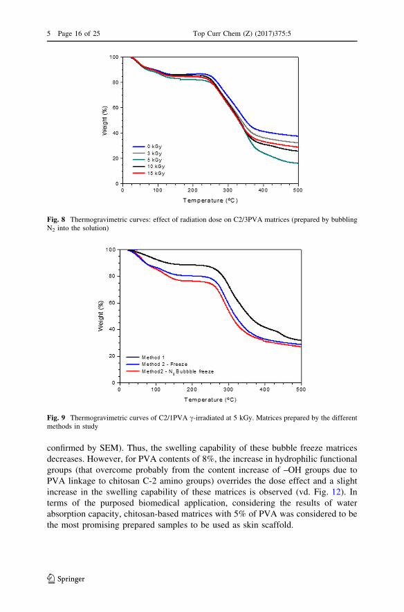
Comparing the dose effect on structural properties of the matrix it is possible to
understand the changes in the structure (vd. Fig. 8). Up to an irradiation dose of
5 kGy the structure stability of the matrices slightly decreases while for higher
irradiation doses it is possible to observe some recovery of the structural stability of
the materials.
In fact, comparing the TGA curves of samples prepared through Method 1 and
Method 2 with and without N2 bubbling (vd. Fig. 9), although showing a similar
thermal profile, Method 2 (freeze-drying of the solutions) leads to matrices with
lower structural stability. This is understandable since the referred methodology
introduces more porosity in the final matrices.
4.2.2 ATR-FTIR Characterization
The ATR-FTIR spectra of samples C2/3PVA and C2/3PVA0.25Gly irradiated at a
total dose of 10 kGy depicted in Fig. 10 show the typical peaks related to chitosan,
PVA, and glycerol previously mentioned. To a better understanding that informa-
tion is also summarized in the figure. A better peak definition and higher intensity
can be observed in the spectrum of the matrix obtained through the freeze
procedure, suggesting a more organized and stable structure when compared with
the one of bubble-freeze procedure. This is in accordance with TGA data.
Fig. 6 SEM micrographs of C2/1PVA matrix c-irradiated at 5 kGy: a surface (scale bar 100 lm);b surface and cross-section (scale bar 10 lm)
5 Page 14 of 25 Top Curr Chem (Z) (2017) 375:5
123

4.2.3 Water Absorption Capacity
It is well known that the water absorption and swelling capacity is one of the
important issues in skin scaffold development. In fact, when the scaffolds are
capable of swelling, they allow their pore size to increase, which facilitates the cells
to attach and penetrate inside the inter-polymeric network. Figure 11 shows the
increase of surface area and volume of bubble freeze (BF) prepared matrices from
dry to swollen state. In the present study, similarly to what occurs in Method 1,
results suggest that the higher the PVA content or the higher the radiation dose is
(within the studied concentration and radiation dose ranges), more crosslinking
between chitosan and PVA chains and/or intermolecular bonds are introduced in
matrices. This confers a stabilizing effect in the structure, which became more
compact and with smaller pores (the pore structures of the matrices were further
Fig. 7 Thermogravimetric curves of Chit/PVA matrices N2 bubbled: a non-irradiated and c-irradiated at5 kGy C2/1PVA; b effect of PVA concentration on matrices c-irradiated at 5 kGy
Top Curr Chem (Z) (2017) 375:5 Page 15 of 25 5
123
confirmed by SEM). Thus, the swelling capability of these bubble freeze matrices
decreases. However, for PVA contents of 8%, the increase in hydrophilic functional
groups (that overcome probably from the content increase of –OH groups due to
PVA linkage to chitosan C-2 amino groups) overrides the dose effect and a slight
increase in the swelling capability of these matrices is observed (vd. Fig. 12). In
terms of the purposed biomedical application, considering the results of water
absorption capacity, chitosan-based matrices with 5% of PVA was considered to be
the most promising prepared samples to be used as skin scaffold.
Fig. 8 Thermogravimetric curves: effect of radiation dose on C2/3PVA matrices (prepared by bubblingN2 into the solution)
Fig. 9 Thermogravimetric curves of C2/1PVA c-irradiated at 5 kGy. Matrices prepared by the differentmethods in study
5 Page 16 of 25 Top Curr Chem (Z) (2017) 375:5
123

4.2.4 In Vitro Stability
The stability of a skin scaffold assumes an important role in the entire wound-
healing process. Generally, it should have a steerable degradability and degradation
rate to match the tissue regeneration. In the particular case of the obtained Chit/PVA
matrices, preliminary studies of its in vitro stability were performed in phosphate-
buffered solution (PBS 1X, pH 7.4) at 37 �C for 24 h. Overall results show that the
higher the radiation dose, the lower the matrices’ mass loss is, since more crosslinks
Fig. 10 ATR-FTIR spectra of C2/3PVA and C2/3PVA0.25Gly matrices irradiated at a total dose of10 kGy
Fig. 11 BF chitosan-based matrices c-irradiated at different radiation doses in a dry and swollen state
Top Curr Chem (Z) (2017) 375:5 Page 17 of 25 5
123

are introduced, conferring more stability to the structure of the material. Comparing
both procedures (vd. Fig. 13), freeze (without bubbling N2) and bubble freeze (with
bubbling N2), the latter one shows higher mass loss. This is observed in all PVA
contents and doses studied and is understandable since in the bubble-freeze
procedure, porosity was introduced in the matrices ‘‘weakening’’ its structure and
helping to washing out eventually free polymer chains. When glycerol is introduced
in theses matrices, the presence of multiple hydroxyl groups and the manifoldness
of its new bonds allows to keep the stability of the polymeric network, fulfilling its
function as plasticizer agent in the blend.
Fig. 12 Water absorption capacity of bubble freeze C2/5PVA and C2/8PVA matrices c-irradiated at 3,10 and 15 kGy
Fig. 13 Mass loss (%) after 1 day of immersion in PBS solution at 37 �C of matrices C2/8PVA non-irradiated and c-irradiated at 3 and 5 kGy
5 Page 18 of 25 Top Curr Chem (Z) (2017) 375:5
123

4.2.5 Morphological Analysis
The morphology of the ‘‘bubble freeze samples’’ was evaluated with SEM.
Figure 14 shows representative SEM micrographs of matrices’ surface morphology
of chitosan-based matrices with different PVA content (without glycerol). It can be
observed that matrices show a good surface homogeneity and an increase in PVA
percentage leads to a decrease in porosity matrices’ surface. Once again, results
suggest that a higher PVA content in Chit/PVA blends leads to a more crosslinked
structure possibly due to more Chit-PVA bonds and OH intramolecular interactions
as already mentioned. Even so, a comparison between Figs. 6 and 14 (SEM
micrographs of C2/1PVA 5 kGy matrix prepared by Method 1) evidences the three-
dimensional porous structure that Method 2 can promote in this type of polymeric
matrix. For the same sample composition, it can be seen that the morphology of
matrices’ surface is also dependent on the radiation dose (vd. Fig. 15). Thus,
radiation dose can be used too to tailor the morphology of the matrices. In the first
irradiation stage, there seems to exist a widespread presence of crosslinking
reactions that lead to a smoother surface due the increasing network density.
However, for higher irradiation doses, some degradation of the terminal chains of
PVA and chitosan ones can occur, promoting ‘‘surface erosion’’ exposing and
Fig. 14 SEM micrographs of the surface of bubble freeze Chit/PVA matrices irradiated at 3 kGy (scalebar 10 lm)
Fig. 15 SEM micrographs of the surface of bubble freeze C2/3PVA matrices when irradiated at differentdoses (scale bar 10 lm)
Top Curr Chem (Z) (2017) 375:5 Page 19 of 25 5
123

accentuating a rougher surface [38]. Together, these results stress out the
importance of this preparation method to achieve in trimming surface properties.
Figure 16 shows the SEM micrographs of the C2/3PVA matrices irradiated at
10 kGy and prepared using the different procedures of Method 2: with N2 bubbling
(bubble freeze) and without (freeze). It can be observed that there is no significant
difference on the surface morphology in the matrices prepared with and without
glycerol. On the other hand, the micrographs also reveal that bubbling of N2 leads to
more open structures, showing to be a good procedure to introduce more porosity in
the matrices.
Additionally, as seen in Fig. 17, despite having some differences in the surface
morphology, all the matrices present a sponge-type ‘‘inner structure’’.
Fig. 16 SEM micrographs of C2/3PVA 10 kGy matrices’ surface using different procedures: with N2
bubbling (bubble freeze) and without (freeze) (scale bar 10 lm)
Fig. 17 Surface and cross-section SEM micrographs of C2/3PVA 10 kGy (scale bar 100 lm)
5 Page 20 of 25 Top Curr Chem (Z) (2017) 375:5
123

4.2.6 In Vitro Cellular Viability
The cellular viability of the HFFF2 fibroblasts was assessed on Chit/PVA matrices
as a means to evaluate their ability to promote cell adhesion and proliferation.
Tested matrices were chosen according to the best structural results previously
obtained. Preliminary cell viability results on Chit/3PVA matrices (bubble freeze,
BF, freeze, F, and with glycerol, Gly) irradiated at different radiation doses (Fig. 18)
show that HFFF2 cells adhere well to the matrices surface but proliferate slower
than control (control = HFFF2 cell cultured under the same condition but without
matrices).
Results point out that only matrices c-irradiated at 10 kGy display cellular
growth on day 1. This is in accordance with SEM analysis, which has revealed that
chitosan-based matrices c-irradiated at 3 and 15 kGy lead to matrices with small
surface pores hindering cells to attach. However, despite showing a good porosity,
the matrix with glycerol (irradiated at 10 kGy) does not show cell growth after the
first day. It appears that cells were able to attach but unable to follow this
attachment with spreading. This might be due to changes in ionic equilibrium (pH)
introduced by glycerol that would be intolerable to cell viability. On the other hand,
still within this group of matrices c-irradiated at 10 kGy, results at day 4 revealed
that only matrices N2 bubbled presented an increase in cell proliferation.
Given the obtained results, only matrices with 3 and 5% of PVA prepared by
bubble freeze and freeze procedure were further evaluated in terms of cell viability.
To comprise all the information concerning irradiation dose (x), PVA content (y),
and preparation procedure [bubble freeze (BF) and freeze (F)], matrices in the
figures of this section are referred to as ‘‘xkGy y BF’’ or ‘‘xkGy y F’’. The results of
matrices irradiated at 5 and 10 kGy (Fig. 19) confirm the HFFF2 cell viability, i.e.,
cells adhesion and proliferation capability into the substrates in study. Once again,
cells adhered to all matrices, which is indicative of the non-cytotoxic nature of the
prepared matrices. However, cells proliferated slower than in controls with the
‘‘freeze procedure’’ matrices presenting a very low proliferation with the matrix C2/
Fig. 18 Cells growing on C2/3PVP and C2/3PVP0.25Gly matrices in culture days 1 and 4. Matricesprepared by bubble freeze (BF) and freeze (F) procedures, and c-radiation doses of 3, 10 and 15 kGy
Top Curr Chem (Z) (2017) 375:5 Page 21 of 25 5
123

5PVA N2 ‘‘bubble freeze’’ presenting the best results. Moreover, the cytochemical
stainings performed showed that cell morphology at day 7 is different to glass
coverslip-seeded fibroblasts (Fig. 20).
Confocal images of control samples at day 7 of culture showed HFFF2 cells with
the expected morphology, a fusiform shape. In confocal images of the Chit2/5PVA
BF 10 kGy c-irradiated matrix (10 kGy 5 BF as referred on Fig. 19), we can only
observe a few cells with round morphology and poor actin cytoskeletal organization
as compared to control cells. This result may indicate that cells are not attaching to
the substrate and growing in the best conditions but, even so, some cells were able
Fig. 19 Cells growing on different Chit/PVA matrices (10 and 5 kGy irradiated; 3 and 5% PVA; bubblefreeze (BF) and freeze (F) preparation procedure) in culture days 1, 4 and 7 (n = 3; mean value ± SD)
Fig. 20 HFFF2 cells growing in culture for 7 days on: a control cells; b 10 kGy 5 BF matrices (out offocus cells asterisk; green actin; blue DNA; scale bar 50 lm)
5 Page 22 of 25 Top Curr Chem (Z) (2017) 375:5
123

to invade the depth of the matrix, as can be seen by the out-of-focus cells in Fig. 20.
This is consistent with the viability assay, which showed reduced cell number.
Huang et al. [39] observed similar results having suggested that inhibition of cell
proliferation on chitosan scaffolds is due to the loss of cell–matrix binding. This
would prevent cells from recognizing and anchoring to new binding sites, and thus
cells would lose some cytoskeleton integrity. Furthermore, the incorporation of
proteins or peptides in the matrix that would allow the presence of multiple cell-
binding ligands or the surface modification of matrix by plasma treatment that
would change the electrostatic interactions may be tested in the future in order to
improve cell affinity for the matrices and to achieve better morphology and behavior
of the fibroblasts growing on these scaffolds.
5 Conclusions
Chitosan-based matrices for tissue regeneration were successfully prepared by
gamma irradiation. Two preparation methods were tested in order to optimize the
methodology. The first one involved the casting of copolymeric solutions and
solvent evaporation at room temperature followed by freeze-drying and irradiation.
The second approach involved the freeze-drying of copolymeric solutions followed
by irradiation.
The matrices were evaluated in terms of methodology, composition, absorbed
dose, structural and functional properties, and in vitro biocompatibility (cellular
viability, morphology and cytochemistry). It was found that c-irradiation and PVA
content can be used to tailor the matrices’ surface in terms of porosity/roughness
with simultaneous sterilization. The bubbling of N2 before freeze-dry showed to be
a good procedure for introducing more porosity in the matrices although it led to
higher matrices’ mass loss. The swelling capability of non-irradiated matrices
increases with the number of hydrophilic groups, i.e., with the PVA content.
Moreover, the swelling properties are also dependent on the density of network
structure.
Concerning the evaluation of cell viability, it was shown that HFFF2 cells
adhered to the surface of all matrices obtained by solutions freeze-drying method,
but do not reveal favorable cellular growth if glycerol is present in the composition.
Bubbling the polymer solution during the homogenization step followed by
freeze-drying and a c-irradiation dose up to 10 kGy seems to be the most promising
procedure. Further modifications to introduce multiple cell-binding ligands in the
matrices should be applied in order to improve cell affinity for the matrices and to
achieve a better morphology and behavior of the fibroblasts growing on these
scaffolds. Nevertheless, the study herein described evidence the versatility of
radiation technology to tailor polymeric materials for specific applications such as
skin regenerative medicine.
Acknowledgements C2TN/IST authors gratefully acknowledge the Fundacao para a Ciencia e
Tecnologia support through the UID/Multi/04349/2013 project. The authors also acknowledge the
International Atomic Energy Agency under the Research Contract No. 18202 for financial support of this
Top Curr Chem (Z) (2017) 375:5 Page 23 of 25 5
123

work. The authors would also like to thank the Erasmus student Reda Paitian (University of Vilnius,
Lithuania) for her collaboration in the preparation and characterization of chitosan-based matrices.
References
1. Ayres CE, Jha BS, Sell SA, Bowlin GL, Simpson DG (2010) Nanotechnology in the design of soft
tissue scaffolds: innovations in structure and function—advanced review. WIREs Nanomed
Nanobiotechnol 2:20–34
2. O’Brien CE (2011) Biomaterials & scaffolds for tissue engineering. Mater Today 14:88–95
3. Van Vlierberghe S, Dubruel P, Schacht E (2011) Biopolymer-based hydrogels as scaffolds for tissue
engineering applications: a review. Biomacromolecules 12:1387–1408
4. Hasirci V, Vrana E, Zorlutuna P, Ndreu A, Yilgor P, Basmanav FB, Aydin E (2006) Nanobioma-
terials: a review of the existing science and technology, and new approaches. J Biomater Sci Polym
Ed 17:1241–1268
5. Shalumona KT, Anulekhaa KH, Chennazhia KP, Tamurab H, Naira SV, Jayakumar R (2011)
Fabrication of chitosan/poly(caprolactone) nanofibrous scaffold for bone and skin tissue engineering.
Int J Biol Macromol 48:571–576
6. Gomes SR, Rodrigues G, Martins GG, Henriques CMR, Silva JC (2013) In vitro evaluation of
crosslinked electrospun fish gelatin scaffolds. Mater Sci Eng C 33:1219–1227
7. Dorozhkin SV (2010) Bioceramics of calcium ortophosphates. Biomaterials 31:1465–1485
8. Liao S, Wang W, Uo M, Ohkawa S, Akasaka T, Tamura K, Cui F, Watari F (2005) A tree-layered
nano-carbonated hydroxyapatite/collagen/PLGA composite membrane for guided tissue regenera-
tion. Biomaterials 26:7564–7571
9. Frohbergh ME, Katsman A, Botta GP, Lazarovici P, Schauer CL, Wegst UGK, Lelkes PI (2012)
Electrospun hydroxyapatite-containing chitosan nanofibers crosslinked with genipin for bone tissue
engineering. Biomaterials 33:9167–9178
10. Martins A, Reis R, Neves N (2008) Electrospinning: processing technique for tissue engineering
scaffolding. Int Mater Rev 53:257–274
11. Safinia L, Datan N, Hohse M, Mantalaris A, Bismarck A (2005) Towards a methodology for the
effective surface modification of porous polymer scaffolds. Biomaterials 26:7537–7547
12. Rodas ACD, Ohnuki T, Mathor MB, Lugao AB (2005) Irradiated PVAl membrane swelled with
chitosan solution as dermal equivalent. Nucl Instrum Methods B 236:536–539
13. Plikk P, Odelius K, Hakkarainen M, Albertsson AC (2006) Finalizing the properties of porous
scaffolds of aliphatic polyesters through radiation sterilization. Biomaterials 27:5335–5347
14. Odelius K, Plikk P, Albertsson AC (2008) The influence of composition of porous copolyester
scaffolds on reactions induced by irradiation sterilization. Biomaterials 29:129–140
15. Cottam E, Hukins DWL, Lee K, Hewitt C, Jenkins MJ (2009) Effect of sterilisation by gamma
irradiation on the ability of polycaprolactone (PCL) to act as a scaffold material. Med Eng Phys
31:221–226
16. Luyen DV, Huong DM (1996) In: Salamone JC (ed) Polymeric Materials Encyclopedia, vol 2. CRC
Press, New York
17. Risbud MV, Hardikar AA, Bhat SV, Bhonde RR (2000) pH-sensitive freeze-dried chitosan-polyvinyl
pyrrolidone hydrogels as controlled release system for antibiotic delivery. J Control Release
68:23–30
18. Ishihara M, Nakanishi K, Ono K, Sato M, Kikuchi M, Saito Y, Yura H, Matsui T, Hattori H,
Uenoyama M, Kurita A (2002) Photocrosslinkable chitosan as a dressing for wound occlusion and
accelerator in healing process. Biomaterials 23:833–840
19. Jayakumar R, Prabaharan M, Reis RL, Mano JF (2005) Graft copolymerized chitosan-present status
and applications. Carbohydr Polym 62:142–158
20. Casimiro MH, Leal JP, Gil MH (2005) Characterisation of gamma irradiated chitosan/pHEMA
membranes for biomedical purposes. Nucl Instrum Methods B 236:482–487
21. Shi C, Zhu Y, Ran X, Wang M, Su Y, Cheng T (2006) Therapeutic potential of chitosan and its
derivatives in regenerative medicine. J Surg Res 133:185–192
22. Casimiro MH, Gil MH, Leal JP (2010) Suitability of gamma irradiated chitosan based membranes as
matrix in drug release system. Int J Pharm 395:142–146
5 Page 24 of 25 Top Curr Chem (Z) (2017) 375:5
123

23. Alves NM, Mano JF (2008) Chitosan derivatives obtained by chemical modifications for biomedical
and environmental applications. Int J Biol Macromol 43:401–414
24. Luyen DV, Huong DM (1996) In: Salamone JC (ed) Polymeric materials encyclopedia chitin and
derivatives. CRC, New York
25. Ulanski P, Rosiak J (1992) Preliminary studies on radiation-induced changes in chitosan. Radiat Phys
Chem 39:53–57
26. Lim LY, Khor E, Koo O (1998) Gamma irradiation of chitosan. J Biomed Mater Res 43:282–290
27. Casimiro MH, Leal JP, Gil MH (2005) Characterisation of gamma-irradiated chitosan/pHEMA
membranes for biomedical purposes. Nucl Instrum Methods B 236:482–487
28. Wiley Billmeyer FW (1984) Textbook of polymer science, 3rd edn. Wiley, New York
29. Hassan CM, Peppas NA (2000) In: Dusek K (ed) Structure and applications of poly(vinyl alcohol),
vol 153. Springer, Berlin
30. Schmedlen RH, Masters KS, West JL (2002) Photocrosslinkable polyvinyl alcohol hydrogels that can
be modified with cell adhesion peptides for use in tissue engineering. Biomaterials 23(22):4325–4332
31. Solaro R, Corti A, Chiellini E (2000) Biodegradation of poly(vinyl alcohol) with different molecular
weights and degree of hydrolysis. Polym Adv Technol 11:873–878
32. Bolto B, Tran T, Hoang M, Xie Z (2009) Crosslinked poly(vinyl alcohol) membranes. Prog Polym
Sci 34:969–981
33. Tamariz E, Rios-Ramırez A (2013) Biodegradation of medical purpose polymeric materials and their
impact on biocompatibility. INTECH. doi:10.5772/56220
34. Zhang H, Grinstaff MW (2014) Recent advances in glycerol polymers: chemistry and biomedical
applications. Macromol Rapid Commun 35:1906–1924
35. Lim L-Y, Khor E, Koo O (1998) c irradiation of chitosan. J Appl Polym Sci 43:290–382
36. Paulino AT, Simionato JI, Garcia JC, Nozaki J (2006) Characterization of chitosan and chitin
produced from silkworm chrysalides. Carbohydr Polym 64:98–103
37. Yue W (2014) Prevention of browning of depolymerized chitosan obtained by gamma irradiation.
Carbohydr Polym 101:857–863
38. Ferreira LM, Leal JP, Casimiro MH, Cruz C, Lancastre JJH, Falcao NA (2014) Evidence of structural
order recovery in LDPE based copolymers prepared by gamma irradiation. Radiat Phys Chem
94:31–35
39. Huang Y, Onyeri S, Siewe M, Moshfeghian A, Madihally SV (2005) In vitro characterization of
chitosan-gelatin scaffolds for tissue engineering. Biomaterials 26:7616–7627
Top Curr Chem (Z) (2017) 375:5 Page 25 of 25 5
123


![Cytocompatibility of Chitosan and Collagen-Chitosan ...forms the highly porous structure of the scaffolds[13] Two percent (w/v) of chitosan was prepared by dissolving chitosan in 0.2](https://img.dokumen.tips/doc/110x75/5e3f1725786dcc56c068fc16/cytocompatibility-of-chitosan-and-collagen-chitosan-forms-the-highly-porous.jpg)