Embed Size (px)
DESCRIPTION
Family and Community Medicine in Rwanda
Citation preview

CHEST X RAY
KABERA René MD, M.Med CandidateResident PGY II, Family and Community Medicine Faculty of Medicine National University of Rwanda

• Differences between radiodence and radiolucent
objects on an x-ray film.
• Four most common x-ray view.
• Steps of reading a chest x-ray.
• Identify major landmarks on chest x-ray.
• Major syndromes on a CXR.
• References .
Objectives
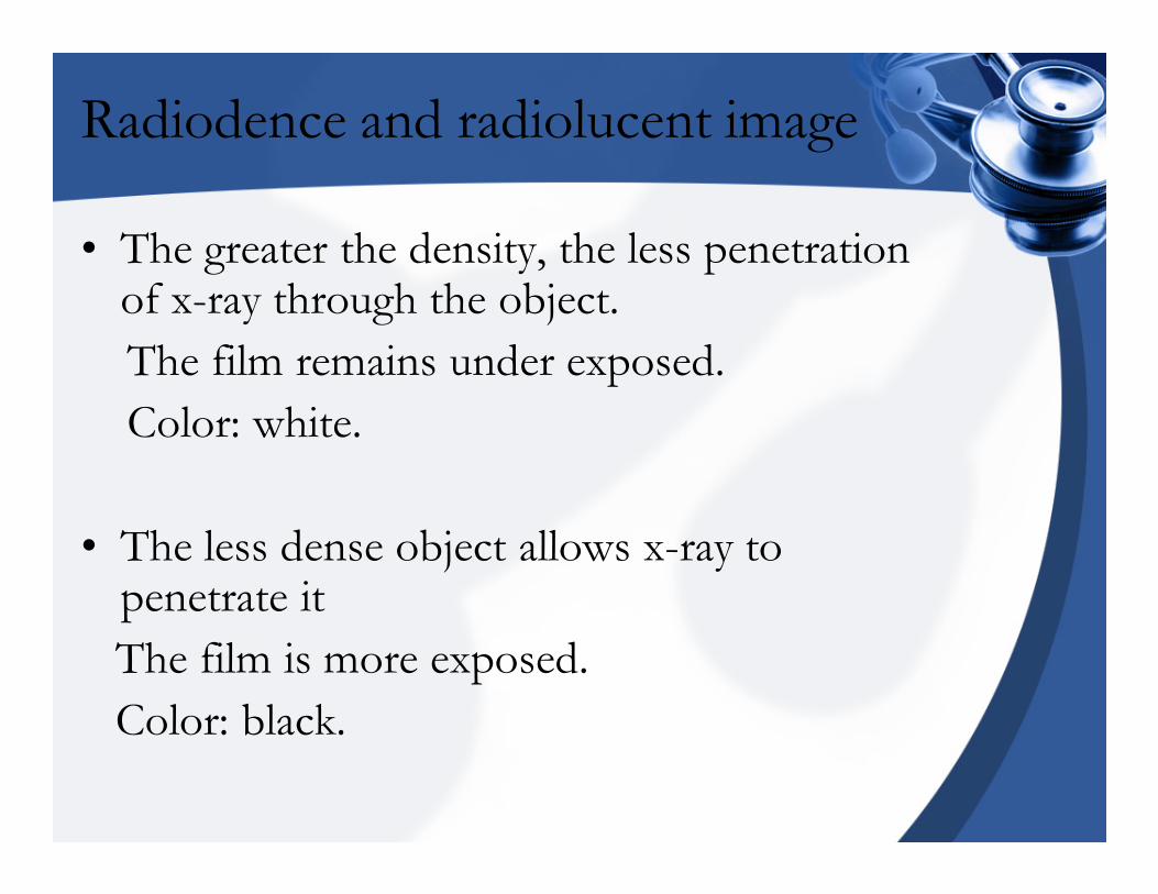
Radiodence and radiolucent image
• The greater the density, the less penetration of x-ray through the object.
The film remains under exposed.
Color: white.
• The less dense object allows x-ray to penetrate it
The film is more exposed.
Color: black.
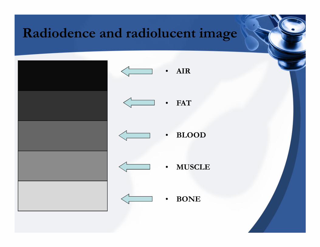
Radiodence and radiolucent image
• AIR
• FAT
• BLOOD
• MUSCLE
• BONE
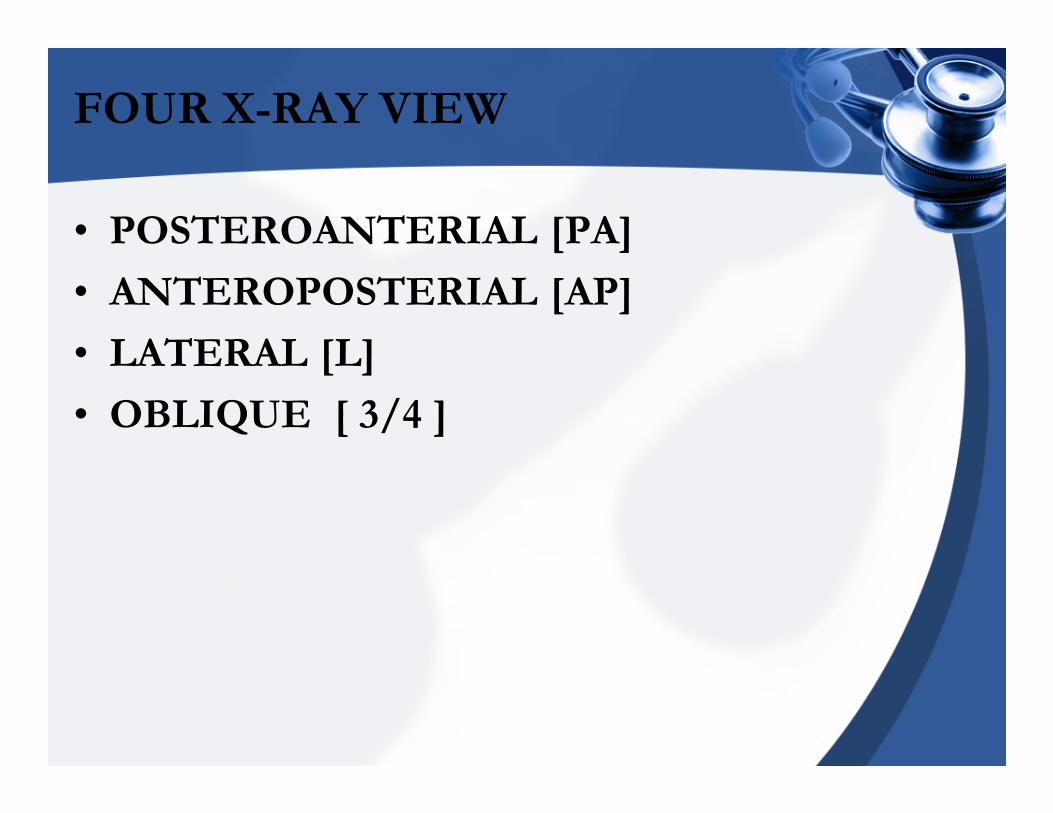
FOUR X-RAY VIEW
• POSTEROANTERIAL [PA]
• ANTEROPOSTERIAL [AP]
• LATERAL [L]
• OBLIQUE [ 3/4 ]
FOUR X-RAY VIEW
• Typical CXR is a PA
• More views are better than one bcs
the body is 3 dimensional.
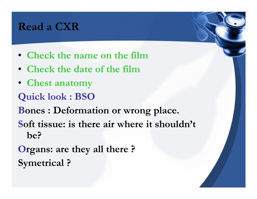
Read a CXR
• Check the name on the film
• Check the date of the film
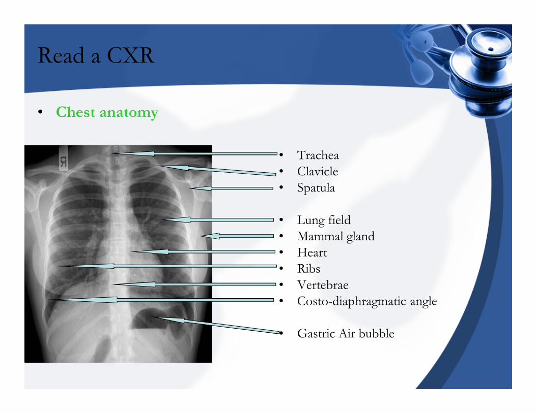
• Chest anatomy
Quick look : BSO
Bones : Deformation or wrong place.
Soft tissue: is there air where it shouldn’t be?
Organs: are they all there ?
Symetrical ?
Read a CXR
• Chest anatomy
• Trachea
• Clavicle
• Spatula
• Lung field
• Mammal gland
• Heart
• Ribs
• Vertebrae
• Costo-diaphragmatic angle
• Gastric Air bubble
Read a CXR
• PA view
• Clavicles are straight ?
• How many ribs ?
�Count ante ribs.
�Count post ribs.
�Count spaces between post ribs.
�7-8 inspiration
�5-6 expiration
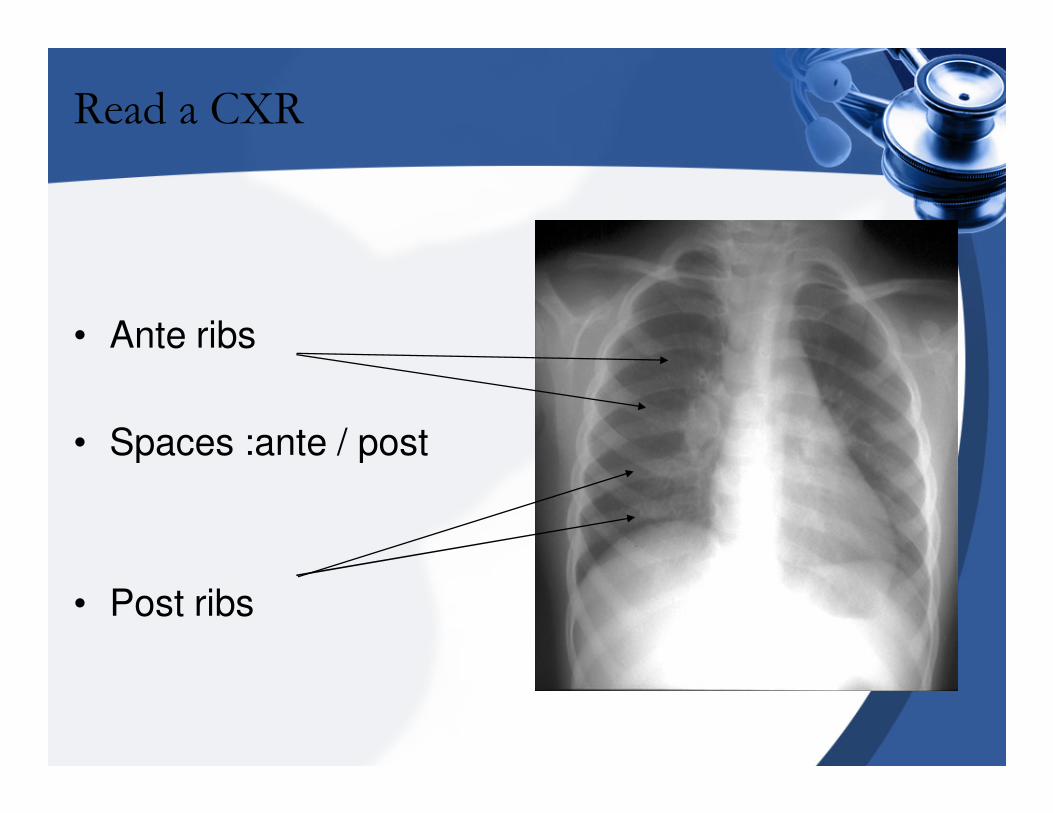
Read a CXR
• Ante ribs
• Spaces :ante / post
• Post ribs
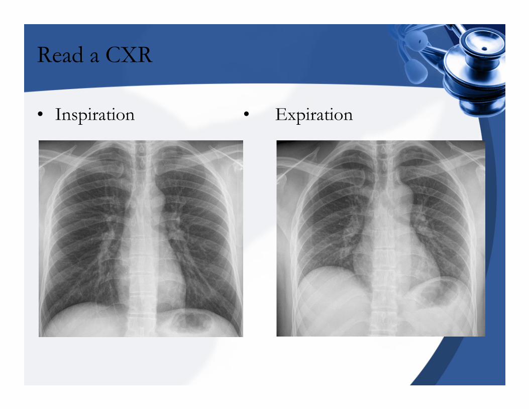
Read a CXR
• Inspiration • Expiration
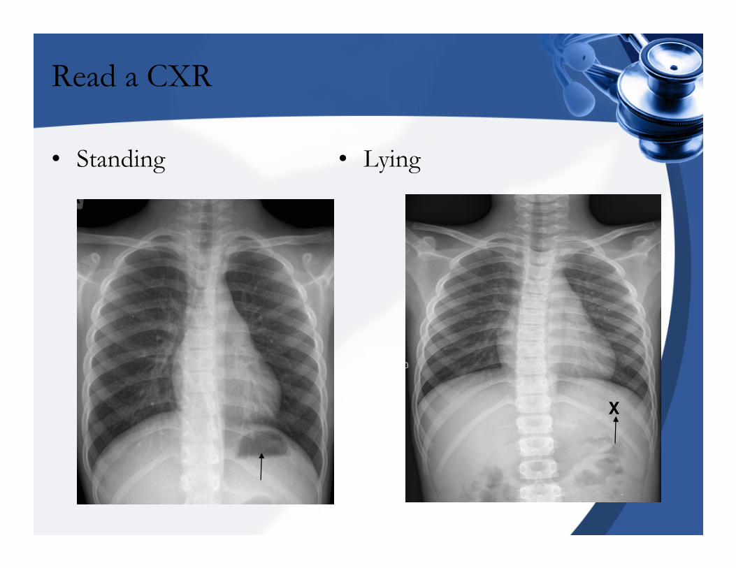
Read a CXR
• Standing • Lying
X
Read a CXR
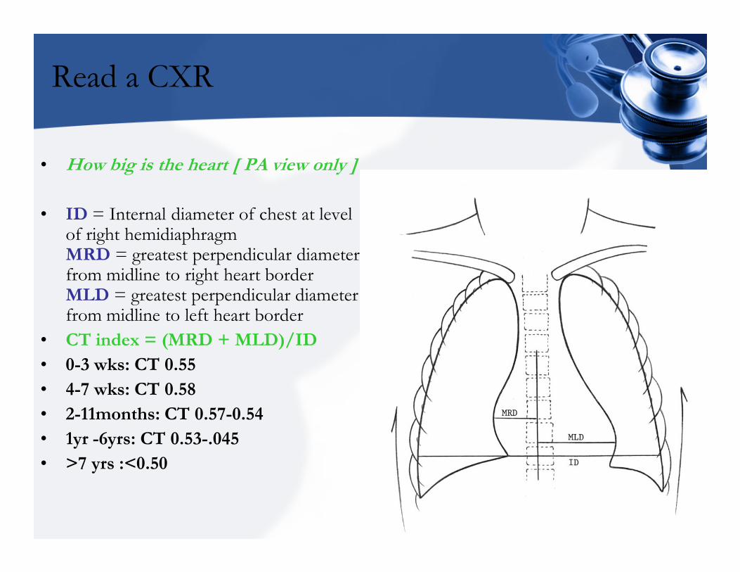
• How big is the heart [ PA view only ]
• ID = Internal diameter of chest at level of right hemidiaphragm MRD = greatest perpendicular diameter from midline to right heart border MLD = greatest perpendicular diameter from midline to left heart border
• CT index = (MRD + MLD)/ID
• 0-3 wks: CT 0.55
• 4-7 wks: CT 0.58
• 2-11months: CT 0.57-0.54
• 1yr -6yrs: CT 0.53-.045
• >7 yrs :<0.50
Read a CXR
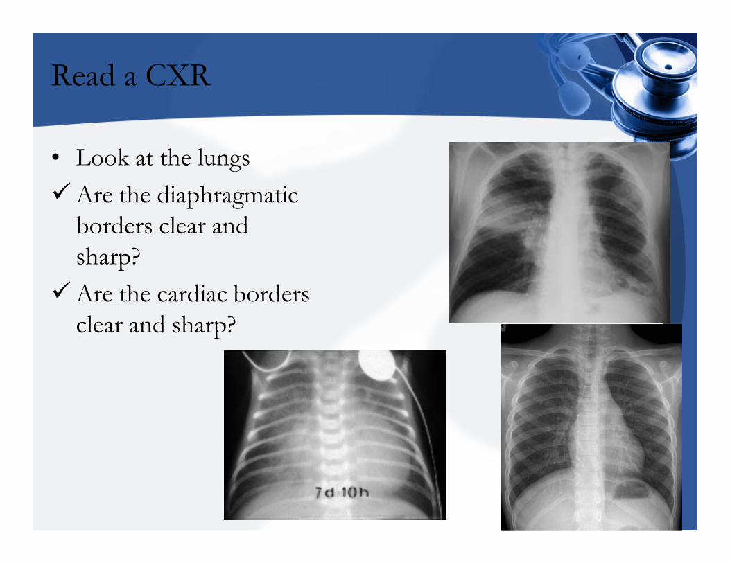
• Look at the lungs
�Are the diaphragmatic
borders clear and
sharp?
�Are the cardiac borders
clear and sharp?
Read a CXR
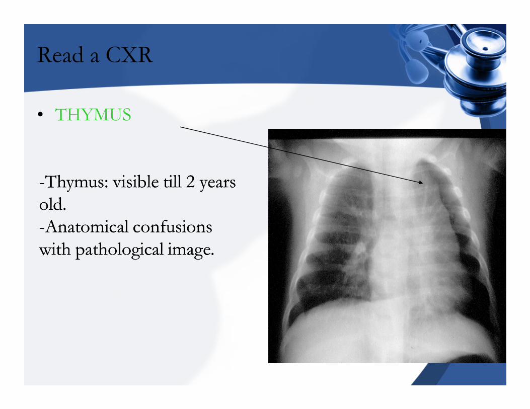
• THYMUS
--Thymus: visible till Thymus: visible till 2 2 years years
old.old.
--Anatomical confusions Anatomical confusions
with pathological image. with pathological image.
Read a CXR
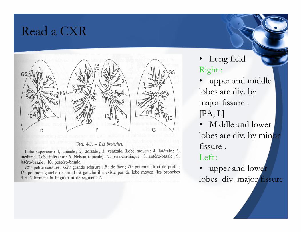
• Lung field
Right :
• upper and middle
lobes are div. by
major fissure .
[PA, L]
• Middle and lower
lobes are div. by minor
fissure .
Left :
• upper and lower
lobes div. major fissure
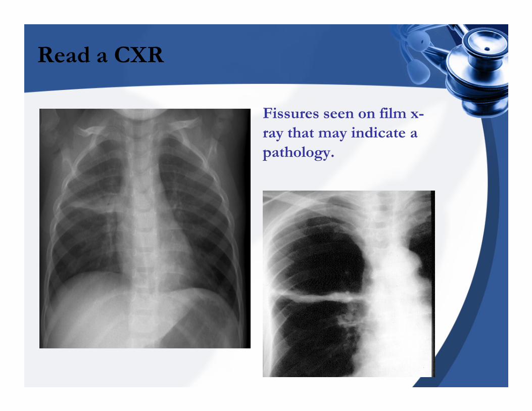
Read a CXR
• Fissures seen on film x-
ray that may indicate a
pathology.
Read a CXR
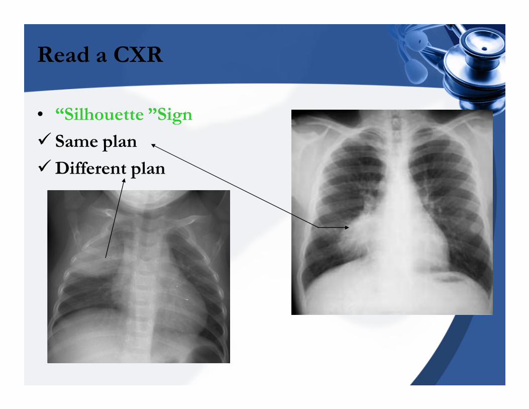
• “Silhouette ”Sign
� Same plan
�Different plan
Read a CXR
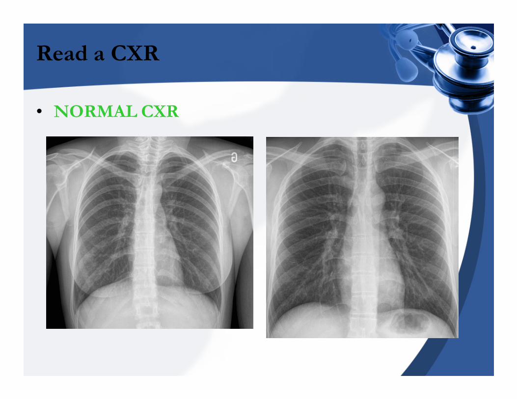
• NORMAL CXR
Major landmarks on CXR
• Alveolar syndrome
• Interstitial syndrome
• Bronchial syndrome
• Pleural syndrome
• Cavity syndrome
• Vascular syndrome
• Parietal syndrome
• Mediastinum syndrome
Major landmarks on CXR
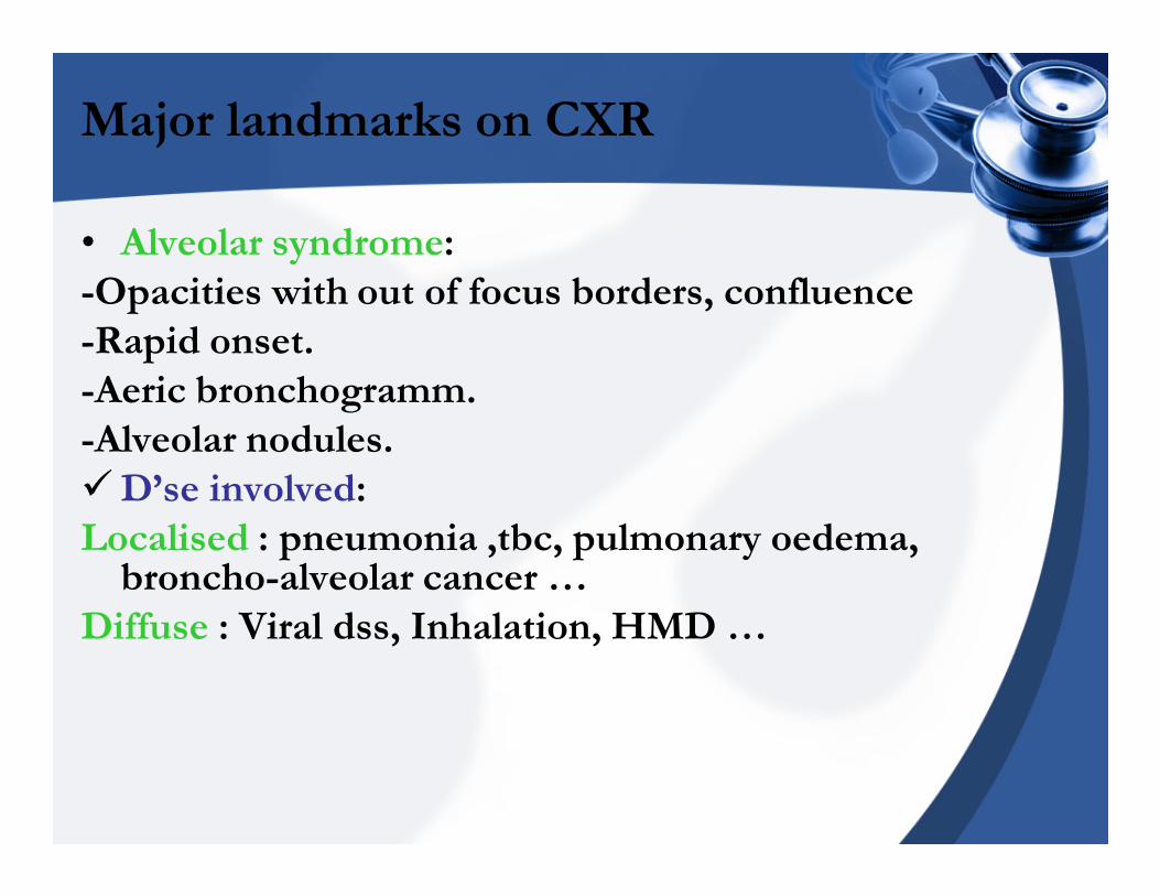
• Alveolar syndrome:
-Opacities with out of focus borders, confluence
-Rapid onset.
-Aeric bronchogramm.
-Alveolar nodules.
�D’se involved:
Localised : pneumonia ,tbc, pulmonary oedema, broncho-alveolar cancer …
Diffuse : Viral dss, Inhalation, HMD …
Major landmarks on CXR
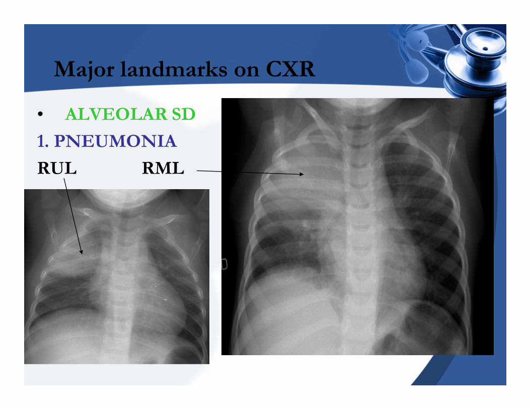
• ALVEOLAR SD
1. PNEUMONIA
RUL RML
Major landmarks on CXR
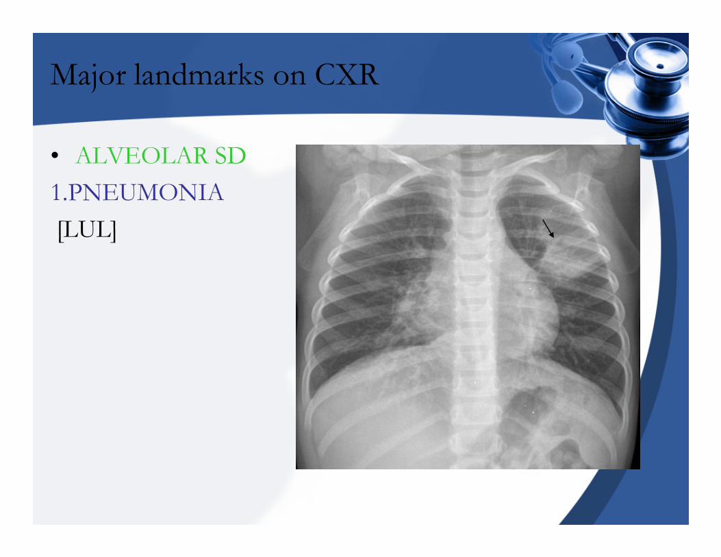
• ALVEOLAR SD
1.PNEUMONIA
[LUL]

Major landmarks on CXR
• ALVEOLAR SD2.BRONCHOPNEUMONIA
Major landmarks on CXR
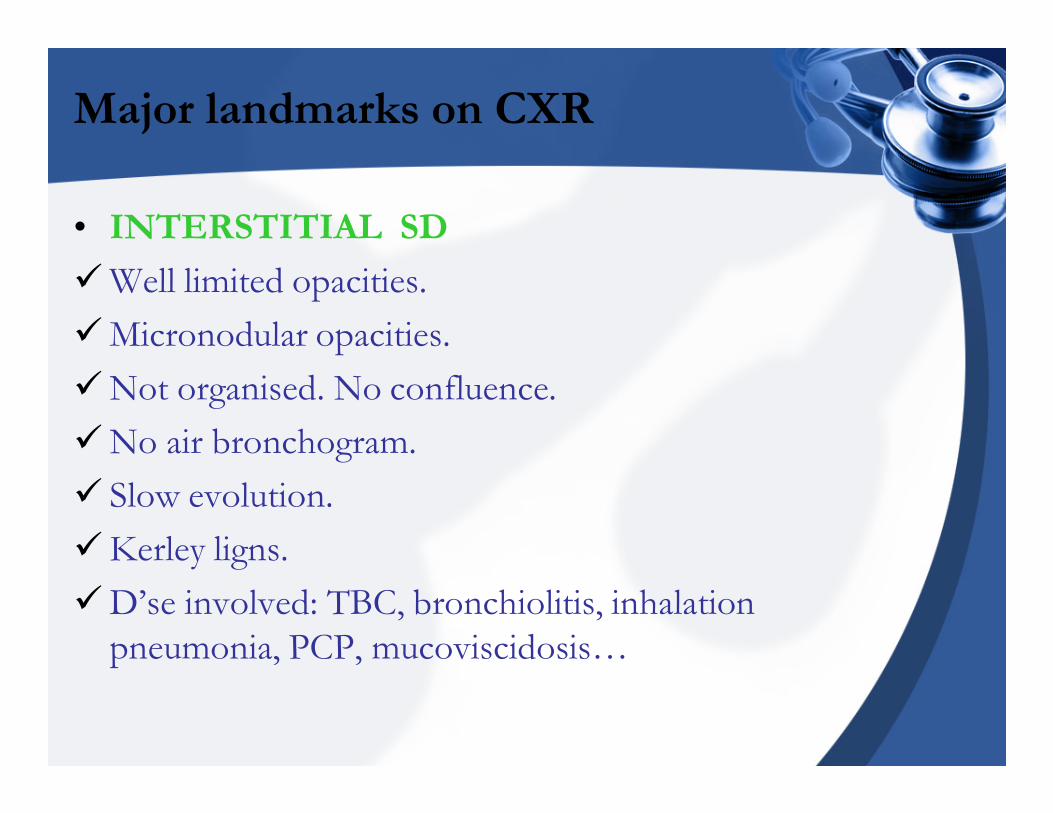
• INTERSTITIAL SD
�Well limited opacities.
�Micronodular opacities.
�Not organised. No confluence.
�No air bronchogram.
� Slow evolution.
�Kerley ligns.
�D’se involved: TBC, bronchiolitis, inhalation
pneumonia, PCP, mucoviscidosis…
Major landmarks on CXR
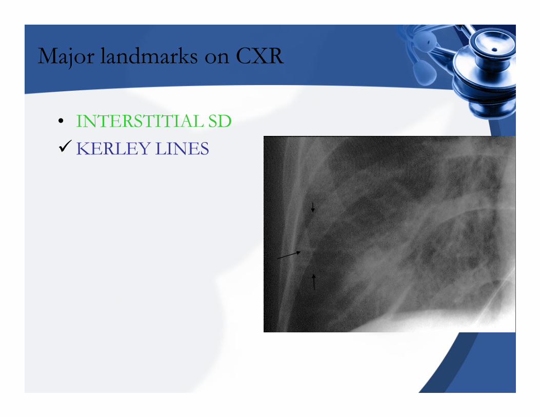
• INTERSTITIAL SD
�KERLEY LINES
Major landmarks on CXR
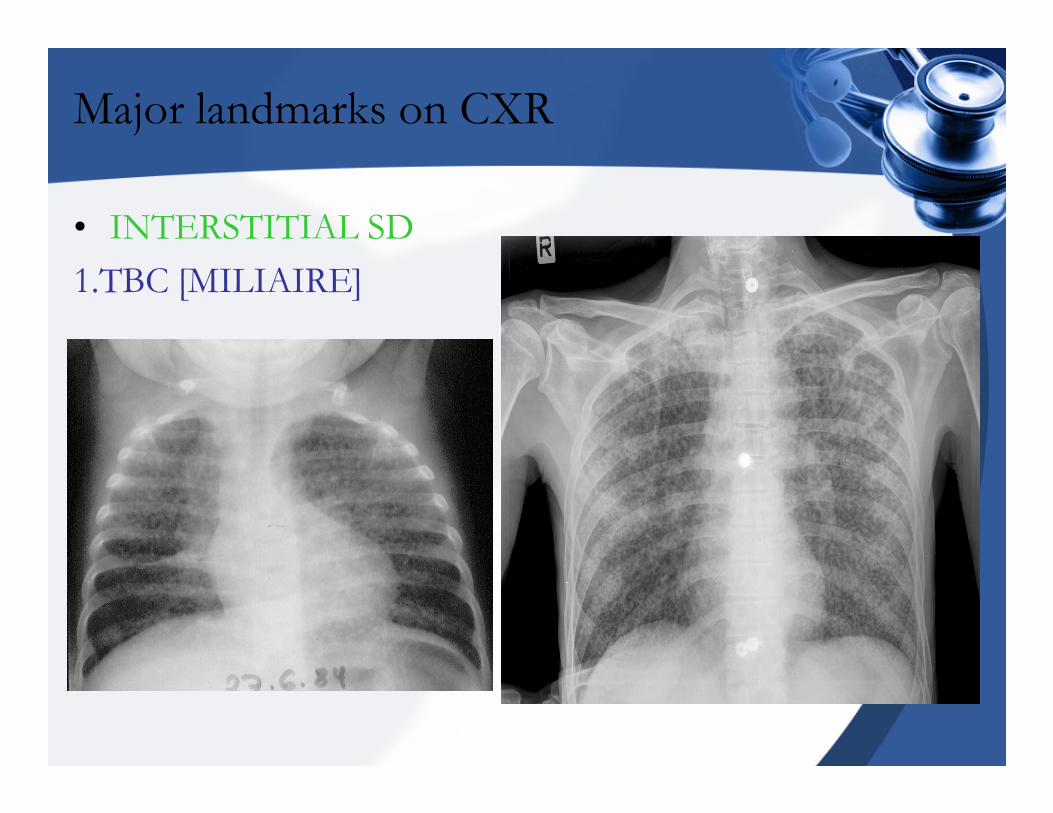
• INTERSTITIAL SD
1.TBC [MILIAIRE]
Major landmarks on CXR
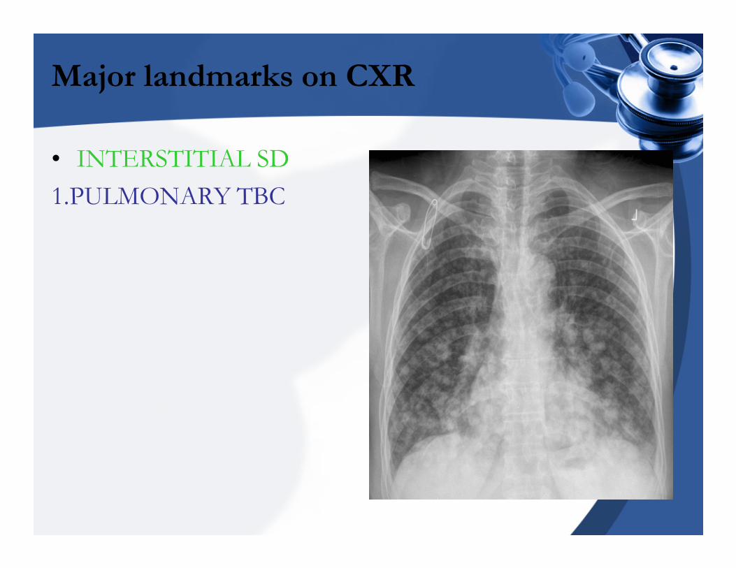
• INTERSTITIAL SD
1.PULMONARY TBC
Major landmarks on CXR
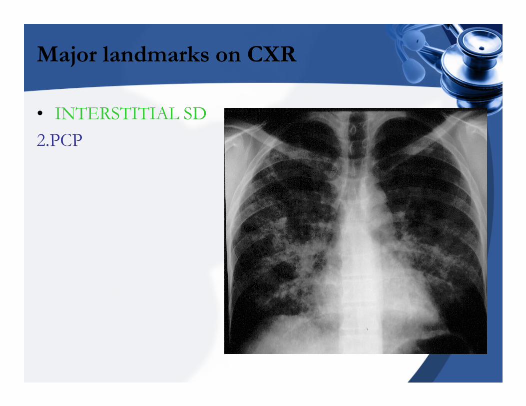
• INTERSTITIAL SD
2.PCP
Major landmarks on CXR
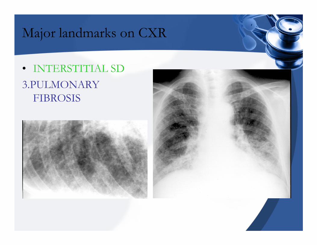
• INTERSTITIAL SD
3.PULMONARY
FIBROSIS
Major landmarks on CXR
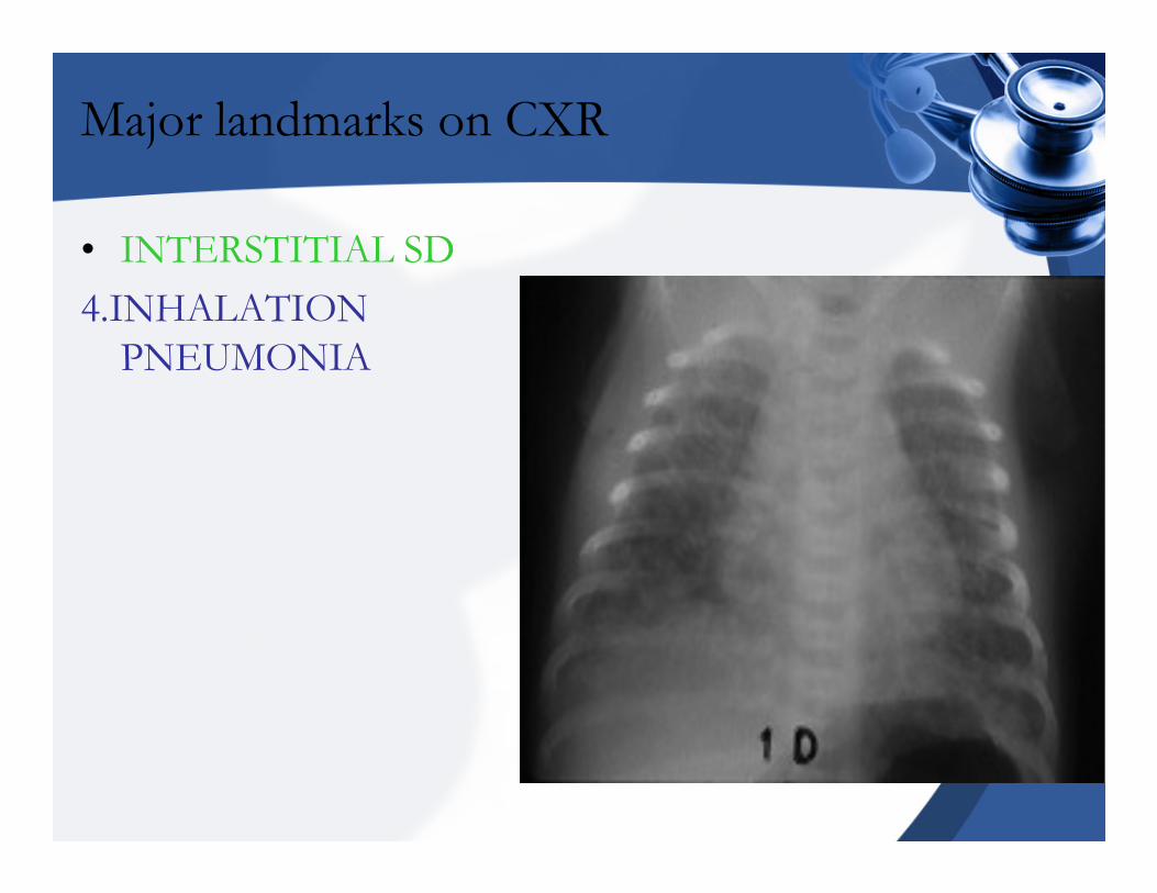
• INTERSTITIAL SD
4.INHALATION
PNEUMONIA
Major landmarks on CXR
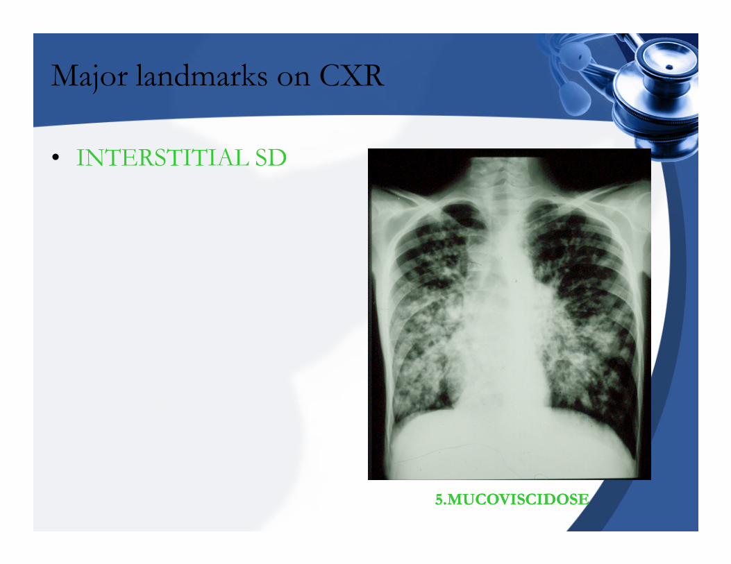
• INTERSTITIAL SD
55.MUCOVISCIDOSE.MUCOVISCIDOSE
Major landmarks on CXR
• BRONCHIAL SD
�Thickening of the bronchial wall .
�Dilatation of the bronchial lumen.
�Hypersecretion ,bronchial obstruction, …
�Obstruction of the bronchial lumen.
�Dse’s involved: chronic bronchitis.
atelectasis
Major landmarks on CXR
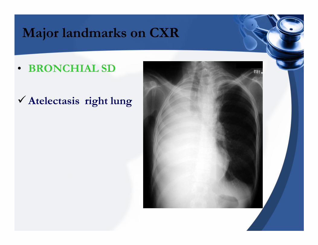
• BRONCHIAL SD
�Atelectasis right lung
Major landmarks on CXR
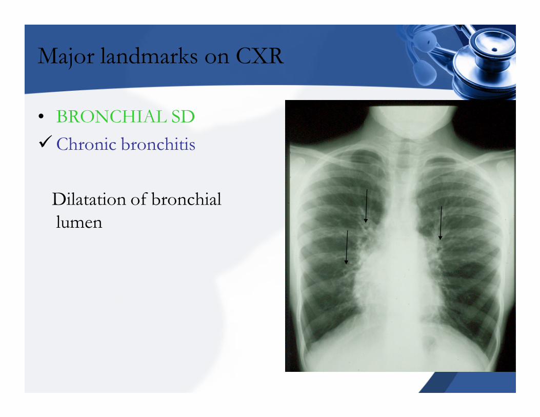
• BRONCHIAL SD
�Chronic bronchitis
Dilatation of bronchial
lumen
Major landmarks on CXR
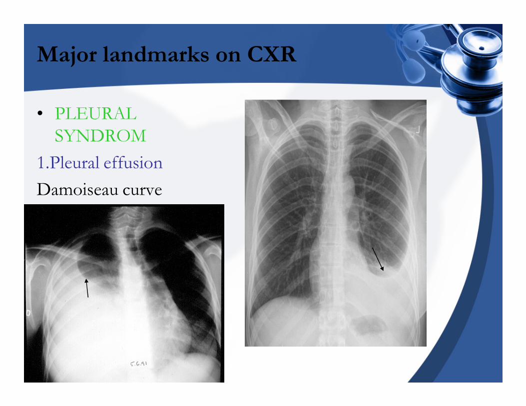
• PLEURAL
SYNDROM
1.Pleural effusion
Damoiseau curve
Major landmarks on CXR
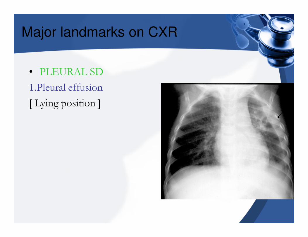
• PLEURAL SD
1.Pleural effusion
[ Lying position ]
Major landmarks on CXR
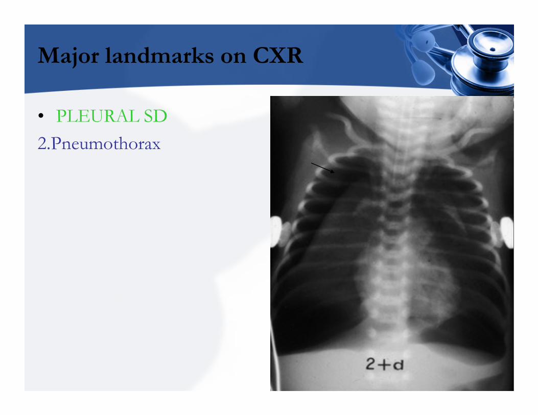
• PLEURAL SD
2.Pneumothorax
Major landmarks on CXR
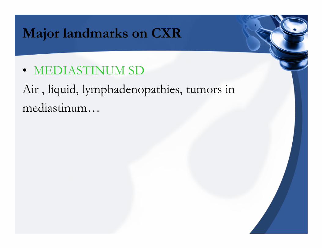
• MEDIASTINUM SD
Air , liquid, lymphadenopathies, tumors in
mediastinum…
Major landmarks on CXR
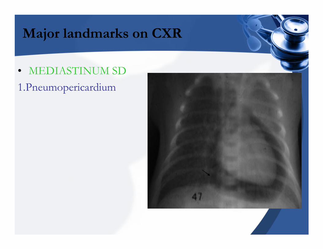
• MEDIASTINUM SD
1.Pneumopericardium
Major landmarks on CXR
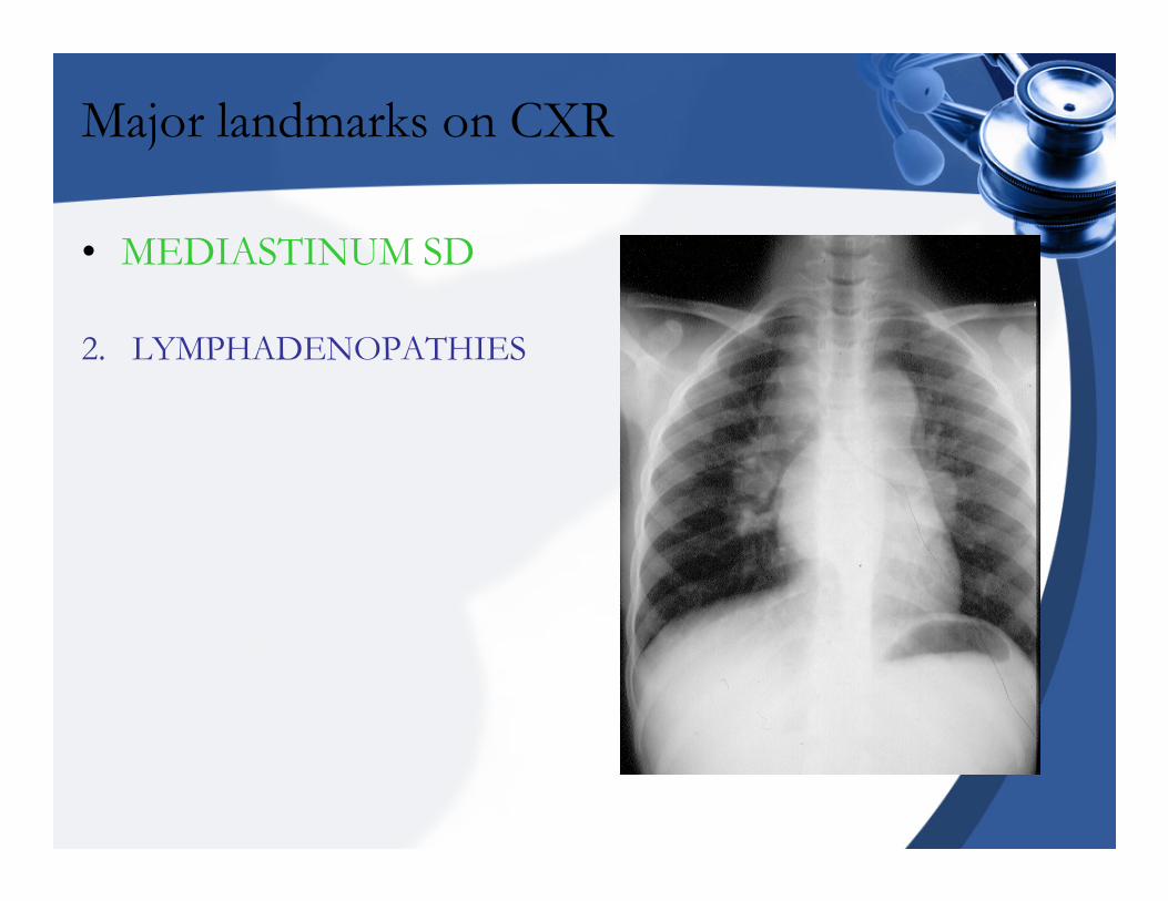
• MEDIASTINUM SD
2. LYMPHADENOPATHIES
Major landmarks on CXR
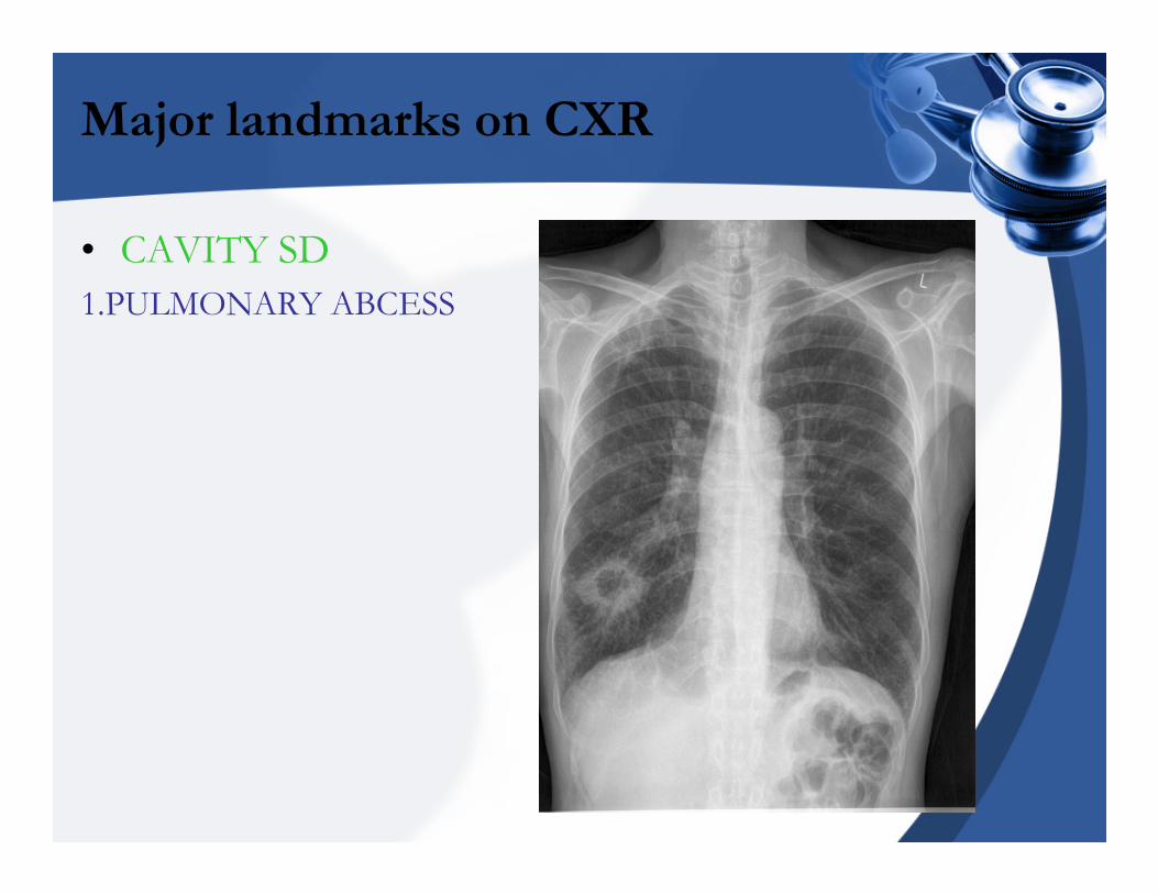
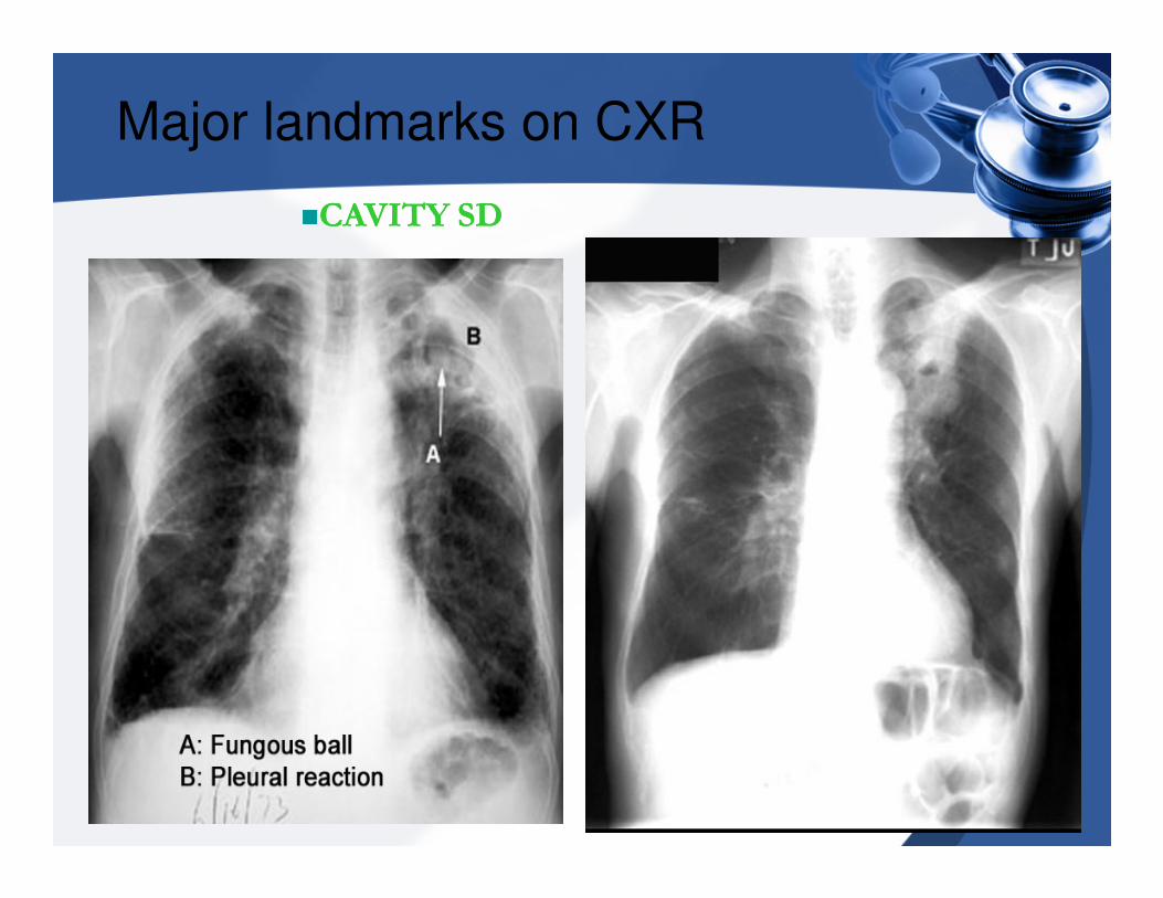
• CAVITY SD
1.PULMONARY ABCESS
Major landmarks on CXR
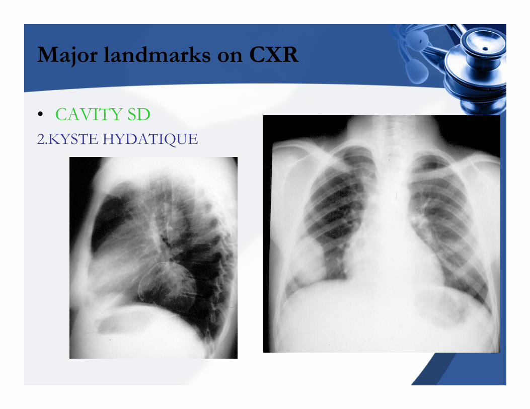
• CAVITY SD
2.KYSTE HYDATIQUE
Major landmarks on CXR
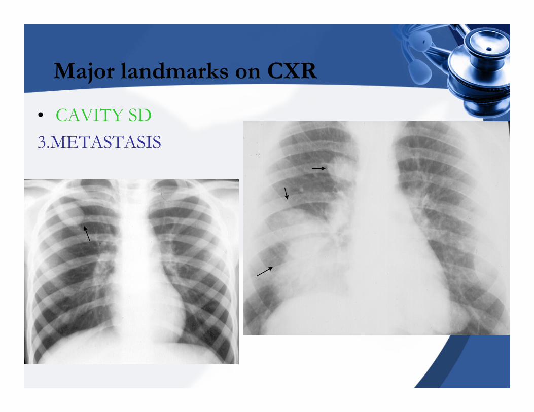
• CAVITY SD
3.METASTASIS
Major landmarks on CXR
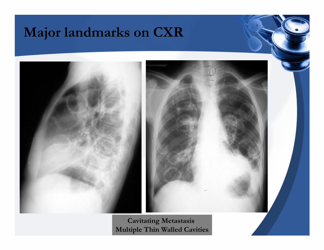
Cavitating Metastasis
Multiple Thin Walled Cavities
Major landmarks on CXR
��CAVITY SDCAVITY SD
Major landmarks on CXR
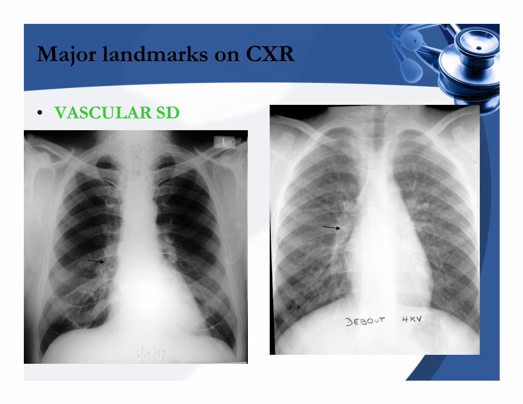
• VASCULAR SD
Major landmarks on CXR
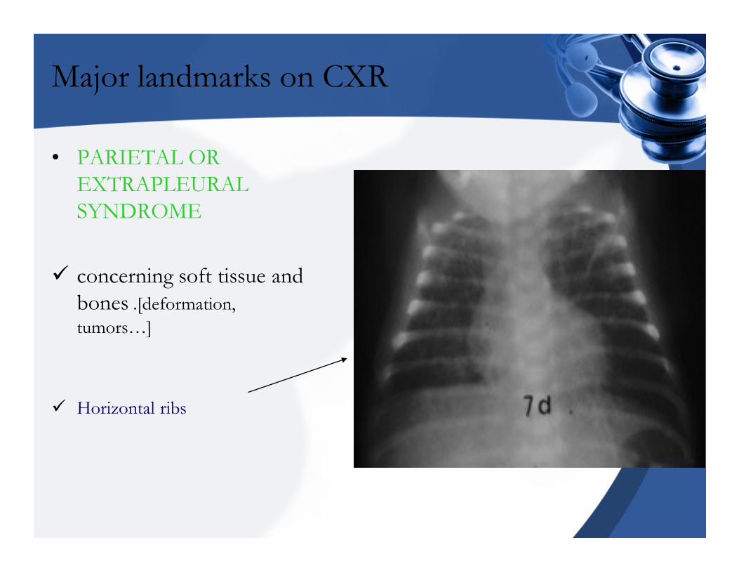
• PARIETAL OR
EXTRAPLEURAL
SYNDROME
� concerning soft tissue and
bones .[deformation,
tumors…]
� Horizontal ribs
Major landmarks on CXR
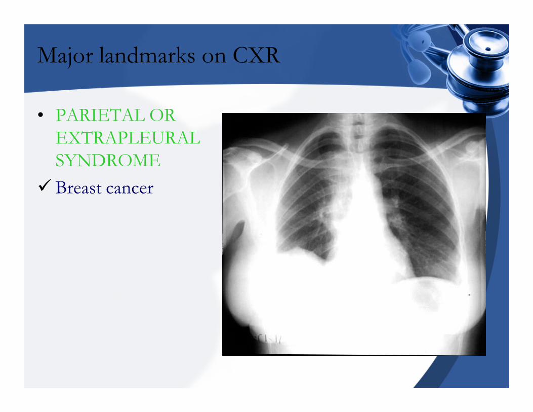
• PARIETAL OR
EXTRAPLEURAL
SYNDROME
�Breast cancer

Major landmarks on CXR
� PARIETAL OR
EXTRAPLEURAL
SYNDROME
� Old fractured ribs
References
• www.uab.edu/pedradpath
• www.pediatricradiology.com
• www.spiral.univ-lyon1.fr/polycops/semeilogie-radiologique
• www.virtualpediatrichospital.org/providers
• Notes de cours “Imagerie médicale”.Doc III Prof SPEHL Marianne.NUR/FACMED
• Chest x-ray Atlas , http://www.meddean.luc.edu/lumen/MedEd/medicine/pulmonar/cxr/atlas/cxratlas_f.htm

THANK YOU
Murakoze