Embed Size (px)
Citation preview

1
A TAF4 coactivator function for E proteins that involves enhanced TFIID binding
Wei-Yi Chen1, Jinsong Zhang1,3, Huimin Geng2, Zhimei Du1,4, Tomoyoshi Nakadai1, and Robert G. Roeder1*
1Laboratory of Biochemistry and Molecular Biology, The Rockefeller University, New York, NY10065, USA
2Laboratory Medicine, UCSF School of Medicine, San Francisco, CA 94143, USA SUPPLEMENTAL INFORMATION Supplemental Materials and Methods Antibodies
Antibodies against TFIIB p55, TFIIEα, and TFIID subunits were described previously (Guermah et al. 2001).
Other antibodies were obtained commercially as follows: anti-E2A, -HEB, -CBP, -p21, -RNA Pol II, and
-TFIIH p89 (Santa Cruz Biotechnology); anti-TAF1 (Abcam; for the ChIP assay in Fig7D); anti-Flag M2 and
M2 agarose (Sigma); anti-His tag (AbD Serotec).
Recombinant proteins and plasmids
GST and GST-tagged HEB [full length (1-682), AD1 (1-99), AD2 (307-548), and AD3 (100-306)], p53,
eTAFH (93-201), and dTAFH (575-706) proteins were expressed in bacteria from pGEX4T-1 vectors and
purified using glutathione-Sepharose beads (Amersham Pharmacia). The 6His-tagged 3xdTAFH (575-706)
domain and the 6His-tagged Gal4-DBD (1-94)-fused AD3 (221-300) and VP16 proteins were expressed in
bacterial from pET28a vectors and affinity purified using Talon beads (Clontech). Flag-tagged full-length
HEB and E47 proteins were cloned in pFASTBac baculovirus vectors, expressed in Sf9 cells and purified
using a heparin-HP column (GE Healthcare Life Sciences) and M2-agarose beads. Recombinant TFIIA was
expressed and purified as previously described (Malik and Roeder 2003). All recombinant proteins were
stored in BC buffer (20 mM Tris-HCl [pH 7.9], 20% glycerol, 0.1 mM EDTA [pH 8.0]) containing 100 mM KCl
(BC100). For mammalian expression constructs, Flag-tagged TAF4 cDNAs were inserted into pIRESneo
vectors and the dTAFH domain (590-675) was deleted by PCR-based site-directed mutagenesis.
Expression vectors for HEB and its derivatives have been described previously (Guo et al. 2009).

2
Pull-down and crosslinking assays
For pull-down assays, recombinant proteins were immobilized on glutathione beads, Ni-NTA resin (Qiagen)
or M2 agarose according to their respective protein tags and then incubated as indicated with nuclear
extract or purified TFIID complex in BC100 buffer containing 0.02% NP40. Reactions were carried out at
4°C for 4 h. Following extensive washing with binding buffer, bound materials were resolved by gel
electrophoresis and visualized by immunoblotting with indicated antibodies. For the DSP crosslinking
experiment, the reaction was carried out in BC300 with 0.1% NP40 according to a previously described
protocol (Wallberg et al. 2003).
Reporter assays
For reporter assays, 293T cells were co-transfected with 20 ng expression constructs for various Gal4-HEB
fragments, 100 ng 5xGal4-UAS-driven firefly luciferase construct, and 5 ng pRL-TK (Promega) using
Lipofectamine 2000 (Invitrogen). Dual luciferase assays (Promega) were performed 24 h post-transfection
according to the manufacturer’s instructions. Firefly luciferase data were normalized to Renilla luciferase
activity (pRL-TK). Data represent the mean ± SD of triplicate experiments and are expressed as fold-
increase over the activity of Gal4-DBD (Vector).
Coimmunoprecipitation assays
For monitoring the endogenous interaction of HEB and TFIID (Figure 1B), HeLa nuclear extract was
incubated with anti-HEB or anti-TAF4 antibodies in BC100 containing 0.02% NP-40 and proteinase inhibitor
cocktail (Roche) at 4°C for 6 h. Immune complexes were precipitated with protein A beads, extensively
washed with binding buffer, and analyzed by gel electrophoresis and immunoblot. Input lanes show 2% of
the samples. For coimmunoprecipitation of the TFIID complex from MEF lines (Figure 7A), extracts were
incubated with anti-TAF1 and anti-TAF4 antibodies in BC300, 0.1% NP40. Input lanes represent 10% of the
samples.
Magnesium-agarose electrophoresis mobility shift assay (Mg-EMSA)
Mg-EMSAs were performed as previously described (Lieberman and Berk 1994) with some modifications.
The DNA probe was prepared by PCR amplification of the pGL3-Gal4-E1B vector, end-labeled, and gel
purified. Binding of Gal4-AD3 (20 ng) and f-TAF4-TFIID complex (containing 25 ng TBP) to the 32P-labeled
DNA probe (6 fmol) was performed in BC70 supplemented with 5 mM MgCl2, 0.5 mg/ml BSA, 40 µg/ml
poly(dG-dC). The reaction was incubated at room temperature for 60 min and followed by native gel
electrophoresis on a 1.4% agarose gel in TBE buffer (45 mM Tris, 45 mM boric acid, and 5 mM magnesium
acetate). The gel was dried and subjected to autoradiography.

3
Immobilized template protein recruitment assay
The immobilized template assay was carried out as described (Black et al. 2006) with modifications. A
biotin-conjugated DNA template was prepared by PCR amplification of pGL3-Gal4-E1B (Figure 5C) or
pGL2-p21 (Figure 6C) vector with biotinylated primers, gel purified, and immobilized on Dynabead M-280
streptavidin (Invitrogen) as instructed by the manufacturer. Blocking was performed in BC100 supplemented
with 0.01% NP40, 1 mg/ml BSA, and 40 µg/ml poly(dG-dC) for 60 min at room temperature. Indicated
proteins were added to the reaction and incubated for 60 min at 4°C with agitation. For dissociation
experiments, beads were washed with binding buffer, suspended in 500 µl binding buffer and incubated at
20°C with agitation for the indicated time. Bound proteins were further washed with binding buffer, eluted in
SDS sample buffer, and detected by immunoblotting. Standard reactions in 100 µl contained 100 ng DNA
probe, 3 µg beads and, as indicated, TAF4wt or TAF4ΔTAFH TFIID complex (containing 50 ng TBP), 20 ng
Gal4-AD3, 100 ng TFIIA, and 100 ng f-HEB or f-E47.
DNase I footprinting assay
DNase I footprinting was performed as previously described (Guermah et al. 2001) with some modifications.
Briefly, the PstI/XbaI-digested DNA fragment (-200 to +27) from pGL2-p21 (Prabhu et al. 1997) was isolated
and end-labeled with 32P by Klenow fill-in reaction. The probe was further purified by gel electrophoresis
and extraction. The binding of f-TAF4-TFIID complexes (containing 25 ng TBP) and TFIIA (100 ng) to the
DNA probe were carried out in the presence or absence of f-HEB (15 ng) in binding buffer [12.5 mM Hepes
(pH 7.9), 12.5% glycerol, 5 mM MgCl2, 70 mM KCl, 0.1 mM EDTA, 1mM DTT, 40 µg/ml poly(dG-dC)] at 4°C
for 60 min. Reactions were then treated with DNase I and DNA fragments were purified and analyzed by gel
electrophoresis and autoradiography.
RNA interference
Lentiviral vectors expressing shRNAs (listed in Table 2) were purchased from OpenBiosystems.
Lentiviruses were prepared according to the recommended protocol on the Addgene website. For knock-
down experiments, 293T cells were infected with indicated viruses and treated with 2 µg/mL puromycin 48-
hr post-infection for 2 days. Total RNA or whole cell lysates were prepared 96-hr post-infection.
RT-qPCR/ChIP-qPCR
Total RNAs were isolated using the RNeasy kit (Qiagen) and cDNAs were made with the qScript cDNA
SuperMix (Quanta Bciosciences) according to manufacturer’s instructions. The SYBR Green PCR Master
Mix (Applied Biosystems) was used in qPCR reactions on a 7300 Real-Time PCR system (Applied

4
Biosystems). The relative levels of mRNA were determined from standard curves and normalized against
GAPDH (human) or Hprt (mouse). For ChIP assays, qPCR reactions were performed with the QuantiTect
SYBR Green PCR kit (Qiagen). The fold-differences were calculated according to the 2-ΔCt method and
presented as percentages of input DNAs.

5
Supplemental Table 1: PCR primers Gene Species Forward primer (5′ -‐> 3′) Reverse primer (5′ -‐> 3′) ChIP Assay: P21 Human TATACAGGGCCGCGCTG GGCTCCACAAGGAACTGACTTC p21 Mouse CCCGGGATCGGTGAAGGAG GCGCCTGACTCCAATTCCC Gata 6 Mouse TGGATCCCTCCTCCTTCTCT TACTGCTCTGCCGGAAAACT Dcn Mouse GGATGCAAAGGGATAAAGCA TCCAGCTGACACCCACATTA Pdgfr-α Mouse GGGGACTTCATTTCCTGACA TTCTCTCCCTCAAGCTCCAA Serpinf1 Mouse AGCACTGGGCCAACTCTCTA AGGCTGCATGGAGAGACTGT Fth1 Mouse ACGAAGTCGCTGTCTGGTGTATGT ACAGAGGGTACCGATGAGGATGAT Mif Mouse TACCTGGAATGCCTCGACAAACCT TGTGCTTCTGGTGCTGTAGGAAGT Itga11 Mouse CAAGAAGCACTGTGGCCTTGACAT TGACAGGTCTTCTTCGGACACGTT Mrpplf3 Mouse CCTGCCACTGTGTAACCTCAGGGT AGGCTTAGCTGTGGATTCCGGGA RT-qPCR : P21 Human GAACTTCGACTTTGTCACCGAGAC TGGAGTGGTAGAAATCTGTCATGCT GAPDH Human CCATGGAGAAGGCTGGGGCT GGAGAGCCCCGCGGCCATCA p21 Mouse ATGTCCAATCCTGGTGATGT TGCAGCAGGGCAGAGGAAGT Gata 6 Mouse GACGGCACCGGTCATTACC ACAGTTGGCACAGGACAGTCC Dcn Mouse GATGCGCTCACGCAGTGAAAC ATGCAGCCCAGGCAAAAGGGTT Pdgfr-α Mouse ATGAGAGTGAGATCGAAGGCA CGGCAAGGTATGATGGCAGAG Serpinf1 Mouse TCCTCACGGGCAACCCTCGA GGATGCTGAGGGCACTGGGC Fth1 Mouse GGGAGAGCGGGCTGAATGCAAT TGGTCACGTGGTCACCCAGTTCT Mif Mouse TCAGGTCCCTGGCTTGGGTCAC CTGCGATGTACTGTGCGGGCT Itga11 Mouse AGCGCAATGGCAGGGATGCC ACTGGTCTCCGCCCTCGTCC Mrpplf3 Mouse GGGCTCAGAGGCAAAAGCCCC TCCAGAGGGCTTTCCCAGGCA Hprt Mouse ACCTCTCGAAGTGTTGGATA CAACAACAAACTTGTCTGGA
Supplemental Table 2: Lentiviral vectors for shRNA
Construct Description Clone ID Mature sense sequence Ctrl-1 Non-hairpin control Ctrl-2 Scramble shRNA HEB-1 HEB shRNA TRCN0000015168 GCTGTGATTATGGTGAACATA HEB-2 HEB shRNA TRCN0000015172 GCAATCATTCAGTCCTGTCTA

6
Supplemental References Black JC, Choi JE, Lombardo SR, Carey M. 2006. A mechanism for coordinating chromatin modification
and preinitiation complex assembly. Mol Cell 23: 809-818.
Guermah M, Tao Y, Roeder RG. 2001. Positive and negative TAF(II) functions that suggest a dynamic TFIID structure and elicit synergy with traps in activator-induced transcription. Mol Cell Biol 21: 6882-6894.
Guo C, Hu Q, Yan C, Zhang J. 2009. Multivalent binding of the ETO corepressor to E proteins facilitates dual repression controls targeting chromatin and the basal transcription machinery. Mol Cell Biol 29: 2644-2657.
Lieberman PM, Berk AJ. 1994. A mechanism for TAFs in transcriptional activation: activation domain enhancement of TFIID-TFIIA--promoter DNA complex formation. Genes Dev 8: 995-1006.
Malik S, Roeder RG. 2003. Isolation and functional characterization of the TRAP/mediator complex. Methods Enzymol 364: 257-284.
Prabhu S, Ignatova A, Park ST, Sun XH. 1997. Regulation of the expression of cyclin-dependent kinase inhibitor p21 by E2A and Id proteins. Mol Cell Biol 17: 5888-5896.
Wallberg AE, Yamamura S, Malik S, Spiegelman BM, Roeder RG. 2003. Coordination of p300-mediated chromatin remodeling and TRAP/mediator function through coactivator PGC-1alpha. Mol Cell 12: 1137-1149.

7

8

9

10
11
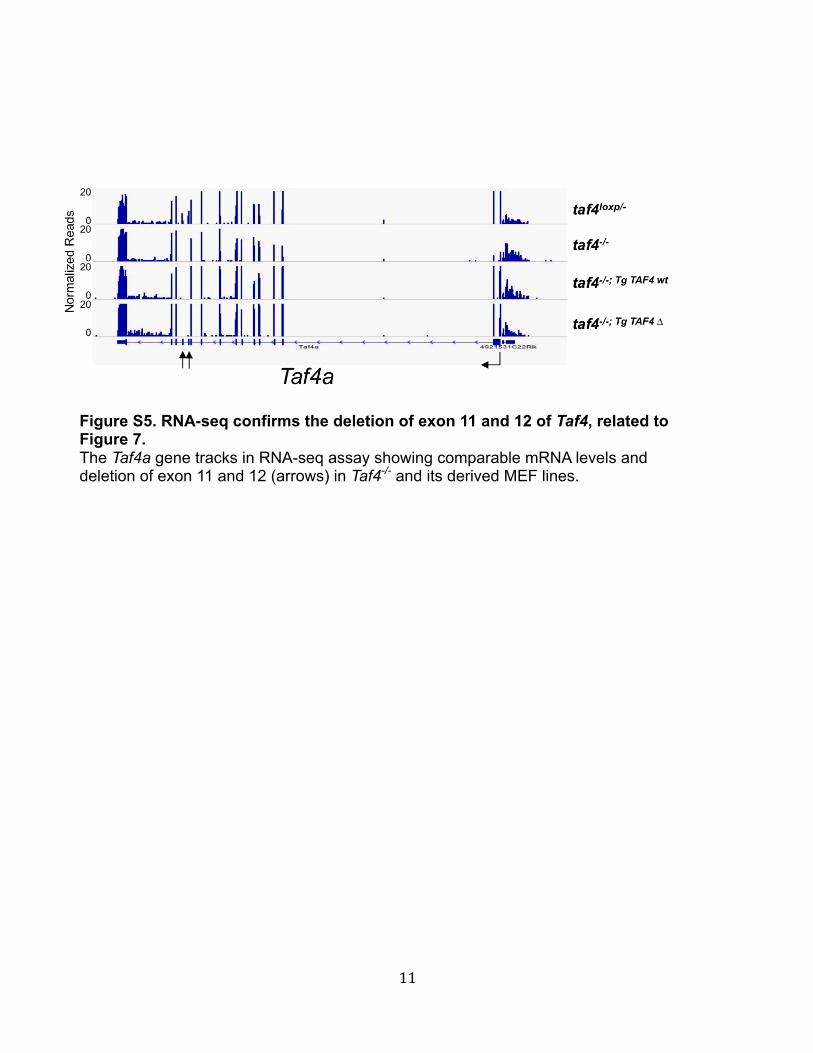
Figure S5. RNA-seq confirms the deletion of exon 11 and 12 of Taf4, related to Figure 7. The Taf4a gene tracks in RNA-seq assay showing comparable mRNA levels and deletion of exon 11 and 12 (arrows) in Taf4-/- and its derived MEF lines.

12
Figure S6. Pairwise gene-expression correlations of stable MEF lines in RNA-seq assay. The colors in the heatmap represent the Pearson's correlation coefficient values with deeper colors indicating higher positive (red) correlation. Genes that differentially expressed (fold change > 2 and FDR < 0.05 compared to Taf4
-/-) in Taf4
loxp/-, Taf4
-/-; tg TAF4wt, or Taf4
-/-; tg
TAF4Δ were used. Note that RNA samples from two clonal f-TAF4wt or f-TAF4Δ-expressing
MEF cells were used.


![OJNJJKINFEMHDGNOLCBAG `WW]ZT … · 0 6 ( @?998>:uNLWEM>nNDE>mANji> wNgBAKfEMi>db>wyc>](https://img.dokumen.tips/doc/110x75/604986452180996e6b5f8d40/ojnjjkinfemhdgnolcbag-wwzt-0-6-998unlwemnndemanji-wngbakfemidbwyc.jpg)