Embed Size (px)
Citation preview
Mar. Drugs 2012, 10, 1799-1811; doi:10.3390/md10081799
Marine Drugs
ISSN 1660-3397
www.mdpi.com/journal/marinedrugs
Article
Chemistry of the Nudibranch Aldisa andersoni: Structure and
Biological Activity of Phorbazole Metabolites
Genoveffa Nuzzo 1, Maria Letizia Ciavatta
1,*, Robert Kiss
2, Véronique Mathieu
2,
Helene Leclercqz 2, Emiliano Manzo
1, Guido Villani
1, Ernesto Mollo
1, Florence Lefranc
3,
Lisette D’Souza 4, Margherita Gavagnin
1 and Guido Cimino
1
1 CNR, Instituto di Chimica Biomolecolare, Via Campi Flegrei 34, I-80078 Pozzuoli, Naples, Italy;
E-Mails: [email protected] (G.N.); [email protected] (E.M.); [email protected] (G.V.);
[email protected] (E.M.); [email protected] (M.G.); [email protected] (G.C.) 2
Laboratoire de Toxicologie, Faculté de Pharmacie, Université Libre de Bruxelles (ULB), Campus
de la Plaine, Boulevard du Triomphe, 1050, Brussels, Belgium; E-Mails: [email protected] (R.K.);
[email protected] (V.M.); [email protected] (H.L.) 3
Service de Neurochirurgie, Hôpital Erasme, ULB, Route de Lennik, 1070 Brussels, Belgium;
E-Mail: [email protected] 4
CSIR—National Institute of Oceanography, 403 004 Dona Paula, Goa, India;
E-Mail: [email protected]
* Author to whom correspondence should be addressed; E-Mail: [email protected];
Tel.: +39-081-867-5243; Fax: +39-081-804-1770.
Received: 25 June 2012; in revised form: 18 July 2012 / Accepted: 8 August 2012 /
Published: 20 August 2012
Abstract: The first chemical study of the Indo-Pacific dorid nudibranch Aldisa andersoni
resulted in the isolation of five chlorinated phenyl-pyrrolyloxazoles belonging to the
phorbazole series. Two new molecules, 9-chloro-phorbazole D and N1-methyl-phorbazole
A, co-occurring with known phorbazoles A, B and D, have been characterized.
Phorbazoles were found to be present mainly in the external part of the mollusc. The
structures of the new compounds were determined by interpretation of spectroscopic data,
mainly NMR and mass spectrometry and by comparison with the literature data.
Evaluation of feeding-deterrence activity as well as in vitro growth inhibitory properties in
human cancer cells was also carried out.
Keywords: Aldisa andersoni; nudibranch mollusc; structural elucidation;
feeding-deterrence; growth inhibitory assays
OPEN ACCESS
Mar. Drugs 2012, 10
1800
1. Introduction
Opisthobranchs are marine molluscs in which the shell is reduced or completely absent. They move
slowly and their soft-bodies often present bright, attractive coloration, with multihued geometric
patterns [1]. However, despite slow movements, the absence of physical attributes and gaudy livery,
opisthobranchs have few documented predators. Field observations and proper ecological assays have
demonstrated that they protect themselves by employing a series of defensive strategies including
chemicals [2]. These compounds that play a fundamental role for their survival are usually small
molecules derived from the diet, which can be accumulated in specialized anatomical parts of the
animal, resulting in their unpleasant taste. In some species the presence of the de novo bio-synthesized
defensive molecules has been clearly demonstrated [3,4]. The ability of opisthobranchs to select
bioactive molecules from nature has resulted in an extraordinary library of compounds with intriguing
framework, which are not found in their terrestrial counterparts and possess the potential as new
pharmaceutical entities [5].
In our ongoing studies on the chemical ecology of marine opisthobranchs, we have examined
the chemical content of six specimens of Aldisa andersoni collected off Muttom coast, India
(Indian Ocean). A. andersoni is a dorid nudibranch (order Nudibranchia, infraorder Anthobranchia,
superfamily Doridoidea) only recorded from Sri Lanka coast (not far from the place of our collection).
The animal presents an oval body, 20–30 mm in length and a series of rows of low tubercles on the
blue dorsum. The tubercles are separated by black areas of pigment, the upper part of the skin is
crossed by a bright yellow saddle behind the rhinophores and other yellow marks on the dorsum [6].
These features appear to be indicative of aposematism (warning coloration), closely resembling the
shape and the color pattern of unpalatable nudibranch species belonging to the genus Phyllidia, which
are known to contain toxic isonitrile molecules [7]. Previous studies on different Aldisa species from
Pacific and Mediterranean areas resulted in the finding of unusual steroids. In particular, two
feeding-deterrent sterols featuring cholic acid side chain have been reported from Aldisa cooperi [8],
whereas an unusual 24-norchol-4-ene-3,22-dione has been isolated from Aldisa smaragdina [9]. It
seems that the source of steroids in Aldisa cooperi should be the sponge Anthoarcuata graceae but the
animal is able to modify the inactive cholestenone into two active compounds. On the other hand, the
absence of the steroid isolated from A. smaragdina in its prey, the sponge Phorbas fictitius, suggests
the modification of a suitable precursor or a biosynthetic origin.
The chemistry of A. andersoni has not been studied previously. We found evidence of the presence
of chlorinated phenyl pyrrolyloxazoles in the ether extract of the mollusc. These molecules were found
to be mainly concentrated in the external part of the mollusc even though they were also detected
in the internal glands. Two new molecules, 9-chloro-phorbazole D (1) and N1-methyl-phorbazole A
(2), co-occurring with the known related phorbazoles A, B and D have been isolated and
characterized from this mollusc. Phorbazoles have been reported to date only from the sponge Phorbas
aff. clathrata [10–12]. The origin of these metabolites in A. andersoni could be ascribed to a diet based
on Phorbas sponges although de novo biosynthesis could not be excluded.
Mar. Drugs 2012, 10
1801
2. Results and Discussion
Six specimens of A. andersoni were caught by scuba diving off Muttom coast (South India) during
November 2009, were immediately frozen at −20 °C and transferred to the laboratory. Unfortunately
no plausible prey was observed in the same area. Anatomical dissection of the body of nudibranchs
was precluded due to the small size and the following procedure was used to obtain the distinct
extracts of external and internal parts. First, the whole animals were immersed in acetone (20 mL) and
submitted to ultrasound vibration for a few minutes. By this procedure only the metabolites present in
the skin (external part) were extracted. The solvent was removed and the animals were crushed by a
pestle and again treated with acetone (20 mL × 3), to extract the digestive gland contents (internal
part). After evaporation of acetone, the aqueous residue of both the extracts was partitioned between
Et2O (50 mL × 4) and H2O. The two ether extracts were analyzed by Thin Layer Chromatography (TLC)
developed in the solvent system (CHCl3/CH3OH, 8:2). Both exhibited similar metabolite pattern with a
series of UV-visible spots significantly more concentrated in the external extract. This extract was then
directly submitted to RP-HPLC purification (MeOH/H2O gradient) which resulted in the novel
compounds 1 and 2 along with known phorbazoles A, B and D, that were identified by comparison of
their spectroscopic data with the literature values [10] (see Experimental Section for details).
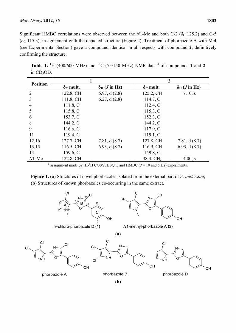
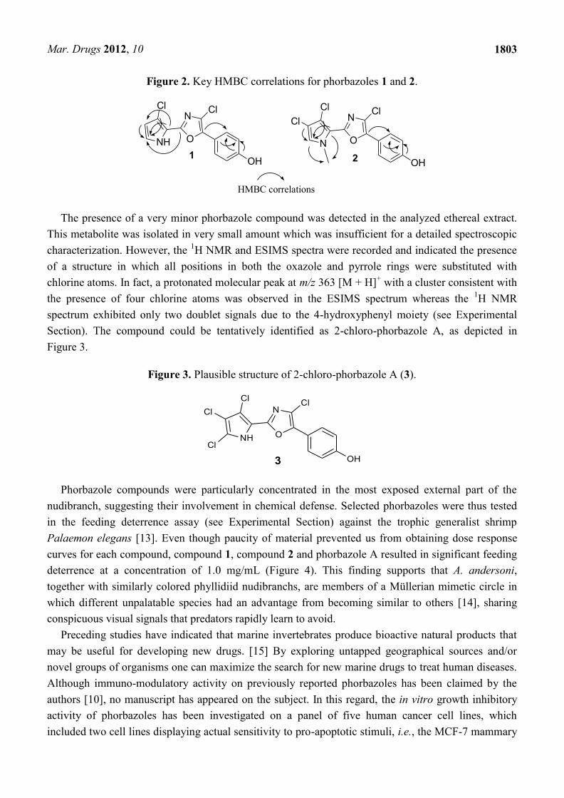
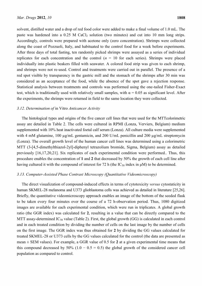
Analysis of NMR spectra of compounds 1 and 2 revealed a close structural relationship with known
phorbazoles however indicating differences in the substitution pattern.
Compound 1 had a molecular formula C13H8Cl2N2O2 deduced by LCMS (m/z 295 [M + H]+) and
HRESIMS (m/z 316.9875 [M + Na]+), that showed a characteristic cluster for two chlorine atoms. The
1H NMR spectrum of 1 exhibited only four sp
2 signals at δ 6.27 (1 H, d, J = 2.8 Hz, H-3), 6.93 (2H, d,
J = 8.7 Hz, H-13 and H-15), 6.97 (1H, d, J = 2.8 Hz, H-2) and 7.81 (2H, d, J = 8.7 Hz, H-12 and H-16),
according to two separated spin systems in the disubstituted pyrrole unit (ring A) and 1,4 disubstituted
benzene ring (ring C). This implied that the two chlorine atoms were located in both the pyrrole and
oxazole rings. Consistent with this, the 13
C NMR spectrum contained 11 sp2 carbon signals in the
range of 111.8–159.6 ppm (Table 1). The 2D NMR experiments aided us to fully characterize the
molecule. In particular, a series of HMBC experiments, recorded with J = 5 Hz and J = 10 Hz, were
crucial to attribute the quaternary carbons and to assign the structure as depicted in Figure 1. In fact,
diagnostic correlations were observed between C-8 (δC 144.2) and H-12/H-16, and between C-6
(δC 153.7) to H-2 indicating the connection of the oxazole ring to both 1,4-disubstituted benzene and
pyrrole. Expected HMBC correlations were also observed between C-5, C-4 and C-3 to H-2 (Figure 2).
Comparison of NMR data with those recorded in the same solvent for phorbazoles (see Experimental
section) confirmed the suggested structure. Compound 1 possessed one more chlorine atom on the
oxazole ring with respect to phorbazole D, thus it was named 9-chloro-phorbazole D.
The ESIMS analysis of compound 2 showed peaks at m/z 343 [M + H]+ with the typical cluster due
to the presence of three chlorine atoms. The HRESIMS spectrum indicated the molecular formula
C14H9Cl3N2O2 as deduced from the sodiated peak at m/z 364.9634 [M + Na]+. The NMR spectra of 2
(Table 1) showed proton and carbon resonances very similar to those of phorbazole A (see Experimental
Section and [10]), indicating the same substitution pattern in the heterocyclic rings. The only
difference was in the presence of a 3H signal resonating at δH 4.00 (δC 38.4), which was attributed to a
methyl linked at the pyrrole nitrogen. Thus compound 2 was suggested to be N1-methyl-phorbazole A.
Mar. Drugs 2012, 10
1802
Significant HMBC correlations were observed between the N1-Me and both C-2 (δC 125.2) and C-5
(δC 115.3), in agreement with the depicted structure (Figure 2). Treatment of phorbazole A with MeI
(see Experimental Section) gave a compound identical in all respects with compound 2, definitively
confirming the structure.
Table 1. 1H (400/600 MHz) and
13C (75/150 MHz) NMR data
a of compounds 1 and 2
in CD3OD.
Position 1 2
δC mult. δH (J in Hz) δC mult. δH (J in Hz)
2 122.8, CH 6.97, d (2.8) 125.2, CH 7.10, s
3 111.8, CH 6.27, d (2.8) 114.7, C
4 111.8, C 112.4, C
5 115.8, C 115.3, C
6 153.7, C 152.3, C
8 144.2, C 144.2, C
9 116.6, C 117.9, C
11 119.4, C 119.1, C
12,16 127.7, CH 7.81, d (8.7) 127.8, CH 7.81, d (8.7)
13,15 116.5, CH 6.93, d (8.7) 116.9, CH 6.93, d (8.7)
14 159.6, C 159.8, C
N1-Me 122.8, CH 38.4, CH3 4.00, s a assignment made by
1H-
1H COSY, HSQC, and HMBC (J = 10 and 5 Hz) experiments.
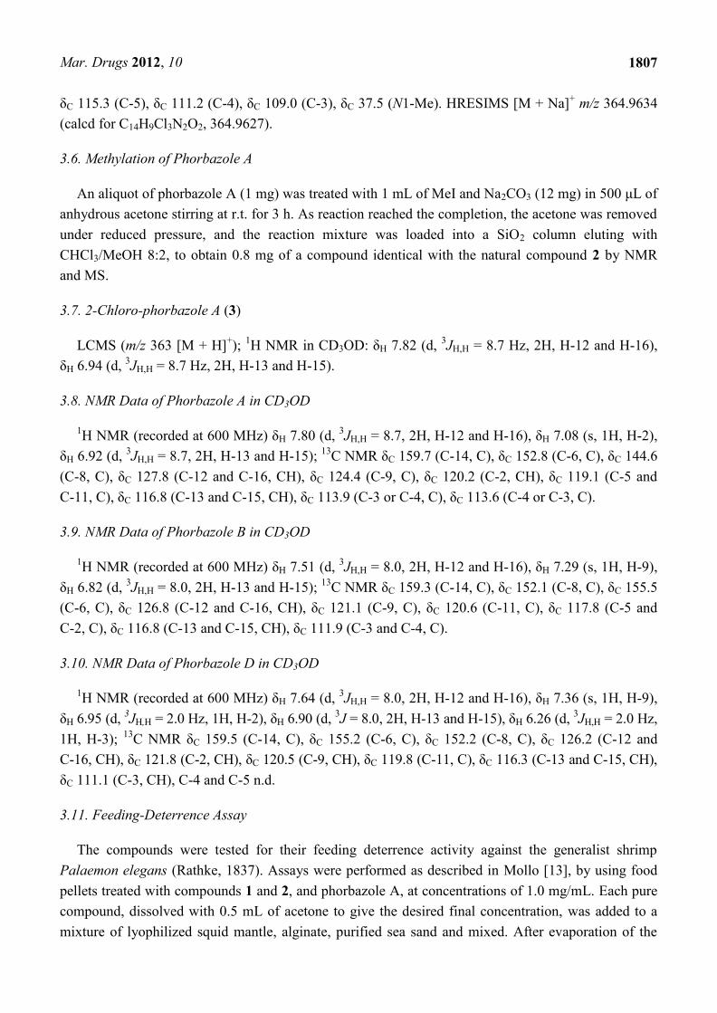
Figure 1. (a) Structures of novel phorbazoles isolated from the external part of A. andersoni;
(b) Structures of known phorbazoles co-occurring in the same extract.
(a)
(b)
N
ONH
OH
phorbazole D
ClN
ONH
OH
ClCl
ClN
ONH
OH
Cl
Cl
Cl
phorbazole A phorbazole B
Mar. Drugs 2012, 10
1803
Figure 2. Key HMBC correlations for phorbazoles 1 and 2.
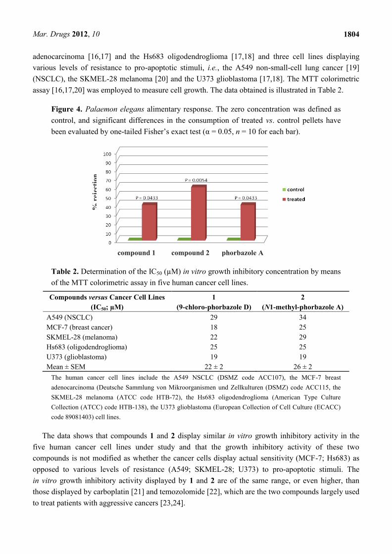
The presence of a very minor phorbazole compound was detected in the analyzed ethereal extract.
This metabolite was isolated in very small amount which was insufficient for a detailed spectroscopic
characterization. However, the 1H NMR and ESIMS spectra were recorded and indicated the presence
of a structure in which all positions in both the oxazole and pyrrole rings were substituted with
chlorine atoms. In fact, a protonated molecular peak at m/z 363 [M + H]+ with a cluster consistent with
the presence of four chlorine atoms was observed in the ESIMS spectrum whereas the 1H NMR
spectrum exhibited only two doublet signals due to the 4-hydroxyphenyl moiety (see Experimental
Section). The compound could be tentatively identified as 2-chloro-phorbazole A, as depicted in
Figure 3.
Figure 3. Plausible structure of 2-chloro-phorbazole A (3).
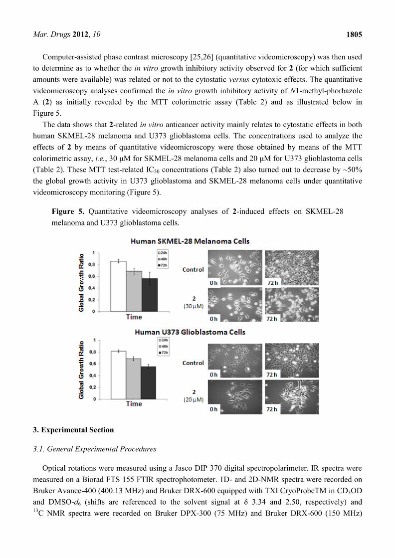
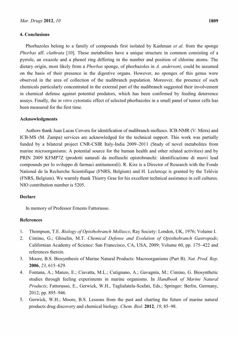
Phorbazole compounds were particularly concentrated in the most exposed external part of the
nudibranch, suggesting their involvement in chemical defense. Selected phorbazoles were thus tested
in the feeding deterrence assay (see Experimental Section) against the trophic generalist shrimp
Palaemon elegans [13]. Even though paucity of material prevented us from obtaining dose response
curves for each compound, compound 1, compound 2 and phorbazole A resulted in significant feeding
deterrence at a concentration of 1.0 mg/mL (Figure 4). This finding supports that A. andersoni,
together with similarly colored phyllidiid nudibranchs, are members of a Müllerian mimetic circle in
which different unpalatable species had an advantage from becoming similar to others [14], sharing
conspicuous visual signals that predators rapidly learn to avoid.
Preceding studies have indicated that marine invertebrates produce bioactive natural products that
may be useful for developing new drugs. [15] By exploring untapped geographical sources and/or
novel groups of organisms one can maximize the search for new marine drugs to treat human diseases.
Although immuno-modulatory activity on previously reported phorbazoles has been claimed by the
authors [10], no manuscript has appeared on the subject. In this regard, the in vitro growth inhibitory
activity of phorbazoles has been investigated on a panel of five human cancer cell lines, which
included two cell lines displaying actual sensitivity to pro-apoptotic stimuli, i.e., the MCF-7 mammary
N
ONH
OH
Cl Cl
HMBC correlations
1
N
ON
OH
Cl Cl
2
Cl
N
ONH
OH
ClCl
Cl
Cl
3
Mar. Drugs 2012, 10
1804
adenocarcinoma [16,17] and the Hs683 oligodendroglioma [17,18] and three cell lines displaying
various levels of resistance to pro-apoptotic stimuli, i.e., the A549 non-small-cell lung cancer [19]
(NSCLC), the SKMEL-28 melanoma [20] and the U373 glioblastoma [17,18]. The MTT colorimetric
assay [16,17,20] was employed to measure cell growth. The data obtained is illustrated in Table 2.
Figure 4. Palaemon elegans alimentary response. The zero concentration was defined as
control, and significant differences in the consumption of treated vs. control pellets have
been evaluated by one-tailed Fisher’s exact test (α = 0.05, n = 10 for each bar).
compound 1 compound 2 phorbazole A
Table 2. Determination of the IC50 (µM) in vitro growth inhibitory concentration by means
of the MTT colorimetric assay in five human cancer cell lines.
Compounds versus Cancer Cell Lines
(IC50; µM)
1
(9-chloro-phorbazole D)
2
(N1-methyl-phorbazole A)
A549 (NSCLC) 29 34
MCF-7 (breast cancer) 18 25
SKMEL-28 (melanoma) 22 29
Hs683 (oligodendroglioma) 25 25
U373 (glioblastoma) 19 19
Mean ± SEM 22 ± 2 26 ± 2
The human cancer cell lines include the A549 NSCLC (DSMZ code ACC107), the MCF-7 breast
adenocarcinoma (Deutsche Sammlung von Mikroorganismen und Zellkulturen (DSMZ) code ACC115, the
SKMEL-28 melanoma (ATCC code HTB-72), the Hs683 oligodendroglioma (American Type Culture
Collection (ATCC) code HTB-138), the U373 glioblastoma (European Collection of Cell Culture (ECACC)
code 89081403) cell lines.
The data shows that compounds 1 and 2 display similar in vitro growth inhibitory activity in the
five human cancer cell lines under study and that the growth inhibitory activity of these two
compounds is not modified as whether the cancer cells display actual sensitivity (MCF-7; Hs683) as
opposed to various levels of resistance (A549; SKMEL-28; U373) to pro-apoptotic stimuli. The
in vitro growth inhibitory activity displayed by 1 and 2 are of the same range, or even higher, than
those displayed by carboplatin [21] and temozolomide [22], which are the two compounds largely used
to treat patients with aggressive cancers [23,24].
% r
ejec
tio
n
Mar. Drugs 2012, 10
1805
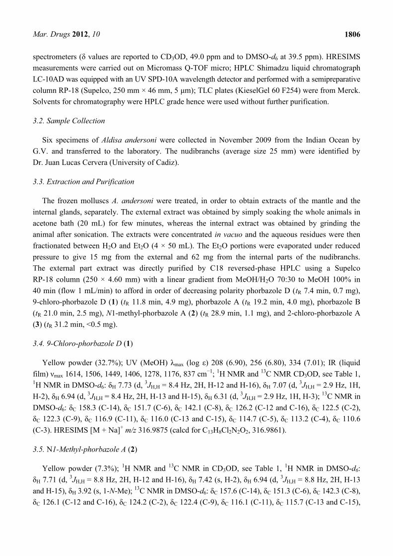
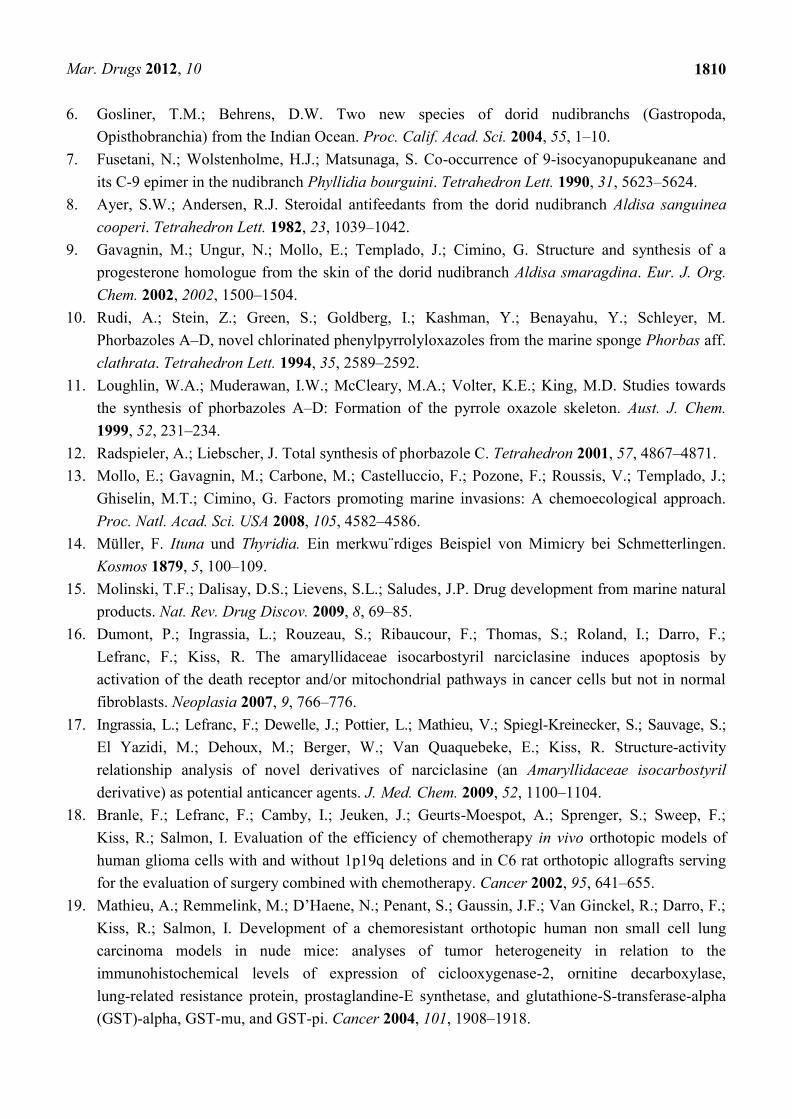
Computer-assisted phase contrast microscopy [25,26] (quantitative videomicroscopy) was then used
to determine as to whether the in vitro growth inhibitory activity observed for 2 (for which sufficient
amounts were available) was related or not to the cytostatic versus cytotoxic effects. The quantitative
videomicroscopy analyses confirmed the in vitro growth inhibitory activity of N1-methyl-phorbazole
A (2) as initially revealed by the MTT colorimetric assay (Table 2) and as illustrated below in
Figure 5.
The data shows that 2-related in vitro anticancer activity mainly relates to cytostatic effects in both
human SKMEL-28 melanoma and U373 glioblastoma cells. The concentrations used to analyze the
effects of 2 by means of quantitative videomicroscopy were those obtained by means of the MTT
colorimetric assay, i.e., 30 μM for SKMEL-28 melanoma cells and 20 μM for U373 glioblastoma cells
(Table 2). These MTT test-related IC50 concentrations (Table 2) also turned out to decrease by ~50%
the global growth activity in U373 glioblastoma and SKMEL-28 melanoma cells under quantitative
videomicroscopy monitoring (Figure 5).
Figure 5. Quantitative videomicroscopy analyses of 2-induced effects on SKMEL-28
melanoma and U373 glioblastoma cells.
3. Experimental Section
3.1. General Experimental Procedures
Optical rotations were measured using a Jasco DIP 370 digital spectropolarimeter. IR spectra were
measured on a Biorad FTS 155 FTIR spectrophotometer. 1D- and 2D-NMR spectra were recorded on
Bruker Avance-400 (400.13 MHz) and Bruker DRX-600 equipped with TXI CryoProbeTM in CD3OD
and DMSO-d6 (shifts are referenced to the solvent signal at δ 3.34 and 2.50, respectively) and 13
C NMR spectra were recorded on Bruker DPX-300 (75 MHz) and Bruker DRX-600 (150 MHz)
Mar. Drugs 2012, 10
1806
spectrometers (δ values are reported to CD3OD, 49.0 ppm and to DMSO-d6 at 39.5 ppm). HRESIMS
measurements were carried out on Micromass Q-TOF micro; HPLC Shimadzu liquid chromatograph
LC-10AD was equipped with an UV SPD-10A wavelength detector and performed with a semipreparative
column RP-18 (Supelco, 250 mm × 46 mm, 5 μm); TLC plates (KieselGel 60 F254) were from Merck.
Solvents for chromatography were HPLC grade hence were used without further purification.
3.2. Sample Collection
Six specimens of Aldisa andersoni were collected in November 2009 from the Indian Ocean by
G.V. and transferred to the laboratory. The nudibranchs (average size 25 mm) were identified by
Dr. Juan Lucas Cervera (University of Cadiz).
3.3. Extraction and Purification
The frozen molluscs A. andersoni were treated, in order to obtain extracts of the mantle and the
internal glands, separately. The external extract was obtained by simply soaking the whole animals in
acetone bath (20 mL) for few minutes, whereas the internal extract was obtained by grinding the
animal after sonication. The extracts were concentrated in vacuo and the aqueous residues were then
fractionated between H2O and Et2O (4 × 50 mL). The Et2O portions were evaporated under reduced
pressure to give 15 mg from the external and 62 mg from the internal parts of the nudibranchs.
The external part extract was directly purified by C18 reversed-phase HPLC using a Supelco
RP-18 column (250 × 4.60 mm) with a linear gradient from MeOH/H2O 70:30 to MeOH 100% in
40 min (flow 1 mL/min) to afford in order of decreasing polarity phorbazole D (tR 7.4 min, 0.7 mg),
9-chloro-phorbazole D (1) (tR 11.8 min, 4.9 mg), phorbazole A (tR 19.2 min, 4.0 mg), phorbazole B
(tR 21.0 min, 2.5 mg), N1-methyl-phorbazole A (2) (tR 28.9 min, 1.1 mg), and 2-chloro-phorbazole A
(3) (tR 31.2 min, <0.5 mg).
3.4. 9-Chloro-phorbazole D (1)
Yellow powder (32.7%); UV (MeOH) λmax (log ε) 208 (6.90), 256 (6.80), 334 (7.01); IR (liquid
film) νmax 1614, 1506, 1449, 1406, 1278, 1176, 837 cm−1
; 1H NMR and
13C NMR CD3OD, see Table 1,
1H NMR in DMSO-d6: δH 7.73 (d,
3JH,H = 8.4 Hz, 2H, H-12 and H-16), δH 7.07 (d,
3JH,H = 2.9 Hz, 1H,
H-2), δH 6.94 (d, 3JH,H = 8.4 Hz, 2H, H-13 and H-15), δH 6.31 (d,
3JH,H = 2.9 Hz, 1H, H-3);
13C NMR in
DMSO-d6: δC 158.3 (C-14), δC 151.7 (C-6), δC 142.1 (C-8), δC 126.2 (C-12 and C-16), δC 122.5 (C-2),
δC 122.3 (C-9), δC 116.9 (C-11), δC 116.0 (C-13 and C-15), δC 114.7 (C-5), δC 113.2 (C-4), δC 110.6
(C-3). HRESIMS [M + Na]+ m/z 316.9875 (calcd for C13H8Cl2N2O2, 316.9861).
3.5. N1-Methyl-phorbazole A (2)
Yellow powder (7.3%); 1H NMR and
13C NMR in CD3OD, see Table 1,
1H NMR in DMSO-d6:
δH 7.71 (d, 3JH,H = 8.8 Hz, 2H, H-12 and H-16), δH 7.42 (s, H-2), δH 6.94 (d,
3JH,H = 8.8 Hz, 2H, H-13
and H-15), δH 3.92 (s, 1-N-Me); 13
C NMR in DMSO-d6: δC 157.6 (C-14), δC 151.3 (C-6), δC 142.3 (C-8),
δC 126.1 (C-12 and C-16), δC 124.2 (C-2), δC 122.4 (C-9), δC 116.1 (C-11), δC 115.7 (C-13 and C-15),
Mar. Drugs 2012, 10
1807
δC 115.3 (C-5), δC 111.2 (C-4), δC 109.0 (C-3), δC 37.5 (N1-Me). HRESIMS [M + Na]+ m/z 364.9634
(calcd for C14H9Cl3N2O2, 364.9627).
3.6. Methylation of Phorbazole A
An aliquot of phorbazole A (1 mg) was treated with 1 mL of MeI and Na2CO3 (12 mg) in 500 μL of
anhydrous acetone stirring at r.t. for 3 h. As reaction reached the completion, the acetone was removed
under reduced pressure, and the reaction mixture was loaded into a SiO2 column eluting with
CHCl3/MeOH 8:2, to obtain 0.8 mg of a compound identical with the natural compound 2 by NMR
and MS.
3.7. 2-Chloro-phorbazole A (3)
LCMS (m/z 363 [M + H]+);
1H NMR in CD3OD: δH 7.82 (d,
3JH,H = 8.7 Hz, 2H, H-12 and H-16),
δH 6.94 (d, 3JH,H = 8.7 Hz, 2H, H-13 and H-15).
3.8. NMR Data of Phorbazole A in CD3OD
1H NMR (recorded at 600 MHz) δH 7.80 (d,
3JH,H = 8.7, 2H, H-12 and H-16), δH 7.08 (s, 1H, H-2),
δH 6.92 (d, 3JH,H = 8.7, 2H, H-13 and H-15);
13C NMR δC 159.7 (C-14, C), δC 152.8 (C-6, C), δC 144.6
(C-8, C), δC 127.8 (C-12 and C-16, CH), δC 124.4 (C-9, C), δC 120.2 (C-2, CH), δC 119.1 (C-5 and
C-11, C), δC 116.8 (C-13 and C-15, CH), δC 113.9 (C-3 or C-4, C), δC 113.6 (C-4 or C-3, C).
3.9. NMR Data of Phorbazole B in CD3OD
1H NMR (recorded at 600 MHz) δH 7.51 (d,
3JH,H = 8.0, 2H, H-12 and H-16), δH 7.29 (s, 1H, H-9),
δH 6.82 (d, 3JH,H = 8.0, 2H, H-13 and H-15);
13C NMR δC 159.3 (C-14, C), δC 152.1 (C-8, C), δC 155.5
(C-6, C), δC 126.8 (C-12 and C-16, CH), δC 121.1 (C-9, C), δC 120.6 (C-11, C), δC 117.8 (C-5 and
C-2, C), δC 116.8 (C-13 and C-15, CH), δC 111.9 (C-3 and C-4, C).
3.10. NMR Data of Phorbazole D in CD3OD
1H NMR (recorded at 600 MHz) δH 7.64 (d,
3JH,H = 8.0, 2H, H-12 and H-16), δH 7.36 (s, 1H, H-9),
δH 6.95 (d, 3JH,H = 2.0 Hz, 1H, H-2), δH 6.90 (d,
3J = 8.0, 2H, H-13 and H-15), δH 6.26 (d,
3JH,H = 2.0 Hz,
1H, H-3); 13
C NMR δC 159.5 (C-14, C), δC 155.2 (C-6, C), δC 152.2 (C-8, C), δC 126.2 (C-12 and
C-16, CH), δC 121.8 (C-2, CH), δC 120.5 (C-9, CH), δC 119.8 (C-11, C), δC 116.3 (C-13 and C-15, CH),
δC 111.1 (C-3, CH), C-4 and C-5 n.d.
3.11. Feeding-Deterrence Assay
The compounds were tested for their feeding deterrence activity against the generalist shrimp
Palaemon elegans (Rathke, 1837). Assays were performed as described in Mollo [13], by using food
pellets treated with compounds 1 and 2, and phorbazole A, at concentrations of 1.0 mg/mL. Each pure
compound, dissolved with 0.5 mL of acetone to give the desired final concentration, was added to a
mixture of lyophilized squid mantle, alginate, purified sea sand and mixed. After evaporation of the
Mar. Drugs 2012, 10
1808
solvent, distilled water and a drop of red food color were added to make a final volume of 1.0 mL. The
paste was hardened into a 0.25 M CaCl2 solution (two minutes) and cut into 10 mm long strips.
Accordingly, controls were prepared with acetone only (zero concentration). Shrimps were collected
along the coast of Pozzuoli, Italy, and habituated to the control food for a week before experiments.
After three days of total fasting, ten randomly picked shrimps were assayed as a series of individual
replicates for each concentration and the control (n = 10 for each series). Shrimps were placed
individually into plastic beakers filled with seawater. A colored food strip was given to each shrimp,
and shrimps were not re-used. Control and treatments were carried out in parallel. The presence of a
red spot visible by transparency in the gastric mill and the stomach of the shrimps after 30 min was
considered as an acceptance of the food, while the absence of the spot gave a rejection response.
Statistical analysis between treatments and controls was performed using the one-tailed Fisher-Exact
test, which is traditionally used with relatively small samples, with α = 0.05 as significant level. After
the experiments, the shrimps were returned in field to the same location they were collected.
3.12. Determination of in Vitro Anticancer Activity
The histological types and origins of the five cancer cell lines that were used for the MTTcolorimetric
assay are detailed in Table 2. The cells were cultured in RPMI (Lonza, Verviers, Belgium) medium
supplemented with 10% heat inactivated foetal calf serum (Lonza). All culture media were supplemented
with 4 mM glutamine, 100 µg/mL gentamicin, and 200 U/mL penicillin and 200 µg/mL streptomycin
(Lonza). The overall growth level of the human cancer cell lines was determined using a colorimetric
MTT (3-[4,5-dimethylthiazol-2yl]-diphenyl tetrazolium bromide, Sigma, Belgium) assay as detailed
previously [16,17,20,21]. Six replicates of each experimental condition were performed.. Thus, this
procedure enables the concentration of 1 and 2 that decreased by 50% the growth of each cell line after
having cultured it with the compound of interest for 72 h (the IC50 index in μM) to be determined.
3.13. Computer-Assisted Phase Contrast Microscopy (Quantitative Videomicroscopy)
The direct visualization of compound-induced effects in terms of cytotoxicity versus cytostaticity in
human SKMEL-28 melanoma and U373 glioblastoma cells was achieved as detailed in literature [25,26].
Briefly, the quantitative videomicroscopy approach enables an image of the bottom of the seeded flask
to be taken every four minutes over the course of a 72 h-observation period. Thus, 1080 digitized
images are available for each experimental condition, which were run in triplicates. A global growth
ratio (the GGR index) was calculated for 2, resulting in a value that can be directly compared to the
MTT assay-determined IC50 value (Table 2). First, the global growth (GG) is calculated in each control
and in each treated condition by dividing the number of cells on the last image by the number of cells
on the first image. The GGR index was thus obtained for 2 by dividing the GG values calculated for
treated SKMEL-28 or U373 cells by the GG values calculated for the control (the data are presented as
mean ± SEM values). For example, a GGR value of 0.5 for 2 at a given experimental time means that
this compound decreased by 50% (1.0 − 0.5 = 0.5) the global growth of the considered cancer cell
population as compared to control.
Mar. Drugs 2012, 10
1809
4. Conclusions
Phorbazoles belong to a family of compounds first isolated by Kashman et al. from the sponge
Phorbas aff. clathrata [10]. These metabolites have a unique structure in common consisting of a
pyrrole, an oxazole and a phenol ring differing in the number and position of chlorine atoms. The
dietary origin, most likely from a Phorbas sponge, of phorbazoles in A. andersoni, could be assumed
on the basis of their presence in the digestive organs. However, no sponges of this genus were
observed in the area of collection of the nudibranch population. Moreover, the presence of such
chemicals particularly concentrated in the external part of the nudibranch suggested their involvement
in chemical defense against potential predators, which has been confirmed by feeding deterrence
assays. Finally, the in vitro cytostatic effect of selected phorbazoles in a small panel of tumor cells has
been measured for the first time.
Acknowledgments
Authors thank Juan Lucas Cervera for identification of nudibranch molluscs. ICB-NMR (V. Mirra) and
ICB-MS (M. Zampa) services are acknowledged for the technical support. This work was partially
funded by a bilateral project CNR-CSIR Italy-India 2009–2011 (Study of novel metabolites from
marine microorganisms: A potential source for the human health and other related activities) and by
PRIN 2009 KFMP7Z (prodotti naturali da molluschi opistobranchi: identificazione di nuovi lead
compounds per lo sviluppo di farmaci antitumorali). R. Kiss is a Director of Research with the Fonds
National de la Recherche Scientifique (FNRS, Belgium) and H. Leclercqz is granted by the Telévie
(FNRS, Belgium). We warmly thank Thierry Gras for his excellent technical assistance in cell cultures.
NIO contribution number is 5205.
Declare
In memory of Professor Ernesto Fattorusso.
References
1. Thompson, T.E. Biology of Opisthobranch Molluscs; Ray Society: London, UK, 1976; Volume I.
2. Cimino, G.; Ghiselin, M.T. Chemical Defense and Evolution of Opisthobranch Gastropods;
Californian Academy of Science: San Franccisco, CA, USA, 2009; Volume 60, pp. 175–422 and
references therein.
3. Moore, B.S. Biosynthesis of Marine Natural Products: Macroorganisms (Part B). Nat. Prod. Rep.
2006, 23, 615–629.
4. Fontana, A.; Manzo, E.; Ciavatta, M.L.; Cutignano, A.; Gavagnin, M.; Cimino, G. Biosynthetic
studies through feeling experiments in marine organisms. In Handbook of Marine Natural
Products; Fattorusso, E., Gerwick, W.H., Taglialatela-Scafati, Eds.; Springer: Berlin, Germany,
2012; pp. 895–946.
5. Gerwick, W.H.; Moore, B.S. Lessons from the past and charting the future of marine natural
products drug discovery and chemical biology. Chem. Biol. 2012, 19, 85–98.
Mar. Drugs 2012, 10
1810
6. Gosliner, T.M.; Behrens, D.W. Two new species of dorid nudibranchs (Gastropoda,
Opisthobranchia) from the Indian Ocean. Proc. Calif. Acad. Sci. 2004, 55, 1–10.
7. Fusetani, N.; Wolstenholme, H.J.; Matsunaga, S. Co-occurrence of 9-isocyanopupukeanane and
its C-9 epimer in the nudibranch Phyllidia bourguini. Tetrahedron Lett. 1990, 31, 5623–5624.
8. Ayer, S.W.; Andersen, R.J. Steroidal antifeedants from the dorid nudibranch Aldisa sanguinea
cooperi. Tetrahedron Lett. 1982, 23, 1039–1042.
9. Gavagnin, M.; Ungur, N.; Mollo, E.; Templado, J.; Cimino, G. Structure and synthesis of a
progesterone homologue from the skin of the dorid nudibranch Aldisa smaragdina. Eur. J. Org.
Chem. 2002, 2002, 1500–1504.
10. Rudi, A.; Stein, Z.; Green, S.; Goldberg, I.; Kashman, Y.; Benayahu, Y.; Schleyer, M.
Phorbazoles A–D, novel chlorinated phenylpyrrolyloxazoles from the marine sponge Phorbas aff.
clathrata. Tetrahedron Lett. 1994, 35, 2589–2592.
11. Loughlin, W.A.; Muderawan, I.W.; McCleary, M.A.; Volter, K.E.; King, M.D. Studies towards
the synthesis of phorbazoles A–D: Formation of the pyrrole oxazole skeleton. Aust. J. Chem.
1999, 52, 231–234.
12. Radspieler, A.; Liebscher, J. Total synthesis of phorbazole C. Tetrahedron 2001, 57, 4867–4871.
13. Mollo, E.; Gavagnin, M.; Carbone, M.; Castelluccio, F.; Pozone, F.; Roussis, V.; Templado, J.;
Ghiselin, M.T.; Cimino, G. Factors promoting marine invasions: A chemoecological approach.
Proc. Natl. Acad. Sci. USA 2008, 105, 4582–4586.
14. Müller, F. Ituna und Thyridia. Ein merkwu r̈diges Beispiel von Mimicry bei Schmetterlingen.
Kosmos 1879, 5, 100–109.
15. Molinski, T.F.; Dalisay, D.S.; Lievens, S.L.; Saludes, J.P. Drug development from marine natural
products. Nat. Rev. Drug Discov. 2009, 8, 69–85.
16. Dumont, P.; Ingrassia, L.; Rouzeau, S.; Ribaucour, F.; Thomas, S.; Roland, I.; Darro, F.;
Lefranc, F.; Kiss, R. The amaryllidaceae isocarbostyril narciclasine induces apoptosis by
activation of the death receptor and/or mitochondrial pathways in cancer cells but not in normal
fibroblasts. Neoplasia 2007, 9, 766–776.
17. Ingrassia, L.; Lefranc, F.; Dewelle, J.; Pottier, L.; Mathieu, V.; Spiegl-Kreinecker, S.; Sauvage, S.;
El Yazidi, M.; Dehoux, M.; Berger, W.; Van Quaquebeke, E.; Kiss, R. Structure-activity
relationship analysis of novel derivatives of narciclasine (an Amaryllidaceae isocarbostyril
derivative) as potential anticancer agents. J. Med. Chem. 2009, 52, 1100–1104.
18. Branle, F.; Lefranc, F.; Camby, I.; Jeuken, J.; Geurts-Moespot, A.; Sprenger, S.; Sweep, F.;
Kiss, R.; Salmon, I. Evaluation of the efficiency of chemotherapy in vivo orthotopic models of
human glioma cells with and without 1p19q deletions and in C6 rat orthotopic allografts serving
for the evaluation of surgery combined with chemotherapy. Cancer 2002, 95, 641–655.
19. Mathieu, A.; Remmelink, M.; D’Haene, N.; Penant, S.; Gaussin, J.F.; Van Ginckel, R.; Darro, F.;
Kiss, R.; Salmon, I. Development of a chemoresistant orthotopic human non small cell lung
carcinoma models in nude mice: analyses of tumor heterogeneity in relation to the
immunohistochemical levels of expression of ciclooxygenase-2, ornitine decarboxylase,
lung-related resistance protein, prostaglandine-E synthetase, and glutathione-S-transferase-alpha
(GST)-alpha, GST-mu, and GST-pi. Cancer 2004, 101, 1908–1918.
Mar. Drugs 2012, 10
1811
20. Van Goietsenoven, G.; Hutton, J.; Becker, J.P.; Lallemand, B.; Robert, F.; Lefranc, F.; Pirker, C.;
Vandenbussche, G.; Van Antwerpen, P.; Evidente, A.; et al. Targeting of eEF1A with
Amaryllidaceae isocarbostyrils as a strategy to combat melanomas. FASEB J. 2010, 24,
4575–4584.
21. Van Goietsenoven, G.; Andolfi, A.; Lallemand, B.; Cimmino, A.; Lamoral-Theys, D.; Gras, T.;
Abou-Donia, A.; Dubois, J.; Lefranc, F.; Mathieu, V.; Kornienko, A.; Kiss, R.; Evidente, A.
Amaryllidaceae alkaloids belonging to different structural subgroups display activity against
apoptosis-resistant cancer cells. J. Nat. Prod. 2010, 73, 1223–1227.
22. Lefranc, F.; Mijatovic, T.; Kondo, Y.; Sauvage, S.; Roland, I.; Debeir, O.; Krstic, D.; Vasic, V.;
Gailly, P.; Kondo, S.; Blanco, G.; Kiss, R. Targeting the alpha 1 subunit of the sodium pump to
combat glioblastoma cells. Neurosurgery 2008, 62, 211–221.
23. Grossi, F.; Kubota, K.; Cappuzzo, F.; de Marinis, F.; Gridelli, C.; Aita, M.; Douillard, J.Y. Future
scenarios for the treatment of advanced non-small cell lung cancer: focus on taxane-containing
regimens. Oncologist 2010, 15, 1102–1112.
24. Lefranc, F.; Facchini, F.; Kiss, R. Pro-autophagic drugs: A novel means to combat a
poptosis-resistant cancers. Oncologist 2007, 12, 1395–1403.
25. Delbrouck, C.; Doyen, I.; Belot, N.; Decaestecker, C.; Ghanooni, R.; de Lavareille, A.; Kaltner, H.;
Choufani, G.; Danguy, A.; Vandenhoven, G.; Gabius, H.J.; Hassid, S.; Kiss, R. Galectin-1 is
overexpressed in nasal polyps under budesonide and inhibits eosinophil migration. Lab. Invest.
2002, 82, 147–158.
26. Mégalizzi, V.; Mathieu, V.; Mijatovic, T.; Gailly, P.; Debeir, O.; De Neve, N.; Van Damme, M.;
Bontempi, G.; Haibe-Kains, B.; Decaestecker, C.; Kondo, Y.; Kiss, R.; Lefranc, F. 4-IBP, a
sigma1 receptor agonist, decreases the migration of human cancer cells, including glioblastoma
cells, in vitro and sensitizes them in vitro and in vivo to cytotoxic insults of proapoptotic and
proautophagic drugs. Neoplasia 2007, 9, 358–369.
Samples Availability: Available from the authors.
© 2012 by the authors; licensee MDPI, Basel, Switzerland. This article is an open access article
distributed under the terms and conditions of the Creative Commons Attribution license
(http://creativecommons.org/licenses/by/3.0/).