Embed Size (px)
Citation preview

1
Chemical Genetic Control of Protein Levels: Selective
in vivo Targeted Degradation
1John S. Schneekloth, Jr., 2Fabiana Fonseca, 2Michael Koldobskiy, 2Amit Mandal‡, 3,4Raymond Deshaies,3,5Kathleen Sakamoto, and 1,2,6Craig M. Crews*
Departments of 1Chemistry, 2Molecular, Cellular, and Developmental Biology, and 6Pharmacology, Yale
University, New Haven, Connecticut 06520-8103. 3Division of Biology and 4Howard Hughes Medical
Institute, California Institute of Technology, Pasadena, California 91125. Department of 5Pediatrics and
Pathology, Mattel Children’s Hospital, David Geffen School of Medicine at UCLA, Gwynn Hazen Cherry
Memorial Laboratories, Molecular Biology Institute, and Jonsson Comprehensive Cancer Center, Los
Angeles, California 90095-1752.
E-mail: [email protected]
RECEIVED DATE (to be automatically inserted after your manuscript is accepted if required
according to the journal that you are submitting your paper to)
ABSTRACT
Genetic loss of function analysis is a powerful approach for the study of protein function.! However, some
cell biological questions are difficult to address using traditional genetic strategies often due to the lack of
appropriate genetic model systems.! Here, we present a general strategy for the design and syntheses of
molecules capable of inducing the degradation of selected proteins in vivo via the ubiquitin-proteasome
pathway.! Western blot and fluorometric analyses indicated the loss of two different targets, green
fluorescent protein (GFP) fused with FK506 binding protein (FKBP12) and GFP fused with the androgen
receptor (AR), after treatment with PROteolysis TArgeting Chimeric moleculeS (PROTACS) incorporating

2
a FKBP12 ligand and dihydrotestosterone, respectively.! These are the first in vivo examples of direct small
molecule-induced recruitment of target proteins to the proteasome for degradation upon addition to
cultured cells. Moreover, PROTAC-mediated protein degradation offers a general strategy to create
“chemical knockouts,” thus, opening new possibilities for the control of protein function.
MANUSCRIPT TEXT
Introduction
The selective loss of critical cellular proteins and subsequent analysis of the resulting phenotypes have
proven to be extremely useful in genetic studies of in vivo protein function. In recent years, genetically
modified knockout cell lines and animals have allowed biological research to advance with unprecedented
speed. Chemical genetic approaches, using small molecules to induce changes in cell phenotype, are
complimentary to traditional genetics. Many chemical genetic strategies use knowledge gained from
natural product mode of action studies1,2,3, while others employ chemical inducers of dimerization to
manipulate intracellular processes4,5,6,7. To date, however, there have been few attempts to design small
molecules which induce the destruction (rather than inhibition) of a targeted protein in an otherwise healthy
cell. Access to such reagents would provide a chemical genetic alternative to the traditional ways of
interfering with protein function, resulting in “chemical knockouts.” Importantly, a small molecule
capable of inducing this process could do so without any genetic manipulation of the organism, thus
allowing one to target proteins that are not readily accessible by traditional genetic means (i.e., genes
essential for proliferation and early development).
In principle, targeted proteolytic degradation could be an effective way to accomplish the removal of
a desired protein. Given the central role of the ubiquitin-proteasome pathway in protein degradation within
the cell8, reagents capable of redirecting the substrate specificity of this pathway would be useful as
experimental tools for modulating cellular phenotype and potentially act as drugs for inducing the
elimination of disease-promoting proteins. We present here a general strategy for designing molecules
capable of inducing the proteolysis of a targeted protein via the ubiquitin-proteasome pathway, as well as
the first evidence that such molecules are effective upon addition to living cells.
Protein degradation, like protein synthesis, is an essential part of normal cellular homeostasis. As
the major protein degradation pathway, the ATP-dependent ubiquitin-proteasome pathway has been

3
implicated in the regulation of cellular processes as diverse as cell cycle progression9, antigen
presentation10, the inflammatory response11, transcription12, and signal transduction13. The pathway
involves two discrete steps: (i) the specific tagging of the protein to be degraded with a polyubiquitin chain
and (ii) the subsequent degradation of the tagged substrate by the 26S proteasome, a multicatalytic protease
complex. Ubiquitin, a highly conserved 76 amino acid protein14, is conjugated to the target protein by a
three part process. First, the C-terminal carboxyl group of ubiquitin is activated by a ubiquitin-activating
enzyme (E1). The thioester formed by attachment of ubiquitin to the E1 enzyme is then transferred via a
transacylation reaction to an ubiquitin-conjugating enzyme (E2). Finally, ubiquitin is transferred to a lysine
(or, less commonly, the amino terminus) of the protein substrate that is specifically bound by an ubiquitin
ligase (E3)15. Successive conjugation of ubiquitin to internal lysines of previously added ubiquitin
molecules leads to the formation of polyubiquitin chains16. The resulting polyubiquitinated target protein
is then recognized by the 26S proteasome, whereupon ubiquitin is cleaved off and the substrate protein
threaded into the proteolytic chamber of the proteasome. Importantly, substrate specificity of the ubiquitin-
proteasome pathway is conferred by the E3 ligases. Each E3 ligase or recognition subunit of a
multiprotein E3 ligase complex binds specifically to a limited number of protein targets sharing a particular
destruction sequence. The destruction sequence may require chemical or conformational modification (e.g.
phosphorylation) for recognition by E3 enzymes17,18.
Recently, we demonstrated a strategy for inducing the ubiquitination and ensuing proteolytic
degradation of a targeted protein in vitro. This approach uses heterobifunctional molecules known as
PROteolysis TArgeting Chimeric moleculeS (PROTACS), which are comprised of a ligand for the target
protein, a linker moiety, and a ligand for an E3 ubiquitin ligase19. In that proof of principle experiment the
degradation of a stable protein, methionine aminopeptidase 2 (MetAP-2), was induced in a cellular lysate
upon the addition of a PROTAC (referred to as PROTAC-1) consisting of the known MetAP-2 ligand,
ovalicin, joined to a peptide ligand for the ubiquitin ligase complex SCFbTrCP. By bridging MetAP-2 and an
E3 ligase, PROTAC-1 initiated the ubiquitination and proteasome-mediated degradation of MetAP-2
(Figure 1). We have also recently shown that an estradiol-based PROTAC (PROTAC-2) could promote
the ubiquitination of the human estrogen receptor (hERa) in vitro. Furthermore, a dihydrotestosterone
(DHT)-based PROTAC (PROTAC-3), when microinjected into cells, was capable of inducing the
degradation of the androgen receptor20. Encouraged by our success with PROTACS-1, -2, and -3, we next

4
directed our efforts toward the design of molecules capable of accomplishing the inducing proteolysis
simply upon addition to cells.
Results
Development of a Cell Permeable PROTAC: PROTAC-4
For the design of PROTAC-4, we used a protein target/ligand pair developed by ARIAD
Pharmaceuticals. The F36V mutation of FK506 Binding Protein (FKBP12) generates a ‘hole’ into which
the artificial ligand AP21998 (1) fits via a hydrophobic “bump,” thus conferring specificity of this
particular ligand to the mutant FKBP over the wild type protein21,22. Inclusion of AP21998 as one domain
of PROTAC-4 thus allows it to target (F36V)FKBP12 proteins orthogonally, without disrupting
endogenous FKBP12 function. Given the lack of small molecule E3 ubiquitin ligase ligands, we chose the
seven amino acid sequence ALAPYIP for the E3 recognition domain. This sequence has been shown to be
the minimum recognition domain for the von Hippel-Lindau tumor suppressor protein (VHL)23, part of the
VBC-Cul2 E3 ubiquitin ligase complex. Under normoxic conditions, a proline hydroxylase catalyzes the
hydroxylation of hypoxia inducible factor 1a (HIF1a) at P56424 (the central proline in our sequence),
resulting in recognition and polyubiquitination by VHL. HIF1a is thus constitutively ubiquitinated and
degraded under normoxic conditions25,26. Finally, we included a poly-D-arginine tag on the carboxy
terminus of our peptide sequence to confer cell permeability and resist nonspecific proteolysis.
Polyarginine sequences fused to proteins have been shown to facilitate translocation into cells27,28 via a
mechanism that mimics that of the Antennapedia29 and HIV Tat proteins30. Because a molecule fused to
the polyarginine sequence should in principle be cell permeable, the necessity of PROTAC microinjection
is circumvented. This design element also allows greater flexibility in the types of ligands that could be
used in future PROTACs, since polarity of the compound is no longer an issue for membrane
permeability. We hypothesized that the PROTAC-4 would enter the cell, be recognized and hydroxylated
by a prolyl hydroxylase, and subsequently bound by both VHL E3 ligase and the mutant FKBP12 target
protein. PROTAC-mediated recruitment of FKBP12 to the VBC-Cul2 E3 ligase complex would be
predicted to induce FKBP12 ubiquitination and degradation as in Figure 1.

5
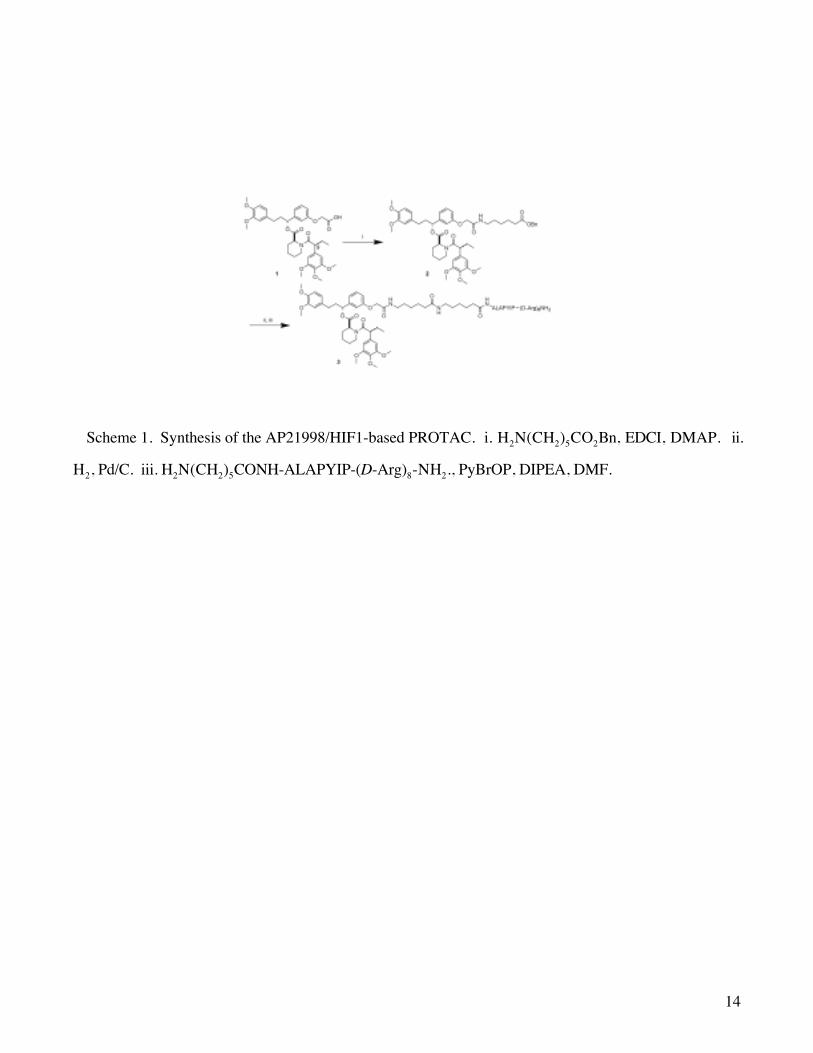
The F36V FKBP12 ligand AP21998 (1) was synthesized as previously described,21,22 as a 1:1
mixture of diastereomers at C9. Treatment of 1 with the benzyl ester of aminocaproic acid followed by
removal of the benzyl group afforded 2 in 85% crude yield after 2 steps. It is important to note that
although this material was carried through as a mixture of two diastereomers at C9, each diastereomer has
previously been shown to bind to the target2 2. Standard peptide coupling conditions were used to label the
peptide sequence. HPLC purification yielded 3 (PROTAC-4) with 17% recovery from 1 (Scheme 1).
In order to monitor the abundance of the targeted protein, we generated a vector capable of
expressing the mutant FKBP12 fused to enhanced green fluorescent protein (EGFP). In this way,
proteolysis of FKBP12 could be monitored by loss of intracellular fluorescence. This vector was then
used to generate a HeLa cell line stably expressing the EGFP-(F36V)FKBP12. Bright field (not shown)
and fluorescent photographs of the cells were taken before and 2.5 hours after treatment with PROTAC-4
(3). As shown in Figure 2 A-D, EGFP-FKBP12 was retained in those cells treated with DMSO, but lost
in cells treated with 25mM PROTAC-4 for 2.5h. Western blot analysis of cells treated with PROTAC-4
also indicated loss of EGFP-FKBP12 relative to an equal number of cells treated with DMSO (Figure 2I).
As a control, cells were treated with uncoupled 1 and the HIF-polyarginine peptide fragment (Figure 2E,
F). These cells retained fluorescence, indicating that the two domains require a chemical bond to each other
in order to exert a biological effect. In order to investigate whether VHL was required for PROTAC-4-
mediated EGFP-FKBP12 degradation, the renal carcinoma cell line 786-O31 was used. 786-O cells fail to
produce VHL protein and thus lacks a functional VBC-Cul2 E3 ligase complex. 786-O cells stably
expressing the degradation substrate EGFP-FKBP12 retained fluorescence despite treatment with 25 mM
PROTAC-2 for 2.5 hours (Figure 2 G, H), confirming that the E3 ligase is required for PROTAC-4
activity.
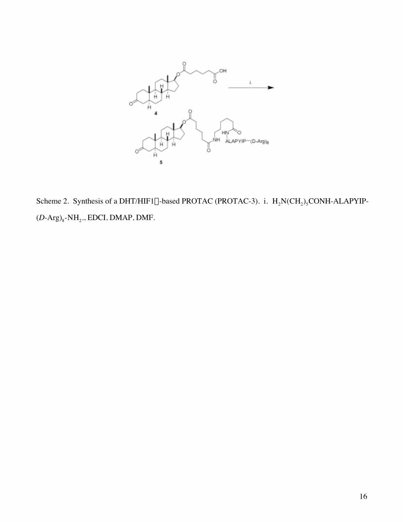
Implementation of a DHT-Based PROTAC: PROTAC-5
To test the robustness of this approach for the induction of intracellular protein degradation, we
next targeted a protein which, although still modified, is closer to being clinically relevant. It has been
shown that the androgen receptor (AR) can promote the growth of prostate tumor cells, even in some
androgen independent cell lines32. We hypothesized that a PROTAC could be utilized to degrade AR,
potentially yielding a novel strategy to repress tumor growth. With this in mind, we designed PROTAC-5,

6
5, which contains dihydrotestosterone (DHT) as the ligand for AR as well as the HIF-polyarginine peptide
sequence which was successful with PROTAC-4. Using standard peptide coupling conditions, we were
able to couple known DHT derivative 433 to the HIF-polyarginine peptide (Scheme 2). In order to monitor
protein degradation by fluorescence analysis, HEK293 cells stably expressing GFP-AR (293GFP-AR) were
treated with increasing concentrations of PROTAC-5. Within one hour, a significant decrease in GFP-AR
signal was observed in cells treated with 100, 50, and 25 mM PROTAC-5, but not in the DMSO control
(Figure 3 A-F). Western blot analysis with anti-AR antisera verified the downregulation of GFP-AR in
cells treated with 25mM PROTAC-5 compared to DMSO control or non-treated cells (Figure 3 G).
PROTAC-5 concentrations lower than 25 mM did not result in GFP-AR degradation (data not shown).
Pretreatment of cells with epoxomicin, a specific proteasome inhibitor34, prevented degradation of GFP-AR
(Figure 3 H), indicating that the observed degradation was proteasome-dependent. Competition
experiments with testosterone also inhibited PROTAC-5 from inducing GFP-AR degradation (Figure 4 A-
D). In addition, cells treated only with testosterone retained all fluorescence, as did cells treated with the
HIF-polyarginine peptide (Figure 4 G, H). Finally, cells treated with both testosterone and the HIF-
polyarginine peptide together also retained fluorescence, indicating again that both domains needed to be
chemically linked in order to observe degradation (Figure 4 F). It is important to note that the cells
survived treatment with PROTAC-5, indicating that the strategy of utilizing the ubiquitin-proteasome
pathway for targeted degradation does not necessarily cause a toxic effect.
Discussion
These experiments highlight the general applicability of our strategy to target and degrade proteins
in vivo. Moreover, the modularity of the PROTAC design offers the possibility to synthesize similar
PROTAC molecules targeting different intracellular targets. We have shown that the ligand for the target
protein can be varied using both natural and synthetic ligands. Although the linker length has not been
fully explored, we have demonstrated that a spacer consisting of two aminocaproic acids (12 atoms) is
flexible enough to accommodate some structural variation in the target and E3 ligase proteins yet bring the
proteins within close proximity. Since ubiquitination occurs most commonly on an exposed lysine,
different spacer lengths may be required to accommodate the structures of different target proteins.

7
Small molecules have previously been implicated in inducing ubiquitination and degradation of
proteins, most notably geldanamycin derivatives act by controlling target interaction with molecular
chaperones35,36,37,38. However, there are often specificity issues with these approaches, and the exact
mechanism of induced degradation is not clear. In addition, no unified strategy has been presented to
confront these issues. Our strategy represents the first attempt to develop a general method for small
molecule-induced targeted proteolysis via the ubiquitin-proteasome pathway in intact cells.
PROTACS could in principle be used to target almost any protein within a cell and selectively
initiate its degradation, resulting in a “chemical knockout” of protein function. A notable advantage to our
strategy is that proteolysis is not dependent on the active site inhibition of the target-any unique site of a
protein may be targeted, provided that there are exposed lysines within proximity for the attachment of
ubiquitin. Because some E3 ligases are expressed in a tissue specific manner, this also raises the
possibility that PROTACS could be used as tissue specific drugs.
Several applications for this technology can be envisioned. First, PROTACS could be used to
control a desired cellular phenotype, for example via the induced degradation of a crucial regulatory
transcription factor which is difficult to target pharmaceutically. “Chemical knockout” of a protein could
prove viable as an alternative for a genetic knockout, which would be extremely valuable in the study of
protein function. This strategy could also provide significantly more temporal control than gene
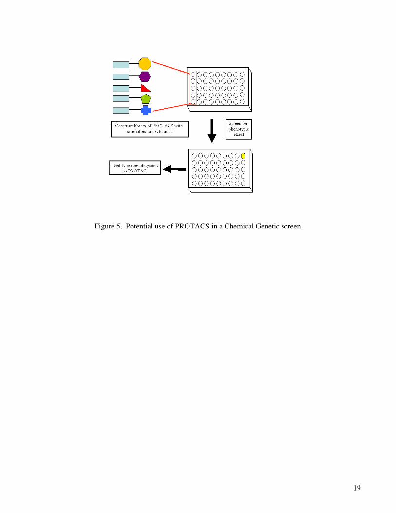
inactivation at the DNA or RNA level. Second, diversified libraries of PROTACS could be used to screen
for phenotypic effects in a chemical genetic fashion. This strategy could be used either to identify novel
ligands for a target by monitoring degradation of the target or to identify new therapeutically vulnerable
protein targets by studying phenotypic change as a result of selective protein degradation (Figure 5). This
chemical genetic strategy would employ a library of PROTAC molecules with identical E3 ubiquitin ligase
domains but chemically diverse target ligands. After PROTAC library incubation with cultured cells and
detection of the desired cellular phenotype (e.g., inhibition of pro-inflammatory signaling), one could
identify the protein that was degraded by incubation with the PROTAC. A number of approaches could be
used to identify the PROTAC-targeted protein, including affinity chromatography and differential
proteomic technologies such as ICAT39. In a modification of this strategy, a library of PROTACS could
be screened to identify a ligand for a particular target by monitoring degradation of the target protein (e.g.,
loss of GFP fusion protein). Finally, PROTACS could be used as drugs to remove toxic or disease-
causing proteins. This strategy is particularly appealing since many diseases, including several cancers, are

8
dependent on the presence or overexpression of a small number of proteins. The large number of potential
uses for this technology coupled with the success of these experiments suggests that PROTACS could
find broad use in the fields of cell biology, biochemistry, and potentially medicine.
Experimental Section
A. Materials (F36V)FKBP12 expression vector was generously provided by ARIAD Pharmaceuticals
(Cambridge, MA), and GFP-AR expression plasmid was a gift from Dr. Charles Sawyers (HHMI,
UCLA). Epoxomicin40 and AP2199821,22 were synthesized as previously described. Dihydrotestosterone
and testosterone were obtained from Sigma-Aldrich (St. Louis, MO). Monoclonal antibody recognizing
VHL was purchased from Oncogene (San Diego, CA); antibodies recognizing GFP and _-Tubulin were
obtained from Santa Cruz Biotech (Santa Cruz, CA); and polyclonal antibody against the androgen
receptor was from United Biomedical, Inc. (Hauppauge, NY). HEK293, 786-O, and HeLa cells were
purchased from the American Type Culture Collection (Manassas, VA). Tissue culture medium and
reagents were obtained from GIBCO-Invitrogen (Carlsbad, CA).
B Tissue culture HeLa cells, 786-O cells, and HEK 293 cells were separately cultured in D-MEM
supplemented with 10% fetal bovine serum, 100 units/mL penicillin, 100 mg/mL streptomycin and 2 mM
L-glutamine. All cell lines were maintained at temperature of 37°C in a humidified atmosphere of 5% CO2.
To generate cells stably expressing a particular fluorescent target protein, the parent cell line was grown to
70% confluency and transfected using calcium phosphate precipitation of the designated cDNA.
Following transfection, cells were split 1:10 into culture medium supplemented with 600 g/mL G418
(GIBCO-Invitrogen). Individual clones which optimally expressed fluorescent target protein were
identified and expanded under selection for further experimentation.
C. Detection of PROTAC-induced degradation by fluorescence microscopy Cells stably
expressing fluorescent target protein were plated into 96 well plates (HeLaEGFP-FKBP cells plated at 4000
cells/well; HEK293GFP-AR cells plated at 60,000-100,000 cells/well). Synthesized PROTACS were
9
dissolved in DMSO vehicle at a final concentration of 1%. Disappearance of target protein in vivo was
monitored by fluorescence microscopy at an excitation wavelength of 488 nm.
D. Detection of PROTAC-induced degradation by western blot Whole cell lysates were prepared
from HeLaEGFP-FKBP cells treated with PROTAC-4 and with HEK293GFP-AR cells treated with
PRTOAC-5 by lysing the cells in hot Laemmli buffer. Lysates were subjected to 8% polyacrylamide gel
electrophoresis and the proteins transferred to nitrocellulose membrane. Membranes were blocked in 3%
nonfat milk in TBS supplemented with 0.1% Triton X-100 and 0.02% sodium azide. Lysates from
HeLaEGFP-FKBP cells treated with PROTAC-4 were probed with anti-GFP (1:1000) and anti-VHL (1:1000)
antibodies; HEK293GFP-AR cells treated with PROTAC-5 were probed with anti-androgen receptor
(1:1000) and anti-ß-tubulin (1:200) antibodies. Blots were developed using chemiluminescent detection.
Acknowledgement J.S.S. thanks the American Chemical Society, Division of Medicinal Chemistry and
Aventis Pharmaceuticals for a predoctoral fellowship. We would like to thank Charles Sawyers (UCLA)
for providing the GFP-AR expression plasmid. The authors would like to thank John Hines for helpful
discussions. This work was supported by the NIH (R21 DK63404 to C.M.C.) UCLA SPORE in Prostate
Cancer Research (P50 CA92131 to K.M.S.), CaPCURE (R.J.D., C.M.C., and K.M.S.), Department of
Defense (DAMD17-03-1-0220 to K.M.S.), UC BioSTAR Project (01-10232 to K.M.S.), Stein-
Oppenheimer Award (K.M.S.), and the Susan G. Komen Breast Cancer Foundation (DISS0201703 to
R.J.D.). RJD is an Assistant Investigator of the HHMI.
Supporting Information Available
Preparation and characterization information for compounds 3 and 5 and the HIF-polyarginine
peptide.

10
1 Harding, M. W.; Galat, A.; Uehling, D. E.; Schreiber, S. L. Nature. 1989 341, 758-60.
2 Sin, N.; Meng, L.; Wang, M. Q. W.; Wen, J. J.; Bornmann W. G.; Crews C. M. Proc. Natl. Acad.
Sci. U.S.A. 1997, 94, 6099-6103.
3 Kwok, B. H. B.; Koh, B.; Ndubuisi, M. I.; Elofsson, M.; Crews, C. M. Chem. Biol., 2001, 14, 1-8.
4 Spencer, D. M.; Wandless, T. J.; Schreiber, S. L.; Crabtree, G. R. Science 1993, 262, 1019-1024.
5 Belshaw, P. J.; Ho, S. N.; Crabtree, G. R.; Schreiber, S.L. Proc. Nat. Acad. Sci. U.S.A., 1996, 93,
4604-4607.
6 Lin, H.; Abdia, W. M.; Sauer, R. T.; Cornish, V. W. J. Am. Chem. Soc. 2000, 122, 4247-4248.
7 Lin, H.; Cornish, V. W. Angew. Chem., Int. Ed. 2001, 40, 871-875.
8 Myung, J.; Kim, K.; Crews, C. M. Med. Res. Rev. 2001, 21, 245-273.
9 Koepp, D. M.; Harper, J. W.; Elledge, S. J. Cell 1999, 97, 431-434.
10 Rock, K. L.; Goldberg, A. L. Annu. Rev Immunol. 1995, 17, 739-779.
11 Ben-Neriah, Y. Nat. Immunol. 2002, 3, 20-26.
12 Muratani, M.; Tansey, W. P. Nat. Rev. Mol. Cell. Biol. 4, 192-201 (2003).
13 Hershko, A.; Ciechanover, A. Annu. Rev. Biochem. 67, 425-479 (1998).
14 Vijay-Kumar, S.; Bugg, C. E.; Wilkinson, K. D.; Vierstra, R. D.; Hatfield, P. M.; Cook, W. J. J. Biol.
Chem. 1987, 262, 6396-6399.
15 Breitschopf, K.; Bengal, E.; Ziv, T.; Admon, A.; Ciechanover, A. EMBO J. 1998, 17, 5964-5973.
16 Pickart, C. M. Annu. Rev. Biochem. 2001, 3, 503-533.
17 Yaron, A.; Hatzubal, A.; Davis, M.; Lavon, I.; Amit, S.; Manning, A. M.; Andersen, J. S.; Mann, M.;
Mercurio, F.; Ben-Neriah, Y. Nature 1998, 396, 590-594.
11
18 Crews, C. M. Curr. Opin. Chem. Biol. (in press) 2003.
19 Sakamoto, K. M.; Kim, K. B.; Kumagai, A.; Mercurio, F.; Crews, C. M.; Deshaies, R. J. Proc. Nat.
Acad. Sci. U.S.A. 2001, 98, 8554-8559.
20 Sakamoto, K.; Kim, K. B.; Verma, R.; Rasnick, A.; Stein, B.; Crews, C. M.; Deshaies, R. J. Mol. Cell.
Prot., accepted.
21 Yang, W.; Roxamus, L. W.; Narula, S.; Rollins, C. T.; Yuan, R.; Andrade, L. J.; Ram, M. K.; Phillips,
T. B.; van Schravendijk, M. R.; Dalgarno, D.; Clackson, T.; Holt, D. J. Med. Chem. 2000, 43, 1135-1142.
22 Rollins, C. T.; Rivera, V. M.; Woolfson, D. N.; Keenan, T.; Hatada, M.; Adams, S. E.; Andrade, L. J.;
Yaeger, D.; van Schravendijk, M. R.; Holt, D. A.; Gilman, M.; Clackson, T. Proc. Nat. Acad. Sci. U.S.A.
2000, 97, 7096-7101.
23 Hon, W.; Wilson, M. I.; Harlos, K.; Claridge, T. D. W.; Schofield, C. J.; Pugh, C. W.; Mazwell, P.
H.; Ratcliffe, P. J.; Stuart, D. I.; Jones, E. Y. Nature 2002, 417, 975-978.
24 Epstein, A. C.; Gleadle, J. M.; McNeill, L. A.; Heritson, K. S.; O’Rourke, J.; Mole, D. R.; Mukherji,
M.; Metzen, E.; Wilson, M. I.; Dhanda, A.; Tian, Y. M.; Masson, M.; Hamilton, D. L.; Jaakkola, P.;
Barstead, R.; Hodgkin, J.; Mazwell, P. H.; Pugh, C. W.; Schofield, C. J.; Ratcliffe, P. J. Cell 2001, 107,
43-54.
25 Ohh, M.; Park, C. W.; Ivan, M..; Hoffmann, M. A.; Kim, T. Y.; Huang, L. E.; Pavletich, N.; Chau, V.;
Kaelin, W. G. Nat. Cell. Biol. 2000, 2, 423-427.
26 Tanimoto, K.; Makino, Y.; Pereira, T.; Poellinger, L. EMBO J. 2000, 19, 4298-4309.
27 Wender, P. A.; Mitchell, D. J.; Pattabiraman K.; Pelkey, E. T.; Steinman L.; Rothbard, J. B. Proc.
Natl. Acad. Sci. U.S.A. 2000, 97, 13003-13008.
28 Kirschberg, T. A.; VanDeusen, C. L.; Rothbard, J. B.; Yang, M.; Wender, P. A. Org. Lett. 2003, 5,
3459-3462.
29 Derossi, D.; Joliot, A. H.; Chassaing, G.; Prochiants, A. J. Biol. Chem. 1994, 269, 10444-10450.

12
30 Fawell, S.; Seery, J.; Daikh, Y.; Moore, C.; Chen. L. L.; Pepinsky, B.; Barsoum, J. Proc. Nat. Acad.
Sci. U.S.A. 1994, 91, 664-668.
31 Baba, M.; Hirai, S.; Yamada-Okabe, H.; Hamada, K.; Tabuchi, H.; Kobayashi, K.; Kondo, K.;
Yoshida, M.; Yamashita, A.; Kishada, T.; Nakaigawa, N.; Nagashima, Y.; Kubota, Y.; Yao, M.; Ohno, S.
Oncogene 2003, 22, 2728-2738.
32 Debes, J. D., Schmidt, L. J., Huang, H. &Tindall, D. J. Cancer Res. 2002, 62, 5632-5636.
33 Stobaugh, M. E.; Blickenstaff, R. Steroids 1990, 55, 259-262.
34 Meng, L., Mohan, R., Kwok, B. H. K., Elofsson, M.; Sin, N.; Crews, C. M. Proc. Natl. Acad. Sci.
U.S.A. 1999, 96, 10403-10408.
35 Kuduk, S. D.; Zheng, F. F.; Sepp-Lorenzino, L.; Rosen, N.; Danishefsky, S. J. Bioorg. Med. Chem.
Lett. 1999, 9, 1233-1238.
36 Kuduk, S. D.; Harris, C. R.; Zheng, F. F.; Sepp-Lorenzino, L.; Ouerfelli, Q.; Rosen, N.; Danishefsky,
S. J.; Bioorg. Med. Chem. Lett., 2000, 10, 1303-1306.
37 Zheng, F. F.; Kuduk, S. D.; Chiosis, G.; M_nster, P. N.; Sepp-Lorenzino, L.; Danishefsky, S. J.;
Rosen, N. Cancer Res. 2000, 60, 2090-2094.
38 Citri, A.; Alroy, I.; Lavi, S.; Rubin, C.; Xu, W.; Grammatikakis, N.; Patterson, C.; Neckers, L.; Fry, D.
W.; Yarden, Y. EMBO J. 2002, 2407-2417.
39 Han D. K.; Eng J.; Zhou, H.; Aebersold, R. Nat Biotechnol. 2001, 19, 946-951.
40 Sin, N.; Kim, K. B.; Elofsson, M.; Meng, L.; Auth, H.; Kwok, B. H. B.; Crews, C. M. Bioorg. Med.
Chem. Lett. 1999, 9, 2283-2288.
13
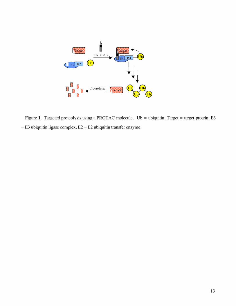
Figure 1. Targeted proteolysis using a PROTAC molecule. Ub = ubiquitin, Target = target protein, E3
= E3 ubiquitin ligase complex, E2 = E2 ubiquitin transfer enzyme.
14
Scheme 1. Synthesis of the AP21998/HIF1-based PROTAC. i. H2N(CH2)5CO2Bn, EDCI, DMAP. ii.
H2, Pd/C. iii. H2N(CH2)5CONH-ALAPYIP-(D-Arg)8-NH2., PyBrOP, DIPEA, DMF.

15
Figure 2. PROTAC-4 (3) mediates EGFP-FKBP degradation in a VHL-dependent manner. No change
in fluorescence is observed before (A) and 2.5 h after (B) treatment in DMSO control, while a significant
change is observed between before (C) and 2.5h after (D) treatment with 25 mM 3. Cells treated with 25
mM 1 and 25 mM HIF-(D-Arg)8 peptide show no difference before (E) and 2.5 h after (F) treatment. 786-
OEGFP-FKBP cells do not lose fluorescence before (G) or 2.5 h after (H) treatment with 25 mM 3. Western
blot analysis (I) with monoclonal anti-GFP antibodies confirms loss of EGFP-FKBP in cells treated with
25 mM 3 (PROTAC-4) for 2.5 h compared to an equal load from vehicle (DMSO) treated cells.
16
Scheme 2. Synthesis of a DHT/HIF1a-based PROTAC (PROTAC-3). i. H2N(CH2)5CONH-ALAPYIP-
(D-Arg)8-NH2., EDCI, DMAP, DMF.

17
Figure 3. DHT-HIF PROTAC-5 (5) mediates GFP-AR degradation in a proteasome-dependent manner.
One hour after treatment, 293GFP-AR cells treated with a 100 mM (B) or 50 mM (C) concentration of 5 lose
fluorescence, while the DMSO control (A) retains fluorescence. Cells treated with 10 mM epoxomicin (D),
and pretreated with 10 mM epoxomicin for four hours followed by treatment with 25 mM 5 for one hour
(E) retain fluorescence, while cells treated only with 25 mM 5 lose fluorescence after one hour (F).
Western blot analysis confirms loss of GFP-AR after treatment with PROTAC 5 (+PT) relative to a
loading control (G), while inhibition of the proteasome with epoxomicin (Epox) inhibits degradation (H).

18
Figure 4. A chemical bond between the HIF-(D-Arg)8 peptide and DHT is required for PROTAC-5-
induced degradation of GFP-AR. Cells were treated with (A) no treatment, (B) DMSO (equal volume), (C)
25 mM PROTAC-3, (D) 25 mM PROTAC-5 + 10-fold molar excess testosterone, (E) 25 mM PROTAC-5
+ 10-fold molar excess (250 mM) HIF-D-Arg peptide, (F) 25 mM HIF-D-Arg peptide + 25 mM
testosterone added separately, (G) 25mM DHT, and (H) 25 mM HIF-D-Arg peptide.
19
Figure 5. Potential use of PROTACS in a Chemical Genetic screen.

20