Embed Size (px)
Citation preview
Chemical and physical properties of iron(III)-oxidehydrateGiessen, van der, A.A.
DOI:10.6100/IR23239
Published: 01/01/1968
Document VersionPublisher’s PDF, also known as Version of Record (includes final page, issue and volume numbers)
Please check the document version of this publication:
• A submitted manuscript is the author's version of the article upon submission and before peer-review. There can be important differencesbetween the submitted version and the official published version of record. People interested in the research are advised to contact theauthor for the final version of the publication, or visit the DOI to the publisher's website.• The final author version and the galley proof are versions of the publication after peer review.• The final published version features the final layout of the paper including the volume, issue and page numbers.
Link to publication
Citation for published version (APA):Giessen, van der, A. A. (1968). Chemical and physical properties of iron(III)-oxide hydrate Eindhoven:Technische Hogeschool Eindhoven DOI: 10.6100/IR23239
General rightsCopyright and moral rights for the publications made accessible in the public portal are retained by the authors and/or other copyright ownersand it is a condition of accessing publications that users recognise and abide by the legal requirements associated with these rights.
• Users may download and print one copy of any publication from the public portal for the purpose of private study or research. • You may not further distribute the material or use it for any profit-making activity or commercial gain • You may freely distribute the URL identifying the publication in the public portal ?
Take down policyIf you believe that this document breaches copyright please contact us providing details, and we will remove access to the work immediatelyand investigate your claim.
Download date: 15. May. 2018
CHEMICAL AND PHYSICAL PROPERTIES OF IRON (III)-OXIDE
HYDRA TE
PROEFSCHRIFT
TER VERKRJJGING VAN DE GRAAD VAN DOCfOR IN DE TECHNISCHE WETENSCHAPPEN AAN DE TECHNISCHE HOGESCHOOL TE EINDHOVEN OP GEZAG VAN DE RECTOR MAGNIFICUS, DR. K. POSTHUMUS, HOOGLERAAR IN DE AFDELING DER SCHEIKUNDIGE TECHNOLOGIE, VOOR EEN COMMISSIE UIT DE SENAAT TE VERDEDIGEN OP DINSDAG 28 MEI 1968, DES
NAMIDDAGS TE 4 UUR
DOOR
AART ANTONIE van der GIESSEN SCHEIKUNDIG INGENIEUR
GEBOREN TE VLAARDINGEN
DIT PROEFSCHRIFT IS GOEDGEKEURD DOOR DE PROMOTOR
PROF. DR. G. C. A. SCHUIT

Aan mijn ouders Aan Magda
CONTENTS
I. HYDROL YSIS IN SOLUTIONS OF IRON(III) NITRATE 1 l .I. Introduetion . . . . . . . . . . . . 1 1.2. Reaction rates and hydrolysis equilibria . 4
1.2.1. Experirnental . . . . . 4 1.2.2. Results and discussion . 5
1.3. Mechanism of the hydrolysis . 10 1.3.1. Introduetion . . . . • 10 1.3.2. Experimental . . . . . 10 1.3.3. Results and discussion . 11
1.4. Summary 12 References . . . . . . . . . . . 13
2. THE HYDROLYSIS OF Fe3+ IONSIN VERY DILUTED SOLU-TIONS . . . . . 14 2.1. Introduetion . 14 2.2. Experimental . 15 2.3. Results . . . 16
2.3.1. Determination of the size distribution 16 2.3.2. Electron micrographs of the sediment . 18 2.3.3. Influence of the precipitation conditions on the size distri-
bution . 19 2.4. Summary 19 References . . . . 20
3. THE DEHYDRATION OF IRON(III)-OXIDE- HYDRATE GELS 21 3.1. Non-destructive removal ofthe capillary water . 21
3.1.1. The low-temperature dehydration process . . . . . . . 21 3.1.2. Experimental . . . . . . . . . . . . . . . . . . . . 21
3.2. A study of the constitution and freezing behaviour of iron(III)-oxide-hydrate gels by means of the Mössbauer effect 22 3.2.1. Introduetion . 22 3.2.2. Experimental . 23 3.2.3. Results . . . 24 3.2.4. Discussion . .
3.2.4.1. Constitution of the gels . 3.2.4.2. The freezing process
3.3. Summary References . . . . . . . . . . . . .
26 26 28 29 29
4. THE STRUCTURE OF IRON(III)-OXIDE HYDRATE 4.1. Crystallographic properties
4.1.1. Introduetion . . . . . 4.1 .2. Experimenta1 . . . . . 4.1.3. Results and discussion .
4.1.3.1. Crystallographic properties 4.1.3.2. Morpho1ogy
4.2. Mössbauer spectroscopy . 4.2.1. Introduetion . . . . 4.2.2. Experimental . . . . 4.2.3. Results and discussion .
31 31 31 31 32 32 35 37 37 38 38
4.3. Magnetic measurements . . . 40 4.3.1. Superparamagnetic behaviour 40 4.3.2. Results and discussion . 42
4.4. Summary 45 References . . . . . . . . . . • . 46
5. RECRYSTALLIZA TION OF THE IRON(IIl)-OXIDE-HYDRA TE GEL IN AQUEOUS SOLUTIONS 47 5.1. Discussion of the literature . . . . . . . . . . . . . . . . 47 5.2. Experimental . . . . . . . . . . • . . . . . . . . • . . . 49 5.3. Ageing phenomena occurring in partially hydrolyzed solutions . 50
5.3.1. Crystallographic properties of the ageing products 50 5.3.2. Morphology . . . . . . . . 50
5.3.2.1. Tyndall effect . . . . 50 5.3.2.2. Electron rnicroscopy . 52 5.3.2.3. Viscosity . . . . . . 53
5.3.3. Magnetic measurements . . . 55 5.4. Recrystallization of the iron(III)-oxide-hydrate gel in alkaline
solution . . . . • . . • . . . . . . . . 57 5.4.1. The rate of crystallization of the gel . . . . . . . . . 57
5.4.1.1. Chemica) characterization . . . . . . . . . . 57 5.4.1.2. Structural and morphological transformations . 58
5.4.2. Mechanism of the ageing 61 5.4.3. Magnetic properties . 64
5.5. Summary 65 References . . . . . . . . . . 66
6. THE LOCALIZA TION OF THE PROTONS IN THE IRON(lli)-OXIDE HYDRATE . . . . . . . . . . . 67 6.1. Introduetion; discussion of the Iiterature . . . . . . . . . . . 67
6.2. Infrared spectroscopy . 68 6.2.1. Introduetion . . 68 6.2.2. Experimental . . 69 6.2.3. Results and discussion . 70
6.3. Nuclear magnetic resonance . 72 6.3.1. Discussion of the literature . 72 6.3.2. Experimental . • . . . 73 6.3.3. Results and discussion . 73
6.4. Summary 75 References . . . . . . . . . . . 76
7. THE RECRYSTALLIZATION OF IRON(III)-OXIDE HYDRATE AT ELEV ATED TEMPERA TURES . . . . . . . . . . . . . 77 7.1. Introduetion . . . . . . . . . . . . . . . . . . . . . . 77 7.2. Thermogravimetrie analysis and differential thermal analysis . 78
7.2.1. Experimental . . . . . . . . . . . . . 78 7.2.2. Results and discussion . . . . . . . . . 79
7.3. Crystallographic transformations ; morphology . 7.4. Magnetic properties of the dehydrated products 7.5. Mechanism of the dehydration . 7.6. Summary References .
Summary
Samenvatting .
82 83 87 88 88
90
93
1.1. Introduetion
l. HYDROL YSIS IN SOLUTIONS OF IRON(III) NITRATE
Precipitation of compounds in solutions usually leads to crystalline products with particles, of a shape and size depending u pon the reaction conditions. A precipitate is formed when the solubility product is exceeded and when at the same time nuclei on which the crystals can grow are formed or are already present. Such a solution contains growing nuclei, together with the ions composing them. There are examples showing that precipitation in solutions does not lead to the formation of a crystalline product. For a number of ions such as Fe3+, AJH, Cr3+, the addition of OH- ions totheir aqueous solutions first gives rise to the formation of new products that remaio in solution - in the case of FeH ions this can already be derived from the darkening of the colour ofthe solution - and ultimately causes the formation of a gelatinous precipitate. Regarding the mechanism it was thought that a series of hydrolytic processes takes place, with the intermediate formation of polymerie ions, which finally uni te to form the gelatinous precipitate. In the case of FeH there is nogeneral agreement in the literature with regard to the size and the composition of the polynuclear complexes.
According to various authors units of the type Fe( OH). ' 3 -•>+ are fust formed which u pon further addition of OH- i ons grow to Jarger complexes containing two or more Fe3+ ions. The size of the complex depends upon the hydralysis conditions: both the concentration of the Fe(III) compound as wel! as that of OH- ions are important. In very dilute solutions, with Fe(III) below 10- 3
gion/1, only Fe(OH)2+ complexes are formed as was concluded by Siddall and Vosburgh from spectrophotometricanalysis 1 - 1 ). In moreconcentrated solutions (l-100.10- 3 gion/1) dimers arealso formed, according to Hedström 1 - 2 ) . The relation between the Fe3+ concentratien (deterrnined with the aid of redoxpotential measurements of the system Fe3+ /Fe2+ ) and the pH is such that the hydralysis equilibria:
nFeH + mH20:? Fe.(OH)mC3n-m)+ + mH 3ü +
can bedescribed with values for n equal to unity or two (formation ofmonomers and dimers). Mulay and Selwood investigated sirnilar solutions by magnetic measurements 1 - 3). These investigators found on increasing pH a considerable decreasein the magnetization (at constant field strength) between pH 1 and 2. Assuming a Curie law, a decrease in the magnetic moment per Fe3+ ion from 5·8 to 3·6 /hs at pH = 1·5 was calculated. This was explained by the formation of a diamagnetic diroer (of zero moment) following the example ofHedström. These dimers, at the pH of precipitation, then uni te to form the gelatineus precipitate.

-2-
The results of Selwood and Hedström are not in accordance with earlier results of Jander and Winkel 1- 4 ). These authors estimated the molecular weight of the particles present in hydrolyzed Fe(CI04h solutions in the pH region 1-3, with the aid of measurements of the diffusion coefficient. In 0·1-molar solutions, at pH = 2, a considerable decrease in the diffusion velocity was observed, indicating the formation of larger particles. The molecular weights of these particles depend upon the pH. Assuming that, in solutions with pH < I only the ions FeCI04 2+ are present (molecular weight about 250) and that the relation D VM = constant is valid, they concluded that at pH = 3 polynuclear particles with a molecular weight of 5000 would be present. Recently Spiro et al., by ultracentrifuging partially hydrolyzed FeH solutions, concluded the presence of even much larger particles with a size of 70 A 1 - 5).
There is apparently no consensus of opinion in the literature regarding the structure and the size of the species formed u pon the hydrolysis. In our opinion this is not surprising as the measurements do not refer to equilibria: the results discussed are all obtained on supersaturated solutions. This immediately appears on checking the experimental conditions against the values of the solubility product of "Fe(OH)J". The latter equals 10- 38'7 , as determined by Biedermann and Schindler for solutions of Fe(Cl04 ) 3 with the same ionic strength as those discussed above 1 - 6). In view of this it is not surprising that a precipitate is formed in such supersaturated solutions in course of time. Feitknecht and Michaelis 1 - 7 ) investigated such precipitates formed in solutions similar to those studied by Hedström, after the solution had stood at room temperature for a long time. The precipitates were isolated from the solution by ultracentrifugating and they appeared to consist of a mixture of needies of o:-FeOOH and spherical particles of 50-70 A.
Mechanism of hydrolysis
A summary of the literature in which possible mechanisms of the hydralysis are discussed bas been given by Rollinson 1 - 8). In aqueous solution the Fe3 + ion is hydrated; the coordination sphere contains six H 20 molecules forming an octahedron. Strongly acidic solutions of Fe(III) salts contain the complexes Fe(H20)63+. These complexes react as an acid by donation of a proton:
Fe(H20)63+ + H20 +±: [Fe(H20)s(OH)F+ + H 3o+. ,
Addition of hydroxyl i ons displaces this equilibrium to the right. The morromers can give dimers by the formation of OH bridges:
2 [Fo(H,O),(OH)]'+ +" fH,O). Fo(~)Fo(H,O). <+ + 2 H,O.

-3-
This reaction is designated by the term olation. The dimer in turn can split off protons according to
~ /0" r+ <0 lH,O), Fe--..,._~/Fe(H,O)J + H,o• .
This is called oxolation; the Fe-0-Fe bond is called an oxo bridge. These reactions which give rise to a decrease of the pH proceed slowly. This applies especially to the oxolation reaction. The final result of these deprotonizing processes is the formation of polynuclear species, linear macromolecules (or ions) as well as chains with side branches (not depicted in the scheme). The reactions are reversible on decreasing pH but it is thought that it is particularly the oxo bridge that is difficult to break. As a consequence a precipitated oxidehydroxide only very slowly redissolves upon the addition of acid.
According to the ideas discussed above the gels are the final products of hydrolytic processes and they consist of a netwerk of polynuclear complexes linked up by -0- and -OH bridges.
Anions largely influence the process of hydrolysis. Anions, such as S04 2 -
which are able to give a coordination bond with the cation may replace H 20 as wellas OH-. This can be perceived from an increase in the pH on addition of Na2S04 to a partially hydrolyzed solution of Fe(N03h. The addition of S04 2 - ions also causes the formation of a precipitate, probably consisting of basic sulphates 1 - 9 •10); a precipitate is also observed 1 - 11) after addition ofP04 3 -.
Reaction rates and hydralysis equilibria
It is stated repeatedly in the literature that after a rapid initia! reaction the subsequent hydrolytic processes occurring in Fe3+ solutions are slow. At room temperature, after the addition of OH- ions, it takes hundreds of hours before a constant pH value is reached. These slow pH changes are one of the main experimental facts underlying tbe olation-oxolation reactions mentioned above. lt is not necessary, however, to assume the gradual formation of large polymers as the cause of these pH changes. Weiser and Milligan assume that, u pon the hydralysis of Fe(III) or AI(III) salts crystalline particles are formed, albeit of exceedingly smal! size compared to that of crystals formed in most other well-known precipitation reactions1 - 12•13 •14). Their arguments, in favour
-4-
of crystallites, are, however, questionable. Yet the idea may be correct. It bas been shown by Onoda and De Bruyn that in aqueous suspension coarse crystals of a-Fe20 3 , after the addition ofacid or base, also give rise to similar retarded changes in the pH, due to the slow ditfusion of protons from the surface towards the inside of the crystals 1 - 15) (see also ref. 1-1 6). Hence the slow ditfusion of protons from the inside of a crystalline material via its surface to the solution could also be a possible reason for the slow pH changes instead of the slowly proceeding olation-oxolation reactions. This mechanism only applies if in deed crystallites are formed upon hydrolysis. As no visible precipitate is formed as Jong as OH-/FeH < 2·5 these then must be very smal!. A diminishing initially present disorder or a slow growth of these small particles could act as the driving forces of the donation of protons to the solution.
In the following sections a number of experiments will he discussed which have been carried out in order to obtain a better knowledge of the hydralysis phenomena of FeH ions. These experiments are all done with N03-
containing solutions. The reason for this is that N0 3 - Uust as CI04 -) has only a weak affin.ity to the FeH coordination shell (contrary to CI- and S04 2 - ions) and thus secondary effects caused not by the OH- ions but by the other ligands are avoided.
1.2. Reaction rates and bydrolysis equilibria
1.2.1. Experimental
The Fe(N03 ) 3 solutions are hydrolyzed by adding NaOH, 1 molar, from a burette at a rate of one drop per second with vigorous agitation, until the desired OH- /FeH ratio is reached. Solutions containing 0· 10 mole/1 Fe(N03) 3 (Merck, p.a.) and 2·8 mole/1 NaN03 (U.C.B., p.a.) are prepared by dissolving the appropriate amounts of these compounds in distilled water. From this solution 100 mi is brought into a 250-ml beaker placed in a thermostat kept at 25 °C. The beaker is covered by a rubber plug in which the pH electredes and the stirrer lead are fastened. The tap end of the burette protrucles into a spare hole lined with a glass tube. Through this also the nitrogen escapes which is bubbled through the solution at a rate of 100 mi/min. The pH is measured by means of a Philips G.A. I 10 glasselectrode and a saturated-calomel reference electrode.
Foranother series of experiments the amount of Fe(N03h is varied so that [FeH] varies between 200.10- 3 and 1 gion/1, whereas the pH is brought to the same value of 2·20. In order to obtain the same pH in all cases, either 0·1-molar NaOH or HN03 is added.
In a number of experiments, after establishing a eertaio OH- fFeH ratio, to 100 ml of the FeH solution an equal volume of a 0·1-molar Na2 S04 solution is added which is brought to the same pH as the hydrolyzed FeH solution, with H 2S04 . After this addition the pH is measured again.

-5-
Determination of the hydrolysis equilibria is carried out with the aid of titrations of the Fe3+ ions with the disodium salt of ethylene diamine tetraacetic acid, EDTA, using KCNS as an indicator. Of the solution to be examined, 25 mi is pipetted into a conical flask of 100 mi. This is titrated by adding an EDTA solution, 0·05 or 0·1 molar, dropwise from a burette until nearly the equivalent point is reached which is indicated by the appearance of a precipitate. When about half ofthe necessarytamount of EDTA is added, a precipitate already appears; it intensifies near the equivaJence point. This precipitate is removed by centrifuging, after which about 20 mg of KCNS indicator is added to the clear yellow solution. The solution is then further titrated with EDT A soiution until the red colour of tbe Fe-(CNS) complex disappears; because of the interference of yellowish colour of the solution with the red colour of the Fe-(CNS) complex the equivaJence point is difficult to determine and it is necessary first to make a rough estimation of the amount of EDT A required.
A number ofviscosity measurements are done, at 25 oe, using an Ubbelohde viscosimeter.
1.2.2. Results and discussion
Titration of iron nitrate with a base
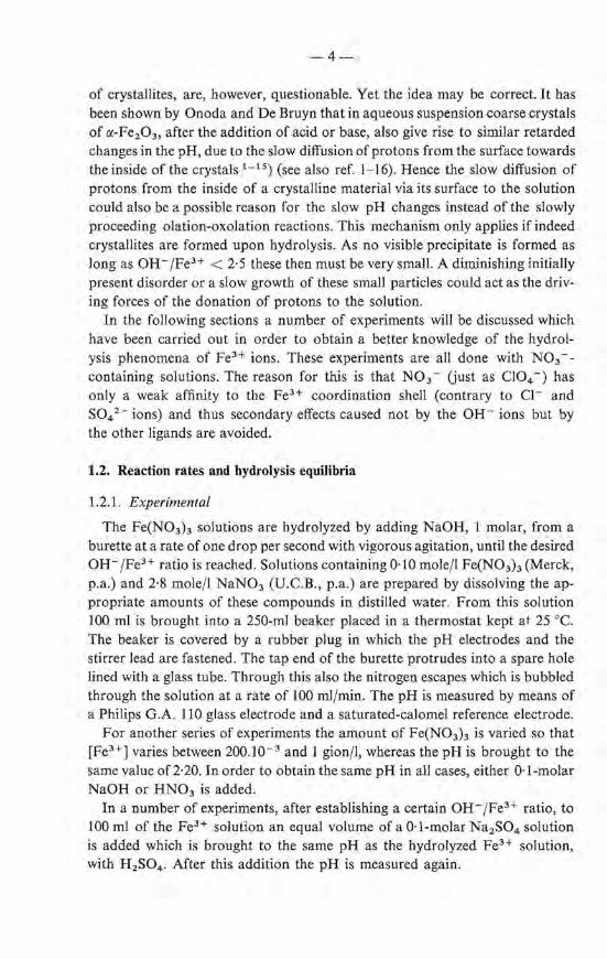
A comparison of the titration curve of a solution of Fe(N03 ) 3 with that of a HN03 solution shows that the Fe(H20)6 3+ ion behaves as a fairly strong acid. Figure 1.1 shows a titration curve of a 0·1-molar Fe(N03) 3 solution together with that of a 0· 3-molar HN03 solution. U pon addition of OH- i ons the Fe3+ solution becomes darker but no visible precipitate appears until about 2·5 OH-
6
4
2
--j I
_}, r-- _..,
i r------ ----------2 3
- OH-/ Fe3+ OW/ 3H+
Fig. l.I. Titration curves of
---- 0·1-molar Fe(N03h, -------- - 0·3-molar HN03 .
-6-
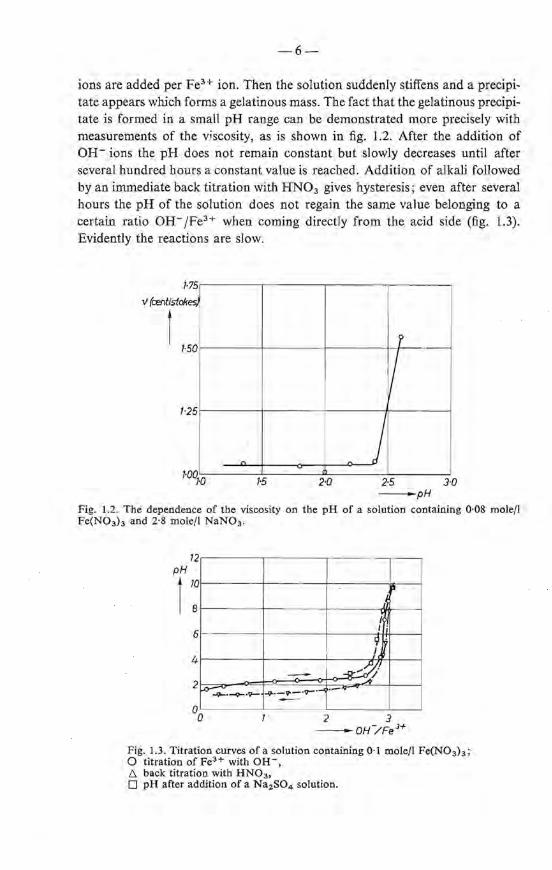
ions are added per FeH ion. Then the solution suddenly stiffens and a precipitate appears which forms a gelatinous mass. The fact that the gelatinous precipitate is formed in a small pH range can be demonstrated more precisely with measurements of the viscosity, as is shown in fig. 1.2. After the addition of OH- ions the pH does not remain constant but slowly decreases until after several hundred hours a constant value is reached. Addition of alkali foliowed by animmedia te back titration with HN03 gives hysteresis; even after several hours the pH of the solution does not regain the same value belonging to a eertaio ratio OH- /FeH when coming directly from the acid side (fig. 1.3). Evidently the reactions are slow.
·75
v (centistok ·~
1, 50 r
1/ 25
j 00 1' 1·0 1·5 2·0 2·5 3·0
-pH
Fig. 1.2. The dependenee of tbe viscosity on the pH of a salution containing 0·08 molefl Fe(N03h and 2·8 mole/1 NaN03 .
p
I
6 I.
9' I
- 4' 2 ~·-9-·t'- -9-·-v--·v-·-< ·-
~
2 3 -oH/Fe 3+
Fig. 1.3. Titration curves of a salution containing 0·1 mole/1 Fe(N03h; 0 titration of Fe3+ with OH-, /',. back titration with HN03 ,
D pH after addition of a Na2 S04 solution.

-7-
The concentration ofthe N03 - i ons is of only minor importance: the titration curve of a 0·1-molar Fe(N03) 3 solution which is 2·8 molar in NaN03 is only slightly shifted to the left for valnes of oH-jFe3+ R:> 2·5 (fig. 1.4) compared to a solution of the same strength to which no NaN03 bas been added. Nearly the same curves are obtained when two titrations are carried out, one by continuously dropping the NaOH into the solution and one by waiting four minutes after each ml NaOH added. When instead of 1-molar NaOH, 10-molar NaOH is used as a titrating agent the curve is shifted slightly to the left (not shown).
~ I
6 ~~
f~ J
4
2 ~-u
2 3 -oH/ FeJ+
Fig. 1.4. A comparison of the titration curves of Fe(N03h; 0 0·1-mole/1 (FeN03h, D 0·1-mole/1 Fe(N03h + 3-mole/1 NaN03 •
Ligands which are able to form complexes with the Fe3+ ions, contrary to N03 - ions, infl.uence the hydralysis processes. Addttion of a solution of Na2S04 of the same pH as a hydrolyzed Fe3 + solution gives a marked increase in the pH (fig. 1.3) under the simultaneons formation of a precipitate. This proves that the S04 2 - ions liberate oH- ions from the species which are formed upon hydrolysis.
The sudden formation of a gel suggests that we are dealing here with the fl.occulation of a colloidal precipitate. This flocculate then consists of an agglomerate formed by either the polynuclear chains as described by the olationoxolation theory or by small crystallites. In this study it will be shown that the latter possibility is the case: crystallites are formed on addition of OHions and remain in colloidal solution until, due to a further addition of OHions the point of electroneutrality is reached and flocculation occurs.
Determination of the hydralysis equilibria
A chemica! metbod has been used for the determination of the concentration ofthe Fe3+ ions in equilibrium with the larger units present in the salution as a result of the hydrolysis. It has been found that, under our experimental con-

-8-
ditions, the Fe3+ ions can be determined by the conventional titration with a solution of the disodium salt of ethylene diamine tetraacetic acid (EDTA); KCNS is used as an indicator. The titration of FeH ions with the complexing agent is well known, see for instanee ref. 1-17; a more detailed discussion regarding the ·theoretica! background of this titration can be found in ref. 1-18. For the present case we used this metbod also for partially hydrolyzed solutions, under the assumption that at least all monomeric species of the type Fe(OH)n<3 -nl+ react under the formation of a complex with the EDTA and that polynuclear ion ie complexes or particles do not consume EDT A, an assumption justified by the results. This titration of Fe3 + in partly hydrolyzed solutions is possible due to the slow rate at wbich the hydralysis processes occur: if the FeH ions are removed from the solution by complexing them with EDTA, FeH ions are only slowly liberated from the larger complexes.
It has been found that, in hydrolyzed solutions, the concentratien of free Fe3 + ionsis only a fraction ofthe total amount of iron presentand independent of the total Fe(III) concentratien at constant pH. This is illustrated with the data listed in table 1-I obtained with solutions of pH = 2·30. After one hour the equilibrium concentratien is a bout 3.10- 3 gion/1. This corresponds toa solubility product ~ of 3.10- 3 x I0- 35 .7 = I0- 38' 2 (pK H 2 0 = 14·22). A solubility product of about the same numerical value can be calculated from FeH titrations of about 0·08-molar Fe(N03) 3 solutions with different pH's, table 1-11. The results are related to solutions obtained after the addition of 0, 1, 2 and 2·6 OH- fFeH, respectively. The value of the solubility product is not entirely constant. The concentratien of Fe3+ ions gradually decreases with
TABLE 1-1
Concentratien (mgion/1) of Fe3+ ions wbich can be complexed with EDTA [Fe3+]. All solutions contain 2·8 mo1e NaN03/l. One hour after the preparation the pH = 2·30. Determinations have been done), 24 and 200 hours after the preparatien
[Fe(III)] [FeH] (mgion/1)
(mmole/1) 1 h 24 h 200h
220 2·0 80 3·0 2·5 0·6 45 2·0 2·4 25 2·4 1·5 0·6 5 3·8 1·9 1 0·5 0·5 0-4
-9-
TABLE 1-11
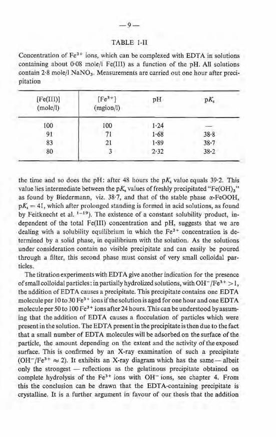
Concentration of FeH i ons, which can be complexed with EDT A in solutions containing about 0·08 mole/1 Fe(III) as a function of the pH. All solutions contain 2·8 mole/1 NaN03 . Measurements are carried out one hourafter precipitation
[Fe(III)] (mole/1)
100 91 83 80
[FeH] (mgion/1)
100 71 21 3
pH
1·24 1·68 1-89 2·32
pK.
38·8 38·7 38·2
the time and so does the pH: after 48 hours the p_K. value equals 39·2. This value lies intermediate between the p_K. values offreshly precipitated "Fe(OH)3 "
as found by Biedermann, viz. 38·7, and that of the stabie phase a:-FeOOH, pK. = 41 , which after prolonged standing is formed in acid solutions, as found by Feitknecht et al. 1 - 19). The existence of a constant solubility product, independent of the total Fe(III) concentration and pH, suggests that we are dealing with a solubility equilibrium in which the FeH concentration is determined by a solid phase, in equilibrium with the solution. As the solutions under consideration contain no visible precipitate and can easily be poured through a filter, this second phase must consist of very small colloidal particles.
The titration experirnents with EDT A give another indication for the presence of small colloidal particles: in partially hydrolized solutions, with OH-/FeH > 1, tbe addition of EDTA causes a precipitate. Tbis precipitate contains one EDTA molecule per I 0 to 30 FeH i ons ifthe solution is aged for one hourand one EDTA moleculeper 50to 100 Fe3 + ions after 24hours. This can be understood byassuming tbat the addition of EDTA causes a fiocculation of particles which were present in the solution. The EDT A present in the precipitate is then due tothefact that a small number of EDT A molecules wiJl be adsorbed on the surface of tbe particle, the amount depending on the extent and the activity of tbe exposed surface. This is confirmed by an X-ray examination of such a precipitate (OH-jFeH ~ 2). It exhibits an X-ray diagram which bas the same- albeit only the strongest - reflections as the gelatinous precipitate obtained on complete hydralysis of the FeH ions with OH- ions, see chapter 4. From this the condusion can be drawn that the EDTA-containing precipitate is crystalline. It is a further argument in favour of our thesis that the addition
-10-
of EDT A causes the flocculation of particles that are already present in the partially hydrolyzed solution.
1.3. Mecbanism of tbe bydrolysis
1.3.1. Introduetion
If the formation of a gel, as mentioned before, may be described as the flocculation of particles formed u pon hydrolysis, a study of these particles can give some idea of the processes occurring when the Fe(H 20)6 3+ ions are deprotonized. Such a study can only be clone provided that the particles can be isolated from the gel without changing their properties. Th is isolation necessarily involves a dehydration procedure; such a dehydration process, described in chapter 3, bas been used for the study described in the next sections.
At this point we shall introduce the characterization with the aid of the magnetic susceptibility preceding a more detailed discussion in chapter 4. Ultrafine antiferromagnetic crystallites with a size below 50 A - the type of material we are dealing with - are superparamagnetic. Powders composed of such particles have a magnetization which is linearly proportional to the field strength. The susceptibility is independent of the size of the particles as long as this is below 50 A and depends only upon the disorder prevailing in the lattice. Based on this knowledge the data presented in the following can assist in a further elucidation of the hydrolysis mechanism and show in which respect Selwood's interpretation of the magnetic properties of hydrolyzed solutions fails.
1.3.2. Experimental
In series of experiments 0·1-molar solutions of Fe(N03)J are hydrolyzed with NaOH solutions varying in concentration (1·0, 3·7 and 9·7 molar) such that OH-/FeH = 2·41, and kept at 25 oe under vigorous agitation for about half an hour, after which the hydrolyzed solutions are brought to pH = 7·5 with NaOH of the desired concentration again by addition at a rate of one drop per secend and while stirring. The time between the addition of the first drop to the freshly prepared solution and the precipitation is 60 minutes. The resulting gel is kept for another five rninutes in the precipitation vessel, with agjtation, and afterwards the pH which bas dropped slightly is readjusted. The gel is then placed on a filter, wasbed with one portion of water brought to pH= 7·5 with NH40H and further dehydrated in a manner described on page 21. The resulting oxide hydrate is driedon P 20 5 to constant weight at room temperature which corresponds to a H 2 0 content of about 16 %. The magnetic susceptibilities, measured according to a metbod described in ref. 1-20 are all reduced to water contents of 16·0%.
-11-
1.3.3. Results and discussion
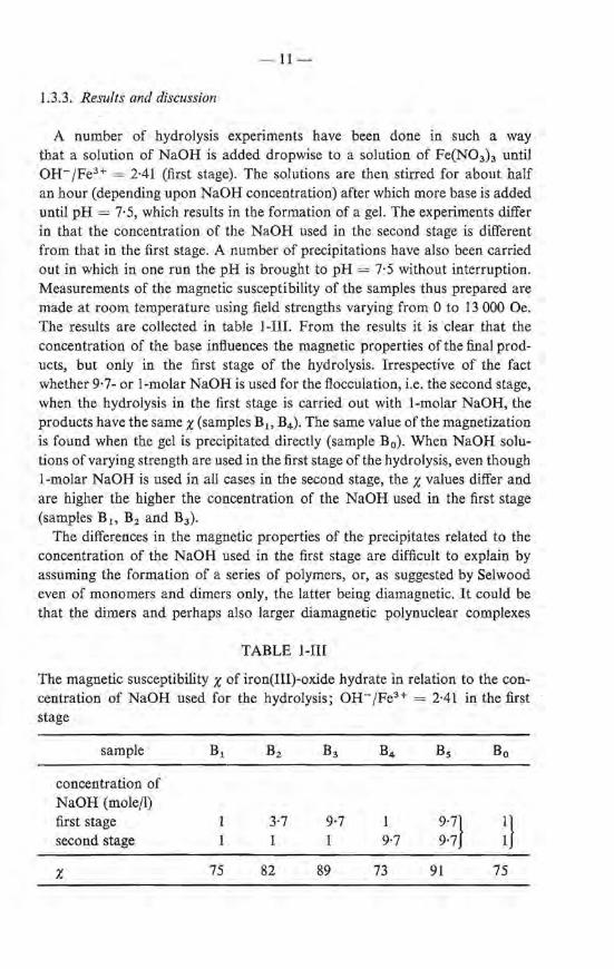
A number of hydrolysis experiments have been clone in such a way that a solution of NaOH is added dropwise to a solution of Fe(N03h until OH-/Fe3+ = 2-41 (first stage). The solutions are then stirred for about half an hour (depending upon NaOH concentration) after which more base is added until pH= 7·5, which results in the formation of a gel. The experiments differ in that the concentration of the NaOH used in the second stage is different from that in the first stage. A number of precipitations have also been carried out in which in one run the pH is brought to pH = 7·5 without interruption. Measurements of the magnetic susceptibility of the samples thus prepared are made at room temperature using field strengtbs varying from 0 to 13 000 Oe. The results are collected in table 1-liL Fr om the results it is clear that the concentration of the base influences the magnetic properties ofthe final products, but only in the first stage of the hydrolysis. Irrespective of the fact whether 9·7- or 1-molar NaOH is used for the flocculation, i.e. the second stage, when the hydralysis in the first stage is carried out with 1-molar NaOH, the products have the same x (samples Bl> B4 ). The same value ofthe magnetization is found when the gel is precipitated directly (sample B0). When NaOH solutions of varying strength are used in the first stage of the hydrolysis, even though l-molar NaOH is used in all cases in the second stage, the x values differ and are higher the higher the concentration of the NaOH used in the first stage (samples B 1> B2 and B3 ).
The differences in the magnetic properties of the precipitates related to the concentration of the NaOH used in the first stage are difficult to explain by assuming the formation of a series of polymers, or, as suggested by Selwood even of monomers and dimers only, the latter being diamagnetic. It could be that the dimers and perhaps also larger diamagnetic polynuclear complexes
TABLE 1-III
The magnetic susceptibility x of iron(III)-oxide hydrate in relation to the con-centration of NaOH used for the hydrolysis; OH-/Fe3+ = 2·41 in the fust stage
sample B1 B2 B3 B4 Bs Bo
concentra:tion of NaOH (mole/1) fust stage 3·7 9·7 9·7} a second stage 9·7 9·7
x 75 82 89 73 91 75
-12-
do not reach their equilibrium distribution within the ageing period used, due to kinetic effects. But, in that case, too, the results are in conflict with the assumption. When a drop of NaOH falls into the solution there is a brief Iocal high concentration of OH- ions which could lead only to a higher degree of complexation than that corresponding to the equilibrium conditions. Hence, at higher NaOH concentrations a smaller x value seems more probable than a larger one. A more satisfactory explanation is offered by the assumption that NaOH addition leads to the formation of particles with a higher degree of disorder the higher the concentration of the base added. The occurrence of this disorder together with the small partiele size could wel! result from the extremely low solubility product of the material under study. Hence, on the addition of a drop of NaOH, there is a brief local excess of OH- ions, giving an enormous supersaturation and thus causing a very rapid precipitation. This leads to crystallites which are not much larger than the critica! nucleus and which may be expected to have a large number of stacking faults. The explanation of the different x values as due to a high degree of disorder is also in accord with the observed slow decrease of the solubility mentioned in sec. 1.2.2. The latter could be attributed to growth of the particles or to an increasing ordering within the particles. In view of the foregoing it is most likely that both processes contribute to the decrease of the solubility.
1.4. Summary
The ideas advanced in the Iiterature regarding the processes that occur on hydrolyzing solutions of Fe(III) salts generally explain the experimental results mainly as due to the formation of deprotonized monoroers Fe(H20)6 _n(OH)n<J-n>+ at low degree of hydralysis or, at higher ratios of OH-/fe3+, as due to the formation of polynuclear complexes with a size distribution depending on the experimental conditions. Hysteresis effects are accounted for by assuming that the dorration or the acceptance of protons of these complexes is rather slow. When, on average, more than 2·5 OH- ions per fe3+ ion are added, a gel is formed, which accordingly must be regarcled as a network of interconnected ebains of polynuclear complexes. Weiser and Milligan, however, have a different opinion. They consider the gel as a fl.occulated colloid composed of crystallites which have previously been formed in the solution.
The results presented in this chapter are in favour of the latter mechanism. As desc_ribed, titrations with an agent that has strong complexing properties to fe3+ ions (EDTA) lead to the idea that there is a solid, colloidally dispersed, phase in the hydrolyzed solutions, which has a constant solubility product.
On addition of a base to a Fe (III) solution a gel is formed when the ratio OH-/fe3+ exceeds 2·5. This formation of a gel in a rather short pH range can thus be considered as a flocculation of the colloidal particles present in the
-13-
solution. Measurements of the magnetic susceptibility x give some idea of the mechanism of the reactions that take place, i.e. the actdition of OH- ions to the FeH salution causes a rapid formation of crystallites with more defects in the lattice the higher the concentratien of the base added. From the knowledge thatthere is a high degree of disorder, the slowand gradual decrease of the solubility of the productsin partially hydrolyzed solutions cao at least partly be attributed to an increased ordering within the particles.
The results mentioned thus far allow the provisional condusion that the precipitation processes occurring in solutions of FeH ions differ from the usually observed precipitation reactions only in that the size of the crystallites is extremely smal!. The number of iron ions composing them is probably oot far beyond the number which is necessary for the formation of a critica! nucleus. The experimental evidence presented in the next chapters will show that this supposition is justified.
REPERENCES 1- 1) Th. H. Siddall and W. C. Vosburgh, J. Am. cbem. Soc. 73, 427, 1951. 1 - 2) B. A. 0. Hedström, Arkiv f. Kemi 6, 1, 1952. 1- 3) L. N. Mulay, P.W. Selwood, J. Am. chem. Soc. 76, 6207, 1954; 77, 2693, 1955. 1 - 4) G. Jander and A. Winkel, Z. anorg. allg. Chem. 193, 1, 1930. 1 - 5) G. Spiro, S. E. Allerton, J. Renner, A. Terzis, R. Bi is and P. Saltman, J. Am.
chem. Soc. 88, 2721, 1966. 1- 6 ) G. Biedermann and P. Schindler, Acta chem. Scand. 11, 731, 1957. 1 - 7 ) W. Feitknecht and W. Michaelis, Helv. chim. Acta 45, 212, 1962. 1 - 8) J . C. Ba ilar and D . H. Busch, The chemistry of the coordination compounds,
Reinhold-Publ. Corp., 1956, p. 448. 1 - 9 ) T. V. Arden, J. chem. Soc. 1951, 350, 1951. 1 - 10) S. R. Gupta and S. Ghosh, Z. anorg. allg. Chem. 279, 212, 1955. 1 - 11) H. Galal-Gorchev, J. inorg. nucl. Chem. 25, 567, 1963. 1 - 12) H. B. Weiser and W. 0. Milligan, Chem. Rev. 25, 1, 1939. 1- 13) H. ;B. Weiser and W. 0. Milligan, J. phys. Chem. 44, 1081, 1940. 1 - 14) H. B. Weiser and W. 0. Mill iga n, Advances Coll. Sci. 1, 227, 1941. 1- 15) G . Y. Onoda and P. L. de Bruyn, Surface Sci. 4, 48, 1966. 1 - 16) R. J. Atkinson, A. M. Posner and J. P. Quick, J. phys. Chem. 71, 551, 1967. 1 - 17) L. T. Bultand N. Strafford, An. chim. Acta 12, 124, 1955. 1- 18) A. Ringbom, Complexation in analytica! chemistry, Interscience Pub!., 1963. 1 - 19) H. Lengwei Ier, W. Buser and W. Feitknecht, Helv. chim. Acta 91, 796, 1961. 1- 20) G. W. van Oos ter ho ut an d L. J. Noord er meer, Philips tech. Rev. 25, 139, 1963.
2. THE HYDROLYSIS OF Fe3 + IONSIN VERY DILUTED SOLUTIONS
2.1. Introduetion
As discussed in chapter I there are two possible mechanisms of the hydrolysis of feH ions; either a more or less continuous series of polynuclear species is generated (with a maximum in the size distribution depending on the degree of hydrolysis) or a precipitation process occurs dillering only from the usually encountered precipitations in that extremely small crystallites are formed. So far our experimental results are in favour of the latter mechanism.
If there would be a series of polynuclear species in the partially hydrolyzed solution, a Smoluchowski type offtocculation will occur on complete hydrolysis giving rise to a precipitate consisting of particles with a continuous size distribution. If, on the other hand, small crystallites with defined size were present, on ftocculation, the precipitate will consist of these crystallites or conglomerates of a number of them and hence will show a discontinuous size dis tribution; moreover, besides the feH ions, particles smaller than these crystallites could not be observed. A determination of the size distribution of the particles formed in very dilute solutions therefore seems interesting. This has been done making use of a sedimentation analysis with an ultracentrifuge. Such a method has also been used by Spiro et al. 2 - 1). In our case the sedimentation velocity of the different particles was determined using a radioactive tracer. These experiments not only lead to knowledge of the size distribution of the particles but at the same time allow the determination of the concentration of feH ions in the solution. A knowledge of the latter is useful for a study of the recrystallization processes occurring on the ageing of the initia! precipitate (chapter 5). The solubility product K., as determined by Biedermann and Schindler 2 - 2 ) in acid medium is very small: K,= I0- 38 . 7 • Other authors found higher Ks values : I0- 35 . 5 2 - 3), I0- 36•5 2 - 4 ) or I0- 3 7 .7 2 - 5 ) , no doubt due to dillerences in the experimental conditions. If these solubility data may be extrapolated to neutral solutions, the concentration of Fe3 + ions at pH = 7 would be i"::! I0- 15 gion/1. Ultracentrifuge experiments using radioactive tracers by Feitknecht et al. 2 - 6), however, show that in neutral or alkaline medium the solubility bas a higher value, possibly as a result of the reaction
fe3+ + 4 OH- =<::±: Fe02 - + 2 H 20 .
The concentration of Fe3 + at pH= 7 estimated by these authors was 2.10- 9
gion/1 or lower, their results being limited by the specific activity of their 59Fe tracer. Recently a 59Fe tracer has become available with a specific activity higher than used by Feitknecht et al. This tracer has been used for the sedimentation analysis to be discussed in the next sections.

-15-
2.2. Experimental
Preparatien of an NH4N03 salution is carried out via precipitating a 1-molar Fe(N03) 3 salution (Merck, p.a.) with concentrated ammonia (prepared by Ieading NH3 gas into distilled water) until pH = 7·5. The precipitate is filtered and the resulting filtrate is centrifuged for 18 hours in a Horoef L.C. 30 centrifuge at 3000 r.p.m. The top of the 50-ml centrifuge vessel was at a distance to the axis of rotation of 6·5 cm, the bottorn 14·5 cm. The pH is adjusted to 7·5. In this way a 3-molar NH4N03 salution is obtained which certainly does not contain nuclei capable of initiating the formation of a precipitate other than those of the compound under study. Only some Fe2 0 3 .nH2 0 is present: [Fe(III)] equals 1·4.10- 7 mole/1 (as determined by gravimetrie analysis).
Preparation of a radioactive Fe(N03h salution is carried out as fellows. Add 3 drops of HN03 (conc.) to 0·050 mi 59FeCl3 (Philips Duphar) and expel the HN03 by heating; this is repeated again. Extract the residue (a bright white powder) with 5 drops of H20. The extract contains 4.10- 5 gion 59Fe3 + /1 and bas a pH of 2·5-3 (solution 1). For a number of experiments a salution of 59Fe(N03) 3 obtained from R.C.C. Amersham Ltd was used (solution 2). The solutions contain some Co as a result of
p-59Fe -> s9co.
The Co content of the 59Fe(N03h never exceeded 5% at the end of an experiment.
Two series of experiments are carried out: (A) 50 mi of the NH4 N03 salution is brought into a polyethylene flask of
250 ml; to this salution 59Fe3+ is added (using salution I) and mixed thoroughly (final concentration 59Fe3+ ~ 3.10-7 gionfl); 5 mi aliquots of this salution are pipetted into nitrocellulose centrifuge tubes (fig. 2.1); the time of centruuging varies;
(B) 5 mi NH4 N03 salution are pipetted into the centrifuge tube; to this is added 0·030 ml 59Fe3 + (solution 2) and mixed by stirring with a glass rod; the centrifuge tubes are placed in a desiccator over a 3-molar salution of NH4 N03 during 3 days; the stirring rate is varied.
Centrif u ging is carried out with a Beekman-Spinca centrifuge, at 36 000 r. p.m. for the desired length of time. The geometry is as depicted in fig. 2. I. After the centrifuging process, an injection needie is punched through the bottorn of a centrifuging tube and the centrifugate is collected in three portions taken from the lower, middle and upper part of the tube.
The radioactivity is counted with a 0 3" x 3" Nai(Tl) weil-type detector and a Philips single-channel pulse-height analyser. In the discrimination channel the two 59Fe y peaks are counted. The count efficiency is 29 %.

-16-
96 47
55 ~ I ~------~------~ I
69 84 i measures in mm
~----------~~----------~
Fig. 2.1. Size and shape of the vessel used for the ultracentrifuge experiments.
Electron micrographs are made from samples prepared as follows. The dried centruuging vessel is partly filled with a polymer ("Technovite") which is hardened. The hardened resin is expelled from the vessel and on the lower side of it carbon is evaporated. The polymer is dissolved in acetone and the carbon film is photographed, using a Philips EM 200 electron microscope.
2.3. Results
2.3.1. Determination of the size distribution
Both the addition of OH- ions to a solution of FeH ions and the reverse in such a way that OH- /FeH ~ 2·5 gives rise to the formation of a number of very smal! particles. The concentration of these particles can be determined by their ra te of sedimentation. This is done by placing the hydrolyzed solution in a gravitational field of an ultracentrifuge. Due to the large forces involved any large polymerie particles take part in the sedimentation process; only for extremely small particles (e.g. ions) does the effect of the Brownian motion counteract the sedimentation velocity 2 - 7). Assuming that the sedimentation velocity of the particles- thought to be spheres- obeys Stokes' law, the size of the particles present in each part of the centrifugation tube can be calculated with the relationship
which can be rearranged to
where
R 2r 2
In - 2 =-Ü!t- rh)w2(t2- t1), R1 9'YJ
'YJ = viscosity ~ 1·25.10- 3 Nsjm 2 ,
r = radius of the partiele (m), v = dRjdt = velocity (m/s),
(h- fh =apparent density ~ 3.10- 3 kg/m3 ,
R = distance from axis of rotation (m).
(I)
-17-
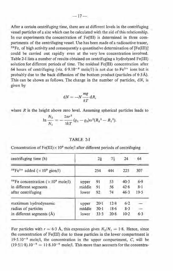
After a eertaio centrifuging time, there are at different levels in the centrifuging vessel particles of a si ze which can be calculated with the aid of this relationship. In our experiments the concentratien of Fe(III) is determined in three compartments of the centrifuging vessel. U se has been made of a radioactive tracer, 59Fe, of high activity and consequently a quantitative determination of [Fe(III)] could be carried out rapidly even at the very low concentratien involved. Table 2-I lists a number ofresults obtained on centrifuging a hydrolyzed Fe(III) solution for different periods of time. The residual Fe(III) concentratien after 64 hours of centrifuging (viz. 6·9.10- 9 mole/1) is not due to FeH ions but is probably due to the back diffussion of the bottorn product (particles of 6·3A). This can be shown as fellows. The change in the number of particles, dN, is given by
mg dN=-N-dR,
kT
where R is the height above zero level. Assuming spherical particles leads to
N 2 2nr 3
In- = - -- (lh - (h)w2(R/- R 1 2).
N 1 3kT
TABLE 2-1
Concentratien of Fe(III) x 109 mole/1 after different periods of centruuging
centrifuging time (h) 2-!- 7-! 24 64
59Fe3+ added ( X 109 gion/1) 254 444 225 307
59Fe concentratien (x 109 mole/1) upper 91 53 40·3 6·9 in different segrnents middle 91 56 42·8 8·1 after centrifuging lower 92 74 46·3 19·5
maximum hydrodynamic up per 20·1 12·8 6·2 radius of particles middle 30·1 18·6 8·3 in different segments (À) lower 33·3 20·8 10·2 6·3
For particles with r = 6·3 A, this expression gives N 2/N 1 = 1·8. Hence, since the concentratien of Fe(IID due to these particles in the Iower cernpartment is 19·5.10- 9 mole/1, tbe concentratien in the upper compartment, C, wiJl be (19·5/1·8).10- 9 = 11·8.10- 9 mole/1. This more than accounts for the concentra-
-18-
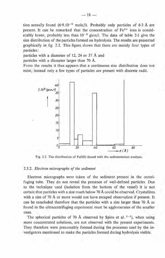
tion actual!y found (6·9.10- 9 mole/1). Probably only particles of 6·3 A are present. lt can be remarked that the concentratien of FeH ions is considerably lower, probably less than I0- 9 gion/1. The data of table 2-I give the size distri bution of the particles formed on hydrolysis. The results are presented graphically in fig. 2.2. This figure shows that there are mainly four types of particles: particles with a diameter of 12, 24 or 37 A and particles with a diameter larger than 70 A. From the results it thus appears that a continuous size distribution does not exist; instead only a few types of particles are present with discrete radü.
r-40
C.109 (gion/1} 35 -
JO
25
20
15
10
5 -n-0-0 20 40 60 80
-d(.R)
Fig. 2.2. The distribution of Fe(III) found with the sedimentation analysis.
2.3.2. Electron micrographs of thesediment
Electron micrographs were taken of the sediment present in the centrifuging tube. They do not reveal the presence of well-defined particles. Due to the technique used (isolation from the bottorn of the vessel) it is not certain that particles with a size much below 70 A could be observed. Crystallites with a size of 70 A or more would not have escaped observation if present. It can be concluded therefore that the particles with a size larger than 70 A as found in the ultraceritrifuging experiment must be agglomerates of the smaller on es.
The spherical particles of 70 A observed by Spiro et al. 2 - 1), when using more concentrated solutions, are not observed with the present experiments. They therefore were presumably formed during the processes used by the învestigators mentioned to make the particles formed during hydralysis visible.
-19-
2.3.3. Jnfluence of the precipitation conditions on the size distribution
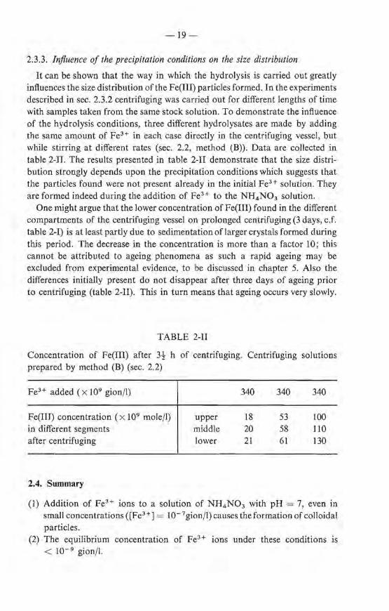
It can be shown that the way in which the hydralysis is carried out greatly influences the size distribution ofthe Fe(III) particles formed. In the experiments described in sec. 2.3.2 centrifuging was carried out for different lengtbs of time with samples taken from the same stock solution. To demonstrate the influence of the hydrolysis conditions, three different hydrolysates are made by adding the same amount of FeH in each case directly in the centrifuging vessel, but while stirring at different rates (sec. 2.2, method (B)). Data are collected in table 2-II. The results presented in table 2-11 demonstrate that the size distribution strongly depends u pon the precipitation conditions which suggests that the particles found were not present already in the initia! feH solution. They are formed indeed during the addition of feH to the NH4 N03 solution.
One might argue that the Iower concentration of Fe(III) found in the different compartments of the centrifuging vessel on prolonged centrifuging (3 days, c.f. table 2-I) is at least partly due to sedimentation oflarger crystals formed during this period. The decrease in the concentratien is more than a factor 10; this cannot be attributed to ageing phenomena as such a rapid ageing may be excluded from experimental evidence, to be discussed in chapter 5. Also the differences initially present do not disappear after three days of ageing prior to centrifuging (table 2-II). This in turn means that ageing occurs very slowly.
TABLE 2-11
Concentratien of Fe(III) after 3-} h of centrifuging. Centrifuging solutions prepared by metbod (B) (sec. 2.2)
feH added ( x 109 gionfl)
Fe(IIJ) concentratien ( x 109 mole/1) in different segments after centrifuging
2.4. Summary
up per middle lower
340
18 20 21
340
53 58 61
340
100 110
130
(!) Addition of FeH ions to a solution of NH4 N03 with pH= 7, even in smal! concentrations ([feH ] = I0- 7 gion/1) causes the formation of colloidal particles.
(2) The equilibrium concentration of feH ions under these conditions is < w-9 gion/l.
-20-
(3) The size distribution of the particles is discontinuous; four different sizes can be distinguished: 12, 24, 40 and > 70 A.
(4) The amount of the different particles is dependent upon the way in which the Fe3+ ions are added to the NH4N03 solution.
(5) The particles constituting the precipitate do not contain crystallites larger than 70 A, as these are not visible on the electron micrographs. The particles · with a diameter larger than 70 A found with the ultracentrifuging experiments must therefore be considered as agglomerates of smaller particles.
REFERENCES
2 - 1) G. Spiro, S. E. Allerton, J. Renner, A. Terzis, R. Bils and P. Saltman, J. Am. chem. Soc. 88, 2721, 1966.
2 - 2) G. Biedermann and P. Schindler, Acta chem. Scand. 11, 731, 1957. 2 - 3) M.R. Evans and M. J. Pryor, J. chem. Soc. SS, 157, 1949. 2 - 4 ) P.A. Kriukov and G. P. Awsejewitsch, Z. elektr. Chem. 39, 884, 1933. 2 - 5 ) H.S. Britton, J. chem. Soc. 127, 2148; 1925. 2 - 6 ) H. Lengweil er, W. Bus er and W. Feitknecht, Helv. chim. Acta 91, 796, 1961 ;
91, 805, 1961. 2 - 7 ) T. Svedberg and K. 0. Pedersen, Die Ultrazentrifuge, Steinkopf Verlag, Leipzig,
1940.
3. THE DEHYDRATION OF IRON(III)-OXIDE-HYDRATE GELS
3.1. Non-destructive removal of the capillary water
3.1.1. The !ow-tempera/ure dehydration process
It was briefly stated in chapter 1 that the ultimate hydrolysis product of Fe3+ solutions is a gelatinous precipitate which contains a large amount of water, a bout 90%. In genera!, gels consist of a network of macromolecules or are built up of chains of loosely aggregated sol particles held together by Van der Waals forces 3 - 1 •2). Iron-oxide- hydrate gel probably belongs to the second class.
De hydration ofthe gel at low temperature may be expected to offer the most reliable method for the isolation of its building units from it without any structural change. This dehydration can be carried out via a freezing process using liquid nitrogen 3 - 3) . When the gel is frozen, it does not retain its initia! appearance after thawing. Instead it is separated into a brown powder and a water phase. Even when the gel has been washed thoroughly before freezing this water phase still contains some N03-.
The powder after fiJtration and drying on P20 5 until constant weight, still contains considerable amounts of water. This water content greatly depends upon the preparation conditions. When the gel is prepared at 90 oe the powder isolated from it usually contains 11-15 % H 20 , when the gel is preparedat 20 oe the powder contains 14-18% H 20. When the dried powder is exposed to the air it rapidly takes up water again. In a typical example the H 2 0 content of a powder dried by exposure to the airwas 25·5 %; after drying on P 2 0 5 the H 2 0 ' content decreased to 11 % and after reexposure it took up its initia! H 2 0 content within one hour. On prolonged air exposure (200 h) there was a further increase of the H 20 content of 10 wt %. The powder, which will be used for a number of studies to be described in the next chapters, wiJl be designated further on as iron(III)-oxide hydrate.
The freezing process is most unlikely to change the partiele properties as follows from a detailed discussion of the processes occurring during freezing and the subsequent thawing of the frozen gel.
3.1.2. Experimental
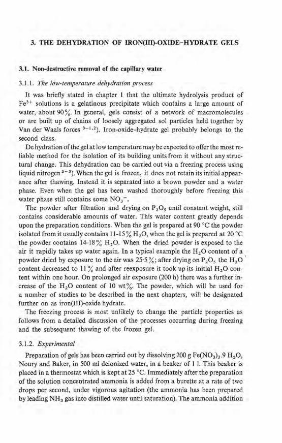
Preparation of gels has been carried out by dissolving 200 g Fe(N03)J.9 H 2 0, Noury and Baker, in 500 rol deionized water, in a beaker of 1 I. This beaker is placed in a thermostat which is kept at 25 oe. Imroediately after the preparation of the solution concentrated ammonia is added from a burette at a rate of two drops per second, under vigorous agitation (the ammonia has been prepared by teading NH3 gas into distilled water until saturation). The ammonia addition

-22-
is stopped at pH = 7·5. Stirring is continued for five minutes after which the pH, which has dropped slightly, is readjusted.
Where the resulting gel is dehydrated for the preparation ofiron(III)-oxide hydra te it is brought into a porcelain dishand frozen by pouring liquid nitrogen over it. The dish is covered with a watch glass and left standing overnight. The gel is then separated into a brown precipitate and an aqueous layer which is filtered off. The oxide hydrate, a compact powder, is washed with water containing a very small amountofammonia (pH= 7·5) until the tiltrateis nitratefree (reaction with sulphanilic acid and a-naphtylamine) and dried in vacuum at room temperature over P 20 5 • The water content is determined by heating at 1000 oe for four hours in air; nitrogen is determined by gravimetrie analysis.
Where the gel is used for a Mössbauer spectroscopy study as described in sec. 3.2, it is washed seven times with ammoniacal water of pH = 7·5 by decantation. The gel is then filtered by suction. lts nitrogen content is less than 0·0001% by wt.
3.2. A study of the constitution and freezing behaviour of iron(III)-oxide-hydrate gels by means of the Mössbauer effect *)
3.2.1. Introduetion
It will be shown that Mössbauer spectroscopy provides a means of studying the gel structure and its freezing behaviour.
Only a short discussion of some Mössbauer phenomena will be given here; for a more detailed introduetion into the Mössbauer spectroscopy the reader is referred to ref. 3- 5.
The basis of Mössbauer spectroscopy is the existence of different energy levels in the nucleus of an a torn, that can be excited by a y quanturn of the appropriate energy. Such a y quanturn is emitted by a souree when one of its nuclei undergoes a transition from an excited state to a lower energy level with an energy difference equal to the transition energy of the receiving atom. Due to the very small naturalline width the difference in transition energy of the souree and the receiving atom must be very smal! in order that resonance absorption can occur. The resonance couple 57Co-57 Fe is very well suited for Mössbauer-resonance study of iron compounds (natura! iron containing about 2% 57Fe). Although, in the case of coarse crystals the positions of the emitting and receiving atoms are fixed by the rigid bounding to the crystallattice, some recoil energy is lost. To correct for this and for the kinetic energy of the photon, the photon energy is shifted by the imparting to the souree of a Doppier velocity (for the experiments under consideration varying between -5 and 5 mm/s). The intensity of the absorption as a function of the Doppier velocity gives the Mössbauerresonance-absorption spectrum.
*) This sectien has already been publisbed elsewhere 3 - 4 ).
-23 -
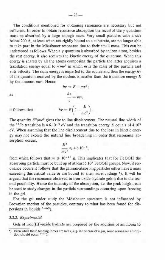
The conditions mentioned for obtaining resonance are necessary but not sufficient. In order to obtain resonance absorption the recoil of the y quanturn must be absorbed by a large enough mass. Very small particles with a size below 200 A, at least when not rigidly bound toa substrate, are no Jonger able to take part in the Mössbauer resonance due to their smal! mass. This can be understood as follows. When a y quanturn is absorbed by an iron a torn, besides the rest energy, it also receives the kinetic energy of the quantum. When this energy is shared by all the atoms composing the partiele the latter acquires a translation energy equal to t mv2 in which m is the mass of the partiele and v its velocity. The same energy is imparted to the souree and thus the energy hv of the quanturn received by the nucleus is smaller than the transition energy E by the amount mv2 • Hence
as
it follows that
hv = E-mv2 ;
hv -=mv, c
hv = E(l - ~). mc2
The quantity E 2 jmc2 gives rise to line displacement. The natura! line width of the 5 7 Fe transition is 4·6. w- 9 e V and the transition energy E equals 14·4.1 03
eV. When assuming that the line displacement due to the loss in kinetic energy may not exceed the natura! line broadening in order that resonance absorption occurs,
Ez - ~ 4·6.10- 9 ,
mc2
from which follows that m ~ 10- 16 g. This implicates that for FeOOH the absorbing partiele must be built up of at least 5.105 FeOOH groups. Now, if resonance occurs it follows that the gamma-absorbing particles either have a mass exceeding this critica! value or are bound to their surroundings *). lt will be argued that the resonance observed in iron-oxide-hydrate gels is due to the secoud possibility. Hence the intensity of the absorption, i.e. the peak height, can be used to study changes in the partiele surroundings occurring upon freezing in the gel.
For the gel under study the Mössbauer spectrum is not inftuenced by Brownian motion of the particles, contrary to what has been found for dispersions in liquids 3 - 6 - 9 ).
3.2.2. Experimental
Gels of iron(III)-oxide hydra te are prepared by the addition of ammonia to
*) Even when these binding forces are weak, e.g. in the case of a gas, some resonance absorption should occur 3 - 10) .
-24-
a solution of Fe(N03 ) 3 at 20 oe as described in sec. 3.1 .2. Samples suitable for investigation in the Mössbauer apparatus are prepared by putting the gel in a flat polystyrene vessel, so that uniform Jayers are formed ; their thickness is varied between 0·5 and 2·5 mm.
Mössbauer spectra have been determined at temperatures between -100 oe and 40° C. The low temperatures can be maintained by cooling the sample holder, isolated with polyfoam, with cold nitrogen gas. Above room temperature the same arrangement is used with heated nitrogen gas. The measurements have been carried out with a constant-velocity Mössbauer spectrometer as described in ref. 3- 11, using 57eo in Pd as a source.
3.2.3. Results
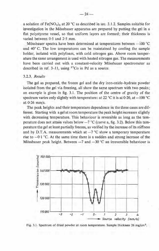
The gel as prepared, the frozen gel and the dry iron-oxide-hydrate powder îsolated from the gel via freezing, all show the same spectrum with two peaks; an example is given in fig. 3.1. The position of the centre of gravity of the spectrum varies only slightly with temperature: at 22 oe ît is at 0·20, at -100 oe at 0·26 mmfs.
The peak heights and their temperature dependenee in the three cases are different. Startîng with a gel at room temperature the peak height increases slightly with decreasing temperature. This behaviour is reversible as long as the temperature does not attain values below -7 oe (curve a, fig. 3.2). Below this temperature the gel at least partially freezes, as verified by the increase ofits stiffness and by D.T.A. measurements which at - 7 oe show a temporary temperature rise to - 0·1 oe. At the same time there is a sudden and strong increase of the Mössbauer peak height. Between -7 and -30 oe an irreversible behaviour is
ïii' /50 000 -j.., c: :::. 0
~
.ê 140000
"' -~ E "' @ 130000
1-
)", IJlJIJ
110000
.,.. -u
-3 -2
- 0
\ ~0 0
; \
~I ~
\ \J V
-I 0· 2 3 4
---- Souree velocity (mm/s)
Fig. 3.1. Spectrum of dried powder at room temperature. Sample thickness 26 mgfcm2 •

-25-
-- T (°K) 200 250 300
0·30 c -..., 0·25 .<::;
·!:?> ~ 0·20
"" 0 D-15 ~
1 D-10
0·08
........... -~ b~
~ ~ ~ ~
'\~ ~
(}06
(}04 J (}03
a" ~ r--0·02 -100 -80 -60 -40 -20 0 20 40
- T(°C)
Fig. 3.2. Typical example of peak height (relative to total number of pulses in the 14-keV channel outside resonance) as a function oftemperature. In the gel state the sample thickness was 320 mgjcm2 •
Curve a: gel as prepared. Curve b: frozen gel. The irreversible behaviour above -30 oe during the first cooling is indicated for a specific temperature cycle: -8 _.. -2 _.. -15->- -2 _.. -20->- -2 _.. -30->-2 - -60 _.. -2 - 100 _.. -2 °C. Curve c: dried powder isolated from the gel.
found: the behaviour of the sample then depends on the lowest temperature it has reached previously. If for example the gel has been cooled down to -15 oe the peaks do not return to their former heights upon reheating to -7 oe, but stay higher, following a curve that can be traeed up to the thawing point at 0·0 oe (fig. 3.2). As long as the gel temperature remains between -15 and 0·0 oe the peak-height vs temperature curve is reversible. Once the sample bas been below - 30 oe the peak height of the frozen gel behaves reversibly up to 0 oe. A gel that has been cooled to -7 oe or lower, upon thawing does not return to the original gel structure but decomposes into a slurry of iron-oxide powder and water. The powder isolated from this slurry has higher Mössbauer peaks than either the original or frozen gel. This is shown in curve c of fig. 3.2. The changes in the peak height with temperature are reversible for this powder.
-26-
3.2.4. Discussion
3.2.4.1. Constitution of the gels
The fact that in all cases, gel, frozen gel and oxide-hydrate powder, similar two-line spectra are found, indicates that we are dealing with the same iron compound. The height of the peaks provides information about the way in which iron nuclei are incorporated in the gel structure. The peak height depends on the probability of resonance absorption - or Mössbauer fraction - J, and this in turn depends on the elastic properties of the environment of the iron nuclei. If the environment is not the same for all iron nuclei the probability of resonance absorption becomes an average of the f's of iron nuclei in the different environments. This is e.g. the case with a colloidal solution of coarse and su bcritical crystallites.
For our experiments thefvalue ofthe absorber was determined in the usual way 3 - 12). The Mössbauer fraction of the souree was taken to be 0·65 3 - 13).
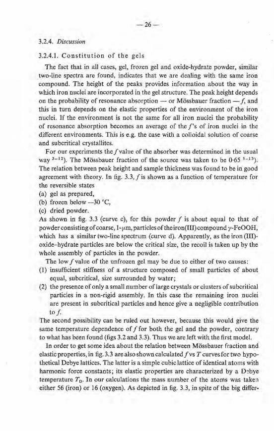
The relation between peak height and sample thickness was found to be in good agreement with theory. In fig. 3.3, fis shown as a function of temperature for the reversible states (a) gel as prepared, (b) frozen below -30 °C, (c) dried powder. As shown in fig. 3.3 (curve c), for this powder f is about equal to that of powder consisting of coarse, 1-fLID, particles of the iron(III) compound y-FeOO H, which bas a similartwo-line spectrum (curve d). Apparently, as the iron (III)oxide-hydrate particles are below the critica! size, the recoil is taken up by the whole assembly of particles in the powder.
The low f value of the unfrozen gel may be due to either of two causes: (1) insufficient stiffness of a structure composed of small particles of about
equal, subcritical, size surrounded by water; (2) the presence of only a small number oflarge crystals or clusters of subcritical
particles in a non-rigid assembly. In this case the remaining iron nuclei are present in subcritical particles and hence give a negligible contri bution tof
The second possibility can be ruled out however, because this would give the same temperature dependenee of f for both the gel and the powder, contrary to what has been found (figs 3.2 and 3.3). Thus we are left with the first model.
In order to get some idea about the relation between Mössbauer fraction and elastic properties, in fig. 3.3 arealso shown calculated fvs T curves for two hypothetical Debye lattices. The latter is a simp Ie cubic lattice of identical atoms with harmonie force constauts; its elastic properties are characterized by a D~bye
temperature T0 . In our calculations the mass number of the atoms was taken either 56 (iron) or 16 (oxygen). As depicted in fig. 3.3, in spite of the big differ-
I
- '27-
-- T (°K) 200 250 300
O·BOr--.-----r-+--.----r--+.----r--;-+-----,
125 -r---1------l -200
o~~--~-----L----L---~----~--~----~mo -100 -80 -60 -40 -20 0 20 40
- T (°C)
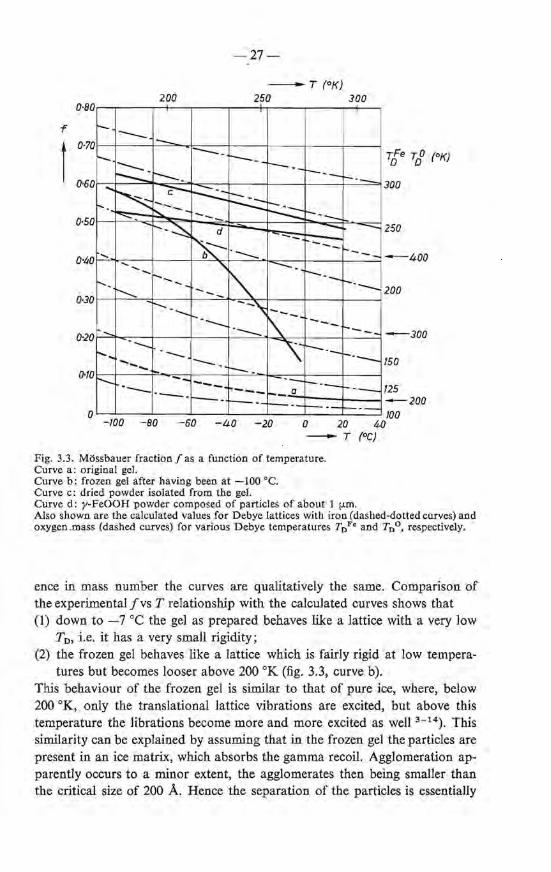
Fig. 3.3. Mössbauer fraction f as a function of temperature. Curve a : original gel. Curve b: frozen gel after having been at -100 oe. Curve c: dried powder isolated from the gel. Curve d: y-FeOOH powder composed of particles of a bout 1 (..lffi. Also shownare the calculated values for Debye lattices with iron (dashed-dotted curves) and oxygen.mass (dashed curves) for various Debye temperatures T0 Fe andT0 °, respectively.
ence in mass number the curves are qualitatively the same. Comparison of the ex perimental fvs T relationship with the calculated curves shows that (1) down to -7 oe the gel as prepared behaves like a Iattice with a very low
T0 , i.e. it has a very small rigidity; (2) the frozen gel behaves like a lattice which is fairly rigid at Iow tempera-
tmes but becomes looser above 200 oK (fig. 3.3, curve b). This behaviour of the frozen gel is similar to that of pure ice, where, below 200 °K, only the translational lattice vibrations are excited, but above this temperature the librations become more and more excited as well 3 - 14). This similarity cao be explained by assuming that in the frozen gel the particles are present in an ice matrix, which absorbs the gamma recoil. Agglomeration apparently occurs to a minor extent, the agglomerates then being smaller than the critica! size of 200 A. Hence the separation of the particles is essentially

-28-
preserved after freezing; the collapse of the netwerk, Ieading to the formation of the powder, does not occur during freezing but upon tbawing.
3.2.4.2. The freezing process
In four respects the freezing behaviour of the iron(III)-oxide-hydrate gel is peculiar: (1) the initia! freezing temperature is -7 oe, although the electrolyte content
is less than w- 5 mole%; (2) freezing is not completed until tbe gel bas been caoled below -30 oe; (3) the freezing process is not reversible; (4) melting occurs at O·O oe; after thawing the gel is separatedintoa powder
and a water pbase. The low initia! freezing temperature cao be ascribed to undercooling; this is
also observed in organic gel systems 3 - 15). We found that after solidification has started, although the sample is caoled steadily, the temperature remains at the constant level of -0·1 oe during a eertaio Iength of time. This points to a depression of the freezing point 3 - 15), but could also be an erroneous result due to the experimental technique 3 - 16).
On further cooling the freezing contillues until at -30 oe all particles are frozen in. The range of freezing points observed in our gels cannot be explained by kinetic effects (nucleation ra te): no change in peak height and its temperature dependenee is observed within 20 hours. A range of freezingpoints down to - 40 oe is also observed in porous glass 3 - 1 7 ). The phenomenon cao be ascribed to the differences in surface energy within a porous system cantairring water in pores with various diameters. As the surface energy of water in a po re differs from that of ice, the freezing point is depressed. According to Kuhn et al. 3 - 15) the relation between freezing-point depression LIT and pore diameter d is: LIT d = 3·7.10- 6 • Thus freezing points of -0·1 and -30 oe observed in our gels correspond to pore diameters of 3700 A and 12 A, respectively, indicating that there is a large variatien in pore sizes.
A difference between freezing temperature and melting temperature - bere 0·1 oe - is also observed in organic gels 3 - 15). Kuhn tried to explain this with a model in which the ice crystals in adjacent capillaries have the same orientation while the intermediate netwerk remains undamaged during freezing. Kanig, however, observed with electron micrographs that after freezing the pare system in the gel was damaged 3 - 16). For inorganic systems, the samewas concluded by Weiss from the increase of the freezing point upon repeated freezing and thawing 3 - 18). Our ex perimental results demonstrate that for the gel under study the latter explanation is the most probable. U pon ice formation the ebains of the particles forming the gel netwerk break in several places. When freezing proceeds more cantacts between the particles are braken. Hence larger capillaries are formed and remelting does not occur until close to 0 oe. The fact that
-29-
only chains break and no agglomeration takes place is concluded from the observed rigidity of the surroundings of the particles in the frozen gel which shows that the particles remain separate in the ice matrix. The final collapse of the network occurs u pon thawing, when the cantacts are notrestored and agglomeration starts. The powder isolated from the gel in this way, thus consists of agglomerates of primary gel particles.
The fact that the agglomerates are composed of the original crystallites, forming the gel, follows from the further evidence that the magnetic susceptibilities of powders either isolated from the gel via freezing or by drying the gel on P20 5 without freezing are the same within the experimental error. Also the X-ray spectrum of the gel and the powder isolated from it upon freezing are identical (chapter 5).
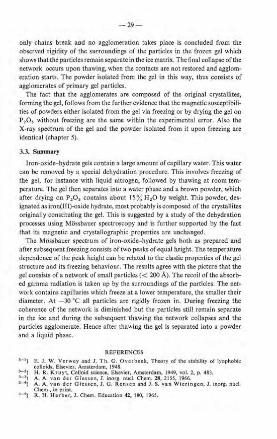
3.3. Summary
Iron-oxide-hydrategels contain a large amount of capillary water. This water can be removed by a special dehydration procedure. This involves freezing of the gel, for instanee with liquid nitrogen, foliowed by thawing at room temperature. The gel then separates into a water phase and a brown powder, which after drying on P 2 0 5 contains a bout 15% H 2 0 by weight. This powder, designated as iron(III)-oxide hydrate, most probably is composed of the crystallites originally constituting the gel. This is suggested by a study of the dehydration processes using Mössbauer spectroscopy and is further supported by the fact that its magnetic and crystallographic properties are unchanged.
The Mössbauer spectrum of iron-oxide- hydrate gels both as prepared and after subsequent freezing consistsof two peaks of equal height. The temperature dependenee of the peak height can be related to the elastic properties of the gel structure and its freezing behaviour. The results agree with the picture that the gel consists of a networkof smal! particles ( < 200 Á). The recoil of the absorbed gamma radiation is taken up by the surroundings of the particles. The network contains capillaries which freeze at a lower temperature, the smaller their diameter. At -30 oe all particles are rigidly frozen in. During freezing the caberenee of the network is diminished but the particles still remain separate in the ice and during the subsequent thawing the network collapses and the particles agglomerate. Hence after thawing the gel is separated into a powder and a liquid phase.
REPERENCES 3 - 1) E. J. W. Verwey and J . Th. G. Overbeek, Theory of the stability of Jyophobic
colloids, Elsevier, Amsterdam, 1948. 3 - 2) H. R. Kruyt, Colloid science, Elsevier, Amsterdam, 1949, vol. 2, p. 483. 3 - 3) A. A. van der Giessen, J. inorg. nucl. Chem. 28, 2155, 1966. 3 - 4 ) A. A. van der Giessen, J. G. Rensen and J. S. van Wieringen, J. inorg. nucl.
Chem., in print. 3- 5) R. H. Herber, J. Chem. Education 42, 180, 1965.
-30-
3 - 6 ) K. S. Singwi and A. Sjölander, Phys. Rev. 120, 1093, 1960. 3 - 7 ) P.P. Craig and N. Sutin, Phys. Rev. Letters 10, 460, 1963. 3- 8) D. St. P. Bunbury, J. A. Elliott, H.E. Halland J. M. Williams, Phys. Letters 6,
34, 1963. 3 - 9 ) T . Bonchev, P. Aidemirski, I. Mandzh udov, N. Nedyalkova, B. Skorchev
and A. Strigachev, J. exptl theor. Phys. (U.S.S.R.) 50, 62, 1966; Sov. Phys. JETP 23, 42, 1966.
3 - 10) L. Eyges, Am. 1. Phys. 33, 790, 1965. 3 - 11) J. S. van Wieringen, Philips tech. Rev. 28, 33, 1967. 3 - 12) R. S. Preston, S. S. Hanna and J. Heber le, Phys. Rev. 128, 2207, 1962. 3 - 13) W. A. Steyert and R. W. Taylor, Phys. Rev. 134A, 716, 1964. 3 - 14) N. Ockman, Adv. Phys. 7, 199, 1958. 3 - 15) W. Kuhn, R . Blochand P. Läuger, Kolloidz. 193, I, 1963. 3 - 16) G. Kanig, Kol!. Z. 173, 97, 1960. 3 - 17) A. A. Antoniou, J. phys. Chem. 68, 2755, 1964. 3 - 18) A. Weiss, Rheologica Acta 2, 292, 1962.

4. THE STRUCTURE OF IRON(III)-OXIDE HYDRATE
4.1. Crystallograpbic properties
4.1.1. Introduetion
The existence of a crystallographic ordering in iron-oxide-hydrate gel has been extensively examined with the aid ofX-ray-diffraction techniques. Nearly all authors arrive at the same conclusion, i.e. that the material is amorphous, see for instance"refs 4-1, 2. Frei and co-workers are of the same opinion, although in their-products, dried at 60 oe, some faint reflections were observed at d = 2·54, 2·23, 1·97, 1·71 and 1·49 A 4 - 3).
Weiset and Milligan however are not convineed of the amorphous character of the gel and more generally speaking of oxide gels: "the gels are believed to consist of agglomerates of extremely minute crystals of oxide or simple hydrate (or hydroxide) which hold large amounts of water by adsorption and capillary forces" 4 - 4 •11). This condusion was supported by results obtained with electron diffraction. This technique has, however, the disadvantage that the hydrate has to be studied in the vacuum of the microscope and, due to heating with the electron beam can easily decompose or recrystallize. This most probably explains the observation of diffraction patterns which could be attributed to a-Fe2 0 3 4 - 4 ). Their evidence for crystallinity in iron-oxide-hydrate gels is therefore believed not to be conclusive.
From the investigations described in the preceding chapters it was concluded that the gel or the oxide hydrate isolated from it is composed of particles with a size considerably below 200 A. Investigation of such small crystallites with radiation of a rather long wavelength with respect to the partiele size, for instance the commonly used CoKa radiation, should lead to an X-ray diagram with such a considerable Iine broadening that one erroneously could conclude to the absence of any ordering. It seemed worth while trying to take X-ray-diffraction patterns using radiation of a short wavelength (MoKct radiation with À = 0·71 A); in that case the diffraction bands become much more distinguishable from the background. Experiments are described below.
4.1.2. Experimental
The preparatien of the oxide hydrate bas been described on page 21. X-raydiffraction patterns are made with the aid of a Philips diffractometer using Co Ka and MoKa radiation and with a Debye-Scherrer camera taking a twelvehours exposure to MoKct radiation. In the latter case a Zr filter is laid on the film.
Electron micrographs and electron-diffraction patterns are made with a Philips E.M. 200 electron microscope with a resolution of 7 A, 80 keV.
The specific surface area, S, is determined by argon adsorption ; the results are
-32-
interpreted with the B.E.T. method assuming that the argon molecule occupies a surface of 18·2 Á2 • A number of samples arealso measured using nitrogen: the same S values were found as with argon.
4.1.3. Results and discussion
4.1.3.1. erystallographic properties
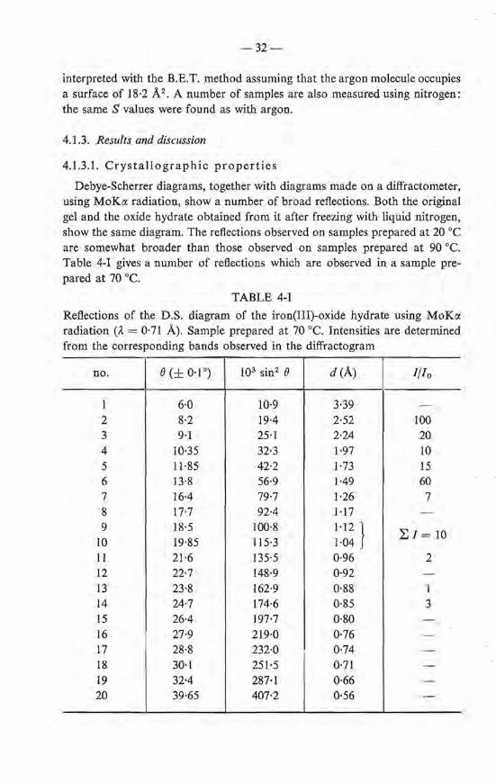
Debye-Scherrer diagrams, together with diagrams made on a diffractometer, using MoKo: radiation, show a number of broad reflections. Both the original gel and the oxide hydrate obtained from it after freezing with liquid nitrogen, show the same diagram. The reflections observed on samples preparedat 20 oe are somewhat broader than those observed on samples prepared at 90 oe. Table 4-I gives a number of reftections which are observed in a sample prepared at 70 oe.
TABLE 4-I
Reftections of the D .S. diagram of the iron(IIl)-oxide hydrate using MoKo: radiation (À = 0·71 À). Sample prepared at 70 oe. Intensities are determined from the corresponding bands observed in the diffractogram
no. 8 (± 0·1 °) 103 sin2 8 d(Á) 1/10
1 6·0 10·9 3·39 -2 8·2 19·4 2·52 100 3 9·1 25·1 2·24 20 4 10·35 32·3 1·97 10 5 11·85 42·2 1·73 15 6 13-8 56·9 1·49 60 7 16·4 79·7 1·26 7 8 17·7 92·4 1·17 -
9 18·5 100·8 1·12 } 2:, I = 10 10 19·85 115·3 1·04 11 21·6 135·5 0·96 2 12 22·7 148·9 0·92 -13 23·8 162·9 0·88 1 14 24·7 174·6 0·85 3 15 26·4 197·7 0·80 -16 27·9 219·0 0·76 -17 28·8 232·0 0·74 -18 30·1 251·5 0·71 -19 32·4 287·1 0·66 -
20 39·65 407·2 0·56 -
-33-
The existence of an X-ray diagram with about twenty lines indicates that the material under investigation is at least partially crystalline. The resolution of the diffraction peaks in the diffractograms appeared to be less than in the D.S. diagram. The shape of the peaks in the diffraction diagram suggests that some reflections are composed of several bands, especially the strongest reflections at d R::J 2·50 A and d R::J 1·50 A. The diffraction patternis shown in fig. 4.1. A comparison is made with the X-ray diagrams of the well-known oxides and oxide hydroxides of Fe(III). A identification of the main reflections of these oxides tagether with their relative intensity are depicted schematically in fig. 4.2. The figure clearly demonstrates that the iron(III)-oxide hydrate is different from the hitherto known oxide hydroxides and oxides. The oxide hydrate shows the greatest resemblance to <'l-FeOOH. There is also some resemblance to the Van Bernmelen hydrate 4 _,;), a crystallographic study of which has been publisbed by Collongues and Thery 4 - 6 ).
50 46 42 38 34 28---
Fig. 4.1. Diffraction pattem of the iron(III)-oxide hydrate; MoKa radiation.
An elucidation of the structure is hampered not only by the fact that the reftections are broad and in some cases possibly consist of several overlapping ones but also by a Jack of knowledge of the composition. It wiJl be argued in the next chapter that thîs is either FeOOH or Fe20 3 .H20; in the latter case probably the water is adsorbed on the surface of the particles. It was stated in a publication by the present author that the D.S. diagram could be indexed by takinga cubic unit cell with a0 = 8·37 A 4 - 7 ) . Accounting for the results to be discussed in the next sections, which indicate that the iron ions are placed in two groups of identical positions, it is questionable however whether the compound really has such a cubic unit cel!: it proved to be impossible to place the iron ions in a cubic arrangement which leads to calculated intensities of the reflections that are in reasonable accord with the observed ones. Another possibility could be that we are dealing with a hexagonal compound, for instanee with a 0 = 3·0 A and c0 = 9·3 A. But also in that case the iron ions could not be placed in the oxygen lattice in such a way that reasonable intensities can be calculated.
It is difficult to judge from these X-ray mearsurements which fraction ofthe gel
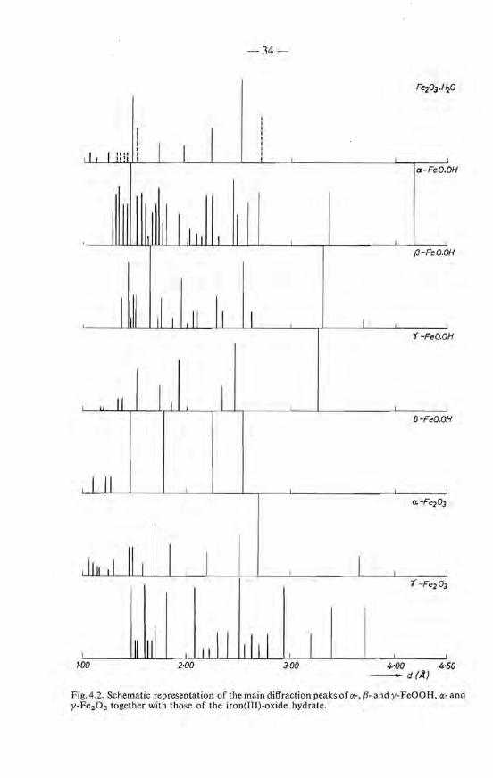
-34;.._
Fe203.H2 0
I I
I I I I I I
I I
I I
I I
I I "" I I I I 111 1 I I
a-FeO. OH
I I l~111 I !I I
H {3-Feo.o
I ~11 ,I, I 11 I, I I I
t -FeO.O H
11
'I I _L
H li-FeO.O
I 11 a:-Fe203
1 I Iu ,I ll1 I I I 7 -Fe203
111 1111 ill j 1111 l 1·00 2·00 3·00 4-·00 4·50
-d(A)
Fig. 4.2. Schematic representation of thema in diffraction peaks of <X·, fl· and y-FeOOH, a· and y-Fe2 0 3 tagether with those of the iron(III)-oxide hydrate.
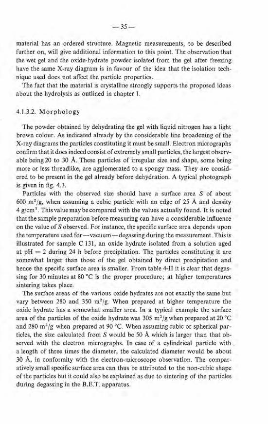
-35-
material bas an ordered structure. Magnetic measurements, to be described further on, will give additional information to this point. The observation that the wet gel and the oxide-hydrate powder isolated from the gel after freezing have the same X-ray diagram is in favour of the idea that the isolation technique used does notaffect the partiele properties.
The fact that the material is crystalline strongly supports the proposed ideas about the hydrolysis as outlined in chapter l.
4.1.3.2. Morphology
The powder obtained by dehydrating the gel with liquid nitrogen has a light brown colour. As indicated already by the considerable line broadening of the X-ray diagrams the particles constituting it must be smal!. Electron micrographs confirm that it doesindeed consist of extremely small partiel es, the largest observable being 20 to 30 A. These particles of irregular size and shape, some being more or less threadlike, are agglomerated to a spongy mass. They are considered to be present in the gel already before dehydration. A typical photograph is given in fig. 4.3.
Particles with the observed size should have a surface area S of about 600 m2/g, when assuming a cubic partiele with an edge of 25 A and density 4 gjcm3 . Thisvaluemaybecompared with the values actually found. It is noted that the sample preparatien befere measuring can have a considerable in:fluence on the value of S observed. For instance, the specific surface area depends u pon the temperature used for - vacuum- degassing during the measurement. Th is is illustrated for sample e 131, an oxide hydra te isolated from a solution aged at pH = 2 during 24 h before precipitation. The particles constituting it are somewhat Iarger than those of the gel obtained by direct precipitation and hence the specific surface area is smaller. From table 4-II it is clear that degassing for 30 minutes at 80 oe is the proper procedure; at higher temperatures sintering takes place.
The surface areas of the various oxide hydrates are not exactly the same but vary between 280 and 350 m2 /g. When prepared at higher temperature the oxide hydrate has a somewhat smaller area. In a typical example the surface area of the particles of the oxide hydra te was 305 m2 jg when prepared at 20 oe and 280 m2 jg when prepared at 90 oe. Wh en assuming cubic or spherical particles, the size calculated from S would be 50 A which is Iarger than that observed-with the electron micrograpbs. In case of a cylindrical partiele with , a length of three times the diameter, the calculated diameter would be about 30 A, in conformity with the electron-microscope observation. The comparatively smal! specific surface area can thus be attributed to the non-cubic shape of the particles but it could a lso be explained as due to sintering of the particles during degassing in the B.E.T. apparatus.
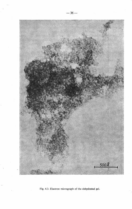
-36-
Fig. 4.3. Electron micrograph of the dehydrated gel.
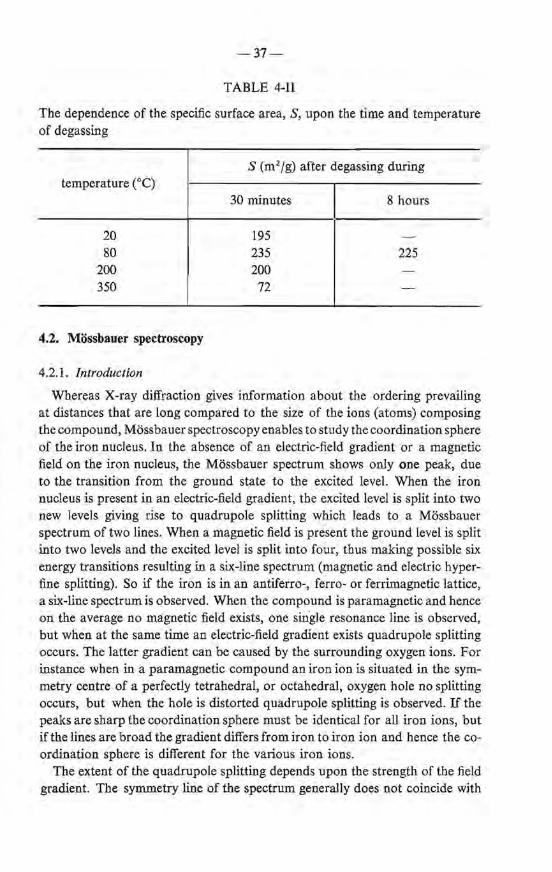
-37-
TABLE 4-11
The dependenee of the specific surface area, S, upon the time and temperature of degassing
temperature (0 C)
20 80
200 350
4.2. Mössbauer spectroscopy
4.2.1. Introduetion
S (m2 /g) after degassing during
30 minutes
195 235 200 72
8 hours
225
Whereas X-ray diffraction gives information about the ordering prevailing at distauces that are long compared to the size of the ions (atoms) composing the compound, Mössbauer spectroscopy enables to study the coordination sphere of the iron nucleus. In the absence of an electric-field gradient or a magnetic field on the iron nucleus, the Mössbauer spectrum shows only one peak, due to the transition from the ground state to the excited level. When the iron nucleus is present in an electric-field gradient, the excited level is split into two new levels giving rise to quadrupale splitting which leads to a Mössbauer spectrum of two lines. When a magnetic field is present the ground level is split into two levels and the excited level is split into four, thus making possible six energy transitions resulting in a six-line spectrum (magnetic and electric hyperfine splitting). So if the iron is in an antiferro-, ferro- or ferrimagnetic lattice, a six-line spectrum is observed. When the compound is paramagnetic and hence on the average no magnetic field exists, one single resonance line is observed, but when at the same time an electric-field gradient exists quadrupale splitting occurs. The latter gradient can be caused by the surrounding oxygen ions. For instanee when in a paramagnetic compound anironion is situated in the symmetry centre of a perfectly tetrahedral, or octahedral, oxygen hole no splitting occurs, but when the hole is distorted quadrupale splitting is observed. If the peaks are sharp the coordination sphere must be identical for all iron ions, but if the lines are broad the gradient differs from iron to iron ion and hence the coordination sphere is different for the various iron ions.
The extent of the quadrupale splitting depends u pon the strengthof the field gradient. The symmetry line of the spectrum generally does not coincide with
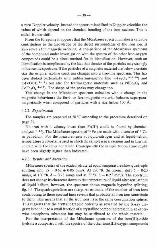
-38-
a zero Doppier velocity. Instead the spectrum is shifted to Doppier veloeities the values of which depend on the chemica! bonding of the iron nucleus. This is called isoroer shift.
From the foregoing it appears that the Mössbauer spectrum makes a valuable contribution to the knowledge of the direct surroundings of the iron ion. It also reveals the magnetic ordering. A comparison of the Mössbauer spectrum of the compound under investigation with the spectra of the other iron-oxygen compounds could be a direct method for its identification. However, such an identification is complicated by the fact that the size ofthe particles may strongly influence the spectrum. If the particles of a magnetic material are below a certain size the original six-line spectrum changes into a two-line spectrum. This has been stuclied particularly with antiferromagnetics like a-Fe2 0 3 4 - 8 - 10) and a-FeOOH 4 - 11) but also for ferrimagnetic materials such as NiFe2 0 4 and CoFe2 0 4 4 - 12). The shape of the peaks may change too.
This change in the Mössbauer spectrum coincides with a change in the magnetic behaviour: the ferri- or ferromagnetic material behaves superparamagnetically when composed of particles with a size below 100 A.
4.2.2. Experimental
The samples are prepared at 20 oe according to the procedure described on page 21.
No iron with a valency lower than Fe(III) could be found by chemica! analysis 4 '--13). The Mössbauer spectra of 57Fe are made with a souree of 57Co in palladium. For the measurements at Jiquid-nitrogen and at liquid-helium temperature a cryostat is used in which the sample is in a vacuum and in thermal contact with the inner container. Consequently the sample temperature might have been slightly higher than indicated.
4.2.3. Results and discussion
Mössbauer spectra of the oxide hydrate, at room temperature show quadrupJe splitting with 2e = 0·62 ± 0·05 mmfs. At 290 oK the isomer shift <5 = 0·20 mm/s, at 130 oK 13 = 0·23 mmfs and at 77 oK o = 0·21 mmfs. The spectrum does notchange its eh araeter down to the temperature of liquid nitrogen; at that of liquid helium, however, the spectrum shows magnetic hyperfine splitting, fig. 4.4. The quadrupale lines are sharp. An estimate of the number of iron ions contributing tothese spectrallines reveals that probably all iron ions contribute to them. This means that all the iron ions have the same coordination sphere. This suggests that the crystallographic ordering as revealed by the X-ray diagram is not due to a smal! fraction of a crystalline compound present in an otherwise amorphous substance but may be attributed to the whole materiaL
For the interpretation of the Mössbauer spectrum of the iron(III)-oxide hydra te a comparison with the spectra of the other iron(III)-oxygen compounds
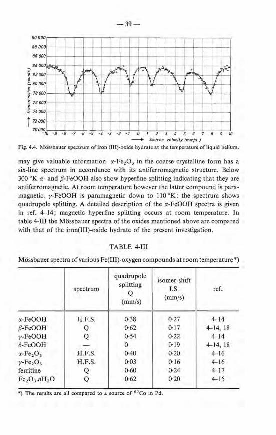
"'
90000
88000
86 000
84 000
<: 82000 :;,
~ 80000
·~ 78000
~ 76000 § ~ 74 000
i 72000
70000
.......
'
~ .~
~ I \ I \ "/> \1
-39-
~ ""'\ j ~ ;A ~ ~ ""' Jf ~
~j, 1\/ i\ ~ ~ I
0
\i \
j
-IQ -9 -8 -7 -6 -5 -4 -3 -2 -1 0 2 3 • 5 6 7 8 9 m - Souree velocity (mmjs )
Fig. 4.4. Mössbauer spectrum of iron (III)-oxide hydrateat the temperature of liquid helium.
may give valuable information. a-Fe2 0 3 in the coarse crystalline form has a six-line spectrum in accordance with its antiferromagnetic structure. Below 300 oK a- and /1-FeOOH also show hyperfine splitting indicating that they are antiferromagnetic. At room temperature however the latter compound is paramagnetic. y-FeOOH is paramagnetic down to 110 °K: the spectrum shows quadrupole splitting. A detailed description of the a-FeOOH spectra is given in ref. 4-14; magnetic hyperfine splitting occurs at room temperature. In table 4-III the Mössbauer spectra of the oxides mentioned above are compared with that of the iron(III)-oxide hydrate of the present investigation.
TABLE 4-III
Mössbauer spectra of various Fe(III)-oxygen compounds at room temperature *)
quadrupale isoroer shift
spectrum splitting
l.S. ref. Q (mmfs)
(mm/s)
et-FeOOH H.F.S. 0·38 0·27 4-14 /1-FeOOH Q 0·62 0·17 4-14, 18 y-FeOOH Q 0·54 0·22 4-14 o-FeOOH - 0 0·19 4-14, 18 a-Fe20 3 H.F.S. 0·40 0·20 4-16 y-Fe203 H.F.S. 0·03 0·16 4-16 ferritine Q 0·60 0·24 4-17 Fe20 3 .nH20 Q 0·62 0·20 4-15
*) The results are all compared to a souree of 57Co in Pd.
-40-
The table shows that y-Fe2 0 3 has a slight isomer shift compared to the other compounds, included the oxide hydrate under study. This compound has about a third of its iron i ons in tetrabedral positions; this could be the reason for the low l.S. The quadrupole splitting of the oxide hydra te is large compared to that of nearly all compounds except {3- and y-FeOOH. The large Q value, according to Kundig and Ando, is probably due to the small size of the particles 4 -t 0 ).
They assumed that the quadrupole spectrum is the sum of two quadrupole spectra: a spectrum caused by the i ons lying in the surface of the particles and another caused by the ions lying in the particles. The smaller the partiele the larger the contribution of the surface i ons; for a-Fe2 0 3 the quadrupole splitting increases whereas the isoroer shift decreases with decreasing partiele size. Based on these results the quadrupele splitting measured for the iron(III)-oxide hydrate-then considered as a hydrated a-Fe2 0 3 - should correspond toa partiele size of about 125 A. Hence it seems probable that the hydrate has a structure different from a-Fe2 0 3 , as the actual observed diameter of the particles lies between 20 and 30 A. Although a correct interpretation of the Mössbauer spectrum is hampered by the effects due to the small partiele size, the tentative condusion can be drawn that all iron ions occupy a similar octahedral position. The condusion that all iron ions occupy the same position could be drawn from the high absorption efficiency (peak height). The condusion that the iron ions oècupy an octahedral position is based on the values of the isomer shift and the quadrupole splitting which most probably are large compared to those of y-Fe20 3 and o-FeOOH where iron also occupies tetrabedral lattice sites.
4.3. Magnetic measurements
4.3.1. Superparamagnetic behaviour
The superparamagnetic behaviour initially observed on ultrafine Ni powders was explained by Néel by assuming that each powder partiele bas a constant but freely rotating magnetic moment 4 - 2 0 ). A powder composed of such particles when placed in a magnetic field attains a magnetization which is described by the Langevin function
a = a. ( coth a - ~). where mH
a= kT '
m is the magnetic moment of a single partiele and a, stands for the saturation magnetization. When a < 1 the function may be simplified by: a = a,a/3. In the case of ferro- and ferrirnagnetic materials the origin of the partiele moment is clear. The fact that also small particles of an antiferromagnetic compound can have a magnetic moment, according to Néel, is aresult of statistica! deviations in the number of sites ofthe sublattices which are occupied by cations 4 - 2 t). According to Néel, when the particles are formed very quickly the difference in the number of ions occupying one sublattice compared to the other equals

-41-
VN, when N is the number of roetal i ons in the particle. Assuming that all i ons interact.with each other, the magnetic moment of each partiele then equals 11 VN, when 11 is the magnetic moment per cation. The saturation (per gramme material) of a powder composed of these particles is given by
NA11VN (Js = MN ,
where NA is Avogadro's number and Mis the mole weight. Hence (a< 1)
NA11VN 11HVN NAI1 2H (]= --= =xH.
MN 3kT 3MkT
The magnetic susceptibility x is thus independent of the number of cations per particle, and is not much different from the value found for the same number of paramagnetic ions in the free state.
In this description it is assumed that the number of iron ions occupying the sites of the two magnetic sublattices is determined by a random process. It coulcl well be, however, that the introduetion of one stacldng fault in the growing partiele activates the formation of another one and hence the di.fference in the number of iron ions occupying the sublattices could well exceed VN. There is also a second possible explanation for a difference of more than jiN ions in the sublattices which especially holds for a somewhat large particle, d R:::l I 00 A. As discussed by Néel 4 - 21 ), when the partiele is perfectly crystalline there still remains a number of unpaired cations when the partiele contains an odd number of Iattice plan es; the difference between the number of occupied sites in both sublattices is then no longer N 112 but instead N 213 . In the latter case
1 N AI1N213 11N213 H (] = - ---'---- ---
2 MN 3kT
the factor t accounting for the fact that only half of the particles contains an unequal number of lattice planes. Accordingly the susceptibility might be exceedingly high for powders composed of smal! single crystals. Yet there are several reasoos whythis will not be thecase. In the first place, when 11N213 Hf kT> I saturation occurs; for FeOOH this would be the case at room temperature and in a field tof 104 Oe for N R:::l 1000 Fe(JII) ions. Second, there is a eertaio temperature below which the magnetic vector is no longerfree to move. This blocking temperature is determined by KV= kT8 , where K is the magnetic-anisotropy energy per unit volume (V). For a-Fe20 3 the value of T8 will be above room temperature for particles with a size larger than 100 A.
Superparamagnetic behaviour bas been found for ultrafine powders ofvarious oxides such as Niü, Fe20 3 , NiFe20 4 and CoFe20 4 by measuring the relatioo between magnetizatioo aod field strength at various temperatures 4 - 22- 24).
These investigations are all performed with oxide particles prepared via de-

-42-
composition of starting compounds (e.g. a-FeOOH). Due to tbe pseudomorphic cbaracter of these reactions the newly formed crystallites are still coherent; the crystallite size as determined from X-ray line broadening gives only an average value of N. Yet the results are in rather good accord with theory.
Magnetic measurements on ultrafine non-coherent particles have hitberto only been carried out on the material ferritine 4 - 17). This is a biologica! material consisting of iron(IIl)-oxide-hydrate particles of 50 A surrounded by a mantie of protein. F or this material also a superparamagnetic behaviour bas been found; the results show that a strong antiferromagnetic magnetic coupling in such small particles is well possible.
The material under investigation also consists of very small discrete particles which are bound together by only weak forces. The theoretica! considerations of Néel may be expected to apply to these particles and a further quantitative treatment based on Néel's theory seems allowed.
On the basis of the theory outlined above it might well be that the superparamagnetic particles, upon recrystallization, show an increase of tbe susceptibility due to the fact that tbey slightly grow and become bounded by perfect lattice planes. Upon further growth, when a certain critica! size is reached (50-100 À) the susceptibility decreases again (blocking). As will be shown in chapters 5 and 7 a study of the magnetic propertiescan give useful information for an elucidation of the mechanism of the recrystallization of iron-oxide hydrate taking place in aqueous solutions or upon heating in air.
4.3.2. Result and discussion
Magnetic measurements are done on iron (III)-oxidehydrate according to a metbod described in the literature 4 - 25).
The experirnental data of the magnetic measurements are given in fig . 4.5. Above 30 oK the curves are reversible. The magnetic moment per gramme, a, is plotted as a function of the field strength H for different temperatures, T. lt appears that the data for temperatures above 30 oK can be brought onto one curve by plottingaas a function of HjT. This curve (fig. 4.6) can be described by the Langevin function. The val u es of a. and m can be found by the following metbod: log a is plottedas a function of log (H/T) and log (coth a- !ja) as a function of log a, on separated sheets, where a varies from 0·01 to 10. One sheet is moved over the other until the curves coincide. In this way all experimental data are used for the determination of the parameters. This leads to a value of a.= 11 gauss cm3/g and m = 1·49.10- 18 erg/Oe, which is large compared to the moment of an iron(IIl) ion. The conesponding Langevin curve bas been drawn in fig . 4.6. From this figure it is clear the material is superparamagnetic.
From the saturation magnetization and the magnetic moment per particle, the number of particles present in one gramme of material can be calculated as
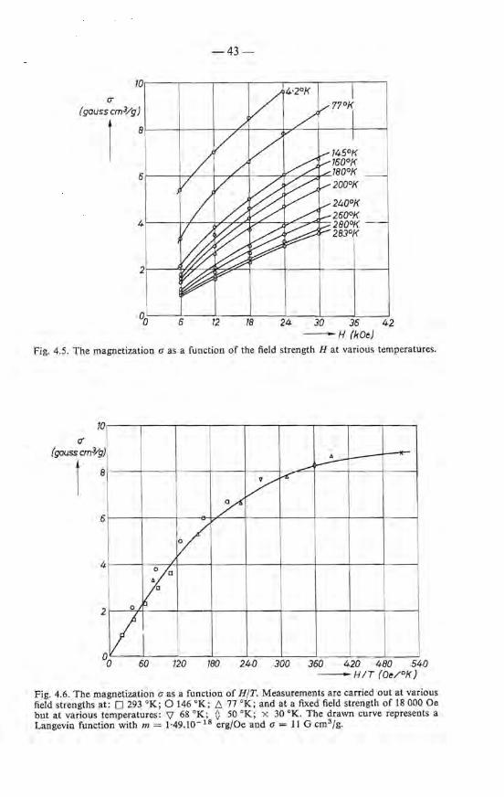
IT
(gauss cm3/g)
1
-43-
0oL---~6~--~12 ____ L_ __ _L __ ~----~--_j 18 24 JO 36 42
-H (kOe)
Fig. 4.5. The magnetization a as a function of the field strength H at various temperatures.
10 (j
(gauss cm3/g)
\ 8
6
4
2 ?
0 ~
VI 60
~ ~ .-'
y / ~
/
120 180 240 300 360 420 480 540 -HIT (Oe/ °K)
Fig. 4.6. The magnetization a as a function of H/T. Measurements are carried out at various field strengths at: 0 293 °K; 0 146 °K; [:,. 77 °K; and at a fixed fieldstrengthof 18 000 Oe but at various temperatures: 'V 68 °K; O 50 oK; x 30 °K. The drawn curve represents a Langevin function with m = 1·49.10- 18 erg/Oe and a = 11 G cm3 /g.

- 44--'-
O's 11 - = = 7·4.1018 particlesfgramme. m 1·49.10- 18
The specific density of the material as measured is about 4 gfcm3 • From the number of particles per gramme the average partiele size, d, cao be calculated:
d = ( 1024 )1 /3 = 30 A 4x7·4.1018
for particles with a cubic shape (when talcing into consideration that the material contains about 15% H 20, probably adsorbed on the partiele surface, the calculated partiele diameter would be somewhat smaller). This average partiele size estimated from the magnetic measurement is in good agreement with that obtained from measurements with the electron microscope, viz. 20-30 A (fig. 4.3).
The average number of iron ions present per partiele (as derived from the magnetic measurements) is equal to
NA 6.1023
- ---- = ~ 1000 iron ionsfpartiele, (a.fm)(M/2) 7·4.1018 x80
where NA = Avogadro's number and M = mole weight Fe2 0 3 • The average contribution of an iron ion to the partiele moment then amounts to
1·49.10- 18
- - -- = 1·49.10-21 ergjOe = 0·16 Bohr magnetons. 1000
This value, low compared to the value of 5 /1s for the iron ion in the paramagnetic state, cao be explained by assurning that there is an antiferromagnetic coupling between the magnetic moments of the iron ions. The existence of a magnetic ordering in the compound under study at the temperature of liquid helium has been demonstrated with the aid of Mössbauer spectroscopy. At these low temperatures the spectra show magnetic hyperfine splitting. The absence of hyperfine splitting at room temperature must then be considered as due to the high frequency of the rotatien of the magnetic vector, compared to the lifetime of the excited level, I0- 8 s. The existence of a magnetic moment in an antiferromagnetic compound cao be explained, according to Néel, as a result ofunequal numbers of ironionsin the sublattices due to the small partiele size. Assuming this difference equal to VN, N being the number of iron ions per partiele, then the calculated number of iron i ons present per partiele should be
(moment per partiele)2 { 1·49.10- 18 } 2
N = . = . = 1050 iron ionsfparticle. momentper FeH wo 5 x 9·3.I0- 21
This is in agreement with the results given above and hence the assumption
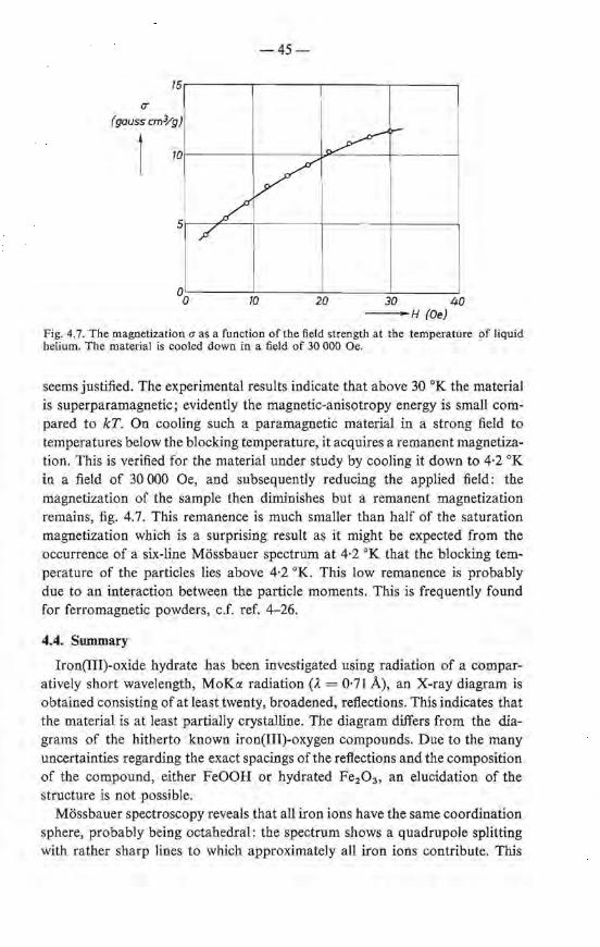
- 45 .,.----
75.-------.-------,-------,-------~
(J"
(gauss cm:Yg)
0o~------7~0-------2~0~----~30------~40 -H(Oe)
Fig. 4.7. The magnetization a as a function of the field strength at the temperature of liquid helium. The material is caoled down in a field of 30 000 Oe.
seems justified. The ex perimental results indicate that above 30 oK the material is superparamagnetic; evidently the magnetic-anisotropy energy is small compared to kT. On cooling such a paramagnetic material in a strong field to temperatures below the blocking temperature, it acquires a remanent magnetization. This is verified for the material under study by cooling it down to 4·2 oK in a field of 30 000 Oe, and subsequently reducing the applied field: the magnetization of the sample then diminishes but a remanent magnetization remains, fig. 4.7. This remanence is much smaller than half of the saturation magnetization which is a surprising resu1t as it might be expected from the occurrence of a six-line Mössbauer spectrum at 4·2 oK that the blocking temperature of the particles lies above 4·2 °K. This low remanence is probably due to an interaction between the partiele moments. This is frequently found for ferromagnetic powders, c.f. ref. 4-26.
4.4. Summary
Iron(III)-oxide hydrate has been investigated using radiation of a comparatively short wavelength, MoKcx radiation (À= 0·71 A), an X-ray diagram is obtained consisting of at least twenty, broadened, refiections. This indicates that the material is at least partially crystalline. The diagram differs from the diagrams of the hitherto known iron(III)-oxygen compounds. Due to the many uncertainties regarding the exact spacings of the refiections and the composition of the compound, either FeOOH or hydrated Fe2 0 3 , an elucidation of the structure is not possible.
Mössbauer spectroscopy reveals that all iron ions have the same coordination sphere, probably being octahedral: the spectrum shows a quadrupole splitting with rather sharp lines to which approximately all iron ions contribute. This
-46-
suggests that all particles may be considered as being crystallites. The quadrupale spectrum is a result of the superparamagnetic character of the materiaL The superparamagnetic character can be verified by measuring the magnetization as a function of H/T. Assuming a random accupation of both lattice sites, it could be estimated from the magnetic results that about 1000 iron ions are present in one crystallite.
REPERENCES 4 - 1) A. Sirnon and Th. Schmidt, Kolloidz. 36, 65, 1925. 4 - 2) 0. G1emser and G. Rieck, Z. anorg. allg. Chem. 297, 175, 1958. 4 - 3 ) V. Cáslavská, V. Frei and A. Blazek, Col!. Czech. chem. Comm. 27, 2168, 1962. 4 - 4 ) H. B. Weiser and W. 0. Milligan, J. phys. Chem. 44, 1081, 1940. 4 - 5 ) J. M. van Bernmelen and E. A. Klobbie, J. prakt. Chem. 46, 497, 1892. 4 - 6 ) R. Collongues and J. Thery, Bull. Soc. chim. France, 1141, 1949. 4 - 7) A. A. van der Giessen, J. inorg. nucl. Chem. 28, 2155, 1966. 4 - 8 ) A. Z. Hrynkiewicz, D. S. Kulgawczuk and K. Tomala, Phys. Letters 17, 93, 1965. 4 - 9 ) T. Nakamura et al., Phys. Letters 12, 178, 1964. 4 - 10) T. Nakamura and S. Shimizu, Bull. Inst. chem. Res. Kyoto Univ. 42, 299, 1962. 4 - 11) A. M. van der Kraan and J. J. van Loef, Phys. Letters 20, 614, 1966. 4 - 12) W. J. Schuele, S. Shtrikman and D. Treves, J. appl. Phys. 36, 1010, 1965. 4 - 13) G. W. van Oo sterhout and J. Vi s ser, Anal. chim. Acta 33, 330, 1965. 4 - 14) M. J. Rossiterand A.S. M. Hodgson, J. inorg. nucl. Chem. 27, 63, 1965. 4 - 15) A. A. van d e r Giessen, J. G. Rensen and J. S. van Wieringen, J. inorg. nucl.
Chem., in print. 4 - 16) J. S. van Wieringen, private communication. 4 - 17) A. Blaise, J. Chappert and J. L. Girardot, C.R. Acad. Sci. Paris 261,2310, 1965. 4-18) I. Dészi, L. Kezthelyi, D. Kulgawczuk, B. Mcinai and N.A. Eissa, Phys. Stat.
sol. 22, 617, 1967. 4-19) W. Kündig and K. J. Ando, Czech. J . Phys. B17, 467, 1967. 4 - 20) L. N éel, C.R. Acad. Sci. Pa ris 228, 664, 1949. 4 - 2 1 ) L. Néel, J. phys. Soc. Japan Suppl. B-1, 17, 676, 1962. 4 - 22) K. M. Creer, J . phys. Soc. Japan Suppl. B-1, 17, 690, 1962. 4 - 23) J. Cohen, K. M. Creer, R. Pauthenet and K. Srivastava, J. phys. Soc. Japan
Suppl. B-1, 17, 685, 1962. 4 - 24) J. T. Richardson and W. 0. Milligan, Phys. Rev. 102, 1289, 1956. 4 - 25) G. W. Rathenau and J. L. Snoek, Philips Res. Repts 1, 239, 1946. 4 - 26) C. P. Bean and J. W. Livingstone, J. appl. Phys. Suppl. 30 (4), 120S, 1959.
5. RECRYSTALLIZATION OF THE IRON(III)-OXIDE-HYDRATE GEL IN AQUEOUS SOLUTIONS
S.t. Discossion of the literature
The crystallographic study of the iron-oxide-hydrate gel, prepared as described in chapter 3, is hampered by the fact that the particles are so small that in the X-ray diagram considerable line broadening occurs. It would therefore be of interest to have this compound in the form of larger crystals. Crystal growth in contact with an aqueous solution might be a possible method for the preparation of larger crystals, provided that nuclei of another phase, a more stabie one with respect to the first compound, are absent. In the presence of such nuclei, instead of crystal growth, recrystallization towards this more stabie phase might well occur. A search ofthe literature shows that the latter crystallization mechanism is dominant in many cases ; ageing of partially hydrolyzed Fe(III) solutions or of gels may finally lead to a-Fe20 3 , a-FeOOH or P-FeOOH, depending on the experimental conditions. Anions greatly determine which compound will be formed. Whereas in the presence of Cl04 - and N03 - ions - which are considered to be unable of complex formation with Fe3+ ionsa-Fe20 3 and a-FeOOH are formed 5-1. 2), the presence of Cl- ions may lead to P-FeOOH 5-3.4). These compounds are formed with particles that are of submicroscopie dimensions even after prolonged ageing in acid solutions. Heller et al. showed with the aid of light scattering that in FeCI 3 solutions colloidal particles are formed with an elongated shape5 - 5 • 6 • 7 ) . Similar results have been found by Lamband Jaques 5- 8 ) . In partially hydrolyzed solutions of Fe(Cl04h, as found by Feitknecht and Michaelis, needle- and platelet-shaped particles of a-FeOOH or y-FeOOH are formed on ageing, together with more or less spherical a-Fe20 3 and spherules with a size of 50 À which are supposed to be amorphous 5 - 9 ).
Gels, when leftin contact with the mother liquor, recrystallize on prolonged standing with the formation of a-Fe20 3 , a-FeOOH or a mixture of both, also with particles of submicroscopie dimensions, independent of the experimental conditions.
Before the introduetion of electron microscopes of sufficient resolution, Krause and co-workers developed a number of chemical methods to obtain some knowledge of the morphology and the structure of the particles present in the geland its ageing products. According to Krause those OH groups of the oxide hydrate which are in contact .with the solution react with Ag+ ions in boiling alkaline solutions to give a ferrite, e.g. Ag2Fe20 4 5 - 10), whereas the OH groups built in the lattice, such as in a-FeOOH, do not react with Ag+ ions. From this kind of experimental evidence the condusion was drawn that the gel consists of small colloidal particles, being linear polymers of the type
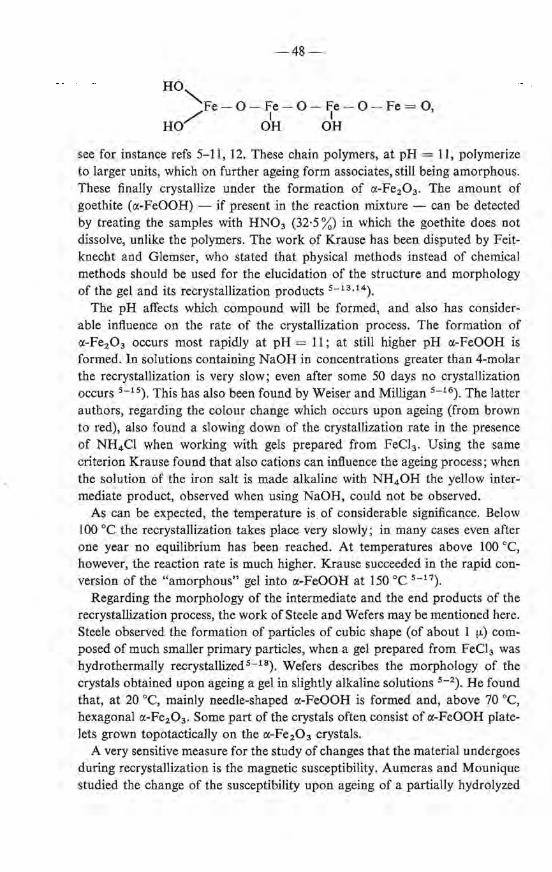
-48-
HO"'-. Fe - 0- Fe-0- Fe- 0- Fe = 0
/ I I ' HO OH OH
see for instanee refs 5-11, 12. These chain polymers, at pH = 11, polymerize to larger units, which on further ageing form associates, still being amorphous. These finally crystallize under the formation of a-Fe2 0 3 • The amount of goethite (a-FeOOH) - if present in the reaction mixture - can be detected by treating the samples with HN03 (32·5 %) in which the goethite does not dissolve, unlike the polymers. The work of Krause has been disputed by Feitknecht and Glemser, who stated that physical methods instead of chemica! methods should be used for the elucidation of the structure and morphology of the gel and its recrystallization products 5 - 13•14).
The pH affects which compound wiJl be formed, and also has considerable influence on tbe rate of the crystallization process. The formation of a-Fe2 0 3 occurs most rapidly at pH = 11; at still higher pH a-FeOOH is formed. In solutions containing NaOH in concentrations greater than 4-molar the recrystallization is very slow; even after some 50 days no crystallization occurs 5 - 15). This bas also been found by Weiser and Milligan 5 - 16). The latter authors, regarding the colour change which occurs upon ageing (from brown to red), also found a slowing down of the crystallization rate in the presence of NH4 Cl when working with gels prepared from Feel3 . Using the same criterion Krause found that also cations can influence the ageing process; when the solution of the iron salt is made alkaline with NH4 0H the yellow intermediate product, observed when using NaOH, could not be observed.
As can be expected, the temperature is of considerable significance. Below 100 oe tbe recrystallization takes place very slowly; in many cases even after one year no equilibrium bas been reached. At temperatures above 100 oe, however, the reaction rate is much higher. Krause succeeded in the rapid conversion of the "amorphous" gel into a-FeOOH at 150 oe 5 - 17).
Regarding the morphology of the intermediate and the end products of the recrystallization process, the workof Steele and Wefers may be mentioned here. Steele observed the formation of particles of cubic shape (of about 1 tJ.) composed of much smaller primary particles, when a gel prepared from Feel3 was hydrothermally recrystallized 5 - 18). Wefers describes the morphology of the crystals obtained upon ageing a gel in slightly alkaline solutions 5 - 2). He found that, at 20 oe, mainly needle-shaped a-FeOOH is formed and, above 70 oe, hexagonal a-Fe2 0 3 • Some part of the crystals often consist of a-FeOOH plateIets grown topotactically on the a-Fe 2 0 3 crystals.
A very sensitive measure for the study of changes that the material undergoes during recrystallization is the magnetic susceptibility. Aumeras and Mounique stuclied the change of the susceptibility upon ageing of a partially hydrolyzed

-49-
FeCl3 solution 5- 19) . Chevallier and Mathieu showed that the magnetic properties of a gel largely depend upon the way the hydrolysis has been carried out 5 - 20) : when precipitating a gel with more concentrated NaOH, preparations are obtained with a higher x value. Upon ageing, the gel's susceptibility increases but after ten days of ageing at room temperature the susceptibility decreases again. The same phenomena have been found by Atbrecht and Wedekind for gels prepared with NH40H s-21 ) . These authors attributed the maximum in the susceptibility to an intermediate product formed during ageing. They did oot succeed in obtaining more details of this alleged compound.
Summarizing it can be said that the ageing of the gel proceeds via a series of intermediate products towards o:-FeOOH and o:-Fe20 3 •
In the next sections investigations are described, which we re carried out to get a better understanding of the mechanism underlying the ageing phenomena occurring in partially hydrolyzed colloidal iron(III) solutions or in iron-oxidehydrate gels.
5.2. Experimental
Ageing experiments
200 g Fe (N03h.9aq. (Merck, p.a.) are dissolved in 500 mi of deionized water in a beaker of 1 I, which is placed in a thermostat kept at 20 °C. After one hour the alkali is added to the solution from a burette at a rate of two drops per second, while vigorously stirring and bubbling N 2 gas through it. Three preparations ~are made with resp. conc. ammonia (prepared as mentioned in chapter 1), 4-molar ammonia (prepared by dilution of the concentrated ammonia) and 3·5-molar NH4 HC03 prepared by leading C02 gas into the ammonia. The alkali is added until the solution has reached a pH = 2·2. The pH meter is equipped with electredes as mentioned in chapter l . The solutions are kept in the thermostat ; at time intervals of 0, 48 and 340 hours portions are taken from it which are further treated with concentrated ammonia. The gel thus obtained is filtered off by suction and dehydrated as described in chapter 3.
In another series of experiments the pH is brought directly to pH = 7·5; the sample is then placed in a conical flask with a ground stopper fitted with a water loek in order to prevent the entrance of co~ .
Physical measurements
Viscosity is measured using an Ubbelohde viscosimeter. Electron rnicrographs, X-ray diffractions and magnetic measurements are all done as described in chapter 4.

-50-
5.3. Ageing phenomena occurring in partiaUy hydrolyzed solutions
5.3.1. Crystallographic properties of the ageing produels
A series of hydrolysis experiments has been carried out by adding, at room temperature, concentrated NH4 0H, 4-molar NH4 0H and 3·5-molar NH4 HC03
toa 1-molar solution of Fe(N03h until pH= 2·2, which corresponds toabout 2 OH- ions per FeH ion. At this pH no precipitate is formed . The solution is kept for 48-340 hours at room temperature. The products which are formed upon ageing are isolated from the solution by brioging the pH to 7·5, which leads to the formation of a gel in which the particles, formed during ageing, are dispersed.
When the hydrolysis is carried out incompletely (OH- /FeH < 3) the solution remains acid . In such an acid solution changes take place on prolonged standing at room temperature and from the decrease of the pH it appeared that even after 340 hours no equilibrium was established. These changes are due to a recrystallization which the primarily formed particles undergo. Already after 48 hours the X-ray diagram is different from that of the gel. After a period of 340 hours the X-ray diagram shows a number of broadened lines which are typical for a-FeOOH ; the observed reflections all could be indexed by taking an orthorhombic unit cell with a0 = 4·64, b0 = 10·0 and c0 = 3·03 A. The X-ray data are presented in table 5-1.
Besides the reflections 1-ll also a number of very weak and very broadened reflections are observed, a-b ,which can be indexed with less certainty due to the large error of 8 involved. It is clear that in the presence of NH4 + and N03 - ions together with or without CO/- /C02 , upon ageing at room temperature only a-FeOOH is formed ; no reflections can be observed which could not be indexed with the unit cell of this compound. Hence the formation of y-FeOOH which was observed by Feitknecht et al. in their experiments 5 - 9 ),
using Fe(Cl04h solutions, may be excluded in the hydrolyzed Fe (N03h solutions. In case the hydrolysis is carried out with NaOH instead of NH4 0H similar results are obtained.
Particles having the same structure as observed in a non-aged gel (chapter 4) are not found in the form of larger crystals in the gels obtained from aged
· partially hydrolyzed solutions. It is conceivable that these primary particles do not grow at all. The formation of larger particles upon ageing is accompanied by a structural transformation, mainly into a-FeOOH.
5.3.2. Morphology
5.3.2.1. Tyndall effect
Valuable additional information about the size and shape of the primary particles present in partially hydrolyzed solutions can be obtained from a
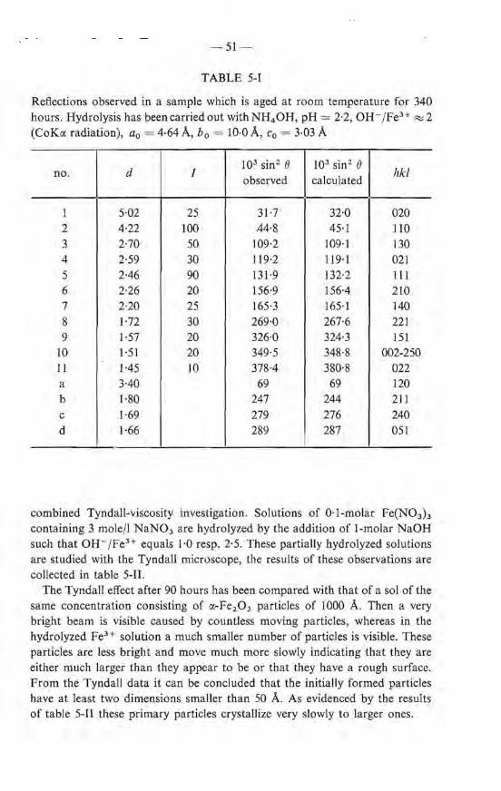
-51-
TABLE 5-1
Reflections observed in a sample which is aged at room temperature for 340 hours. Hydrolysis has been carried out with NH4 0H, pH= 2·2, OH-/FeH ~ 2 (Co Ka radiation), a0 = 4·64 A, b0 = 10·0 A, c0 = 3·03 A
d I 103 sin2 (} 103 sin2 (}
hkl no. observed calculated
1 5·02 25 31·7 32·0 020 2 4·22 100 44·8 45·1 110 3 2·70 50 109·2 109·1 130 4 2·59 30 119·2 119·1 021 5 2·46 90 131·9 132·2 lil 6 2·26 20 156·9 156-4 210 7 2·20 25 165·3 165·1 140 8 1·72 30 269·0 267·6 221 9 1·57 20 326·0 324·3 151
lO 1·51 20 349·5 348·8 002-250 I I 1·45 10 378·4 380·8 022 a 3·40 69 69 120 b l-80 247 244 211 c 1·69 279 276 240 d 1·66 289 287 051
combined Tyndall-viscosity investigation. Solutions of 0·1-molar Fe(N03 ) 3
containing 3 mole/1 NaN03 are hydrolyzed by the addition of 1-molar NaOH such that OH- /Fe3+ equals 1·0 resp. 2·5. These partiaiiy hydrolyzed solutions are stuclied with the Tyndall microscope, the results of these observations are collected in table 5-II.
The Tyndall effect after 90 hours has been compared with that of a soJ.of the same concentration consisting of a-Fe2 0 3 particles of 1000 A. Then a very bright beam is visible caused by countless moving particles, whereas in the hydrolyzed Fe3 + solution a much smaller number of particles is visible. These particles are less bright and move much more slowly indicating that they are either much larger than they appear to be or that they have a rough surface. From the Tyndall data it can be concluded that the initia lly formed particles have at least two dimensions smaller than 50 A. As evidenced by the results of table 5-11 these primary particles crystallize very slowly to larger ones.
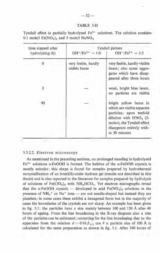
- 52-
TABLE 5-11
Tyndall effect in partially hydrolyzed Fe3+ solutions. The salution contains 0·1 molejl Fe(N03h and 3 mole/1 NaN03
time elapsed after hydrolyzing (h)
0
3
90
Tyndall picture oH-/ Fe3 + = 1·0 I OH-/Fe3+ = 2·5
very feeble, hardly visible beam
very feeble, hardly visible beam; also some aggregates which have disappeared after three hours
weak, bright blue beam, no particles are visible
bright yellow beam in which are visible separate particles; upon tenfold dilution with HN03 (2-molar), the Tyndall effect disappears entirely within 30 minutes
5.3.2.2. Electron microscopy
As mentioned in the preceding sections, on prolonged standing in hydrolyzed Fe3+ solutions o:-FeOOH is formed. The habitus of the o:-FeOOH crystals is mostly acicular; this shape is found for samples prepared by hydrothermal recrystallization of an iron(III)-oxide-hydrate gel (results not described in this thesis) and is also reported in the literature for samples prepared by hydralysis of solutions of Fe(Cl04 ) 3 with NH4 HC03 . Yet electron micrographs reveal that the o:-FeOOH crystals - developed in acid Fe(N03 ) 3 solutions in the presence of NH4 + or Na+ ions - are not needle-sbaped but instead they are platelets; in some cases these exhibit a hexagonal form but in the majority of cases the boundaries of the crystals are not sharp. An example has been given in fig. 5.1 ; the particles have a size mainly between 100 and 150 A after 48 hours of ageing. From the line broadening in the X-ray diagram also a size of the particles can be estimated; correcting for the line broadening due to the apparatus from the relation d = 0·9À {3 112 cos 8 a partiele size of 100 A is calculated for the same preparatien as shown in fig. 5.1. After 340 bours of

-53-
Fig. 5.1. Electron micrographof particles which are formed in a partially hydrolyzed 1-molar Fe(N03h solution (OH-jFe3+ ""' 2) after ageing at room temperature for 48 hours. Hydrolysis has been carried out with 4-molar NH4 0H.
ageing the crystallite size is 130 A. After 116 hours of. ageing in a partially hydrolyzed solution containing Na+ ions (NaOH/Fe3+ ~ 1) the crystals are smaller: ~ 100 A, see fig. 5.2.
5.3.2.3. Viscosity
It could well be that the primary particles are very thin acicular a-FeOOH crystals which, slowly, recrystallize under the formation of larger ones. If the initia! particles were of acicular shape then the presence of these particles may give rise to a viscosity which is large compared to the viscosity of the solution containing the same amount of iron in the form of free ions. A number of viscosity data is graphically represented in fig. 5.3; the curves show that up to the point when on the average the ratio OH- fFe3+ is adjusted at about 2·5, the viscosity hardly changes. This indicates that the primary particles are more or less spherical, anyhow not acicular. This result agrees with the electron micrographs of the gel. Only on prolonged standing does the viscosity increase. It is remarkable that when the ratio OH-/Fe3+ is smaller than 2·5 the crystallization takes place very slowly, whereas when OH-/Fe3 + > 2·65 a relatively rapid crystallization takes place, as indicated by the viscosity change. This evidently must be correlated with the fact that the colloid in the latter case is
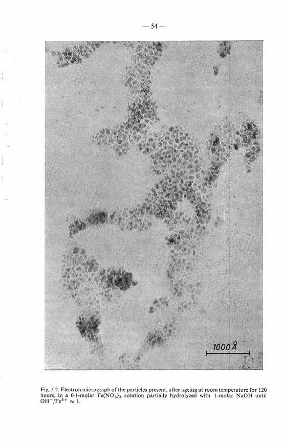
-54-
Fig. 5.2. Electron micrographof the particles present, after ageing at room temperature for 120 hours, in a 0·1-molar Fe(N0 3h solution partially hydrolyzed with 1-molar NaOH until OH- /Fe3+ ""' I.

v (centistokes
f 1·75
1·50
1·25
L~
-55-
/
~ ~ -1·00
0 50 100
~
750 200 250 -Time(h)
Fig. 5.3. The change of the viscosity at various degrees of hydrolysis upon ageing at room temperature :
OH- /Fe3+ 0 2-42 0 2-48 • 2·54 17 2-65
more close to its point of e!ectroneutrality. The increase of the viscosity points to the fact that recrystallization leads to particles with at least one dimeosion smaller than the other two, i.e. platelets or needles.
5.3.3. Magnetic measurements
In chapter 4 it bas been discussed that for small particles the magnetic susceptibility is a valuable measure for their characterization. In antiferromagnetic ultrafine particles with random occupation of the sublattices the susceptibility is independent of the size of the particles. When, however, the particles grow and become bounded by Iattice planes, the susceptibility increases until the partiele bas attained such a size that its blocking temperature lies above room temperature. As discussed in chapter 4, the critica! size is probably 50-100 A. Hence, aftera eertaio ageing period, depending on the rate of crystal growth, one may find a higher as wel! as a Iower value of the susceptibility compared to the starting value. The occurrence of either one of these possibilities can be demonstrated with the aid of two series of experiments.
One series of hydrolysis experiments is done by using alkaline solutions of various strengtbs in such a way that in all cases the ratio OH-/FeH equals about 2. The still acid solutions are aged at room temperature. The magnetic susceptibility is measured after various ageing times. The results of these experiments are col!ected in table 5-III. The table shows that upon precipitation
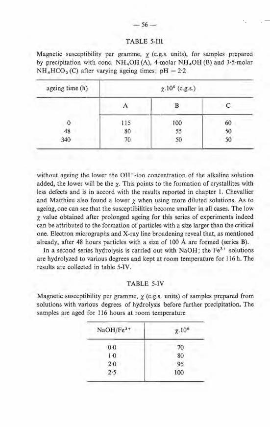
-56-
TABLE 5-111
Magnetic susceptibility per gramme, x (c.g.s. units), for samples prepared by precipitation with conc. NH 4 0H (A), 4-molar NH 40H (B) and 3·5-molar NH 4 HC03 (C) after varying ageing times ; pH= 2·2
ageing time (h)
0 48
340
A
115 80 70
x.106 (c.g.s.)
B
100 55 50
c
60 50 50
without ageing the lower the OH--ion concentration of the alkaline solution added, the lower wiJl be the X· This points to the formation of crystallites with less defects and is in accord with the results reported in chapter 1. Chevallier and Matthieu also found a lower x when using more diluted solutions. As to ageing, one cao see that tbe susceptibilities become smaller in all cases. The low x value obtained after prolonged ageing for this series of experiments 'indeed can be attributed to the formation of particles with a size larger than the critica! one. Electron micrographs and X-ray line broadening reveal that, as mentioned already, after 48 hours particles with a size of 100 A are formed (series B).
In a second series hydrolysis is carried out with NaOH; the Fe3+ solutions are hydrolyzed to various degrees and kept at room temperature for 116 h. The results are collected in table 5-IV.
TABLE 5-IV
Magnetic susceptibility per gramme, x (c.g.s. units) of samples prepared from solutions with various degrees of hydrolysis before further precipitation. The samples are aged for 116 hours at room temperature
Na0H/Fe3 + x.I06
0·0 70 l·O 80 2·0 95 2·5 100
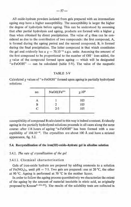
-57-
All oxide-hydrate powders isolated from gels prepared with an intermediate ageing step have a higher susceptibility. The susceptibility is larger the higher
· the degree of hydrolysis before · ageing. This can be understood by assuming tbat after partial hydralysis and ageing, products are formed with a higher x than when obtained by direct precipitation. The value of x thus can be considered as due to the con tribution of two compounds: the first compound, A, is formed during the ageing period and the second compound, B, is formed during the final precipitation. The latter compound is that which constitutes the gel and evidently has a x = 10.10- 6 c.g.s. units. Assuming the amount of the first compound to be proportional to the number of OH- ions added, the x value of the compound formed upon ageing - which will be designated "a-FeOOH" - can be calculated (table 5-V). The value of the magnetic
TABLE 5-V
Calculated x values of "a-FeOOH" formed upon ageing in partially hydrolyzed solutions
no.
A B c
NaOH/Fe3+
1 2 2·5
103 103 105
susceptibility of compound B calculated in this way is indeed constant. Evidently ageing in the partially hydrolyzed solutions proceeds in all cases along the same course: after 116 hours of ageing "a-FeOOH" has been formed with a susceptibility of 104.10-6 . Tbe crystallites are about 100 A and have a striated appearance, fig. 5.2.
5.4. Recrystallization of the iron(lll)-oxide-hydrate gel in alkaline solution
5.4.1. The rate of crystallization of the gel
5.4.1.1. Chemica! characterization
Gels of iron-oxide hydrate are prepared by adding ammonia to a solution of Fe(N03h, until pH= 7·5. Two gels are prepared: one at 20 oe, the other at 90 oe. Ageing is performed at 70 oe in the mother liquor.
In order to follow the ageing process quantitatively we characterize tbe extent of the ageing by the amount of material insoluble in nitric acid, as originally proposed by K.rause5 - 22 •23). The resu1ts of the solubility tests are collected in
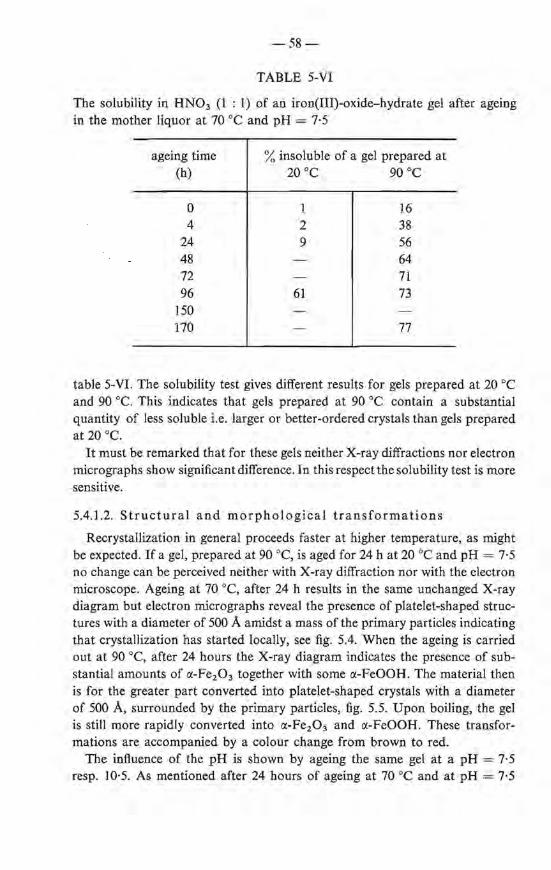
-58-
TABLE 5-VI
The solubility in HN03 (1 : 1) of an iron(III)-oxide-hydrate gel after ageing in the mother liquor at 70 oe and pH = 7·5
ageing time (h)
0 4
24 48 72 96
150 170
% insoluble of a gel preparedat 20 oe 90 oe
1 2 9
61
16 38 56 64 71 73
77
table 5-Vl. The solubility test gives different results for gels prepared at 20 oe and 90 oe. This indicates that gels prepared at 90 oe contain a substantial quantity of less soluble i.e. larger or better-ordered crystals than gels prepared at 20 oe.
It must be remarked that for these gels neither X-ray diffractions nor electron micrographs show significant difference. In this respect the solubility test is more sensitive.
5.4.1.2. Structural and morphological transformations
Recrystallization in general proceeds faster at higher temperature, as might be expected. lf a gel, preparedat 90 oe, is aged for 24 h at 20 oe and pH = 7·5 no change can be perceived neither with X-ray diffraction nor with the electron microscope. Ageing at 70 oe, after 24 h results in the same unchanged X-ray diagram but electron micrographs reveal the presence of platelet-shaped structures with a diameter of 500 A amidst a ma ss of the primary particles indicating that crystallization has started locally, see fig. 5.4. When the ageing is carried out at 90 oe, after 24 hours the X-ray diagram indicates the presence of substantial amounts of ct-Fe20 3 together with some ct-FeOOH. The material then is for the greater part converted into platelet-shaped crystals with a diameter of 500 A, surrounded by the primary particles, fig. 5.5. Upon boiling, the gel is still more rapidly converted into ct-Fe20 3 and ct-FeOOH. These transformations are accompanied by a colour change from brown to red.
The influence of the pH is shown by ageing the same gel at a pH = 7·5 resp. 10·5. As mentioned after 24 hours of ageing a t 70 oe and at pH = 7·5
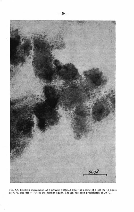
-59-
sóoB
Fig. 5.4. Electron micrograph of a powder obtained after the ageing of a gel for 48 hours at 70 oe and pH = 7·5, in the mother liquor. The gel has been precipitated at 20 oe.

-60-
Fig. 5.5. Electron micrograph of a sample obtained by the ageing of a gel (precipitated at 20 oe) at 90 oe for 24 hours at pH = 7·5.
no change is observed in the X-ray diagram but after ageing during the same length of time at pH = 10·5 a well-developed X-ray diagram is observed which can be attributed to a mixture of a-FeOOH and a-Fe2 0 3 •
When the ageing is carried out at high pH (> 10), the morphology of the crystals is different from those obtained at pH = 7. The a-Fe2 0 3 crystals then mostly have the shape of diamonds; part of them exhibit bars intergrown with
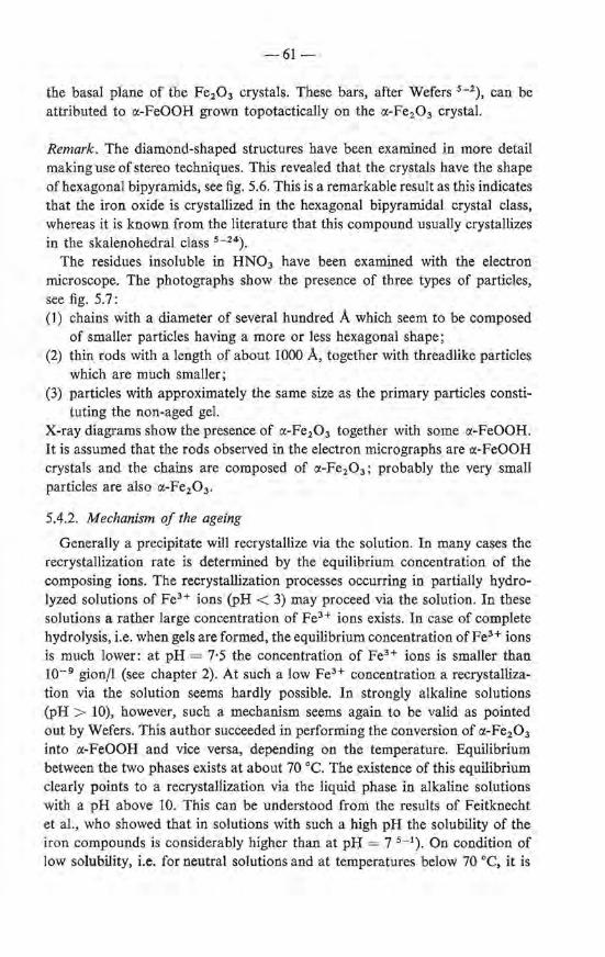
-61-
the basal plarie of the Fe20 3 crystals. These bars, after Wefers 5 - 2), can be attributed to ct-FeOOH grown topotactically on the ct-Fe2 0 3 crystal.
Remark. The diamond-shaped structures have been examined in more detail making u se of stereo techniques. This revealed that the crystals have the shape of hexagona1 bipyramids, see fig. 5.6. This is a remarkable result as this indicates that the iron oxide is crystallized in the hexagonal bipyramidal crystal class, whereas it is known from the literature that this compound usually crystallizes in the skalenohedral class 5 - 24).
The residues insoluble in HN03 have been examined with the electron microscope. The photographs show the presence of three types of particles, see fig. 5.7: (1) chains with a diameter of several hundred A which seem to be composed
of smaller particles ha ving a more or less hexagonal shape ; (2) thin rods with a length of about 1000 A, tagether with threadlike particles
which are much smaller; (3) particles with approximately the same size as the primary particles consti-
tuting the non-aged gel. X-ray diagrams show the presence of ct-Fe2 0 3 together with some ct-FeOOH. It is assumed that the rods observed in the electron micrographs are ct-FeOOH crystals and the ebains are composed of ct-Fe20 3 ; probably the very small particles are also ct-Fe20 3 •
5.4.2. Mechanism of the ageing
Generally a precipitate will recrystallize via the solution. In many cases the recrystallization rate is determined by the equilibrium concentration of the composing ions. The recrystallization processes occurring in partially hydrolyzed solutions of Fe3+ ions (pH < 3) may proceed via the solution. In these solutions a rather large concentration of Fe3+ ions exists. In case of complete hydrolysis, i.e. when gels are formed, the equilibrium concentration of Fe3+ ions is mi.Jch lower: at pH = 7·5 the concentration of FeH ions is smaller than w- 9 gion/1 (see chapter 2). At such a low Fe3 + concentration a recrystallization via the solution seems hardly possible. In strongly alkaline solutions (pH > 10), however, such a mechanism seems again to be valid as pointed out by Wefers. This author succeeded in performing the conversion of ct-Fe20 3
into ct-FeOOH and vice versa, depending on the temperature. Equilibrium between the two phases exists at about 70 oe. The existence of this equilibrium clearly points to a recrystallization via the liquid phase in alkaline solutions with a pH above 10. This can be understood from the results of Feitknecht et al., who showed that in solutions with such a high pl-I the solubility of the iron compounds is considerably higher than at pH = 7 5 - 1). On condition of low solubility, i.e. for neutral solutions and at temperatures below 70 oe, it is
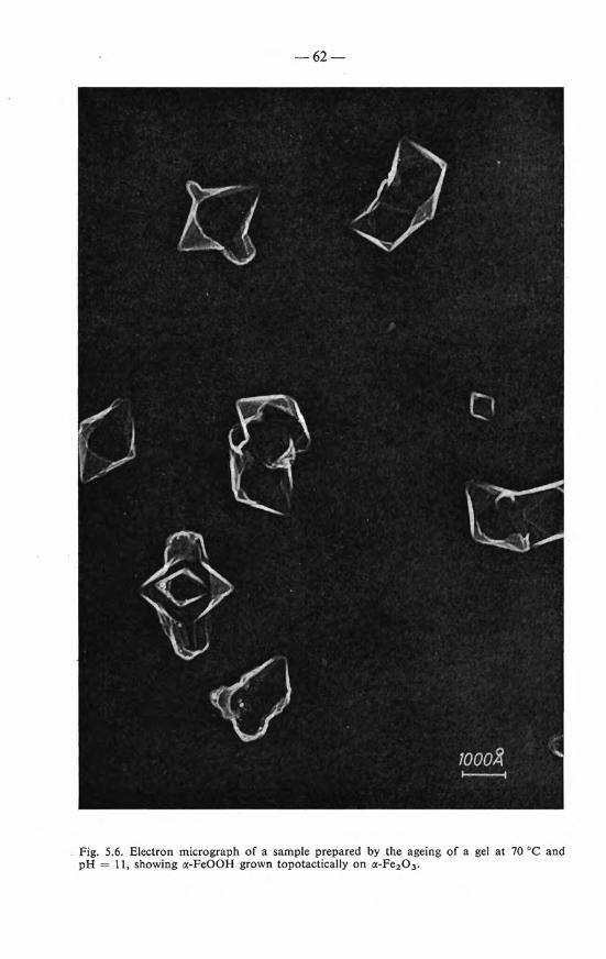
-62-
Fig. 5.6. Electron micrograph of a sample prepared by the ageing of a gel at 70 oe and pH = 11, showing a-FeOOH grown topotactically on a-Fe2 0 3 .
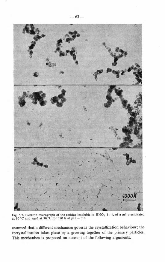
• • .1 11 ~~ ..... ,.,
r
,.f
-63-
Ï . •1000~
· - ~ .. .
Fig. 5.7. Electron micrograph of the residue insoluble in HN0 3 1 : I, of a gel precipitated at 90 oe and aged at 70 oe for 170 h at pH = 7·5 .
assumed that a different mechanism governs the crystallization behaviour; the recrystallization takes place by a growing together of the primary particles. This mechanism is proposed on account of the following arguments.
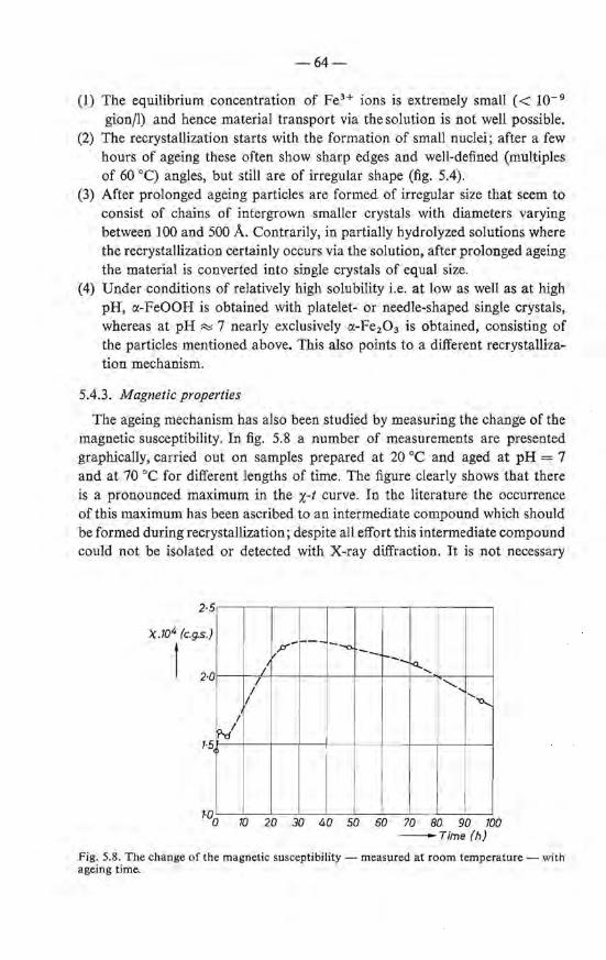
-64-
(1) The equilibrium concentratien of Fe.H ions is extremely small ( < I0- 9
gion/1) and hence material transport via the solution is not well possible. (2) The recrystallization starts with the formation of small nuclei; after a few
hours of ageing these often show sharp edges and well-defined (multiples of 60 oq angles, but still are of irregular shape (fig. 5.4).
(3) After prolonged ageing particles are formed of irregular size that seem to consist of chains of intergrown smaller crystals with diameters varying between 100 and 500 A. eontrarily, in partially hydrolyzed solutions where the recrystallization certainly occurs via the solution, after prolonged ageing the material is converted into single crystals of equal size.
(4) Under conditions of relatively high solubility i.e. at low as wellas at high pH, o:-FeOOH is obtained with platelet- or needle-shaped single crystals, whereas at pH ::::::; 7 nearly exclusively o:-Fe2 0 3 is obtained, consisting of the particles mentioned above. This also points to a different recrystallization mechanism.
5.4.3. Magnetic properties
The ageing mechanism has also been stuclied by measuring the change of the magnetic susceptibility. In fig. 5.8 a number of measurements are presented graphically, carried out on samples prepared at 20 oe and aged at pH = 7 and at 70 oe for different lengths of time. The figure clearly shows that there is a pronounced maximum in the x-t curve. In the literature the occurrence of this maximum has been ascribed to an intermedia te compound which should be formed during recrystallization; despite all effort this intermediate compound could not be isolated or detected with X-ray diffraction. It is not necessary
2·5
X .104 (c.g.s.)
t 2·0
1·5
I 'f'dl
p--
/ I
I I
r--- --o. -- ~---- -<l..., '
............. ''0.....
i
1'0o 10 20 30 40 50 60 70 80 90 700 -Time(h)
Fig. 5.8. The change of the magnetic susceptibility- measured at room temperature- with ageing time.
-65-
however to assume the formation of a new magnetic compound to account for the increase of tbe susceptibility X· As discussed in chapter 4 the latter can be explained as a consequence of the growth of superparamagnetic particles.
The fact that only partiele growth causes the increase of x and oot the formation of a new compound is further confirmed by X-ray diagrams which reveal that also in preparations having the maximum x value only c.:-Fe2 0 3 and ct-FeOOH are present.
5.5. Summary
In solutions of ferric nitrate which are partially hydrolyzed by the addition of NH4 0H, NH4 HC03 , or NaOH, at room temperature slow transformations occur due to recrystallization of the initially formed products. At pH = 2 the final product is mainly c.:-FeOOH consisting of particles that are plateletshaped with a diameter of about 100 A as shown by electron micrographs. Viscosity measurements agree with tb is picture. The particles show a superparamagnetic behaviour which can be expected for c.:-FeOOH crystals of that size. The recrystallization of gels, which are formed upon complete hydralysis of the Fe(III) solution, elapses very slowly. On standing for several days at pH = 7·5, ct-Fe2 0 3 and some ct-FeOOH are formed in the mother liquor.
In strongly alkaline solutions (pH > 9) the ageing is faster than at pH = 7; the final product is ct-FeOOH. At high temperature (70 oq and pH= I I, ct-FeOOH and ct-Fe2 0 3 are formed, which are intergrown topotactically.
Recrystallization in acid and strongly alkaline solutions is assumed to proceed via transport of i ons through the solution. It is questionable whether recrystallization in neutral solutions follows the same path, the concentradon of FeH ions being extremely low, viz. < 10-9 gion/1 in this case. An alternative mechanism might be, in neutral solutioos, that the formation of the comparatively large ct-Fe20 3 crystals occurs mainly through agglomeration of the primary particles. This mechanism is suggested by the morphology of the growing particles. The fact that the final product is ct-Fe2 0 3 may be also an indication for a mechanism different from that prevailing at pH < 3 and pH > 8, where ct-FeOOH is formed.
When following the ageing at pH R::; 7 with measurements of the magnetic susceptibility a strong increase in the latter is observed foliowed by a decrease upon prolonged ageing. The maximum x value found is very high, viz. about 250.10- 6 c.g.s. units, which may be compared with the value 16.10- 6 of coarse crystalline ct-Fe2 0 3 . The magnetic results may be understood by assuming that the growing particles in the gel are superantiferromagnetic ct-Fe2 0 3 crystals.
Growth of the primary partiel es, constituting the gels formed by direct preeipita ti on without ageing, towards larger crystals has not been observed.
-66-
REFERENCES 5 - 1) H. Lengweiler, W. Buser and W. Feitknecht, Helv. chim. Acta 44, 766, 1961. 5 - 2 ) K. Wefers, Ber. dt. ker. Ges. 43, 703, 1966. 5 - 3) H. B. Weiser and W. 0. Milligan, J. phys. Chem. 40, I, 1936; 40, 1195, 1936. 5 - 4 ) J. H.L. Watson and W . Heller, J. phys. Chem. 66, 1757, 1962. 5 - 5 ) W. Heller, C.R. Acad. Sci. Paris 201, 831 , 1935. 5 - 6 ) W. Hellerand H. Zocher, Z . phys. Chem. A166, 365, 1933. 5- 7) J. H.L. Watson, W. Hellerand L. E. Poplawski, Proc. 3rd Eur. regionat Conf.
Electr. Mier., Czech. Acad. Sci., Prague, 1964, p. 315. 5 - 8) A.B. Lamband A.G. Jaques, J. Am. chem. Soc. 50, 967, 1938 ; 50, 1215,1938. 5 - 9 ) W. Feitknecht and W. Michael is, Helv. chim. Acta 45, 213, 1962. 5 - 10) A. Krause, Z. anorg. allg. Chem. 306, 216, 1960; 306, 223, 1960. 5- 11) A. Krause and M. Ciokowna, Z. anorg. allg. Chem. 204, 20, 1932. 5 - 12) A. Krause and H. Torno, Z. anorg. allg. Chem. 211, 98, 1933. 5 - 13) W. Feitknecht and K . Ma ser, Z. anorg. allg. Chem. 304, 181, 1960. 5 - 14) 0. Glemser, Z. anorg. allg. Chem. 306, 228, 1960. 5 - 15) A. Krause and A. Lewandowski, Z. anorg. allg. Chem. 206, 328, 1932. 5 - 16) H. B. Weiser and W. 0. Milligan, J. phys. Chem. 39, 30, 1935. 5 - 17) A. Krause, Z. anorg. allg. Chem. 149, 212, 1925. 5 - 18) F. A. Steele, J. Coll. Sci. 9, 167, 1954. 5 - 19) M. Aumeras and M. Mounic, Bull. Soc. chim. franç. 3, 1823, 1936. 5 - 20) R. Chevallier and S. Mathieu, C.R. Acad. Sci. Paris 206, 1469, 1938; 206, 1955,
1938. 5 - 21) W. H. Atbrecht and E. Wedekind, Ber. dt. chem. Ges. 60, 2239, 1927. 5 - 22) A. Krause and I. Garboczówna, Z. anorg. allg. Chem. 211, 297, 1933. 5 - 23) A. Krause, Z. anorg. allg. Chem. 219, 217, 1934. 5 - 24) E. S. Dan a, A textbook of mineralogy, John Wiley and Sons, 4th ed., p. 483.
6. THE LOCALIZATION OF THE PROTONS IN THE IRON(Ill)-OXIDE HYDRATE
6.1. Introduetion; discussion of the literature
The main constituent of the iron(JII)-oxide-hydrate gel resulting from the treatment of a solution of a ferric salt with alkali, is water. As discussed in chapter 3 the greater part of this water is capillary water. The idea that not all the water is present as capillary water but that part of it is bonded with the iron oxide in such a way that hydrates or hydroxides of definite composition exist bas been postulated by several authors already in the beginning of this century. Sirnon et al. tried to establish the preserree of such compounds in a wet gel with the aid of measurements of the dependenee of the vapour pressure on the temperature 6 - 1) (see also sec. 7.1). Foote and Saxton measured the di lation of the gel u pon freezing 6 ·- 2); the idea underlying these experiments is that only the water which is not chemically bonded wiJl contribute to the dilation. Indications of the occurrence of definite compounds were not found in this way. The work of Krause who tried to establish the preserree of OH groups with chemica! methods, bas been mentioned already in chapter 5. Willstätter et al.
_ tried to reptace the capillary water by organic liquids 6 - 3) . This metbod bas been used more recently by Frei who treated the gel with formamide 6 - 4 ). In this way compounds of the composition FeOOH.nHCONH 2 were obtained indicating that the original gel probably may be represented _ by the general formula FeOOH. nH20 . Similar experiments where carried out by the same author with aqueous solutions containing F- ions, which gave rise to compounds of the composition FeOF . n H 20 6 - 4 ) .
The capillary water of the gel can be removed, for instanee by drying at elevated temperatures or by freezing with liquid nitrogen (see chapter 3). In the latter case a powder results which after prolonged drying on P 20 5 still contains a considerable amount of water, the average composition being Fe 20 3 .1·6 H 20 . There are three possible bonding states for this water: (I) Adsorption water, present on the surface of the particles. A monomolecular
layer of water on the surface of particles with a size of 25 A corresponds already to a bout 7% by weigbt.
(2) Hydrate water built in the crystallattice. The material then would have the composition Fe20 3 .m H 20 or more probably Fe20 3 .H 20 with some adsorption water. To the author's knowledge such hydrates of Fe(III) oxide or of the closely allied AI(III) oxide are not known *).
*) Thiessen reported the existence of a large number of hydrates in gels prepared by slow hydrolysis of Fe(Ill) etbylate 6 - 1 0 •11); these are the disputed Thiesseo compounds 6 - 12).
However his results could not be reproduced by Weiser et al. 6 - 13 • 14).
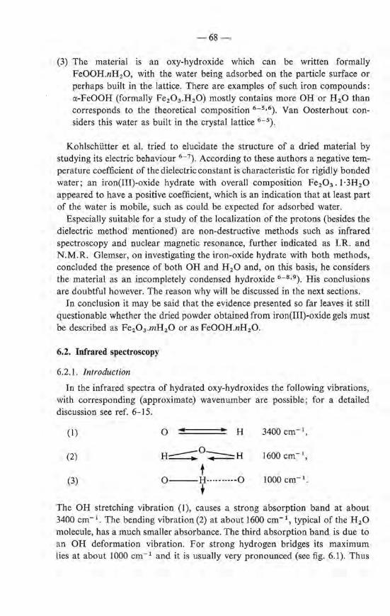
-68-
(3) The material is an oxy-hydroxide which cao be written formally FeOOH.nH20, with the water being adsorbed on the partiele surface or perhaps built in the lattice. There are examples of such iron compounds: a-FeOOH (formally Fe20 3 .H20) mostly contains more OH or H 20 than corresponds to the theoretica! composition 6 - 5 •6). Van Oosterhout considers this water as built in the crystal lattice 6 - 5 ).
Kohlschütter et al. tried to elucidate the structure of a dried material by studying its electric behaviour 6 - 7). According tothese authors a negative temperature coefficient of the dielectric constant is characteristic for rigidly bonded water; an iron(III)-oxide hydrate with overall composition Fe20 3 • 1·3H20 appeared to have a positive coefficient, which is an indication that at least part of the water is mobile, such as could be expected for adsorbed water.
Especially suitable for a study of the localization of the protons (besides the dielectric method mentioned) are non-destructive methods such as infrared spectroscopy and nuclear magnetic resonance, further indicated as LR. and N.M.R. Glemser, on investigating the iron-oxide hydrate with both methods, concluded the presence of both OH and H 2 0 and, on this basis, he considers the material as an incompletely condensed hydroxide 6 - 8 •9 ). His conclusions are doubtful however. The reason why will be discussed in the next sections.
In condusion it may be said that the evidence presented so far leaves it still questionable whether the dried powder obtained from iron(III)-oxide gels must be described as Fe20 3 .mH20 or as FeOOH.nH20.
6.2. Infrared spectroscopy
6.2.1. Introduetion
In the infrared spectra of hydrated oxy-hydroxides the following vibrations, with corresponding (approximate) wavenumber are possible; for a detailed discussion see ref. 6-15.
(I) 0 H 3400 cm- 1 ,
(2) H--0 :;::::::::: H 1600 cm- 1,
t (3) 0--H······----0 1000 cm- 1 .
+ The OH stretching vibration (!), causes a strong absorption band at about 3400 cm- 1 . The bending vibration (2) at about 1600 cm- 1 , typical of the H 20 molecule, has a much smaller absorbance. The third absorption band is due to an OH deformation vibration. For strong hydrogen bridges its maximum lies at about 1000 cm- 1 and it is usually very pronounced (see fig. 6.1). Thus
-69-
an absorption band at about 1600 cm- 1 idicates the presence of H 2 0 and a band at 1000 cm- 1 points to the presence of OH.
Bath OH and H20 give astrong absorption band at about 3400 cm- 1 . The wavelength at maximum absorbance as well as the band width are dependent upon the bonding state of the protons. For instanee the maximum occurs at 3657 cm- 1 for H 2 0 in the vapour state; in the liquid state two maxima can be distinguished at 3450 resp. 3580 cm- 1 and ice has a maximum at 3256 cm- 1 .
The H2 0 in NaBr.2H20 gives a number of maxima between 3200 and 4000 cm- 1 • In MnS04 .7 H 2 0 three maxima cao be distinguished at 3236, 3400 and 3477 cm- 1• Moreover the positions of these maxima (those given befare are all measured at room temperature) are temperature-dependent.
A compilation of the literature dealing with the dependenee of the wavenumber of OH stretching vibrations on interatomie distauces has been given by Nakamato et al. for many organic and inorganic compounds 6 - 16). Glemser and Hatert have presented a sim.ilar study fora large number of hydroxides 6 - 17).
These authors could establish a definite relationship between the frequency of the OH stretching vibration and the 0-0 distance: for d = 2·66 A, u "' ' 2900 cm- 1 (a-AIOOH); ford= 2·90 A, u = 3500 cm- r, see also ref. 6- 18.
In view of these results it is questionable whether to the presence of OH groups in the iron-oxide hydrate may be concluded from the position of "the centre of gravity and tbe extension to smaller wavenumbers" of tbe absorbtion band at about 3400 cm- 1 , as Glemser did 6 - 8 •9). On the contrary, the spectrum published by Glemser even suggests that the material under study does not contain OH groups: the strong band or couple of bands typical for oxide hydroxides around 1000 cm- 1 is oot present in the spectrum of the iron-oxide hydrate. It is stated in the literature, however, that the crystal habit influences the extinction of these deformation bands considerably 6 - 6 ).
6.2.2. Experimental
Infrared spectra are recorded of the oxide hydrate, dispersed in paraffin oil or in KBr, using a Hitachi infrared spectrometer (double-beam apparatus, E.P.I.G.). The spectra appeared to be similar in the two media. Since paraffin oil has a number of absorptions in the wavelength region of interest, all spectra referred to are made using KBr as a dispergent Samples consisting of 0·5 mg Fe20 3-aq. and 300 mg KBr were dried above P 2 0 5 and milled in an agate ball mil! of 3 cm3 , provided with two balls of 3 mm diameter, during ten minutes. The powder is transferred into a steel mould placed in a glove box through which dried nitrogen is blown and is subsequently compacted at a pressure of 15 to 20.103 kg; the resulting tablet has a diameter of 15 mm and a thickness of about 1 mm.
In order to carry out D 20-exchange experiments the tablet is mounted in a measuring cell provided with two CaF2 windows and gas-inlet and -out1et tube.
-70-
0 20 exchange bas been carried out by brioging inside this cell (placed in an exsiccator) a droplet (0·2 mi) of 0 20. After being left for halfan hour the cell is mounted in the pbotometer and nitrogen (containing less than 10 ppm H 20), is passed through the cell during ten minutes after which the absorption spectrum is determined .
6.2.3. Results and discussion
The infrared-absorption spectrum of the oxide hydra te is depicted in fig . 6.1. It exhibits a band at 3400 cm- 1 which is extended to long wavelengtbs (2000 cm- 1) together with a rather strong band at 1630 cm-I; in the region of 1000 cm-I only very weak bands are observed . There are also bands at 1490 and 1360 cm- 1 • The spectrum at large wavelengtbs shows the presence of strong broad bands at 460 and 580 cm-I with a shoulder at 690 cm- I. On 0 20 exchange, besides the bands at 3400 and 1600 cm- I, a lso the band at 1490 cm -I diminishes in intensity which indicates that the latter also must be attributed to H20 but then bonded in a somewhat different manner. The band at 1360 cm-I is notsubject to change on 0 20 treatment. lt might be an overtone of the Jattice vibrations absorbing at long wavelengtbs (such overtones have been found frequently in oxide systems 6 - 19 )), but it could also be due to OH groups that do not exchange protons with deuterons. These protons then must be bonded in a manoer different from the other protons. This could be a distinction between protons present in the lattice and adsorbed as water on the surf ace.
The spectrum may be compared with the spectra of some well-known ironoxide hydroxides, notably a-, {3- and y-FeOOH (fig. 6.1). These have strong bands at 905 and 800 cm- 1 , a doubletatabout 830 cm- 1 and a strong doublet at 670 cm- 1 and weak bands at 1020 and 745 cm- 1 resp., which can be attributed to OH bending vibrations.
The infrared spectrum clearly indicates the presence of H 20 in our iron-oxide hydrate. Although the I.R. spectrum suggests that OH groups are absent, it is not certain that this conclusion holds. In fact the question may be asked whether it is possible that OH bending vibrations are shifted towards Jonger wavelengtbs and herree are overlapping the bands due to the lattice vibrations. Unfortunately these bands could not be investigated with 0 20 exchange as the CaF2 windows used in our experiments have strong absorption in this region.
A second attempt has been made by trying to performa quantitative analysis. This has been carried out by a comparison of tbe ratio of the integrated intensities of the bands at 3400 and 1600 cm-I for a number of hydrates such as KBr.aq., CuS04 .5H20 and MgS04 .aq. on the one hand and those of the iron(III)-oxide hydrate on the other hand. Due to the uncertainty of the base line, particularly for the latter material, this metbod did not allow of a definite conclusion.
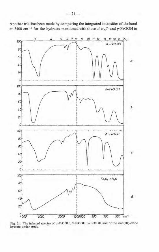
- 71 -
Another trial has been made by camparing the integrated intensities of the band at 3400 cm- 1 for the hydrates mentioned with those of 11.-, {3- and y-FeOOH in
100 3 4 5 14 16 18 21 25 J1
a-FeO.OH
80
60 a
40
20
0
100 13-FeO.OH
40 b
20
'1-FeO.OH
c
80
60
4 d
20
~000 3000 2000 72001100 900 700
Fig. 6.1. The infra red spectra of a -FeOOH, P-FeOOH, y-FeOOH and of the iron(III)-oxide hydrate under study. •

-72-
order to investigate whether the extinction of the OH band is the same for all oxy-hydroxides and differs by a constant factor from that of the hydrates. In this way a distinction between hydrates and hydroxides could not be made, however, and hence the problem remains unsolved.
Glemser tried to distinguish between adsorbed water and OH groups by heating the sample at 120 oe 6 - 8 ). After five hours the H 20 band at 1630 cm- 1
had disappeared, whereas at 3400 cm- 1 a rather strong band stilt existed. The Jatter, according to Glemser, should be due to OH. It is not certain, however, whether the experiments allow this condusion as upon heating the material rnight have changed. ln chapter 7 it will be shown that upon heating at 100 oe within a few hours changes occur indeed ; ho wever, these may be only of minor importance. The suggestion that protons are present which are rigidly bonded to tbe iron-oxygen lattice seems justified by our measurements of dispersions of the iron-oxide hydrate in KBr tablets, after prolonged drying at room temperature *), show that the absorbance of the band at 1630 cm- 1 decreases much sharper than that of the OH stretching band at 3400 cm- 1 . A quantitative estimation of the number of protons contributing to the remaining band at 3400 cm- I, by taking the average extinction of the OH bands of a.-, {3- and y-FeOOH, teaches that about half of the protons take part in it. From tbe absence of the band at 1630 cm- 1 it could be thought that these protons are not present as H 2 0; more information is necessary to draw a definite condusion about the bonding state of these protons. N.M.R. measurements, as discussed in the next section, could provide some more information on this point.
From the spectrum at long wavelengtbs (10-25 f1.) valuable additional information can be obtained regarding the iron-oxygen lattice. eomparison with the spectra of a.-Fe20 3 , y-Fe20 3 and a.-, {3- and y-FeOOH shows that the compound under investigation is different from these, although there is some resemblance to {3-FeOOH (see fig. 6.1).
6.3. Nuclear magnetic resonance
6.3.1. Discussion of the literature
Only a few data are available on N.M.R. spectra of the iron-oxide hydrate. From the observed line broadening, about 5 Oe, Glemser concluded to the presence of OH groups 6 - 9). This conclusion is, however, open to criticism; H 20, when rigidly böund, such as in well-crystallized hydrates, for instanee eaS04 .2H20, exhibits a line width of 10 Oe, but when adsorbed on a partiele surface, the H 20-line width can be considerably smaller, due to the mobility of the H 2 0 molecules, which is large especially on a surface with small radius6 - 21 • 22).
*) Carried out by passing a stream of nitrogen gas over it contaming Jess than 1 ppm H 2 0.
-73-
Even when the water molecules are localized and can only rotate, the eerresponding line width should be 5 Oe 6 - 23).
Moreover, Glemser did nottake into account the superparamagnetic character of the material; the high magnetic moment present in the particles interferes with the applied field and hence influences the line width considerably, as will be discussed further on.
6.3.2. Experimental
The N.M.R. measurements were carried out on a sample consisting of a pressed pellet of 2 g powder of Fe2 0 3 .1·5 H2 0, placed in a saeled glass tube 10 mm in diameter. A coil of insulation-free copper wire is wound around the tube. The number of turns of the coil was between 10 and 20, depending on the eperation frequency range. The coil with sample could be shielded and covered by a brass vessel. It is mounted in a Dewar vessel. A coaxiallead consisting of a stainless-steel shield and an inner wire of copper provided for the conneetion of the top of the co i! with the top of the cryostat. With a piece of f!exible coaxial cable the system was connected to the electronic detection system. The coil formed part of the circuit of a marginal autodyne oscillator. The conventional methods oflow-frequency magnetie-field modulation and phase-sensitive amplification of the N.M.R. signa! were applied.
For the low-temperature measurements a mixture of ethanol and solid e02
was used for cooling the sample system.
6.3.3. Results and discussion
The proton nucleus has a spin I = -! which means that when placed in a - static- magnetic field two energy levels can be occupied, differing in energy by 2 MzHz where Mz = !yh (y = constant) is the magnetic moment in the direction of the applied field H •. Now when a second alternating magnetic field Hx perpendicular to the field H. is applied, resonance absorption can occur when its frequency is equal to the Larmor precession frequency of the nuclear spins, given by w = yH.; for protons y = 2·68.104 radjOe s. In the material to be investigated other fields can act on the nucleus, in actdition to the applied field, for instanee fields due to neighbouring atoms.
Hence, when measuring the absorption as a function of the applied field H. resonance occurs not at a definite frequency but in a certain frequency range giving rise to line broadening !JH.
For the iron(III)-oxide hydrate the line broadening !JH has been measured as a function of H. at two different temperatures; the results of these measurements are presented graphically in fig. 6.2. The results show that at 20 oe the line broadening extrapolated to zero field strength equals about 0·5 Oe and, at -70 oe the line broadening is equal toabout 1·5 Oe. lt will be shown that
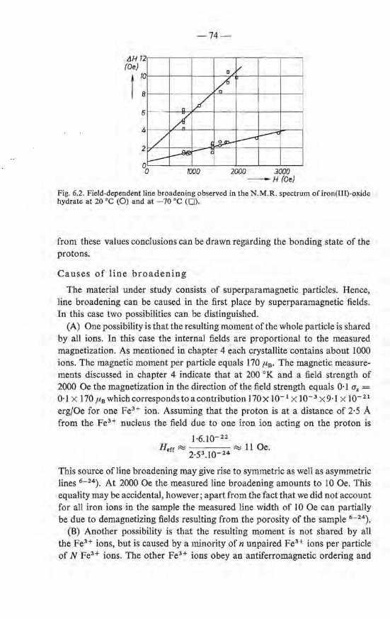
LlH 12 (Oe)
t 1:
6
4
2
r--·
/ ~
-74-
D /
~ tl L
7 .--c ~
J~ ~ - è
1000 2000 3000 -H(Oe)
Fig. 6.2. Field-dependent line broadening observed in the N. M.R. spectrum of iron(III)-oxide hydrateat 20 oe (0) and at -70 oe (0).
from these valnes conclusions can be drawn regarding the bonding state of the protons.
Causes of line broadening
The material under study consists of superparamagnetic particles. Hence, line broadening can be caused in the first place by superparamagnetic fields. In this case two possibilities can be distinguished.
(A) One possibility is that the resulting moment of the whole partiele is shared by all ions. In this case the internal fields are proportional to the measured magnetization. As mentioned in chapter 4 each crystallite contains a bout 1000
ions. The magnetic moment per partiele equals 170 p,8 . The magnetic measurements discussed in chapter 4 indicate that at 200 oK and a field strength of 2000 Oe the magnetization in the direction of the field strength equals 0·1 a, = 0·1 X 170 ftB Which COfrespondS tOa COntribution )70X l0- 1 X lQ- 3 X 9·) X lQ- 21
erg/Oe for one Fe3+ ion. Assuming that the proton is at a distance of 2·5 A from the Fe3+ nucleus the field due to one iron ion acting on the proton is
1·6.10-22
Herr R::! R::J 11 Oe. 2·53.1Q-24
This souree of line broadening may give rise to symmetrie as well as asymmetrie lines 6 - 24). At 2000 Oe the measured line broadening amounts to 10 Oe. This equality may be accidental, ho wever; apart from the fact that we did notaccount for all iron ions in the sample the measured line width of 10 Oe can partially be due to demagnetizing fields resulting from the porosity of the sample 6 - 24).
(B) Another possibility is that the resulting moment is not shared by all the Fe3+ ions, but is caused by a minority of n unpaired Fe3+ ions per partiele of N Fe3+ ions. The other Fe3+ ions obey an antiferromagnetic ordering and
-75-
do not contribute to the external magnetization. The average moment of the Fe3+ ion then equals under the same conditions as mentioned before: 0·1 x 5 x 9·1 x w- 21 ~ 5.10- 21 erg/Oe. The field on the proton then equals
5.10-21 Herr ~ = 300 Oe.
2·53.10-24
At fint view this possibility should be excluded. However, the field at the proton is a summation of the fields due to all the iron i ons in the sample ; the resulting field thus depends on the crystallographic surroundings of the proton. In fact if the proton should be surrounded symmetrically by iron ions this field should be zero.
(C) Dipole fields between neighbouring-protonscan also be the cause of line broadening. It can be calculated that, at a elistance of 1·6 A between the protons, i.e. the distance in H20, the value for iJH amounts to 10 Oe 6 - 23). This line broadening is independent of the applied field. However, as mentioned before, if for instanee the H20 molecules are adsorbed on the surface of the particles in such a way that the H20 molecule can rotate around its symmetry axis instead of a broadening of 10 Oe, a iJH equal to 5 Oe should be observed, which again is independent of the field strength 6 - 23).
(D) A fourth possible reason for the observed line width could be that the H20 molecules, being adsorbed on the partiele surface, are still moving very rapidly along the surface. The line broadening should then be caused by the spatial inhomogeneity in the magnetic field as arising from the magnetized crystallites. In this case it should be probable that the observed lines should be asymmetrie, which has notbeen found.
Taking all arguments together it is tentatively concluded that the observed line widths must be attributed to OH groups. An estimation of the total amount of protons contributing to the supposed OH line gives a quantity of at least 50% or more. Therefore in conneetion with the LR. results it is most probable that the iron-oxide hydrate under study- with the composition Fe20 3 .1·6 H20 - can bedescribed as Fe00H.0·3H20.
6.4. Summary
(1) The infrared spectrum of the iron(III)-oxide hydrate shows a band at 1630 cm-\ typical for H20. The spectrum does not exhibit bands which are typicai for OH-O deformation vibrations, but on drying at room temperature the band at 1630 cm- 1 disappears airoost completeiy, whereas a strong band at 3400 cm- 1 remains. This band is attributed to protons present in the iron-oxygen lattice. From the intensity of this band it is estimated that about half of the protons contribute to it.
-76-
(2) The line broadening in nuclear-magnetic-resonance spectra equals about 1 Oe. This points to the preserree of OH. An estimation shows that at least 50% of the protons must be present as OH.
(3) Combining the infrared and N.M.R. data it is proposed to describe the hydrate Fe20 3 . l ·6 H20 as FeOOH.0·3H20; in general FeOOH.nH 20.
REFERENCES
6 - 1) A. Sirnon and Th. Schmidt, Kolloidz. 36, 65, 1925. 6 - 2 ) H. W. Foote and B. Saxton, J. Am. chem. Soc. 38, 588, 1916; 39, 1103, 1917. 6 - 3 ) R. Willstätter, H. Kraut and W. Fremery, Ber. dt. chem. Ges. 57, 1491, 1924. 6 - 4 ) V. Frei, Col!. Czech. chem. Comm. 27, 775, 1962; 27, 782, 1962. 6 - 5) G. W. van Oosterhout, Proc. int. Conf. Magn. (Nottingham 1964), The InsL of
Physics and The phys. Soc., Londen, 1965, p. 529. 6 - 6 ) V. M. Zolotarev, L. D. Kislovskii and L. V. Riskin, Opties and Spectroscopy
(trans!. from Russ.) Suppl. 2, 142, 1962. 6 - 7 ) H. W. Kohlschütter, H.H.Stammand G. Kämpf, Z. anorg. allg. Chem. 278,
264, 1954; 278, 270, 1954. 6 - 8) 0. Glemser and G. Rieck, Z. anorg. allg. Chem. 297, 175, 1958. 6-9 ) 0. Glemser, Nature 4666, 943, 1959. 6-10) P. A. Thiessen and R. Köppen, z. anorg. allg. Chem. 200, 18, 1931. 6- 11) P.A. Thiessen and R. Köppen, Z. anorg. allg. Chem. 228, 57, 1936. 6 - 12) R. Fricke and G. Hüttig, Hydroxide und Oxid-hydrate, Akad. Verlagsges., Leipzig,
1937, p. 523. 6 - 13) H. B. Weiser and W. 0. Milligan, J. phys. Chem. 39, 25, 1935. 6 - 14) H. B. Weiser, W. 0. Milligan and W. J. Coppoc, J. phys. Chem. 43,1109, 1939. 6 - 15) J. Lecomte, Encyclopedia of physics, Springer Verlag, Berlin, 1958, Vol. 26, p. 244. 6 - 16) K. Nakamoto, M. Margoshes and R. E. RundJe, J. Am. chem. Soc. 77, 6480,
1955. 6 - 17) 0. Glemser and E. Hatert, Z. anorg. allg. Chem. 283, 111, 1956. 6 - 18) E. Schwarzrnann and H. Marsrnann, Naturwiss. 53, 349, 1966. 6 - 19) S. Hafner and F. Lages, Z. f. Krist. 115, 321, 1961. 6 - 20) S. Hafner, z. f. Krist. 115, 331, 1961. 6 - 21) R. K. Webster and T. L. Jones, Proc. Brit. ceram. Soc. 153, 1965 6 - 22) W. S. Brey and K. D. Lawson, J. phys. Chern. 68, 1474, 1963. 6 - 23) G. Hardernao, private comrnuoication. 6-24) D. J. Kroon, Philips Res. Repts 15, 501, 1960.
7. THE RECRYSTALLIZATION OF IRON(III)-OXIDE HYDRATE AT ELEVATED TEMPERATURES
7.1. Introduetion
In the preceding chapter the results have been discussed of an investigation of the iron(III)-oxide hydrate with respect to the occurrence of stoichiometrie hydrated compounds with infrared spectroscopy and nuclear magnetic resonance. Another approach to the examinatien of the existence of hydrates could be a study of its dehydration behaviour upon heating. In the literature there are quite a number of papers dealing with such experiments.
Heating of a wet gel under a vapour pressure of 10 mm Hg up to 400 oe leads to a continuousdehydration as shown by Sirnon 7 - 1); this was taken as an indication that no definite hydrate exists. If the wet gel is dried in air at slightly elevated temperature (40-60 oq the greater part of the capillary water evaporates resulting in products containing 25-35% H 20 7 - 2 •3) . From the thermobalance experiments carried out by Frei et al. on dried gels it was suggested that the dehydration behaviour depends upon the anion present during the hydrolysis and u pon the way in which the hydralysis of the ferric salt bas been carried out. When precipitated from so ... z- -containing solutions the resulting oxide hydrate shows a continuous dehydration diagram, the dehydration occurring at a maximum ra te at a bout 150 oe 7 - 2 ). On the other hand similar products prepared from N03 - -containing solutions show a discontinuous curve. It must be emphasized however, that the oxide hydrates of Frei et al. were prepared in boiling solutions and dried at 60 oe in air; under these conditions the precipitate initially formed could easily recrystallize. The influence of the precipitation temperature on the dehydration behaviour of the gel has been demonstrated by Mackenzie 7 - 4 ) . This author found a relationship between the precipitation temperature and the temperature at which the highest dehydration rate occurs.
Another attempt to demonstrate the existence of a hydrate or hydroxide has been done by Albrecht and Wedekind by measuring the magnetic susceptibility X· These authors found a continuous increase in the susceptibility (per gramme of oxide) on decreasing water content 7 - 6) in the dried gel. At water contents between 11 and 15%, x is at maximum, viz. 300-400.10- 6 cm3 fg 7 - 5 •6),
which according to the opinion of the authors must be due to a definite hydrate or hydroxide different from the well-known ct- and y-oxy-hydroxides (water content ~ 10%) as these compounds have much lower x values, ~ 20.10-6
cm3/g 7 - 7). Tbe authors did not succeed in identifying this alleged compound: they did not find arelation between X-ray-diffraction patterns oftbe dehydrated products and the value of the susceptibility. As will be discussed in sec. 7.4 it is not necessary to assume the formation of a new, hitherto unknown hydrate,
-78-
to explain the changes in the susceptibility. A study of the dehydration behaviour of iron(III)-oxide hydra te has been per
formed by Fricke et al. 7 - 9 •10) with the aid of calorimetry together with X-ray diffractrometry. They arrived at the following conclusions. (1) Upon heating the energy content of the material increases and attains a
maximum value between 150 and 200 oe. (2) Above 250 oe a rapid decrease of the energy content occurs. (3) Samples (prepared at 20 oq after heating at 250 oe show the presence of
ct-Fe20 3 with a size of the crystallites of about 80 A. ( 4) Above 350 oe rapid crystal growth occurs; after one hour at 600 oe
ct-Fe20 3 crystals with a size of about 250 A are present. The formation of ct-Fe20 3 at 300 oe has also been reported by Frei who
found, however, in the X-ray diagram also the spacing d:::::::: 2·0 A which is not observed in ct-Fe2 0 3 in the coarse crystalline form.
Summarizing it can be said that from studying the dehydration behaviour of gels prepared from aqueous solutions of a ferric salt (nitrate, sulphate, chloride and perchlorate) no indication has been found for the occurrence of stoichiometrie compounds between Fe20 3 and H 20. Yet the existence of such a compound is suggested by the results of the preceding chapter. For an elucidation of its composition-tentatively described by FeOOH.nH20-a more precise knowledge of the quantities of hydrate or adsorption water and of hydroxyl is necessary. For that purpose a number of D.T.A. and T.G.A. experiments are done which are described in ~the:foUowing.
"' ..
7.2. Thermogravimetrie analysis and differential tbermal analysis
7.2.1. Experimental
Dijferential thermogravimetry is carried out using a eahn microbalance. The sample weight is continuously registered on a recorder. The sample, about 90 mg Fe20 3 .aq., in an alumina crucible measuring lO x 5 mm2 (wall thickness 0·5 mm), is placed on an alumina support fastened to the arm of the balance with an alumina rod of 0·3 mm diameter. The temperature is measured with a Pt-Rh thermocouple, placed in the vicinity of the sample. The temperature is increased at a rate of 520 oejh. The heating furnace has a zone of constant temperature with a length of 15 mm. The balance and its furnace system is vacuum-pumped. The H 20 pressure in the balance system is kept constant by connecting the latter with a reservoir containing melting ice. A blank gives no measurable weigbt changes in the temperature region of interest.
Differential thermal analysis. Samples of 50 mg, are placed in a cylindrical container of 10 x 3·5 mm2 , wal! thickness 0·1 mm. The Pt sample holder together with the reference - an identical crucible containing Al2 0 3 - are placed
-79-
on top of an alumina rod which is kept carefully centred in the furnace by means of alumina rings. Over the crucibles a Pt cylinder (height 5 cm) is placed which in turn is surrounded by a closed alumina tube provided with four holes (diameter 3 mm) for the passage of nitrogen gas. This tube just fits in the furnace tube. The temperature is measured with a Pt-Rh thermocouple and the temperature difference between the sample and the reference is measured by similar couples which are placed at the bottorn of the crucibles. The heating rate is 500 °Cjh. The water vapour formed during the reaction is removed by passing air through the furnace (inner tube width 28 mm) at a velocity of 0·5 I/min. Measurements are also clone using an apparatus constructed by Bureau de Liaison, Paris. The detecting head of this apparatus has sample bolders formed by the junction of the detecting thermocouple 7 - 11 ). The results obtained with both apparatuses were essentially the same; fig. 7.2 is obtained with that of B.D.L.
7.2.2. Results and discussion
Thermogravimetrie and differential thermal analysis
As mentioned already, the oxide hydrate prepared at 90 oe after drying on P20 5 contains about 13 % H 20 and after re-exposure to the air at room temperature the water content easily rises to 25 %. The dehydration-rehydration behaviour of a sample has been stuclied at higher temperatures by heating the oxide hydrate on a thermobalance in an H 20 atmosphere with a constant vapour pressure of 0·4 mm Hg. lf the temperature is increased a gradual decrease in the weight of the sample results up to 70 oe. Already at 60 oe a rapid loss of H 20 occurs, at 200 oe 60 % and at 300 oe 90 % of the total quantity of water is given off. When the temperature is increased stepwise and kept at a constant value for a certain time an equilibrium etablishes. If the temperature is then lowered and kept constant again a new equilibrium results by adsorption of H 2 0 from the atmosphere. The results are summarized in table 7-I and shown graphically in fig. 7.1. The dehydration is nearly complete at 300 oe; it elapses gradually and thus the existence of a definite compound is not indicated. This, ho wever, does not exclude the existence of such a compound. For, even when having OH groups on definite lattice sites, small crystallites, on decomposition, can loose their protons gradually due to the formation of H 2 0, which must evaparate from the surface, this being the equilibrium-determining step 7 - 12).
When the temperature is lowered water is taken up again. This will be due to adsorption on the partiele surface; a restoration of OH groups inside the partiele is not probable. The initia! water content is not reached even after 10 hours. This decrease in adsorption capacity is not evidence for an initia! existence of OH groups as it could also be explained as being due to the fact that, at the
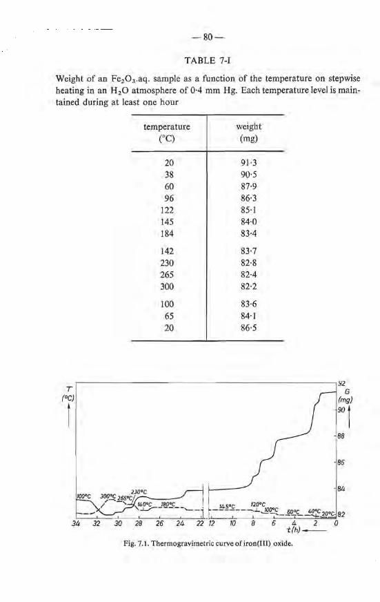
-80-
TABLE 7-I
Weight of an Fe2 0 3 .aq. sample as a function of the temperature on stepwise heating in an H 20 atmosphere of 0·4 mm Hg. Each temperature level is maintained during at least one hour
temperature weight (OC) (mg)
20 91·3 38 90·5 60 87·9 96 86·3
122 85·1 145 84·0 184 83·4
142 83·7 230 82·8 265 82·4 300 82·2
100 83·6 65 84·1 20 86·5
,------------------------------------------------.92 T G.
(°C) (rng) I 90 I 88
86
84
Fig. 7.1. Thermogravimetrie curve of iron(III) oxide.

-81-
temperature of 300 oe, sintering bas taken place (the specific surface area decreases 35 %).
T.G.A. only gives information regarding those processes which involve a weight loss. D.T.A. allows the detection of processes occurring during thermal dehydration which are accompanied by a change in the enthalpy, e.g. recrystallization processes. The -same preparatien as used for the T.G.A. has been stuclied by means of D.T.A. The results are presented graphically in fig. 7.2. A rather broad endothermic peak exists between 20 and 200 oe, with a maximum at 150 oe, no doubt due to the evaporation of water. Between 350 and 450 oe a sharp exothermic peak is observed which may be related to the recrystallization of the dehydrated iron oxide (see also sec. 7.3). e Jearly, the dehydration (20-200 oq and the recrystallization (350-450 oq are separate processes. The anhydrous product formed upon dehydration is stabie from 200 to 350 oe until recrystallization starts. This does not prove that the expelled water was adsorbed on the partiele surface. Similar phenomena are observed on dehydration of well-crystallized oxide hydroxides 7 - 13). The fact that the lattice does not necessarily collapse has been explained by Freund 7 - 12) as due to a stabilization of vacancies created by the disappearance of OH groups; the energy thus stared in the lattice is liberated suddenly in a narrow temperature range, in our case around 430 oe.
The combined D.T.A. and D.T.G. results show the following. (I) It is not possible to get a definite answer with these techniques regarding the
bonding state of the protons; OH groups as wel! as H 20 built in the lat-
\ \ \
\ \ 450°C
\ \ \
\250°C
\ \
\ \200°C
\ \
\4oooc
\ \ 'psooc
\ \ \
\ v oooc \ \
T ime(h) - -
Fig. 7.2. Thermogram of iron(III)-oxide hydrate.
.JT
r
-82-
tice or adsorbed on the surface of the particles should be able to cause the phenomena observed.
(2) On heating already below 200 oe the gel dehydrates nearly completely. This dehydrated gel is stabie in a rather large temperature traject, 200-350 oe.
(3) On rehydration this product takes up H 2 0 again, but less than 50% of the amount originally present. This does not prove that the expelled H 2 0 was originally present as OH groups; the phenomenon can also be explained as due to a decreased adsorbing capacity.
7.3. erystallographic transformations; morphology
The results presented thus far clearly show that in the temperature region of 200-350 oe a dehydrated compound exists in a form which is stabie under our experimental conditions. For a further elucidation of the dehydration mechanism a knowledge of the chemica} and physical properties of the products existing in this intermediate state is of special interest.
Fora crystallographic study two samples are chosen that are obtained from gels preparedat 20 or 90 oe. Each is heated at 201, 227 or 302 oe, respectively. The mai.Q. reflections observed in the corresponding X-ray diffractions of these compounds are listed in table 7-II. Alllilles are broad and overlapping. The d values given in table 7-II are therefore approximations. A comparison of the d values of the various "intermediate products" with those of the wellknown iron-oxide hydroxides shows that (2) contains a-Fe2 0 3 as indicated by the reflections d = 3·70, 2·69, 2·44 and 1·84 A. These reflections arenotpresent in the samples (3) and (4) obtained from starting compounds prepared at 90 oe; when heated at 227 oe some change is observed; there is probably a band at d = 2·83, whereas the value of d = 1·70 could not be observed. When the material is heated at 302 oe, however, a strong band is observed at d = 2·71 A. This indicates again the presence of a-Fe20 3 • In the latter case the other bandscan be attributed to a-Fe2 0 3 as wellas to the structure of the starting compound; the absence of the reflection belonging to d = 1·99 A makes it plausible that this material mainly exists of a-Fe20 3 . When heated at 450 oe, all samples exhibit the diagram of rather well crystallized a-Fe20 3 .
elearly on dehydration some crystallographic transformations take place; there are indications ofthe formation of a-Fe2 0 3 . Only slight crystal growth, if any at all, occurs up to 250 oe. At still higher temperatures and particularly at about 430 oe (the maximum of the exotherrnic peak in the D.T.A. diagram) a rapid crystallization towards a-Fe30 2 takes place under the formation of crystals with a comparatively large size, as derived from the observed line broadening. Electron rnicroscopy confirms these conclusions. As pictured in fig. 7.3, when heated to 227oe the particles have a size below 50 A, but when heated to about 450 oe they attain a size of about 300 A.
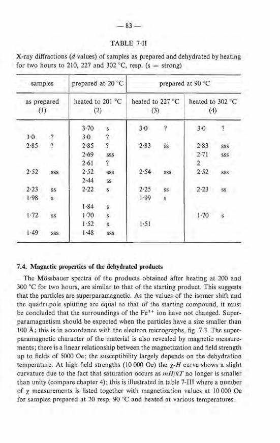
-83-
TABLE 7-11
X-ray diffractions (d values) of samples as prepared and dehydrated by heating for two hours to 210, 227 and 302 oe, resp. (s = strong)
samples prepared at 20 oe 1 prepared at 90 oe
as prepared heated to 201 oe heated to 227 oe heated to 302 oe (1) (2) (3) (4)
3·70 s 3·0 ? 3·0 ? 3·0 ? 3·0 ? 2·85 ? 2·85 ? 2·83 ss 2·83 sss
2·69 sss 2·71 sss 2·61 ? 2
2·52 sss 2·52 sss 2·54 sss 2·52 sss 2·44 ss
2·23 ss 2·22 s 2·25 ss 2·23 ss 1·98 s 1·99 s
1·84 s 1·72 ss 1·70 s 1·70 s
1·52 s 1·51 1·49 sss 1·48 sss
7.4. Magnetic properties of the dehydrated products
The Mössbauer spectra of the products obtained after heating at 200 and 300 oe for two hours, are similar to that of the starting product. This suggests that the particles are superparamagnetic. As the values of the isomer shift and the quadrupale splitting are equal to that of the starting compound, it must be concluded that the surroundings of the FeH ion have not changed. Superparamagnetism should be expected when the particles have a size smaller than 100 A; this is in accordance with the electron micrographs, fig. 7.3. The superparamagnetic character of the material is also revealed by magnetic measurements; there is a linear relationship between the magnetization and field strength up to fields of 5000 Oe ; the susceptibility largely depends on the dehydration temperature. At high field strengtbs (10 000 Oe) the x-H curve shows a slight curvature due to the fact that saturation occurs as mHjkT no Jonger is smaller than unity (compare chapter 4); this is illustrated in table 7-111 where a number of x measurements is listed tagether with magnetization values at 10 000 Oe for samples prepared at 20 resp. 90 oe and heated at various temperatures.

-84-
Fig. 7.3. Electron micrographs ofiron(III)-oxide hydrate (a) as prepared, (b) 227 oe, (c) 302 oe and (d) 350 oe. The oxide hydrate has been prepared at 20 oe.
a
b
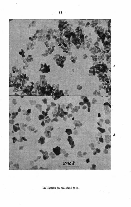
-85-
c
d
See caption on preceding page.
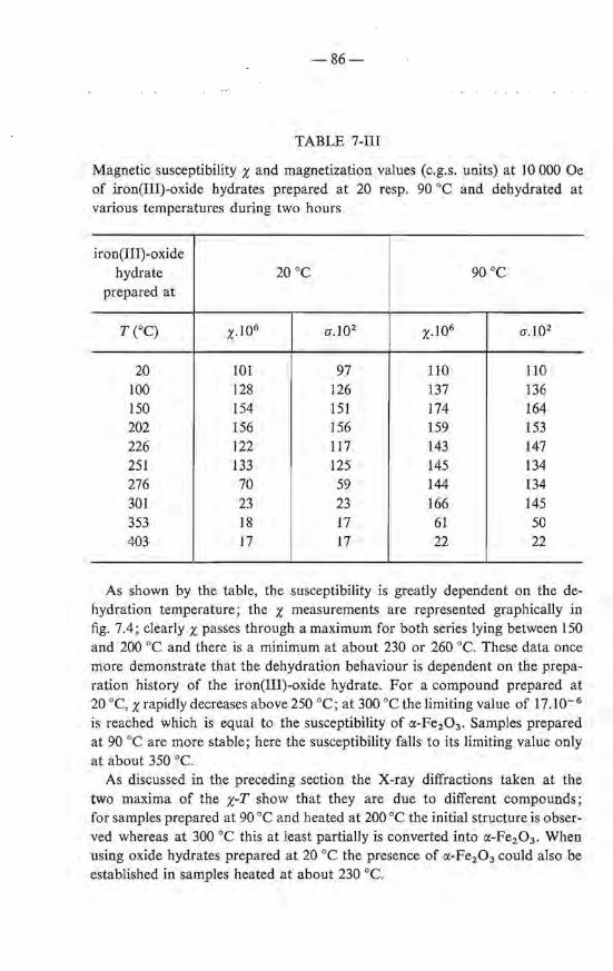
-86-
TABLE 7-III
Magnetic susceptibility x and magnetization values (c.g.s. units) at 10 000 Oe of iron(III)-oxide hydrates prepared at 20 resp. 90 oe and dehydrated at various temperatures during two hours
iron(III)-oxide hydra te 20 oe 90 oe
preparedat
T(OC) x.Io6 0".102 x.I06 a.l02
20 101 97 110 110 100 128 126 137 136 150 154 151 174 164 202 156 156 159 153 226 122 117 143 147 251 133 125 145 134 276 70 59 144 134 301 23 23 166 145 353 18 17 61 50 403 17 17 22 22
As shown by the table, the susceptibility is greatly dependent on the dehydration temperature; the x measurements are represented graphically in fig. 7.4; clearly x passes through a maximum for both series lying between I 50 and 200 oe and there is a minimum at about 230 or 260 oe. These data once more demonstrate that the dehydration behaviour is dependent on the preparation history of the iron(III)-oxide hydrate. For a compound prepared at 20 oe, x rapidly decreases above 250 oe; at 300 oe the limiting value of 17.10- 6
is reached which is equal to the susceptibility of a-Fe20 3 . Samples prepared at 90 oe are more stabie; here the susceptibility fa IJs to its limiting value only at about 350 oe.
As discussed in the preceding section the X-ray diffractions taken at the two maxima of the x-T show that they are due to different compounds; for samples prepared at 90 oe and heated at 200 oe the initia! structure is observed whereas at 300 oe this at least partially is converted into ct-Fe2 0 3 . When using oxide hydrates prepared at 20 oe the presence of ct-Fe2 0 3 could also he established in samples heated at about 230 oe.
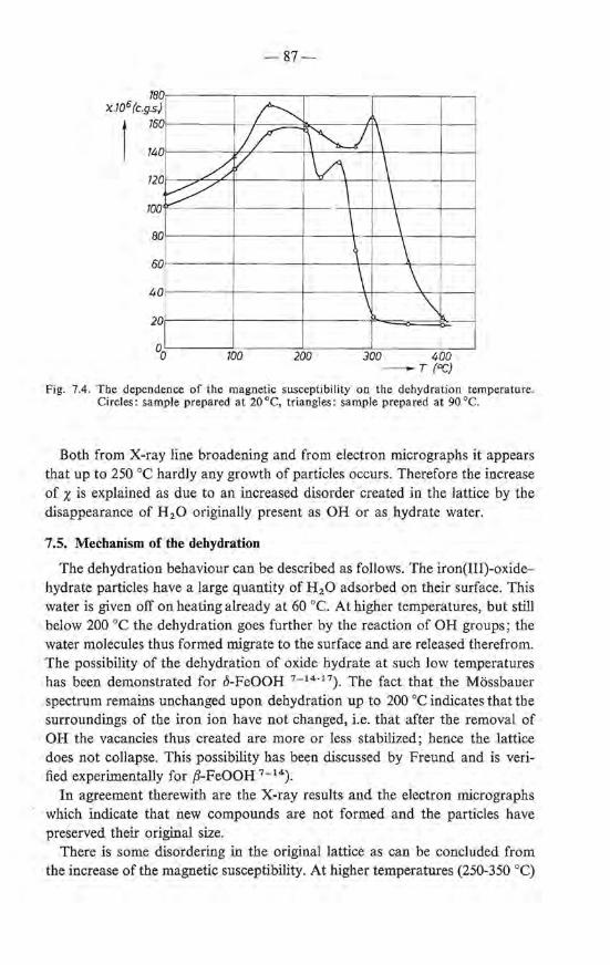
180 'X..105 (c.g.s)
t 760
140
120
100
80
60
40
20
h ~
-87-
b--'// ~"-J \ / \/\ _\
\ \ \ \ \ \ \ \ \ '\
i 100 200 300 400
-T (OC)
Fig. 7.4. Tbe dependenee of the magnetic susceptibility on the dehydration temperature. Circles: sample prepared at 20 oe, triangles: sample prepared at 90 oe.
Both from X-ray line broadening and from electron micrographs it appears that up to 250 oe hardly any growth of particles occurs. Therefore the increase of x is explained as due to an increased disorder created in the lattice by the disappearance of H 20 originally present as OH or as hydrate water.
7.5. Mechanism of the debydration
The dehydration behaviour can bedescribed as follows. The iron(III)-oxidehydrate particles have a large quantity of H20 adsorbed on their surface. This water is given off on heating already at 60 oe. At higher temperatures, but still below 200 oe the dehydration goes further by the reaction of OH groups; the water molecules thus formed migrate to the surface and are released therefrom. The possibility of the dehydration of oxide hydrate at such low temperatures has been demonstrated for ö-FeOOH 7 - 14- 17) . The fact that the Mössbauer spectrum remains unchanged u pon dehydration up to 200 oe indicates that the surroundings of the iron ion have not changed, i.e. that after the removal of OH the vacancies thus created are more or less stabilized; hence the lattice does not collapse. This possibility has been discussed by Freund and is verified experimentally for ,8-FeOOH 7 - 14).
In agreement therewith are the X-ray results and the electron micrographs which indicate that new compounds are not formed and the particles have preserved their original size.
There is some disordering in the original lattice as can be concluded from the increase of the magnetic susceptibility. At higher temperatures (250-350 oq
-88-
the dehydration is nearly completed. Depending upon the preparation temperature of the starting material in our series, 20 or 90 oe, the susceptibility decreases considerably above 260 or 300 oe. Electron micrographs reveal that at the same time partiele growth occurs leading to the formation of small particles which according to X-ray measurements have the cx:-Fe2 0 3 structure. Finally between 350 and 450 oe the surface energy is given off as shown by the D.T.A. measurements and rapid sintering occurs leading to well-crystallized cx:-Fe2 0 3 consisting of crystallites with a size mainly between 300 and 400 A. The calorimetrie and X-ray results of Fricke et al. mentioned in the introduetion give a further support for the scheme outlined above.
7 .6. Summary
(1) Iron-oxide hydrate looses the greater part of its water, present both as OH and H 20, between 60 and 200 oe.
(2) u pon dehydration below 200 oe the iron-oxygen lattice is preserved; the particles keep their original size (20-30 À). Only some disordering occurs leading to an increase of the magnetic susceptibility.
(3) In the temperature region 250-350 oe the dehydration is completed under the formation of cx:-Fe2 0 3 . Simultaneously a slight partiele growth occurs; the superparamagnetic character disappears but the particles remain small, < IOO A. At 400-450 oe rapid sintering occurs under the formation of well-crystallized cx:-Fe2 0 3 with particles of 300 to 400 A.
REFERENCES 7 - 1) A. Sirnon and Th. Schmidt, Kolloidz. 36, 65, 1925. 7 - 2 ) V. Caslavska, V. Frei and A. Bla;ek, Coll. Czech. cbem. Comm. 27, 2168, 1962. 7 - 3) 0. Glemser and G. Rieck, Z. anorg. allg. Chem. 297, 175, 1958. 7-4) R. C. Mackenzie, Nature 164, 244, 1949. 7 - 5) E. Wedekind and W. H. Albrecht, Ber. dt. chem. Ges. 60, 2239, 1927. 7 - 6 ) E. Wedekind and W. H. Albrecht, Ber. dt. chem. Ges. 59, 1726, 1926. 7 - 7 ) W. H. Albrecht, Ber. dt. chem. Ges. 62, 1475, 1929. 7 - 8 ) W. H. Albrecht and E. Wedekind, Z. anorg. allg. Chem. 202, 209, 1931. 7 - 9 ) W. F r icke and L. Klenk, Z. Elektr. 41, 617, 1935. 7 - 10) R. Fricke and K. Meyring, Z. anorg. allg. Chem. 230, 357, 1937. 7 - 11) C. Mazières, Anal. Chem. 36, 602, 1964. 7 - 12) F. Freund, Ber. dt. ker. Ges. 42, 23, 1965. 7 - 13) R. Fricke and P. Ackermann, Z. Elektr. 40, 630, 1934. 7 - 14) J . Dézsi, L. Keszthelyi, D. Kulgawvzuk, B. Molnár and N.A. Eissa, Phy,.
Stat. sol. 22, 617, 1967. 7 - 15) M. J. Ridge and G. R. Boell, Inorg. Phys. Theor. 1147, 1967. 7 - 16) J. D. Bernal ,D. R. Dasgupta and A. L. Mackay, Clay Minerals Bull. 4, 15, 1959. 7 - 17) A.W. Simpson, J. appl. Phys. Suppl. 33, 1203, 1962.

- 89-
Acknowledgement
The investigations described in this thesis were carried out at the Philips Research Laboratories. I thank the directors of these laboratories for their permission to publish the results as a thesis.
The work has been accomplished in a close and highly appreciated cooperation with several colleagues. Many of the points investigated originated during stimulating discussions with Drs G. W. van Oosterhout. The analytica! work has been clone jointly with Mr J. Visser, the radio-tracer work with Mr P. N. Kuin, the Mössbauer spectroscopy with Dr J. S. van Wieringen, the X-ray work with Mr C. Langereis, the infrared spectroscopy with Dr J. Bakker, the N.M.R. experiments with Dr G. E.G. Hardeman and the D.T. and T.G. analysis with Drs P. J. L. Reynen. Thanks are also due to F. Huizinga for experimental assistance and to Mr J. M. Nieuwenhuizen and Mrs J. R. M. Gijsbers-Dekker for the electron micrographs.
Special thanks are due to Dr J. de Jonge for his criticisms of the manuscript.
Mrs A. Meyer-Huisman gave valuable assistance during the proof-reading stages.

-90-
Summary
On the neutralization of aqueous solutions of ferric salts a series of reactions takes place, the end product being a gelatineus precipitate. Hydralysis of the Fe(H20) 63+ ions occurs as soon as OH- ions are brought into the solution. Thereupon, at first no precipitate is formed; when the degree of hydrolysis, defined as the ratio of the number of equivalents of OH- added to FeH, exceeds the value of 2·5, the formation of a gel suddenly starts. There is no consensus of opinion in the literature regarding the nature of the processes that take place in the solution. Whereas in the older literature it is assumed that a continuous series of polynuclear species is formed, it is stated in the more recent Iiterature that only dimers are formed, which in a short pH traject unite, with the formation of a gel.
From our investigations, carried out with solutions of ferric nitrate, it follows that hydralysis of the ferric ions, even at low degree of hydrolysis, gives rise to the formation of colloidal particles. Upon further actdition of OH- ions when the point of electroneutrality is reached, fiocculation occurs of these particles with the formation of a gel. This is derived partly from the fact that the ionic product [FeH] [OH-p is constant in a large concentration region. Fora determination of this ionic product it is necessary to determine [FeH]. This bas been achieved by making use of a titration of the Fe3+ ions using EDT A with KCNS as an indicator. Ultracentrifugation experiments, carried out with the aid of tracer techniques, confirm the view that, u pon the addition of OH- ions, small discrete particles are formed instead of a continuous series of polymers. The precipitation processes occurring in solutions of Fe3+ ions differ from the precipitation reactions usually observed only in that the size of the particles is extremely smal!.
In order to isolate the particles from the gel a procedure has been used in which the gel is frozen at temperatures below 250 oK. During the subsequent thawing the gel disintegrates and a very fine powder is obtained, further indicated as iron(III)-oxide hydrate. The way in which the disintegration of the gel occurs has been stuclied with the aid of Mössbauer spectroscopy. U se has been made of the phenomenon that particles with a very small mass, when absorbing a y quantum, acquire such a velocity that resonance is no Jonger possible. If the surroundings have a great rigidity, e.g. after freezing, these take up the impulse. In this way the freezing of the gel cao be stuclied accurately. From these investigations it proved to be pro bable that during the freezing no essential changes take place within the particle.
The iron(III)-oxide hydrate, prepared in the way as described above, bas been investigated with the aid of several methods: (1) Electron rnicroscopy shows that the particles have a size of 20-30 A. (2) Measurements ofthe magnetic susceptibility reveal thattbematerialissuper-
-91-
paramagnetic. From these measurements it can be concluded that the particles contain about 1000 iron ions, in good agreement with the electronmicroscopie evidence.
(3) Crystallographic investigations using MoKet radiation demonstrate that the particles are crystaJiine, contrary to the prevailing assumption that the material should be amorphous. The crystallographic structure is different from that of the well-known iron oxides and oxide hydroxides.
These results which show that the particles may be considered as crystallites greatly support the mechanism of the hydrolysis outlined before. Due to the small size of the particles and consequently the considerable line broadening, it has not been possible to elucidate the crystallographic structure.
The preparation of larger crystals by recrystallization in solutions did not succeed; instead of growth of the crystallites originally present, recrystaJlization takes place with the formation of et-Fe20 3 or et-FeOOH. This recrystallization has been stuclied with the aid of magnetic measurements; a series of intermedia te products is formed which have a high magnetic susceptibility. This can be explained on the basis of the superparamagnetic character of the particles: on increase of the partiele size the moment per partiele first increases, goes through a maximum value and decreases again due to a coupling of the magnetic vector with a preferred crystallographic direction ("blocking"). The chemica) composition of the oxide hydrate is not constant. After having beendriedon P2 0 5
the material contains about 15% H 20. This corresponds to the formula Fe20 3 .1·6 H20. Infrared spectroscopy has demonstrated that about half of the water is present as H 20. This water is adsorbed on the partiele surface as it eau be removed at room temperature with dry nitrogen gas. The other half of the water is bonded in a different manner, probably in the form of OH, as the typical H 20 band at 1630 cm- 1 is not observed in the spectrum. Investigations with nuclear-magnetic-resonance spectroscopy show that at least 50 % of the water is bonded in a manner such as to give rise to a Iine broadening of about 1 Oe. This points to OH groups. In combination with the infrared investigations it seems most probable therefore that the material with composition Fe2 0 3 .1·6H2 0 may be indicated formally by FeOOH. 0·3H2 0, more generally FeOOH.nH20. Hence we are dealing with an oxide hydroxide with H 20 adsorbed on the surface of the particles.
On thermal dehydration up to 200 oe, nearly all the water is expelled, the composition then still being about Fe20 3 . 0·2H20. Electron microscopy together with X-ray diffractometry confirm that, under these circumstances, neither partiele growth nor recrystallization (for instanee to et-Fe20 3) take place. The magnetic susceptibility increases, indicating an enhanced disorder. Mössbauer spectroscopy suggests that the coordination shell of the iron ion does not change. Apparently on the release of the OH groups the structure of the partiele largely remains unchanged.
-92-
U pon heating to temperatures above 300 °C, recrystallization occurs with the formation of a-Fe2 0 3 • The magnetic susceptibility decreases considerably due to the formation of comparatively large a-Fe20 3 crystals.

-93-
Samenvatting
Bij neutralisatie van waterige oplossingen van ferrizouten vindt een reeks reacties plaats met als eindproduct een gelatineus neerslag. Hydrolyse van het Fe(H2 0)6 3+ ion treedt op zodra OH- ionen in de oplossing gebracht worden. Daarbij wordt aanvankelijk geen neerslag gevormd; eerst wanneer de hydrolysegraad, uitgedrukt in de verhouding van het aantal toegevoegde equivalenten OH- tot Fe3 +, de waarde 2·5 overschrijdt, zet plotseling de vorming van het gel in. Over de aard van de processen die zich in de oplossing afspelen bestaat geen overeenstemming. Terwijl in de oudere literatuur de vorming van een continue reeks polynucleaire producten aangenomen wordt, volgt uit meer recente onderzoekingen dat hoogstens dimeren worden gevormd waaruit dan in een kort pH traject het gel ontstaat.
Uit onze onderzoekingen uitgevoerd met oplossingen van ferrinitraat blijkt dat hydrolyse van het ferri ion reeds bij geringe hydrolysegraad leidt tot de vorming van colloidale deeltjes, die als bij verder toevoegen van oH-ionen het ladingsnulpunt wordt bereikt aanleiding geven tot een gelatineus neerslag. Dit volgt o.a. uit het feit dat het ionenproduct [FeH] [OH-p constant is over een zeer groot concentratietraject. Voor de bepaling hiervan is het noodzakelijk [FeH] te kennen. Daartoe is gebruik gemaakt van een titratie met behulp van EDT A met KCNS als indicator. Ultracentrifuge-experimenten, welke zijn uitgevoerd met behulp van tracer-technieken bevestigen de opvatting dat toevoegen van OH- ionen leidt tot de vorming van discrete kleine deeltjes en niet tot een continue serie van polymeren. De hydrolyse van FeH ionen verschilt dus slechts daarin van geWone precipitatiereacties dat de gevormde deeltjes uitzonderlijk klein zijn.
Om de deeltjes welke de bouwstenen van het gel vormen er uit te isoleren is een werkwijze toegepast waarbij het gel wordt bevroren tot temperaturen beneden 250 °K. Tijdens het daarop volgende ontdooien desintegreert het gel en wordt een zeer fijn poeder verkregen aangeduid met de term ijzeroxydehydraat De wijze waarop de desintegratie van het gel plaats vindt is bestudeerd met behulp van Mössbauer-spectroscopie, waarbij gebruik gemaakt is van het verschijnsel dat, bij uitstraling van een y-quantum (afkomstig van een energieovergang in de kern) aan deeltjes met een zeer kleine massa een zodanige snelheid wordt medegedeeld dat resonantie onmogelijk wordt. Bij grote stijfheid van de omgeving, zoals na bevriezen, is deze in staat om de impuls op te vangen; aldus kan het bevriezen van het gel nauwkeurig bestudeerd worden. Uit deze onderzoekingen is gebleken dat zeer waarschijnlijk geen essentiele veranderingen in het deeltje plaats vinden.
Het ijzer(III)-oxide-hydraat, met behulp van bovengenoemde werkwijze verkregen, is onderzocht met behulp van verschillende onderzoekmethodes :
-94-
(1) Electronenmicroscopie toont dat de deeltjes afmetingen hebben van 20 à 30 A.
(2) Uit metingen van de magnetische susceptibiliteit van het zich superparamagnetisch gedragende materiaal kan worden afgeleid dat de deeltjes zijn opgebouwd uit circa 1000 Fe3+ ionen, in overeenstemming met de electroDenmicroscopische gegevens.
(3) Kristallografisch onderzoek met behulp van MoKtX-straling toont aan dat de deeltjes kristallijn zijn, in tegenstelling met de heersende opvatting dat het materiaal amorf zou zijn. De kristalstructuur verschilt van die van de bekende ijzeroxyden en oxyden-hydroxyden.
Het feit dat de deeltjes mogen worden opgevat als kristallieten is een krachtige ondersteuning voor het boven omschreven beeld van de hydrolyse. Als gevolg van de geringe afmetingen der deeltjes en de hiermede gepaard gaande zeer sterke lijnverbreding is het niet mogelijk gebleken de kristalstructuur te bepalen.
De bereiding van grotere kristallen door rekristallisatie in oplossing is niet gelukt; in plaats van groei der oorspronkelijk aanwezige kristallen vindt een omkristallisatie plaats tot tX-Fe2 0 3 of tX-FeOOH. Deze rekristallisatie is met behulp van magnetische metingen te volgen; er wordt een serie tussenproducten gevormd die een zeer hoge magnetische susceptibiliteit hebben. Deze kan worden verklaard uit het superparamagnetisch karakter der deeltjes: bij toenemende deeltjesgrootte neemt het moment per deeltje eerst toe, gaat door een maximum en neemt daarna weer af tengevolge van koppeling van de magnetische vector aan een kristallografische voorkeursrichting. De chemische samenstelling van het oxide-hydraat is niet constant. Het materiaal bevat na drogen boven P 20 5 meestal ongeveer 15% water. Dit komt overeen met de samenstelling Fe20 3 • 1·6H20. Uit onderzoekingen met behulp van infrarood-spectroscopie is gebleken dat ongeveer de helft van het gebonden water in de vorm van H 2 0 aanwezig is. Vermoedelijk is dit aan het oppervlak geadsorbeerd daar het door middel van droge stikstof bij kamertemperatuur is te verwijderen. De andere helft van het aanwezige water is op een andere manier gebonden, vermoedelijk in de vorm van OH, daar de typische H 2 0 band bij 1630 cm- 1 ontbreekt in het spectrum. Onderzoek met behulp van kernspinresonantie toont aan dat ten minste 50% van het water op een manier gebonden is welke aanleiding geeft tot lijnverbredingen van circa 1 Oe, hetgeen wijst op hydroxylgroepen. In combinatie met de infraroodonderzoekingen lijkt het daarom het meest voor de hand liggend het materiaal met de samenstelling Fe2 0 3 .1·6H20 aan te duiden als FeOOH. 0· 3H20: in het algemeen dus als FeOOH. nH20. Het betreft hier dus een oxide-hydroxide met het H2 0 geadsorbeerd aan het oppervlak der deeltjes.
Bij thermische dehydratie tot 200 oe wordt bijna al het gebonden water afgegeven, de samenstelling is dan nog ongeveer Fe20 3 • 0·2H2 0. Hierbij treedt nog geen deeltjesgroei of rekristallisatie op (b.v. tot tX-Fe2 0 3), zoals is vastgesteld
-95-
met behulp van electronenmicroscopie en röntgenopnamen. Er is een toename van de magnetische susceptibiliteit hetgeen wijst op een toegenomen wanorde. Uit het gelijk blijven van het Mössbauer-spectrum volgt dat de omgeving van het ijzer-ion niet merkbaar verandert. Blijkbaar blijft de structuur van het deeltje bij uittreden van OH groepen grotendeels behouden.
Bij verhitten tot boven 300 oe treedt rekristallisatie op tot a-Fe2 0 3 . De magnetische susceptibiliteit neemt hierbij sterk af als gevolg van de vorming van relatief grote a-Fe2 0 3 kristallen.

STELLINGEN
A. A. van der GIESSEN
STELLINGEN
I
Het "angle of incidence" effect dat soms is waargenomen bij het opdampen van metaallagen op substraten in de beginstadia van de filmgroei, moet worden toegeschreven aan de impuls van de invallende atomen en niet aan een schaduweffect van reeds aanwezige kiemen.
D. 0. Smith, M. S. Cohen en P. G. Weiss, J. appl. Phys. 31, 1755, l960.
V. Kambersky, S. Ma lek, Z. Frait en M. Ondris, Czech. J. Phys. 11, 171, 1961.
J. G. W. van de Waterbeemd enG. W. van Oosterhout, Philips Res. Repts 22, 375, 1967.
TI
De bewering van Spiro c.s., dat in waterige oplossingen van ferrinitraat bij hydrolyse bolvormigedeeltjes worden gevormd met een diameter van 70 A, wordt niet bevestigd door hun experimenten.
G. Spiro, S. E. Allerton, J. Renner, A. Terzis, R. Bils en P. Saltman, J. Am. chem. Soc. 88, 2721, (1966).
III
Voor de analyse van kleine hoeveelheden koolstof in anorganische materialen verdient de conductometrische of titrimetrische methode de voorkeur boven de in enkele vooraanstaande normbladen voorgeschreven gravimetrische bepalingswijze.
A.S.T.M. Book 1966, part 32, pag. 12. B.S. 1121, part IJ, 1967. N.E.N. 1033-1, 1966. J. J. Engelsman, A. Meyer en J. Visser, Talanta 13,409, 1966.
TV
De door Indira enDossgegeven verklaring van de wigvorming die optreedt bij het aanleggen van een spanning aan een kwik-water grensvlak is juist, maar wordt niet gesteund door hun experimenten.
K. S. Indira enK. S.G. Doss, Elektrochimica Acta 12, 741, 1967.
V
Het is waarschijnlijk dat bij het glanzend galvanisch neerslaan van metalen op substraten de vorming van metaal oxy-hydroxyden een essentiële rol speelt.
VI
De bewering van Criscuoli en Turelli dat de reversibele veranderingen van de coercitiefkrac4t. bij cyclische warmtebehandelingen, van de legering met samenstelling Fe 0 . 50Al0 . 24Ni 0 . 23Cu0 . 03 moeten worden toegeschreven aan ordewanorde verschijnselen wordt niet bevestigd door hun experimenten.
R. Cri scuoli en M. T urelli, Z.f. angew. Physik 23, 219, 1967.
VII
De magnetische eigenschappen van de door Johnston, Heikes en Petrolo bereide zeer fijn verdeelde ijzerpoeders zijn vermoedelijk beter dan door deze onderzoekers in hun publicatie vermeld .
W. D. Johnston, R. R. Heikes en J. Petrolo, J. Am. chem. Soc. 79, 5390, 1957.
VIII
De pyrophoriciteit van metaalpoeders wordt, behalve door de deeltjesgrootte en de reactiewarmte bij oxydatie, mede bepaald door de diffusiesnelheid van het metaalion in de op de deeltjes gevormde oxydhuid.
W. Feitknecht en A. Durtschi, Helv. chim. Acta 47, 176, 1963.
IX
De verklaring van Kuhn c.s. betreffende de irreversibiliteit van het gedrag van gelen bij bevriezen en smelten is aanvechtbaar.
W. Kuhn, R. Blochen P. Lä uger, Kolleid Z. 193, I, 1963.
x [n de discussie van Piggot over het effect van de golflengte op diffractiediagrammen van kleine kristallieten is ten onrechte het facet van de signaal-ruis verhouding buiten beschouwing gelaten.
M. R. Piggot, J. appl. Phys. 37, 2927, 1966.
XI
De door de overheid gesubsidieerde consumentenvoorlichting moet tot bredere lagen van de bevolking worden gericht dan thans het geval is.
A . A van der G iessen 28 m ei 1968


![The Uses of Iron (II) Oxide By: Isabel Rimando. Iron (II) oxide [FeO] Not to be confused with iron (III) oxide (rust)](https://img.dokumen.tips/doc/110x75/5a4d1bbd7f8b9ab0599d1c20/the-uses-of-iron-ii-oxide-by-isabel-rimando-iron-ii-oxide-feo-not-to.jpg)