Embed Size (px)
Citation preview
8/8/2019 Chem Erin
http://slidepdf.com/reader/full/chem-erin 1/11
of August 16, 2010This information is current as
doi:10.4049/jimmunol.0903378online Apr 2, 2010;
2010;184;5315-5324; originally published J. Immunol.GreavesJenna L. Cash, Annabel R. Christian and David R.
Syk-Dependent MannerPhagocytosis in a ChemR23- andChemerin Peptides Promote
http://www.jimmunol.org/cgi/content/full/184/9/5315
DataSupplementary
C1http://www.jimmunol.org/cgi/content/full/jimmunol.0903378/D
References http://www.jimmunol.org/cgi/content/full/184/9/5315#BIBLat:
, 24 of which can be accessed freecites 50 articlesThis article
Subscriptions http://www.jimmunol.org/subscriptions/ online at
isThe Journal of ImmunologyInformation about subscribing to
Permissions http://www.aai.org/ji/copyright.html
Submit copyright permission requests at
Email Alerts http://www.jimmunol.org/subscriptions/etoc.shtmlup at
Receive free email alerts when new articles cite this article. Sign
Print ISSN: 0022-1767 Online ISSN: 1550-6606.Immunologists, Inc. All rights reserved.Copyright ©2010 by The American Association of Rockville Pike, Bethesda, MD 20814-3994.The American Association of Immunologists, Inc., 9650
is published twice each month byThe Journal of Immunology
8/8/2019 Chem Erin
http://slidepdf.com/reader/full/chem-erin 2/11
The Journal of Immunology
Chemerin Peptides Promote Phagocytosis in a ChemR23- and
Syk-Dependent Manner
Jenna L. Cash, Annabel R. Christian, and David R. Greaves
Chemerin peptides represent a recently identified component of the endogenous anti-inflammatory network that act via the
G protein-coupled receptor ChemR23. The role of the chemerin peptide/ChemR23 pathway in phagocytosis, the clearance of
apoptotic cells (efferocytosis), and the resolution of inflammation is unknown. In this article, we report that low picomolar
concentrations of the chemerin peptide chemerin15 (C15) enhance macrophage (MF) phagocytosis of microbial particles and
apoptotic cells by up to 360% in vitro. These prophagocytic effects of C15 are significantly impaired in ChemR232 / 2 MFs and are
associated with increased actin polymerization and localization of F-actin to the phagocytic cup. Importantly, pharmacological
inhibition of Syk activity completely abrogates the prophagocytic activities of C15 and associated changes in actin polymerization
and phagocytic cup formation, suggesting that C15 promotes phagocytosis by facilitating phagocytic cup development in a Syk-
dependent manner. During peritoneal inflammation, C15 administration (8 pg/mouse) enhances microbial particle clearance and
apoptotic neutrophil ingestion by MFs in wild-type but not ChemR232 / 2 mice, such that levels of apoptotic and necrotic cells at
the inflammatory site are profoundly reduced. In contrast, neutralization of endogenous chemerin species during peritoneal
inflammation significantly impairs MF ingestion of apoptotic neutrophils and zymosan. Our data identify a key role of thechemerin peptide/ChemR23 axis in the efficient clearance of foreign material, efferocytosis, and, hence, the resolution of in-
flammation. Manipulation of the chemerin peptide/ChemR23 axis may represent a novel therapeutic approach for the treatment
of inflammatory pathologies, especially if failure to efficiently clear phagocytic targets has been implicated in their pathogenesis.
The Journal of Immunology, 2010, 184: 5315–5324.
Macrophages (MFs) are innate immune cells that can
recognize, phagocytose, and kill microbial pathogens;
as such, they represent an important component of the
body’s defense against infection (1–3). Efficient clearance of
pathogenic material by MFs is important in limiting the magni-
tude and duration of the ensuing inflammatory response, although
recognition and engulfment of microbial particles by MFs typi-cally results in their activation and the secretion of inflammatory
cytokines (4, 5). In contrast, MF ingestion of apoptotic cells is
nonphlogistic (noninflammatory) because it does not provoke in-
flammatory mediator expression and is associated with active
suppression of proinflammatory mediator release and upregulation
of anti-inflammatory mediator expression, including TGF-b (6–9).
Thus, apoptotic cell phagocytosis (efferocytosis) plays an impor-
tant role in the resolution of inflammation and the maintenance of
peripheral immune tolerance (1, 2, 7, 9, 10). Inefficient clearance
of apoptotic cells, resulting in the accumulation of secondary
necrotic cells, can result in the exacerbation of inflammation,
because necrotic cell ingestion elicits MF activation, and necrotic
cell lysis releases cytotoxic, proinflammatory, and immunogenic
material (11–17). Thus, failure to efficiently clear apoptotic cells
favors inflammatory and autoimmune reactions rather than in-
flammatory resolution, and it promotes a persistent state of in-
flammation seen in systemic lupus erythematosus (SLE), as well as
in atherosclerosis and diabetes mellitus (18–22). Studies in exper-imental animal models combined with clinical evidence from
human inflammatory diseases, including SLE, highlight the im-
portance of efficient phagocytosis of apoptotic material during in-
flammation and also suggest its potential as a therapeutic target for
the treatment of certain inflammatory diseases (12, 19, 20, 22–24).
We previously reported that picogram quantities of C-terminal
peptides derived from the chemoattractant chemerin, in particular
chemerin15 (C15; AGEDPHGYFLPGQFA), inhibit MF activation
and suppress peritonitis induced by the yeast cell wall component
zymosan (25). We hypothesized that chemerin peptides may
achieve this effect, in part, by enhancing MF phagocytosis of the
inciting stimulus, zymosan, and/or modulating the nonphlogistic
ingestion of apoptotic cells at the site of inflammation.
In this study, we show for the first time that chemerin peptides
potently and profoundly enhance MF clearance of microbial
particles and apoptotic cells in a nonphlogistic and ChemR23-
dependent manner, a process that requires Syk-dependent changes
in F-actin polymerization and phagosome formation.
Materials and Methods Animals
All animal studies were conducted with ethical approval from the DunnSchool of Pathology Local Ethical Review Committee and in accordancewith the U.K. Home Office regulations (Guidance on the Operation of Animals, Scientific Procedures Act, 1986). ChemR232 / 2 mice on anSv129Ev background were a kind gift of Takeda (Cambridge, U.K.).
Sir William Dunn School of Pathology, University of Oxford, Oxford, United King-dom
Received for publication October 14, 2009. Accepted for publication March 3, 2010.
This work was supported by the British Heart Foundation Grants RG/05/011 (toD.R.G.) and FS/05/121 (to J.L.C.).
Address correspondence and reprint requests to Dr. David R. Greaves, Sir WilliamDunn School of Pathology, Oxford University, South Parks Road, Oxford OX1 3RE,United Kingdom. E-mail address: [email protected]
The online version of this paper contains supplemental material.
Abbreviations used in this paper: ChAb, neutralizing anti-chemerin Ab; C15, chem-erin15; C15-S, scrambled C-15 peptide; GeoMFI, geometric mean fluorescence in-tensity; MF, macrophage; OpZ, serum-opsonized zymosan; PEC, peritoneal exudatecell; PI, propidium iodide; PIC, piceatannol; RRI, relative recognition index; SLE,systemic lupus erythematosus.
CopyrightÓ 2010by The American Association of Immunologists, Inc. 0022-1767/10/$16.00
www.jimmunol.org/cgi/doi/10.4049/jimmunol.0903378
g ,
j
g
8/8/2019 Chem Erin
http://slidepdf.com/reader/full/chem-erin 3/11
M F culture and zymosan phagocytosis assays
MFs were obtained and cultured, as previously described (25). Briefly,Bio-Gel P100 polyacrylamide beads (1 ml 2% w/v in PBS; Bio-Rad,Hemel Hempstead, U.K.) were injected into the peritoneal cavities of 8–12-wk-old C57BL/6 mice. Mice were killed 4 d later, and peritoneal ex-udate cells (PECs) were collected in PBS 2 mM EDTA (Lonza, Slough,U.K.). Harvested cells were centrifuged and resuspended in OptiMEMsupplemented with 2 mM L-glutamine, 50 U/ml penicillin, and 50 mg/mlstreptomycin (all from Invitrogen, Paisley, U.K.). Cells were plated in 24-well suspension plates (0.4 3 106 /well; Greiner Bio-One, Stonehouse,U.K.) and allowed to adhere for 1 h at 37˚C in a humidified atmospherecontaining 5% CO2 to purify MF populations by adherence. Nonadherentcells and Bio-Gel beads were removed by washing with PBS after 1 h.MFs were preincubated with 10213–1029 M C15 (Biosynthesis, Lewis-ville, TX), C15-S (scrambled C15 peptide; GLFHDQAGPPAGYEF; Bio-synthesis), or chemerin (R&D Systems, Minneapolis, MN) for 45 min at37˚C and then transferred to ice for 10 min prior to the addition of zy-mosan-FITC (Invitrogen) or serum-opsonized zymosan (OpZ)-FITC ina 10:1 ratio (zymosan: MF). Cells were challenged with zymosan/OpZ-FITC for 5, 15, 30, 45, or 60 min at 37˚C, and the assay was terminated bytransferring cells to ice and vigorously washing off noningested materialwith ice-cold PBS four times. Cells were lifted from tissue culture plasticusing lidocaine-EDTA (Sigma-Aldrich, St. Louis, MO) and scraping andwere fixed using 4% v/v formalin. In control experiments, fluorescencefrom extracellularly bound FITC particles was neutralized by quenchingwith trypan blue. The addition of trypan blue and the reconstruction of
individual Z-series confocal images confirmed that extracellularly boundparticles were not present following stringent washing. Opsonized FITC-zymosan was generated by incubating zymosan-FITC (20 mg/ml) and100% mouse serum in a 1:1 v/v ratio for 1 h at 37˚C with gentle shaking.The OpZ was then washed three times in PBS to remove excess serum.Where appropriate, the Syk inhibitor, piceatannol (PIC; 10 mM; TocrisCookson, Bristol, U.K.), was administered 20 min before C15 pre-treatment. Results obtained with PIC were confirmed using a second Syk inhibitor, BAY 61-3606 (1 mM; Sigma-Aldrich; data not shown). Lack of cytotoxicity of 10 mM PIC and 1 mM BAY 61-3606 was determined usingCellTiter-Glo assay (Promega, Madison, WI). In separate experiments, theeffect of C15 treatment on MF binding of zymosan was determined bypretreating with C15 at 37˚C and then performing the above phagocytosisassay at 4˚C. All in vitro experiments were performed in OptiMEM sup-plemented as described above, and all peptides were reconstituted in filter-sterilized PBS 0.1% BSA. Data from all in vitro phagocytosis assays are
expressed as a relative recognition index (RRI), as described below.For collection of MF supernatants, the above zymosan-phagocytosis
assays were carried out in six-well plates (1 3 106 cells/well; Costar,Loughborough, U.K.) with C15 pretreatment (45 m), followed by stimu-lation with unlabeled zymosan (Sigma-Aldrich) in a 10:1 ratio for 15 min.Noningested zymosan was removed by washing on ice with cold PBS,after which 2 ml supplemented OptiMEM was added to each well, andcells were incubated for 15 h at 37˚C. MF supernatants were then col-lected and stored at 280˚C until use in Luminex assays (Bio-Rad).
Measurement of apoptosis
Jurkat cells were cultured in RPMI 1640 (PAA Laboratories, Yeovil, U.K.)with 10% FCS (PAA Laboratories), 1 mM sodium pyruvate (Invitrogen),10 mM HEPES (Invitrogen), 0.05 mM b-mercaptoethanol (Sigma-Al-drich), 50 U/ml penicillin, and 50 mg/ml streptomycin. Apoptotic Jurkatcells were generated by UV irradiation (20–30 mJ/cm2), followed by a 4-h
incubation at 37˚C in serum-free media. The percentage of apoptosis wasassessed by staining 5 3 105 cells for 15 min with 0.5 mg propidium iodide(PI; Sigma-Aldrich) and 5 ml Annexin V-FITC (BD Biosciences) andperforming FACS analysis in accordance with published protocols. Typi-cally, 70% of Jurkat cells had undergone apoptosis using this protocol;therefore, they were Annexin-V+PI2.
Apoptotic cell phagocytosis assays
Following the induction of apoptosis, Jurkat cells werestainedwith CFSE(1mM; Sigma-Aldrich) for 5 min and delivered to the MFs in a 1:1 ratio after45 min of pretreatment with 10214–10211 M C15, C15-S, or chemerin.MFs were exposed to apoptotic Jurkat cells for 1 h, and the assay wasterminated as described above. In some cases, the serine and cysteineprotease inhibitor leupeptin (15 mg/ml; Sigma-Aldrich) was added 20 minbefore chemerin pretreatment. For collection of MF supernatants, apo-
ptotic cell-phagocytosis assays were carried out, as described above but insix-well plates (1 3 106 cells/well; Costar). MF supernatants were col-lected 15 or 24 h (TGF-b only) after removal of the phagocytic targets.
Calculation of the RRI
RRI was calculated using the equation RRI = % 3 (unknown geometricmean fluorescence intensity [GeoMFI])/(vehicle GeoMFI), where % is thepercentage of zymosan-FITC+ MFs, OpZ-FITC+ MFs, or CFSE-labeledapoptotic cell+ MFs, and “unknown” GeoMFI is the GeoMFI of C15-,C15-S–, or chemerin-treated samples.
Detection of secreted proteins by ELISA and Luminex
TNF-a, IL-6 and -12 p40, and JE (MCP-1) concentrations in cell super-
natants were assessed by Luminex multiplex bead assay (Bio-Rad), ac-cording to the manufacturer’s instructions. Detection limits were 5–20 pg/ ml, depending on the cytokine in question. TGF-b concentrations weredetermined by ELISA (R&D Systems; detection limit was 5 pg/ml).
F-actin staining and phagocytic cup formation
MFs (1 3 106 /well) were plated in six-well tissue culture plates (Costar)containing a 22-mm thickness 1 coverslip (VWR, Leighton, U.K.). FollowingMF adherence and removal of nonadherent cells, MFs were treated with C15for 45 min at 37˚C; challenged with zymosan-FITC, as described above, for5 min; and then transferred to ice where media were removed; cells werewashed; and actin staining solution (4% v/v formalin in PBS, 5 U/ml AlexaFluor 546-phalloidin [Invitrogen] and 5 mg/ml lysopalmitoylphosphati-dylcholine [Sigma-Aldrich]) was added for 20 min. After 20 min on ice, thestaining solution was removed, and cells were washed three times with coldPBS. Coverslips were mounted, and polymerized actin, phagocytic cups, and
phagosomes were visualizedby confocalmicroscopy. Early andlate phagocyticcups and early phagosomes were judged from phalloidin-Alexa Fluor 546–stained images. Early phagocytic cups were defined as the presence of an actincup-like structure around ∼10–25% of the circumference of the zymosan par-ticle.Late phagocytic cups were defined as the presence of an actin cuparound∼50%of the zymosanparticle. Phagosomes weredefinedby 100%enclosure of the zymosan particle by F-actin where early phagosomes were located at thecells periphery, and late phagosomes were found near the center of the cell.
In vivo phagocytosis
C57BL/6J mice were administered 500 ml C15 (0.32 ng/kg), C15-S (0.32 ng/ kg), neutralizing anti-murine Chemerin Ab (ChAb, R&D Systems; 100 ng/ mouse), control IgG (R&D Systems; 100 ng/mouse),or vehicle (PBS; Lonza)i.p. 1 h before injection of 500 ml 10 mg unlabeled zymosan (for Ly6G+ cellphagocytosis; Sigma Aldrich) or zymosan-FITC (for zymosan phagocytosis;Invitrogen). For Ly6G+ cell phagocytosis, PECs were collected by lavage with
5 ml PBS, 2 mM EDTA after 2, 4, 8, 16, or 24 h. For zymosan phagocytosis,PECs were collected after 0.5, 1, 2, or 4 h. PECs (13 106) were fixed (4%v/vformalin), permeabilized, and blocked (3% BSA [Sigma-Aldrich], 0.1%Triton X-100 [Sigma-Aldrich], 2.5 mg/ml 2.4G2 FcgII/III [AbD Serotec,Kidlington, U.K.])prior to staining with F4/80-Alexa Fluor 647 (4mg/ml; BDBiosciences, Oxford, U.K.) and Ly6G-PE (2 mg/ml, only for Ly6G+ cellphagocytosis; BD Biosciences). Zymosan+, F4/80+, and Ly6G+F4/80+ cells,as a percentage of the total PECs and total F4/80+ cells, were obtained usingFlowJo (Tree Star, Ashland, OR).
ChAb effect on leukocyte recruitment
C57BL/6J mice were administered 500 ml ChAb (100 ng/mouse) or isotypecontrol IgG (100 ng/mouse; both from R&D Systems) i.p. 1 h before i.p.injection of 500 ml 10 mg zymosan (Sigma-Aldrich). After 2, 4, 8, 16, 24, or48 h and humane sacrifice, PECs were collected by peritoneal lavage with 5ml sterile PBS-2 mM EDTA. Total cell counts were determined, and thepercentage of neutrophils and monocytes in the peritoneal lavage fluid wereobtained by FACS analysis, as previously described (25). Briefly, cells (1 3105) were blocked with anti-mouse 2.4G2 FcgII/III (2.5 mg/ml) for 10 minand stained for 15 min with FITC-conjugated anti-mouse 7/4 (10 mg/ml;AbD Serotec) and PE-conjugated anti-mouse Ly6G (4 mg; BD Bio-sciences). Gates were constructed around two populations: the neutrophils(7/4high, Ly6Ghigh) and inflammatory monocytes (7/4high, Ly6Glow).
Statistics
Values for all measurements are expressed as mean 6SEM. The Student t test,one-wayANOVAwith the Dunnettpost hoc test, andtwo-way ANOVAwith theBonferroni post hoc test were performed to determine the levels of significancebetweengroupsusingGraphPadPrism5.0 software (GraphPad, SanDiego, CA).
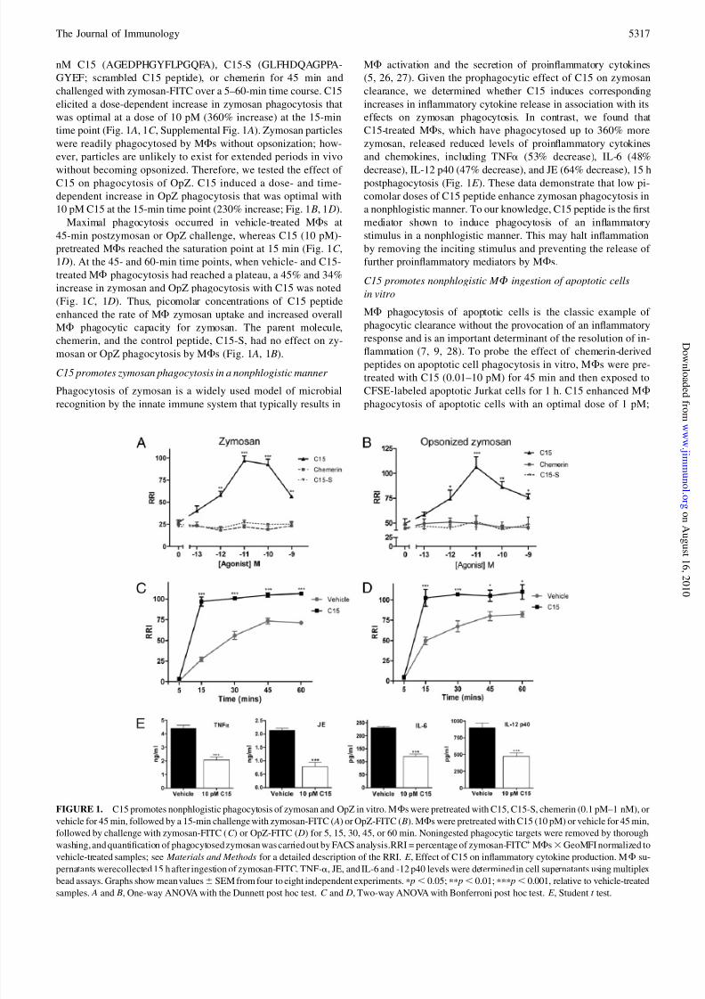
ResultsC15 promotes phagocytosis of zymosan and OpZ in vitro
To determine the effect of C15 and control agents on MF
phagocytosis of zymosan, MFs were pretreated with 0.1 pM–1
5316 CHEMERIN PEPTIDES ARE POTENT PROPHAGOCYTIC MEDIATORS
g ,
j
g
8/8/2019 Chem Erin
http://slidepdf.com/reader/full/chem-erin 4/11
nM C15 (AGEDPHGYFLPGQFA), C15-S (GLFHDQAGPPA-
GYEF; scrambled C15 peptide), or chemerin for 45 min and
challenged with zymosan-FITC over a 5–60-min time course. C15
elicited a dose-dependent increase in zymosan phagocytosis that
was optimal at a dose of 10 pM (360% increase) at the 15-min
time point (Fig. 1 A, 1C , Supplemental Fig. 1 A). Zymosan particles
were readily phagocytosed by MFs without opsonization; how-
ever, particles are unlikely to exist for extended periods in vivo
without becoming opsonized. Therefore, we tested the effect of C15 on phagocytosis of OpZ. C15 induced a dose- and time-
dependent increase in OpZ phagocytosis that was optimal with
10 pM C15 at the 15-min time point (230% increase; Fig. 1 B, 1 D).
Maximal phagocytosis occurred in vehicle-treated MFs at
45-min postzymosan or OpZ challenge, whereas C15 (10 pM)-
pretreated MFs reached the saturation point at 15 min (Fig. 1C ,
1 D). At the 45- and 60-min time points, when vehicle- and C15-
treated MF phagocytosis had reached a plateau, a 45% and 34%
increase in zymosan and OpZ phagocytosis with C15 was noted
(Fig. 1C , 1 D). Thus, picomolar concentrations of C15 peptide
enhanced the rate of MF zymosan uptake and increased overall
MF phagocytic capacity for zymosan. The parent molecule,
chemerin, and the control peptide, C15-S, had no effect on zy-
mosan or OpZ phagocytosis by MFs (Fig. 1 A, 1 B).
C15 promotes zymosan phagocytosis in a nonphlogistic manner
Phagocytosis of zymosan is a widely used model of microbial
recognition by the innate immune system that typically results in
MF activation and the secretion of proinflammatory cytokines
(5, 26, 27). Given the prophagocytic effect of C15 on zymosan
clearance, we determined whether C15 induces corresponding
increases in inflammatory cytokine release in association with its
effects on zymosan phagocytosis. In contrast, we found that
C15-treated MFs, which have phagocytosed up to 360% more
zymosan, released reduced levels of proinflammatory cytokines
and chemokines, including TNFa (53% decrease), IL-6 (48%
decrease), IL-12 p40 (47% decrease), and JE (64% decrease), 15 hpostphagocytosis (Fig. 1E ). These data demonstrate that low pi-
comolar doses of C15 peptide enhance zymosan phagocytosis in
a nonphlogistic manner. To our knowledge, C15 peptide is the first
mediator shown to induce phagocytosis of an inflammatory
stimulus in a nonphlogistic manner. This may halt inflammation
by removing the inciting stimulus and preventing the release of
further proinflammatory mediators by MFs.
C15 promotes nonphlogistic M F ingestion of apoptotic cells
in vitro
MF phagocytosis of apoptotic cells is the classic example of
phagocytic clearance without the provocation of an inflammatory
response and is an important determinant of the resolution of in-
flammation (7, 9, 28). To probe the effect of chemerin-derivedpeptides on apoptotic cell phagocytosis in vitro, MFs were pre-
treated with C15 (0.01–10 pM) for 45 min and then exposed to
CFSE-labeled apoptotic Jurkat cells for 1 h. C15 enhanced MF
phagocytosis of apoptotic cells with an optimal dose of 1 pM;
FIGURE 1. C15 promotes nonphlogistic phagocytosis of zymosan and OpZ in vitro. MFs were pretreated with C15, C15-S, chemerin (0.1 pM–1 nM), or
vehicle for 45 min, followed by a 15-min challenge with zymosan-FITC ( A) or OpZ-FITC ( B). MFs were pretreated with C15 (10 pM) or vehicle for 45 min,
followed by challenge with zymosan-FITC (C ) or OpZ-FITC ( D) for 5, 15, 30, 45, or 60 min. Noningested phagocytic targets were removed by thorough
washing, and quantification of phagocytosed zymosan was carried out by FACS analysis.RRI = percentage of zymosan-FITC+ MFs3GeoMFI normalized to
vehicle-treated samples; see Materials and Methods for a detailed description of the RRI. E , Effect of C15 on inflammatory cytokine production. MF su-
pernatants werecollected 15 h after ingestion of zymosan-FITC. TNF-a, JE, and IL-6 and -12 p40 levels were determined in cell supernatants using multiplex
bead assays. Graphs show mean values6 SEM from four to eight independent experiments. p p, 0.05; pp p, 0.01; ppp p, 0.001, relative to vehicle-treated
samples. A and B, One-way ANOVA with the Dunnett post hoc test. C and D, Two-way ANOVA with Bonferroni post hoc test. E , Student t test.
The Journal of Immunology 5317
g ,
j
g

8/8/2019 Chem Erin
http://slidepdf.com/reader/full/chem-erin 5/11
a 247% increase in phagocytosis was observed (Fig. 2 A, Supple-
mental Fig. 1 B). However, similar prophagocytic effects were still
seen with 0.1 pM C15: doses that are 103 and 1003 lower than
that required for optimal enhancement of zymosan and OpZ
phagocytosis (Fig. 1 A, 1 B). We postulate that reduced C15 con-
centrations are required for optimal enhancement of apoptotic cell
phagocytosis because C15 may act in concert with prophagocytic
mediators that are known to be released by apoptotic cells, in-
cluding nucleotides and Annexin A1 (29–31). Intriguingly, theparent protein chemerin, which failed to enhance zymosan in-
gestion (Fig. 1 A), promoted phagocytosis of apoptotic cells with an
optimal dose of 0.1 pM (Fig. 2 B). These prophagocytic effects were
abolished when chemerin was administered in the presence of the
serine and cysteine protease inhibitor (leupeptin), demonstrating
that chemerin promotes phagocytosis in a proteolysis-dependent
manner (Fig. 2 B). These data mirror our previous observations that
chemerin suppresses MF activation in a proteolysis-dependent
manner (25), suggesting that classically activated MFs and apo-
ptotic cells are capable of releasing proteases that cleave chemerin
to generate anti-inflammatory and prophagocytic peptides. We
postulate that chemerin is cleaved during this assay by apoptotic
cell-derived proteases, because chemerin did not exert any pro-
phagocytic effects on zymosan and OpZ phagocytosis (Fig. 1 A,1 B). In addition, C15-treated MFs, which had ingested up to 2.5-
fold more apoptotic cells, released reduced levels of proin-
flammatory cytokines TNF-a (63%; Fig. 2C ) and JE (54%; Fig.
2 D) and exhibited increased TGF-b expression (108%; Fig. 2E )
with an optimal dose of 1 pM C15.
ChemR232 / 2
M Fs exhibit impaired phagocytosis in response
to C15
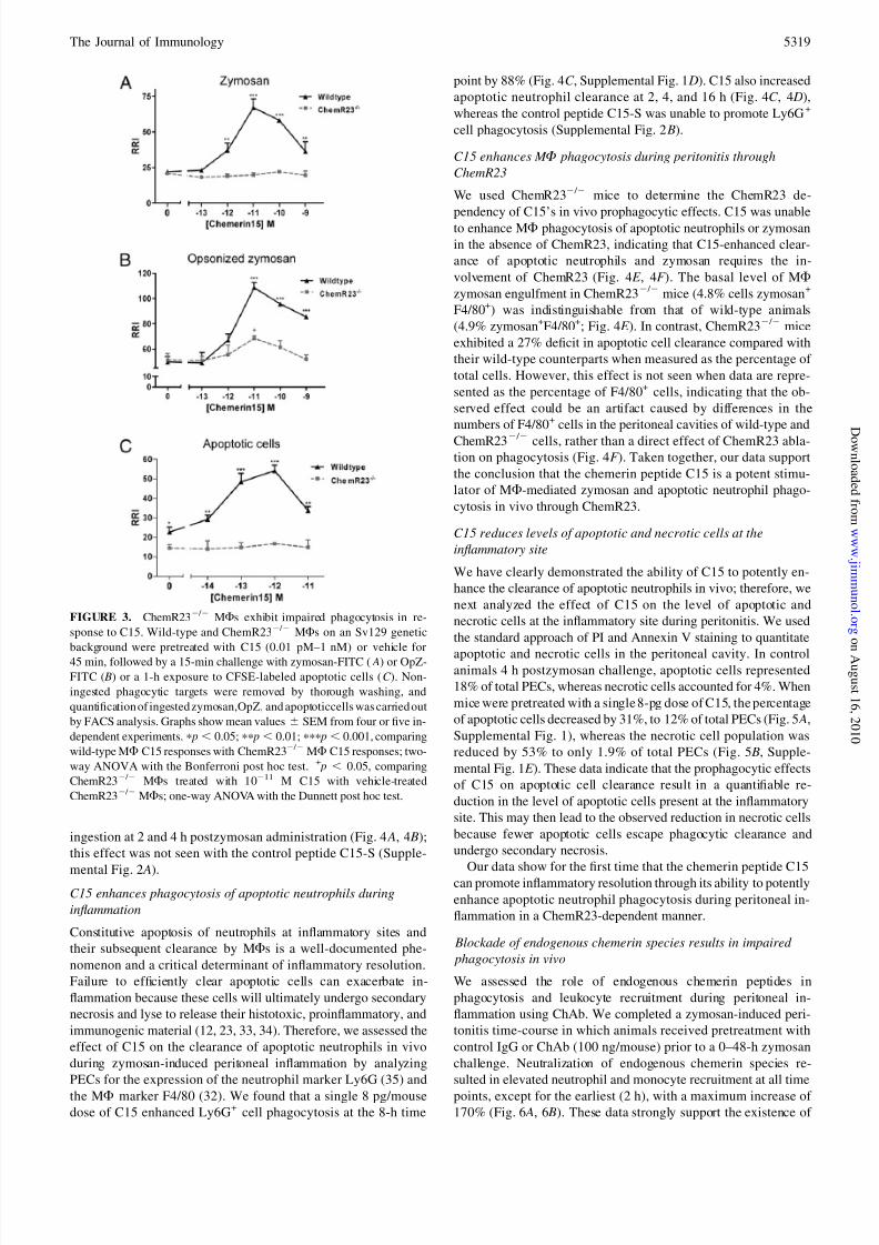
To assess the involvement of the G-protein coupled receptor
ChemR23 in mediating C15 enhancement of zymosan and OpZ
phagocytosis, we compared the effect of C15 on phagocytosis of
these targets by wild-typeand ChemR232 / 2MFsatthe15-mintime
point. The prophagocytic effects of C15 on zymosan phagocytosis
were completely abolished in ChemR232 / 2 MFs, indicating that
C15-enhanced clearance of zymosan requires the involvement of
ChemR23 (Fig. 3 A). In contrast, ChemR232 / 2 MFs displayed
a 71% reduction in C15 enhancement of OpZ phagocytosis at the
optimal 10-pM dose (Fig. 3 B). These data suggest the existence of
an additional C15R that may be required for optimal enhancement
of OpZ clearance in vitro (Fig. 3 B). Furthermore, the level of zy-
mosan (+/+,22RRI;2 / 2,20RRI;Fig.3 A)andOpZ(+/+,49RRI;2 / 2,
51 RRI; Fig. 3 B) phagocytosis in untreated wild-type andChemR232 / 2 MFs was of equivalent magnitude.
We next assessed the involvement of ChemR23 in mediating C15
enhancement of apoptotic cell clearance, finding that the pro-
phagocytic effects of C15 on apoptotic cell phagocytosis were
completely abolished in ChemR232 / 2 MFs, indicating that C15-
enhanced clearance of this phagocytic target also requires the
involvement of ChemR23 (Fig. 3C ). In addition, the basal level of
apoptotic cell ingestion in ChemR232 / 2 MFs (14.5 RRI) was
significantly lower than that of wild-type MFs (22.7 RRI), pro-
viding the first indication of a phenotype for ChemR232 / 2 MFs
in the absence of an inflammatory stimulus (25).
C15 enhances M F zymosan phagocytosis during peritonitis
We previously showed that C15 ameliorates zymosan-inducedperitonitis, reducing leukocyte recruitment by up to 65%, with
a concomitant suppression of inflammatory mediator expression
(25). Having demonstrated that C15 is capable of enhancing zy-
mosan phagocytosis in vitro, we next assessed the effect of C15
(8 pg/mouse; 0.32 ng/kg) on MF zymosan phagocytosis in vivo in
the zymosan peritonitis model. PECs were analyzed for uptake of
zymosan-FITC and expression of the MF marker F4/80 (32). The
maximum level of MFs engaged in the clearance of zymosan
occurred in control animals 1 h postzymosan challenge, where
23% of total cells were zymosan+F4/80+ (Fig. 4 A, Supplemental
Fig. 1C ). A single 8-pg dose of C15 enhanced MF zymosan
clearance by 38%, increasing the percentage of zymosan+F4/80+
PECs to 34% (Fig. 4 A). C15 also enhanced MF zymosan
FIGURE 2. C15 and chemerin promote nonphlogistic phagocytosis of apoptotic cells in vitro. MFs were pretreated with vehicle, C15, or C15-S (0.01–10
pM) ( A) or chemerin (0.01–10 pM) 6 leupeptin (10 mM) ( B) for 45 min and then exposed to CFSE-labeled apoptotic Jurkat cells for 1 h. C –E , Effect of C15
on MF cytokine production following ingestion of apoptotic cells. TNF-a (C ), JE ( D), TGF-b (E ), IL-6, and IL-12 p40 levels were determined in cell
supernatants 15 or 24 h (TGF-b) following phagocytosis of apoptotic cells using multiplex bead assays and ELISA. IL-6 and -12 p40 levels were below the
limit of detection for this assay (5 pg/ml). Noningested CFSE-labeled apoptotic cells and cell debris were removed by thorough washing, and quantification of
ingested CFSE-labeled apoptotic cells was carried out by FACS analysis. Bar graphs show mean values 6 SEM from four independent experiments. p p ,
0.05; pp p, 0.01; ppp p, 0.001; relative to vehicle-treated ( A, C –E ) or leupeptin-treated ( B) samples. A, One-way ANOVA with the Dunnett post hoc test. B,
Two-way ANOVA with Bonferroni post hoc test. C –E , Student t test.
5318 CHEMERIN PEPTIDES ARE POTENT PROPHAGOCYTIC MEDIATORS
g ,
j
g
8/8/2019 Chem Erin
http://slidepdf.com/reader/full/chem-erin 6/11
ingestion at 2 and 4 h postzymosan administration (Fig. 4 A, 4 B);
this effect was not seen with the control peptide C15-S (Supple-
mental Fig. 2 A).C15 enhances phagocytosis of apoptotic neutrophils during
inflammation
Constitutive apoptosis of neutrophils at inflammatory sites and
their subsequent clearance by MFs is a well-documented phe-
nomenon and a critical determinant of inflammatory resolution.
Failure to efficiently clear apoptotic cells can exacerbate in-
flammation because these cells will ultimately undergo secondary
necrosis and lyse to release their histotoxic, proinflammatory, and
immunogenic material (12, 23, 33, 34). Therefore, we assessed the
effect of C15 on the clearance of apoptotic neutrophils in vivo
during zymosan-induced peritoneal inflammation by analyzing
PECs for the expression of the neutrophil marker Ly6G (35) and
the MF marker F4/80 (32). We found that a single 8 pg/mousedose of C15 enhanced Ly6G+ cell phagocytosis at the 8-h time
point by 88% (Fig. 4C , Supplemental Fig. 1 D). C15 also increased
apoptotic neutrophil clearance at 2, 4, and 16 h (Fig. 4C , 4 D),
whereas the control peptide C15-S was unable to promote Ly6G+
cell phagocytosis (Supplemental Fig. 2 B).
C15 enhances M F phagocytosis during peritonitis through
ChemR23
We used ChemR232 / 2 mice to determine the ChemR23 de-
pendency of C15’s in vivo prophagocytic effects. C15 was unableto enhance MF phagocytosis of apoptotic neutrophils or zymosan
in the absence of ChemR23, indicating that C15-enhanced clear-
ance of apoptotic neutrophils and zymosan requires the in-
volvement of ChemR23 (Fig. 4E , 4F ). The basal level of MF
zymosan engulfment in ChemR232 / 2 mice (4.8% cells zymosan+
F4/80+) was indistinguishable from that of wild-type animals
(4.9% zymosan+F4/80+; Fig. 4E ). In contrast, ChemR232 / 2 mice
exhibited a 27% deficit in apoptotic cell clearance compared with
their wild-type counterparts when measured as the percentage of
total cells. However, this effect is not seen when data are repre-
sented as the percentage of F4/80+ cells, indicating that the ob-
served effect could be an artifact caused by differences in the
numbers of F4/80+ cells in the peritoneal cavities of wild-type and
ChemR232 / 2 cells, rather than a direct effect of ChemR23 abla-tion on phagocytosis (Fig. 4F ). Taken together, our data support
the conclusion that the chemerin peptide C15 is a potent stimu-
lator of MF-mediated zymosan and apoptotic neutrophil phago-
cytosis in vivo through ChemR23.
C15 reduces levels of apoptotic and necrotic cells at the
inflammatory site
We have clearly demonstrated the ability of C15 to potently en-
hance the clearance of apoptotic neutrophils in vivo; therefore, we
next analyzed the effect of C15 on the level of apoptotic and
necrotic cells at the inflammatory site during peritonitis. We used
the standard approach of PI and Annexin V staining to quantitate
apoptotic and necrotic cells in the peritoneal cavity. In controlanimals 4 h postzymosan challenge, apoptotic cells represented
18% of total PECs, whereas necrotic cells accounted for 4%. When
mice were pretreated with a single 8-pg dose of C15, the percentage
of apoptotic cells decreased by 31%, to 12% of total PECs (Fig. 5 A,
Supplemental Fig. 1), whereas the necrotic cell population was
reduced by 53% to only 1.9% of total PECs (Fig. 5 B, Supple-
mental Fig. 1E ). These data indicate that the prophagocytic effects
of C15 on apoptotic cell clearance result in a quantifiable re-
duction in the level of apoptotic cells present at the inflammatory
site. This may then lead to the observed reduction in necrotic cells
because fewer apoptotic cells escape phagocytic clearance and
undergo secondary necrosis.
Our data show for the first time that the chemerin peptide C15can promote inflammatory resolution through its ability to potently
enhance apoptotic neutrophil phagocytosis during peritoneal in-
flammation in a ChemR23-dependent manner.
Blockade of endogenous chemerin species results in impaired
phagocytosis in vivo
We assessed the role of endogenous chemerin peptides in
phagocytosis and leukocyte recruitment during peritoneal in-
flammation using ChAb. We completed a zymosan-induced peri-
tonitis time-course in which animals received pretreatment with
control IgG or ChAb (100 ng/mouse) prior to a 0–48-h zymosan
challenge. Neutralization of endogenous chemerin species re-
sulted in elevated neutrophil and monocyte recruitment at all time
points, except for the earliest (2 h), with a maximum increase of 170% (Fig. 6 A, 6 B). These data strongly support the existence of
FIGURE 3. ChemR232 / 2 MFs exhibit impaired phagocytosis in re-
sponse to C15. Wild-type and ChemR232 / 2 MFs on an Sv129 genetic
background were pretreated with C15 (0.01 pM–1 nM) or vehicle for
45 min, followed by a 15-min challenge with zymosan-FITC ( A) or OpZ-
FITC ( B) or a 1-h exposure to CFSE-labeled apoptotic cells (C ). Non-
ingested phagocytic targets were removed by thorough washing, and
quantification of ingested zymosan,OpZ, and apoptoticcells was carried out
by FACS analysis. Graphs show mean values 6 SEM from four or five in-
dependent experiments. p p , 0.05; pp p , 0.01; ppp p , 0.001, comparing
wild-type MF C15 responses with ChemR232 / 2 MF C15 responses; two-
way ANOVA with the Bonferroni post hoc test. + p , 0.05, comparing
ChemR232 / 2 MFs treated with 10211 M C15 with vehicle-treated
ChemR232 / 2 MFs; one-way ANOVA with the Dunnett post hoc test.
The Journal of Immunology 5319
g ,
j
g

8/8/2019 Chem Erin
http://slidepdf.com/reader/full/chem-erin 7/11
anti-inflammatory chemerin-derived peptides in vivo that are in-
volved in modulating leukocyte recruitment during peritoneal
inflammation. In light of the potent prophagocytic effects of the
chemerin peptide C15 on zymosan and apoptotic cell phagocy-
tosis, we postulated that endogenous chemerin peptides maymodulate the inflammatory response by promoting clearance of
these phagocytic targets, in addition to suppressing leukocyte re-
cruitment. Neutralization of endogenous chemerin species re-
sulted in a reduction of up to 46% in zymosan phagocytosis
(Fig. 6C , 6 D) and Ly6G+ cell (neutrophil) phagocytosis (Fig. 6E ,
6F ). These data provide evidence that endogenous chemerin
species are an important component of the endogenous resolution
system, involved in mediating the clearance of microbial particles
and apoptotic cells in vivo.
C15 enhances phagocytosis and phagocytic cup formation
through Syk-dependent changes in F-actin polymerization
Because C15 enhances the phagocytosis of multiple phagocytic
targets, which are cargo for a variety of MF receptors, includingdectin-1 for zymosan ingestion (36) and CD36 for apoptotic cell
engulfment (37), it is likely that C15 enhances a common pathway
of phagocytosis rather than having an effect on specific phagocytic
receptor expression. This is particularly likely because FACS
analysis showed no significant C15-elicited changes in dectin-1
expression during the course of the zymosan phagocytosis assay(data not shown), and C15 exerted no effect on MF binding of
zymosan, as gauged by phagocytosis assays performed at 4˚C
(Supplemental Fig. 3).
Remodeling of the actin cytoskeleton is a prerequisite for
phagocytosis, enabling the formation of phagocytic cups and,
subsequently, the phagosome to internalize the phagocytic target
(38–41). Therefore, we evaluated the effect of C15 on phagocytic
cup formation and F-actin polymerization and localization. In
addition, we further probed the mechanism by which C15 pro-
motes phagocytosis using the Syk inhibitor PIC. Syk is a tyrosine
kinase required for lysosome–phagosome fusion in unopsonized
yeast phagocytosis (42), but it is not required for yeast or apo-
ptotic cell internalization (43–45). In agreement with these ob-
servations, Syk inhibition had no effect on basal MF zymosan orapoptotic cell phagocytosis (Fig. 7 A, 7 B). However, pretreatment
FIGURE 4. C15 enhances MF phagocytosis of zymosan and apoptotic neutrophils during peritonitis through ChemR23. A and B, C57BL/6 mice were
dosed i.p with vehicle or C15 (0.32 ng/kg), followed by injection with zymosan-FITC 1 h later. PECs were harvested by peritoneal lavage at multiple time
points postzymosan-FITC injection, permeabilized, and stained with anti-F4/80-Alexa Fluor 647. Zymosan+F4/80+ cells are expressed as the percentage of
total cells ( A) and as percentage of MFs (F4/80+ cells) ( B), as determined by FACS analysis. C and D, C57BL/6 mice were dosed i.p with vehicle or C15
(0.32 ng/kg), followed by injection with zymosan 1 h later. PECs were harvested by peritoneal lavage at multiple time points postzymosan injection,permeabilized, and stained with anti–Ly6G-PE and anti–F4/80-Alexa Fluor 647. Ly6G+F4/80+ cells are expressed as the percentage of total cells (C ) and as
the percentage of MFs (F4/80+ cells) ( D), as determined by FACS analysis. Graphs in A– D show mean values6 SEM, with 5–10 mice per treatment group
(vehicle or C15) used at each time point. p p , 0.05; pp p , 0.01; ppp p , 0.001, relative to vehicle-treated mice; two-way ANOVA with Bonferroni post
hoc test. Sv129 and ChemR232 / 2 (Sv129) mice were dosed i.p with vehicle or C15 (0.32 ng/kg), followed by injection with zymosan-FITC (E ) or zymosan
(F ) 1 h later. PECs were harvested by peritoneal lavage 4 h postzymosan-FITC administration to quantify zymosan +F4/80+ populations and 8 h post-
zymosan administration to evaluate Ly6G+F4/80+ cell populations. PECs were permeabilized, stained, and analyzed as described in A– D. Graphs show
mean values 6 SEM for 6–12 mice per group. pp p , 0.01; ppp p , 0.001; ++ p , 0.01, relative to vehicle-treated wild-type mice.
5320 CHEMERIN PEPTIDES ARE POTENT PROPHAGOCYTIC MEDIATORS
g ,
j
g

8/8/2019 Chem Erin
http://slidepdf.com/reader/full/chem-erin 8/11
of MFs with PIC prior to C15 treatment resulted in complete
abrogation of C15-induced enhancement of zymosan (Fig. 7 A)
and apoptotic cell phagocytosis (Fig. 7 B). Therefore, these datademonstrate that C15 enhances zymosan and apoptotic cell
phagocytosis in a Syk-dependent manner.
Subsequently, we probed the effect of C15 on phagocytic cup
formation, finding that C15 treatment elicited a significant increase
in the percentage of cells with phagocytic cups, from 25% of cells
with vehicle treatment up to 40% of cells following treatment with
10 pM C15. This increase in phagocytic cup formation was
completely abrogated by treatment with the Syk inhibitor PIC
(Fig. 7C ). Importantly, vehicle-treated and C15 + PIC-treated
MFs predominantly possessed early phagocytic cups, whereas
higher levels of early phagosomes and late phagocytic cups were
observed following C15 treatment (Supplemental Fig. 4). These
data suggest that C15 may increase the kinetics of phagosome
formation in a Syk-dependent manner. C15 also enhanced F-actin
polymerization at the early phagocytic cup (Fig. 7 D), the late
phagocytic cup (Fig. 7E ), and the early phagosome (Fig. 7F ). C15-
mediated alterations in local F-actin polymerization were in-
hibited by PIC, indicating an essential role for Syk in this process(Fig. 7 D–F ). In light of these observations, we assessed the effect
of C15 on Syk activation, finding that C15 (10 pM) triggered
phosphorylation of Syk (Tyr352) in a time-dependent manner
(Supplemental Fig. 5). Since phagocytosis, phagocytic cup for-
mation, and F-actin polymerization at the phagocytic cups and
early phagosomes were enhanced by C15, and C15-mediated
effects were abrogated by the Syk inhibitor PIC, we suggest that
C15 enhances phagocytosis through Syk-mediated alterations in
F-actin polymerization, which likely aid phagocytic cup formation
and, thus, enhances MF phagocytosis.
Discussion
In this study, we identify the chemerin peptide C15 as a newmediator capable of promoting inflammatory resolution following
microbial challenge by stimulating MF clearance of the inciting
stimulus and apoptotic neutrophils in a nonphlogistic manner.
Efficient clearance of invading microorganisms at sites of in-
flammation is an indispensible role of MFs. The ability of C15 to
potently enhance this process, in vitro and in vivo, further high-
lights the dynamic nature of the MF and its capacity for phago-
cytosis while providing novel insights into the regulation of innate
immune responses by endogenous anti-inflammatory pathways.
FIGURE 6. Neutralization of endoge-nous chemerin species results in elevated
leukocyte recruitment and impaired phago-
cytosis in vivo. C57BL/6J mice were dosed
i.p with ChAb (100 ng/mouse) or control
IgG (100 ng/mouse), followed by injection
with zymosan (10 mg/cavity) 1 h later.
PECs were harvested at multiple time
points postzymosan injection ( A, B, E , F ) or
postzymosan-FITC injection (C , D). A and
B, Total PECs were quantified, and cellular
composition (neutrophils versus mono-
cytes) was determined by FACS analysis.
Cells were stained with Ly6G-PE and 7/4-
FITC, and gates were constructed around
two populations: neutrophils ( A; 7/4high
Ly6Ghigh) and monocytes ( B; 7/4highLy6-
Glow). C and D, PECs were permeabilized
and stained with anti–F4/80-Alexa Fluor
647, and the percentage of zymosan+F4/80+
cells was determined by FACS analysis. E
and F , PECs were permeabilized and
stained with anti–Ly6G-PE and anti–F4/80-
Alexa Fluor 647, and the percentage of
Ly6G+, F4/80+ cells was determined
by FACS analysis. Graphs show mean val-
ues 6 SEM for 5–10 mice per treatment
group (IgG control or ChAb) per time point.
p p , 0.05; pp p , 0.01; ppp p , 0.001,
relative to control IgG-treated mice. Two-way ANOVA with Bonferroni post hoc test.
FIGURE 5. C15 reduces levels of apoptotic and necrotic cells at the in-
flammatory site. A and B, C57BL/6 mice were dosed i.p with vehicle or C15
(0.32ng/kg),followed by injection with10 mg zymosan 1 h later. PECs were
harvested by peritoneal lavage 4 h postzymosan challenge, and the per-
centages of live, apoptotic ( A) and necrotic ( B) cells were determined by
staining with annexin V-FITC and propidium iodide. Live cells were An-
nexin V2PI2, apoptotic cells were Annexin+PI2, and necrotic cells were
Annexin V+PI+. Graphsshowmeanvalues6 SEM for five tosevenmice per
treatment group. pp p, 0.01, relative to vehicle-treated mice; Student t test.
The Journal of Immunology 5321
g ,
j
g

8/8/2019 Chem Erin
http://slidepdf.com/reader/full/chem-erin 9/11
Furthermore, this study indicates that manipulation of the chemerin
peptide/ChemR23 axis may be an attractive avenue for therapeuticintervention in inflammation.
Perhaps the most striking characteristic of C15 is the dose at
which it exerts its effects. C15 suppresses leukocyte recruitment
by up to 65% (25) and enhances phagocytosis in vivo by up to
100% when administered in low picogram quantities. The lipid-
resolution mediators resolvin E1, protectin D1, and lipoxin A4
exert less dramatic effects at much higher doses (27). Although
these mediators enhance zymosan phagocytosis (27), it is un-
known whether this occurs in a nonphlogistic manner, whereas the
prototypical anti-inflammatory drug dexamethasone inhibits MF
zymosan phagocytosis (46). To our knowledge, C15 peptide is the
first mediator shown to induce MF phagocytosis of an in-
flammatory stimulus in a nonphlogistic manner. This may halt
inflammation by removing the inciting stimulus and preventingthe release of further proinflammatory mediators by MFs.
Interestingly, the dose required for optimal enhancement of
apoptotic cell phagocytosis in vitro by C15 was 10-fold lower thanthat required for other phagocytic targets. This observation may
reflect differences in the nature of these assays; phagocytosis of
zymosan can be viewed as a proinflammatory event because MF
activation and the release of proinflammatory mediators ensues,
whereas apoptotic cell engulfment is a nonphlogistic process.
Alternatively, apoptotic cells were shown to release prophagocytic
signals, including Annexin A1 and Annexin-derived peptides
(30), which could act in concert with the C15 peptide administered
in this assay to promote MF clearance of apoptotic cells.
We have described for the first time a mechanism through which
C15 enhances MF phagocytosis, which involves Syk-dependent
changes in phagocytic cup formation and F-actin localization.
Syk 2 / 2 MFs, along with DAP122 / 2 MFs, have been shown to
display a phenotype characterized by elevated proinflammatorycytokine production in comparison with their wild-type counterparts
FIGURE 7. C15 enhances phagocytosis and phagocytic cup formation through Syk-dependent changes in F-actin polymerization. A and B, MFs were
pretreated with vehicle or PIC (10 mM) for 30 min followed by treatment with C15 (10214–1029 M) or vehicle for 45 min. MFs were subsequently
challenged with zymosan-FITC for 15 min ( A) or CFSE-labeled apoptotic Jurkat cells for 1 h ( B). Samples were processed as in Fig. 1. C –F , MFs were
pretreated with vehicle or PIC for 30 min, followed by treatment with C15 (10 pM) or vehicle for 45 min and subsequent challenge with zymosan-FITC for
15 min (C ) or 5 min ( D–F ). Cells were fixed and stained with Alexa Fluor 546-phalloidin to visualize polymerized actin, phagocytic cups, and phagosomes
and viewed using a 360 magnification. C , Early and late phagocytic cups were quantified and expressed as the percentage of total cells. Representative
images of early phagocytic cups ( D), late phagocytic cups (E ), and early phagosomes (F ) are shown. Figures show mean values 6 SEM from three in-
dependent experiments. A and B, pp p , 0.01; ppp p , 0.001, relative to PIC-treated samples; two-way ANOVA with the Bonferroni post hoc test. C , pp p,
0.01, relative to vehicle-treated samples; # p, 0.05, relative to C15-treated samples. One-way ANOVA with the Bonferroni posttest. Results were confirmed
with a second Syk inhibitor BAY 61-3606 (data not shown).
5322 CHEMERIN PEPTIDES ARE POTENT PROPHAGOCYTIC MEDIATORS
g ,
j
g
8/8/2019 Chem Erin
http://slidepdf.com/reader/full/chem-erin 10/11
(47). It is plausible that C15 and endogenous chemerin species may
harness this endogenous DAP12/Syk signaling pathway to elicit
their anti-inflammatory and prophagocytic effects. If this is the
case, 0Syk-deficient mice could be rendered unresponsive to C15
treatment.
We have demonstrated that the anti-inflammatory and propha-
gocytic effects of C15 are absent in ChemR232 / 2 MFs and
ChemR232 / 2 mice, but we were unable to detect a significant
difference between zymosan-elicited neutrophil and monocyte re-cruitment in wild-type and ChemR232 / 2 at the 4-h time point (25).
Therefore, it is intriguing that ChemR23 ablation also has no dis-
cernable effect on phagocytosis in vitro or in vivo, when we have
clearly demonstrated that neutralization of endogenous chemerin
species results in deficits in zymosan and apoptotic neutrophil
phagocytosis. Our previous studies demonstrated that the full-
length protein chemerin is cleaved into anti-inflammatory peptides
that act on ChemR23, in addition to other unidentified receptor(s)
(25). Thus, the absence of impaired phagocytosis in ChemR232 / 2
mice may be due to redundancy in the receptors for chemerin
peptides in the anti-inflammatory/proresolution system. Additional
receptors for chemerin, and potentially the chemerin peptides, are
GPR1 and CCRL2 (48, 49). Therefore, ablation of the chemerin
gene (RARRES-2) may result in mice with a more overt phenotypethan the ChemR232 / 2 mice through the removal of chemerin-de-
rived ligands for ChemR23 and other potential receptors.
Taken together, our novel data suggest that C15’s potent anti-
inflammatory effects in vivo are a result of a combination of direct
suppression of MF activation (25) and enhancement of MF
phagocytosis of the inciting stimulus and apoptotic neutrophils.
We provide compelling evidence for the role of chemerin peptides
and ChemR23 in MF phagocytosis and the resolution of in-
flammation. Therefore, manipulation of the chemerin peptide/
ChemR23 axis may represent a novel therapeutic approach for the
treatment of inflammatory pathologies, such as atherosclerosis and
SLE, in which failure to efficiently clear apoptotic cell debris has
been implicated in their pathogenesis (50).
AcknowledgmentsWe thank E. McNeil, P. Taylor, and G. White for advice.
DisclosuresThe authors have no financial conflicts of interest.
References1. Metchnikoff, E. 1887. Ueber den Kampf der Zellen gegen Erysipelkokken, ein
Beitrag zur Phagocytenlehre. Arch. Pathol. Anat. [Virchow’s Archiv.] 107: 209–
249.2. Gordon, S. 2007. The macrophage: past, present and future.Eur. J. Immunol. 37
(Suppl. 1): S9–S17.3. Mosser, D. M., and J. P. Edwards. 2008. Exploring the full spectrum of mac-
rophage activation. Natl. Rev. Immunol. 8: 958–969.4. Rossi, A. G., D. A. Sawatzky, A. Walker, C. Ward, T. A. Sheldrake, N. A. Riley,
A. Caldicott, M. Martinez-Losa, T. R. Walker, R. Duffin, et al. 2006. Cyclin-
dependent kinase inhibitors enhance the resolution of inflammation by pro-
moting inflammatory cell apoptosis. Nat. Med. 12: 1056–1064.5. Taylor, P. R., S. V. Tsoni, J. A. Willment, K. M. Dennehy, M. Rosas, H. Findon,
K. Haynes, C. Steele, M. Botto, S. Gordon, and G. D. Brown. 2007. Dectin-1 is
required for beta-glucan recognition and control of fungal infection. Nat. Im-
munol. 8: 31–38.6. Fadok, V. A., P. P. McDonald, D. L. Bratton, and P. M. Henson. 1998. Regulation
of macrophage cytokine production by phagocytosis of apoptotic and post-
apoptotic cells. Biochem. Soc. Trans. 26: 653–656.7. Fadok, V. A., D. L. Bratton, A. Konowal, P. W. Freed, J. Y. Westcott, and
P. M. Henson. 1998. Macrophages that have ingested apoptotic cells in vitro
inhibit proinflammatory cytokine production through autocrine/paracrine
mechanisms involving TGF-beta, PGE2, and PAF. J. Clin. Invest. 101: 890–898.
8. Fadok, V. A., D. L. Bratton, and P. M. Henson. 2001. Phagocyte receptorsfor apoptotic cells: recognition, uptake, and consequences. J. Clin. Invest. 108:
957–962.
9. Huynh, M. L., V. A. Fadok, and P. M. Henson. 2002. Phosphatidylserine-dependent ingestion of apoptotic cells promotes TGF-beta1 secretion and theresolution of inflammation. J. Clin. Invest. 109: 41–50.
10. Neumann, J., S. Sauerzweig, R. Ronicke, F. Gunzer, K. Dinkel, O. Ullrich,M. Gunzer, and K. G. Reymann. 2008. Microglia cells protect neurons by directengulfment of invading neutrophil granulocytes: a new mechanism of CNSimmune privilege. J. Neurosci. 28: 5965–5975.
11. Silva, M. T., A. do Vale, and N. M. dos Santos. 2008. Secondary necrosis inmulticellular animals: an outcome of apoptosis with pathogenic implications.
Apoptosis 13: 463–482.12. Savill, J., I. Dransfield, C. Gregory, and C. Haslett. 2002. A blast from the past:
clearance of apoptotic cells regulates immune responses. Natl. Rev. Immunol. 2:965–975.
13. Vandivier, R. W., P. M. Henson, and I. S. Douglas. 2006. Burying the dead: theimpact of failed apoptotic cell removal (efferocytosis) on chronic inflammatorylung disease. Chest 129: 1673–1682.
14. Serhan, C. N., and J. Savill. 2005. Resolution of inflammation: the beginningprograms the end. Nat. Immunol. 6: 1191–1197.
15. Haslett, C. 1992. Resolution of acute inflammation and the role of apoptosis inthe tissue fate of granulocytes. Clin. Sci. (Lond.) 83: 639–648.
16. Zhang, Z., G. Cherryholmes,and J. E. Shively. 2008. Neutrophil secondarynecrosisis induced by LL-37 derived from cathelicidin. J. Leukoc. Biol. 84: 780–788.
17. Rock, K. L., and H. Kono. 2008. The inflammatory response to cell death.Annu.
Rev. Pathol. 3: 99–126.18. O’Brien, B. A., W. E. Fieldus, C. J. Field, and D. T. Finegood. 2002. Clearance
of apoptotic beta-cells is reduced in neonatal autoimmune diabetes-prone rats.Cell Death Differ. 9: 457–464.
19. Cohen, P. L., R. Caricchio, V. Abraham, T. D. Camenisch, J. C. Jennette,R. A. Roubey, H. S. Earp, G. Matsushima, and E. A. Reap. 2002. Delayed ap-optotic cell clearance and lupus-like autoimmunity in mice lacking the c-mer
membrane tyrosine kinase. J. Exp. Med. 196: 135–140.20. Munoz, L. E., U. S. Gaipl, S. Franz, A. Sheriff, R. E. Voll, J. R. Kalden, and
M. Herrmann. 2005. SLE—a disease of clearance deficiency? Rheumatology
(Oxford) 44: 1101–1107.21. Gaipl, U. S., R. E. Voll, A. Sheriff, S. Franz, J. R. Kalden, and M. Herrmann.
2005. Impaired clearance of dying cells in systemic lupus erythematosus. Au-
toimmun. Rev. 4: 189–194.22. Gaipl, U. S., L. E. Munoz, G. Grossmayer, K. Lauber, S. Franz, K. Sarter,
R. E. Voll, T. Winkler, A. Kuhn, J. Kalden, et al. 2007. Clearance deficiency andsystemic lupus erythematosus (SLE). J. Autoimmun. 28: 114–121.
23. Schrijvers, D. M., G. R. De Meyer, M. M. Kockx, A. G. Herman, andW. Martinet. 2005. Phagocytosis of apoptotic cells by macrophages is impairedin atherosclerosis. Arterioscler. Thromb. Vasc. Biol. 25: 1256–1261.
24. Ait-Oufella, H., V. Pouresmail, T. Simon, O. Blanc-Brude, K. Kinugawa,R. Merval, G. Offenstadt, G. Leseche, P. L. Cohen, A. Tedgui, and Z. Mallat.2008. Defective mer receptor tyrosine kinase signaling in bone marrow cellspromotes apoptotic cell accumulation and accelerates atherosclerosis. Arte-
rioscler. Thromb. Vasc. Biol. 28: 1429–1431.
25. Cash, J. L., R. Hart, A. Russ, J. P. Dixon, W. H. Colledge, J. Doran,A. G. Hendrick, M. B. Carlton, and D. R. Greaves. 2008. Synthetic chemerin-derived peptides suppress inflammation through ChemR23. J. Exp. Med. 205:767–775.
26. Damazo, A. S., S. Yona, R. J. Flower, M. Perretti, and S. M. Oliani. 2006. Spatialand temporal profiles for anti-inflammatory gene expression in leukocytes duringa resolving model of peritonitis. J. Immunol. 176: 4410–4418.
27. Schwab, J. M., N. Chiang, M. Arita, and C. N. Serhan. 2007. Resolvin E1and protectin D1 activate inflammation-resolution programmes. Nature 447:869–874.
28. Ren, Y., and J. Savill. 1998. Apoptosis: the importance of being eaten. Cell
Death Differ. 5: 563–568.29. Maderna, P., S. Yona, M. Perretti, and C. Godson. 2005. Modulation of
phagocytosis of apoptotic neutrophils by supernatant from dexamethasone-treated macrophages and annexin-derived peptide Ac(2-26). J. Immunol. 174:3727–3733.
30. Scannell, M., M. B. Flanagan, A. deStefani, K. J. Wynne, G. Cagney, C. Godson,and P. Maderna. 2007. Annexin-1 and peptide derivatives are released by apo-
ptotic cells and stimulate phagocytosis of apoptotic neutrophils by macrophages. J. Immunol. 178: 4595–4605.
31. Elliott, M. R., F. B. Chekeni, P. C. Trampont, E. R. Lazarowski, A. Kadl,S. F. Walk, D. Park, R. I. Woodson, M. Ostankovich, P. Sharma, et al. 2009.Nucleotides released by apoptotic cells act as a find-me signal to promotephagocytic clearance. Nature 461: 282–286.
32. McKnight, A. J., A. J. Macfarlane, P. Dri, L. Turley, A. C. Willis, and S. Gordon.1996. Molecular cloning of F4/80, a murine macrophage-restricted cell surfaceglycoprotein with homology to the G-protein-linked transmembrane 7 hormonereceptor family. J. Biol. Chem. 271: 486–489.
33. Baumann, I., W. Kolowos, R. E. Voll, B. Manger, U. Gaipl, W. L. Neuhuber,T. Kirchner, J. R. Kalden, and M. Herrmann. 2002. Impaired uptake of apoptoticcells into tingible body macrophages in germinal centers of patients with sys-temic lupus erythematosus. Arthritis Rheum. 46: 191–201.
34. Ren, Y., J. Tang, M. Y. Mok, A. W. Chan, A. Wu, and C. S. Lau. 2003. Increasedapoptotic neutrophils and macrophages and impaired macrophage phagocyticclearance of apoptotic neutrophils in systemic lupus erythematosus. Arthritis
Rheum. 48: 2888–2897.
35. Daley, J. M., A. A. Thomay, M. D. Connolly, J. S. Reichner, and J. E. Albina.2008. Use of Ly6G-specific monoclonal antibody to deplete neutrophils in mice.
J. Leukoc. Biol. 83: 64–70.
The Journal of Immunology 5323
g ,
j
g
8/8/2019 Chem Erin
http://slidepdf.com/reader/full/chem-erin 11/11
36. Brown, G. D., and S. Gordon. 2001. Immune recognition. A new receptor for
beta-glucans. Nature 413: 36–37.37. Fadok, V. A., M. L. Warner, D. L. Bratton, and P. M. Henson. 1998. CD36 is
required for phagocytosis of apoptotic cells by human macrophages that use
either a phosphatidylserine receptor or the vitronectin receptor (alpha v beta 3).
J. Immunol. 161: 6250–6257.38. Sheterline, P., J. E. Rickard, and R. C. Richards. 1984. Fc receptor-directed
phagocytic stimuli induce transient actin assembly at an early stage of phago-
cytosis in neutrophil leukocytes. Eur. J. Cell Biol. 34: 80–87.39. Greenberg, S., J. el Khoury, F. di Virgilio, E. M. Kaplan, and S. C. Silverstein.
1991. Ca(2+)-independent F-actin assembly and disassembly during Fc receptor-
mediated phagocytosis in mouse macrophages. J. Cell Biol. 113: 757–767.40. Greenberg, S. 1999. Modular components of phagocytosis. J. Leukoc. Biol. 66:
712–717.41. May, R. C., and L. M. Machesky. 2001. Phagocytosis and the actin cytoskeleton.
J. Cell Sci. 114: 1061–1077.42. Majeed, M., E. Caveggion, C. A. Lowell, and G. Berton. 2001. Role of Src
kinases and Syk in Fcgamma receptor-mediated phagocytosis and phagosome-
lysosome fusion. J. Leukoc. Biol. 70: 801–811.43. Canetti, C., B. Hu, J. L. Curtis, and M. Peters-Golden. 2003. Syk activation is
a leukotriene B4-regulated event involved in macrophage phagocytosis of IgG-
coated targets but not apoptotic cells. Blood 102: 1877–1883.
44. Herre, J., A. S. Marshall, E. Caron, A. D. Edwards, D. L. Williams,E. Schweighoffer, V. Tybulewicz, C. Reis e Sousa, S. Gordon, and G. D. Brown.2004. Dectin-1 uses novel mechanisms for yeast phagocytosis in macrophages.
Blood 104: 4038–4045.45. Underhill, D. M., E. Rossnagle, C. A. Lowell, and R. M. Simmons. 2005. Dectin-
1 activates Syk tyrosine kinase in a dynamic subset of macrophages for reactiveoxygen production. Blood 106: 2543–2550.
46. Mlambo, G., and L. B. Sigola. 2003. Rifampicin and dexamethasone havesimilar effects on macrophage phagocytosis of zymosan, but differ in their ef-fects on nitrite and TNF-alpha production. Int. Immunopharmacol. 3: 513–522.
47. Hamerman, J. A., N. K. Tchao, C. A. Lowell, and L. L. Lanier. 2005. Enhancedtoll-like receptor responses in the absence of signaling adaptor DAP12. Nat.
Immunol. 6: 579–586.48. Zabel, B. A., S. Nakae, L. Zuniga, J.-Y. Kim, T. Ohyama, C. Alt, J. Pan, H. Suto,
D. Soler, S. J. Allen, et al. 2008. Mast cell-expressed orphan receptor CCRL2binds chemerin and is required for optimal induction of IgE-mediated passivecutaneous anaphylaxis. J. Exp. Med. 205: 2207–2220.
49. Barnea, G., W. Strapps, G. Herrada, Y. Berman, J. Ong, B. Kloss, R. Axel, andK. J. Lee. 2008. The genetic design of signaling cascades to record receptoractivation. Proc. Natl. Acad. Sci. USA 105: 64–69.
50. Aprahamian, T., I. Rifkin, R. Bonegio, B. Hugel, J. M. Freyssinet, K. Sato,J. J. Castellot, Jr., and K. Walsh. 2004. Impaired clearance of apoptotic cells promotessynergy between atherogenesisand autoimmune disease. J.Exp.Med.199: 1121–1131.
5324 CHEMERIN PEPTIDES ARE POTENT PROPHAGOCYTIC MEDIATORS
g ,
j
g


![[CHEM] Chem Nomenclature](https://img.dokumen.tips/doc/110x75/577dabac1a28ab223f8ccaec/chem-chem-nomenclature.jpg)