Embed Size (px)
Citation preview

J ALLERGY CLIN IMMUNOL
VOLUME 127, NUMBER 1
LETTERS TO THE EDITOR 277
3 years of life, but not thereafter. The observed sex differenceswere larger for asthmatic wheeze than for total wheeze, suggest-ing that sex differences are stronger for asthma than for transientsymptoms.Young boys are thought to have smaller airway diameters in
proportion to their total lung volume than girls, predisposing themto airway obstruction and wheeze.1,6 Our results suggest that sexdifferences in asthma may partly be explained by the higherprevalence of atopy in boys and cannot be explained by a strongereffect of perinatal risk factors in boys.
Alet Wijga, PhDa
Cora Tabak, PhDa
Dirkje S. Postma, MD, PhDb
Marjan Kerkhof, MD, PhDc
Marjan H. Wieringa, PhDd
Maarten O. Hoekstra, MD, PhDf
Bert Brunekreef, PhDg,h
Johan C. de Jongste, MD, PhDe
Henriette A. Smit, PhDh
From athe Center for Prevention and Health Services Research, National Institute of
Public Health and the Environment, Bilthoven; the Departments of bPulmonology
and cEpidemiology, University Medical Center Groningen, University of Groningen;dthe Department of Otorhinolaryngology, Head and Neck Surgery, Erasmus Univer-
sity Medical Center, and ethe Department of Pediatrics/Respiratory Medicine, Eras-
mus University Medical Center—Sophia Children’s Hospital, Rotterdam; fthe
Department of Pediatrics, University Medical Center St Radboud, Nijmegen; gthe
Institute for Risk Assessment Sciences, University of Utrecht; and hthe Julius Center
for Health Sciences and Primary Care, University Medical Center Utrecht, The
Netherlands. E-mail: [email protected].
The PIAMA study is supported by The Netherlands Organization for Health Research
and Development; The Netherlands Organization for Scientific Research; The Nether-
lands Asthma Fund; The Netherlands Ministry of Spatial Planning, Housing, and the
Environment; and The Netherlands Ministry of Health, Welfare, and Sport.
Disclosure of potential conflict of interest: D. S. Postma is a consultant for Nycomed and
receives research support from Top Institute Pharma and AstraZeneca.The rest of the
authors have declared that they have no conflict of interest.
REFERENCES
1. Almqvist C, Worm M, Leynaert B. Working Group of GA2LEN WP 2.5 Gender.
Impact of gender on asthma in childhood and adolescence: a GA2LEN review.
Allergy 2008;63:47-57.
2. Martinez FD, Godfrey S. Wheezing disorders in the preschool child: pathophysiol-
ogy and management. London and New York: Martin Dunitz; 2003.
3. Postma DS. Gender differences in asthma development and progression. Gend Med
2007;4(suppl B):S133-46.
4. Brunekreef B, Smit J, de Jongste J, Neijens H, Gerritsen J, Postma D, et al. The
Prevention and Incidence of Asthma and Mite Allergy (PIAMA) birth cohort study:
design and first results. Pediatr Allergy Immunol 2002;13(suppl 15):55-60.
5. Scholtens S, Wijga AH, Seidell JC, Brunekreef B, de Jongste JC, Gehring U, et al.
Overweight and changes in weight status during childhood in relation to asthma
symptoms at 8 years of age. J Allergy Clin Immunol 2009;123:1312-8, e2.
6. Becklake MR, Kauffmann F. Gender differences in airway behaviour over the
human life span. Thorax 1999;54:1119-38.
Available online November 20, 2010.doi:10.1016/j.jaci.2010.09.022
Characterization of pollen antigen–inducedIL-31 production by PBMCs in patients withallergic rhinitis
To the Editor:Japanese cedar/cypress pollinosis (JCCP) is the major pheno-
type of allergic rhinitis in Japan and has a prevalence of 29.8%,with a substantial impairment of quality of life (QOL).1 JCCP ismainly caused by exposure to Japanese cedar (Cryptomeria
japonica) pollen and Japanese cypress (Chamaecyparis obtusa)pollen. The cypress pollen disperses after the cedar pollen inspring. Because cedar and cypress pollen contain several cross-reactive components, pollinosis-related symptoms can last foras long as 4 months, from February to May. On the other hand,species-specific components and epitopes for IgE, T cells, orboth have been identified.2
IL-31 is a novel cytokine produced by CD41 T cells, particu-larly TH2 cells and skin-homing CD45RO1 cutaneous lympho-cyte–associated antigen–positive cells.3,4 Thus the role of IL-31in patients with pruritic skin diseases, including atopic dermatitis,has been examined.3-6 On the other hand, the role of IL-31 in thepathogenesis of respiratory allergic diseases remains unclear.7-9
IL-31 enhances epidermal growth factor, vascular endothelialgrowth factor, and CCL2 production by human bronchial epithe-lial BEAS-2B cells.8 However, a murine model of TH2-biasedpulmonary inflammation suggests that IL-31 is a negative regula-tor in this type of inflammation.7
In the present study we investigated the production of IL-31 inpollen antigen–stimulated PBMCs from subjects with and with-out JCCP. Details on the methods are available in the Methodssection and Fig E7 of this article’s Online Repository at www.jacionline.org.
PBMCs from the healthy control group did not produce IL-31in response to pollen antigens. On the other hand, the JCCP groupincluded both positive and negative responders. The detectionlimit of the ELISA (7.8 pg/mL) was used as a cutoff fordiscriminating IL-312 from IL-311 JCCP. Of PBMCs from pa-tients with JCCP not treated with specific immunotherapy(SIT), 62.1% (P 5 .002 compared with control subjects, Fisherexact probability test), 63.0% (P 5 .002), and 34.6% (P 5 .060)produced IL-31 in response to Cry j 1, cedar crude antigen, andcypress crude antigen, respectively. This might be the first reportof the induction of IL-31 protein production by means of allergenstimulation in human subjects. Among the SIT-treated patientswith JCCP, only 25.0% (P5.011 compared with patients not trea-ted with SIT), 21.1% (P5 .005), and 16.7% (P5 .166) of PBMCsproduced IL-31 in response to Cry j 1, cedar crude antigen, andcypress crude antigen, respectively. Overall, the median amountsof IL-31 produced in response to Cry j 1 and cedar crude antigen,but not cypress crude antigen, were significantly higher in patientswith JCCP not treated with SIT compared with those seen inhealthy control subjects and SIT-treated patients with JCCP(Fig 1). Increased expression levels of IL-31 protein, mRNA, orboth in sera, PBMCs, and inflamed tissues in other allergic dis-eases have been reported for both human subjects and mice.4-6,9
The present results are consistent with the previous reports andsuggest that the increased expression of IL-31might be a commonfeature in patients with atopic allergic diseases.The amounts of Cry j 1–induced, cedar crude antigen–induced,
and cypress crude antigen–induced IL-31 production were sig-nificantly and positively correlated with the production of IL-5and IL-13, but not IFN-g, in response to the respective antigens inpatients with JCCP without SIT treatment (see Fig E1 in this ar-ticle’s Online Repository at www.jacionline.org). In addition,PBMCs from patients who produced IL-31 in response to Cryj 1, cedar crude antigen, and cypress crude antigen produced sig-nificantly higher amounts of IL-5 and IL-13 by means of stimula-tion with the respective antigens compared with PBMCs frompatients who did not produce IL-31 (see Fig E2, A, B, D, E, G,and H, in this article’s Online Repository at www.jacionline.
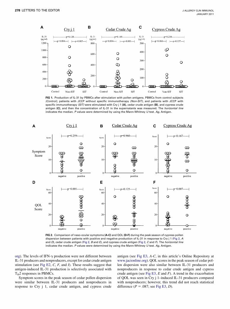
FIG 1. Production of IL-31 by PBMCs after stimulation with pollen antigens. PBMCs from control subjects
(Control), patients with JCCP without specific immunotherapy (Non-SIT), and patients with JCCP with
specific immunotherapy (SIT) were stimulated with Cry j 1 (A), cedar crude antigen (B), and cypress crude
antigen (C), and then the concentration of IL-31 in the supernatants was measured. The horizontal line
indicates the median. P values were determined by using the Mann-Whitney U test. Ag, Antigen.
FIG 2. Comparison of naso-ocular symptoms (A-C) and QOL (D-F) during the peak season of cypress pollen
dispersion between patients with positive and negative production of IL-31 in response to Cry j 1 (Fig 2, A
and D), cedar crude antigen (Fig 2, B and E), and cypress crude antigen (Fig 2, C and F). The horizontal line
indicates the median. P values were determined by using the Mann-Whitney U test. Ag, Antigen.
J ALLERGY CLIN IMMUNOL
JANUARY 2011
278 LETTERS TO THE EDITOR
org). The levels of IFN-g production were not different betweenIL-31 producers and nonproducers, except for cedar crude antigenstimulation (see Fig E2, C, F, and I). These results suggest thatantigen-induced IL-31 production is selectively associated withTH2 responses in PBMCs.Symptom scores in the peak season of cedar pollen dispersion
were similar between IL-31 producers and nonproducers inresponse to Cry j 1, cedar crude antigen, and cypress crude
antigen (see Fig E3, A-C, in this article’s Online Repository atwww.jacionline.org). QOL scores in the peak season of cedar pol-len dispersion were also similar between IL-31 producers andnonproducers in response to cedar crude antigen and cypresscrude antigen (see Fig E3, E and F). A trend in the exacerbationof QOL was seen in Cry j 1–induced IL-31 producers comparedwith nonproducers; however, this trend did not reach statisticaldifference (P 5 .087; see Fig E3, D).
J ALLERGY CLIN IMMUNOL
VOLUME 127, NUMBER 1
LETTERS TO THE EDITOR 279
Symptom scores in the peak season of cypress pollen disper-sion were also similar between IL-31 producers and nonproducersin response to Cry j 1, cedar crude antigen, and cypress crudeantigen (Fig 2, A-C). However, QOL scores, in which a high scoremeans a lowQOL, in the peak season of cypress pollen dispersionwere significantly higher in IL-31 producers in response to Cry j 1and cypress crude antigen, but not cedar crude antigen, comparedwith those seen in the respective nonproducers (Fig 2, D-F).Together with the finding that PBMCs that produced IL-31 in re-sponse to pollen antigens produced higher amounts of IL-5 andIL-13 in response to the respective antigens, these results suggestthat the induction of IL-31 production might lead to a deteriora-tion of JCCP.The amount of IL-31 production in response to pollen antigens
did not correlate with symptom or QOL scores in the peak seasonof cedar pollen dispersion (see Fig E4 in this article’s OnlineRepository at www.jacionline.org). However, the amounts, espe-cially in response to Cry j 1 (r 5 0.641, P < .001) and cypresscrude antigen (r 5 0.658, P 5 .002), significantly and positivelycorrelated with QOL scores in the peak season of cypress pollendispersion (see Fig E5, D-F, in this article’s Online Repository atwww.jacionline.org). In addition, there was a trend for a positivecorrelation between cypress crude antigen–induced IL-31production and symptom scores in the season (r 5 0.451,P 5 .070; see Fig E5, C). In contrast, the amounts of IL-5 orIL-13 production after stimulation with pollen antigens did notcorrelate with the QOL scores (see Fig E6 in this article’s OnlineRepository at www.jacionline.org). This result suggests that theinduction of pollen antigen–induced IL-31 production by PBMCsis associated with the severity of allergic rhinitis. Detailed discus-sion is available in this article’s Discussion section and Figs E8and E9 in this article’s Online Repository at www.jacionline.org.The present study provides evidence that, unlike other TH2-
type cytokines, including IL-5 and IL-13, IL-31 displays a uniqueand independent role in the pathophysiology of allergic rhinitis.The amount of pollen antigen–induced IL-31 production byPBMCs is selectively associated with the severity of QOL in pa-tients with JCCP. These observations might provide a basis for fu-ture therapeutic approaches targeting IL-31 in the managementand alleviation of allergic rhinitis.
Mitsuhiro Okano, MDa
Tazuko Fujiwara, BSa
Takaya Higaki, MDa
Seiichiro Makihara, MDa
Takenori Haruna, MDa
Yohei Noda, MDa
Kengo Kanai, MDa
Shin Kariya, MDa
Hiroshi Yasueda, PhDb
Kazunori Nishizaki, MDa
From athe Department of Otolaryngology–Head & Neck Surgery, Okayama University
Graduate School of Medicine, Dentistry and Pharmaceutical Sciences, Okayama,
Japan, and bthe Clinical Research Center for Allergy and Rheumatology, Sagamihara
National Hospital, Sagamihara, Japan. E-mail: [email protected].
Supported in part by grants from the Ministry of Education, Culture, Sports, Science and
Technology, Japan (20592001).
Disclosure of potential conflict of interest: The authors have declared that they have no
conflict of interest.
REFERENCES
1. Okubo K, Goto M, Fujieda S, Okano M, Yoshida H, Morikawa H, et al. A
randomized-double-blind comparative study of sublingual immunotherapy for cedar
pollinosis. Allergol Int 2008;57:265-7.
2. Sone K, Dairiki K, Morikubo K, Shimizu K, Tsunoo H, Mori T, et al. Recognition of
T cell epitopes unique to Cha o 2, the major allergen in Japanese cypress pollen, in
allergic patients cross-reactive to Japanese cedar and Japanese cypress pollen.
Allergol Int 2009;58:237-45.
3. Dillon SR, Sprecher C, Hammond A, Bilsborough J, Rosenfeld-Franklin M, Presnell
SR, et al. Interleukin 31, a cytokine produced by activated T cells, induces dermatitis
in mice. Nat Immunol 2004;5:752-60.
4. Bilsborough J, Leung DYM, Maurer M, Howell M, Boguniewcz M, Yao L, et al.
IL-31 is associated with cutaneous lymphocyte antigen-positive skin homing T cells
in patients with atopic dermatitis. J Allergy Clin Immunol 2006;117:418-25.
5. Neis MM, Peters B, Dreuw A, Wenzel J, Bieber T, Mauch C, et al. Enhanced expres-
sion levels of IL-31 correlate with IL-4 and IL-13 in atopic and allergic contact
dermatitis. J Allergy Clin Immunol 2006;118:930-7.
6. Raap U, Wichmann K, Bruder M, Stander S, Wedi B, Kapp A, et al. Correlation of
IL-31 serum levels with severity of atopic dermatitis. J Allergy Clin Immunol 2008;
122:421-3.
7. Perrigoue JG, Li J, Zaph C, Goldschmidt M, Scott P, de Sauvage FJ, et al. IL-31-IL-
31R interactions negatively regulate type 2 inflammation in the lung. J Exp Med
2007;204:481-7.
8. Ip WK, Wong CK, Li MLY, Li PW, Cheung PFY, Lam CWK. Interleukin-31 induces
cytokine and chemokine production from human bronchial epithelial cells through
activation of mitogen-activated protein kinase signaling pathways: implications
for the allergic diseases. Immunology 2007;122:532-41.
9. Lei Z, Liu G, Huang Q, Lv M, Zu R, Zhang GM, et al. SCF and IL-31 rather than
IL-17 and BAFF are potential indicators in patients with allergic asthma. Allergy
2007;63:327-32.
doi:10.1016/j.jaci.2010.09.029
Sequence variation in the IL4 gene and resis-tance to Trypanosoma cruzi infection inBolivians
To the Editor:Chagas disease, caused by the parasite Trypanosoma cruzi, af-
fects 10 to 12million people each year in Latin America, with Bo-livia having the highest prevalence of infection (see ‘‘Outlook:Chagas disease’’1 and references therein). In the chronic phase,Chagas infection may present as an indeterminate form in which60% of infected individuals remain asymptomatic despite havingpositive serologic reactions for T cruzi. In the remaining 40% ofpatients with Chagas disease, tissue inflammation leads to organdamage, affecting the cardiac, digestive, or nervous systems up to25 years after initial infection. Several studies identified geneticmarkers for disease establishment and progression in Venezue-lans, Brazilians, Peruvians, Colombians, and Mexicans,2 but nogenetic studies have been conducted previously in Bolivians.Cytokines produced in response to T cruzi infection appear to
modulate disease progression by enhancing or inhibiting parasitereplication in a variety of cell types. In particular, the TH2 cytokineIL-4maintains inflammationandparasite persistence inChagasdis-ease,3 whereas TH1 cytokines maintain control of parasitism4 butcan also contribute to the development of chronic myocarditis.5
To determine whether genetic variation at the IL4 gene is asso-ciated with T cruzi infection in Bolivians, we performed a rese-quencing study of an approximately 12-kb region around theIL4 locus, including 470 base pairs (bp) of coding (exon) se-quence, 368 bp of 59 untranslated region, 82 bp of 39 untranslatedregion, and 11,453 bp of intronic sequence. The study included110 individuals from the Department of Cochabamba, Bolivia,with infection status serologically confirmed by 2 different diag-nostic tests (HAI Chagas Polychaco; Laboratorio Lemos, S.R.L.,Buenos Aires, Argentina, and IFI Biocientifica S.A., BuenosAires, Argentina). Each subject was classified according to the se-rologic results as a case (positive serology) or a control (negative

METHODS
Antigens and reagentsCrude antigens of Japanese cedar pollen and Japanese cypress pollen were
extracted from Cryptomeria japonica pollen and Chamaecyparis obtuse
pollen, respectively, as described previously.E1,E2 Cry j 1 was purified and
concentrated from Japanese cedar crude antigen, as previously described.E1
PatientsForty-nine patients with JCCP (15 men and 34 women; age range, 29-75
years; mean age, 51.0 years) were enrolled in the study. Written informed
consentwas obtained from each subject. Sensitization to Japanese cedar pollen
was confirmed by the presence of specific IgE antibodies (range, 0.73 to >100UA/mL; mean, 20.54 6 22.39 UA/mL), as determined by means of
ImmunoCAP (Phadia AB, Uppsala, Sweden). Twenty patients received SIT
with a standardized extract of C japonica pollen (Torii Co, Tokyo, Japan) over
a period of at least 2 years. The mean maintenance dose of the extract was 468
JAU. None of the patients had used immunosuppressive drugs, including oral
steroids, during the pollen season. The control group consisted of 8 healthy
subjects with no sensitization to Japanese cedar pollen, as confirmed by means
of ImmunoCAP (3 men and 5 women; age range, 34-62 years; mean age,
46.5 years). No significant differences in age or sex existed among the
3 groups. The study was approved by the Human Research Committee of
OkayamaUniversity Graduate School ofMedicine, Dentistry and Pharmaceu-
tical Sciences.
Antigen-specific cytokine production by PBMCsHeparinized blood was collected from May to June 2009. PBMCs
were isolated and cultured as previously described.E3 In brief, PBMCs (2 3106/mL) were incubated in the presence or absence of 10 mg/mL cedar crude
antigen, cypress crude antigen, or Cry j 1 at 378C in a 5% CO2/air mixture for
72 hours. PBMCs from all 29 patients with JCCP who were not treated with
SIT were examined for cytokine production in response to Cry j 1. However,
because of limited sample volumes, PBMCs from 27 and 26 patients with
JCCP not treated with SITwere examined for cytokine production in response
to cedar crude antigen and cypress crude antigen, respectively. All PBMCs
from SIT-treated patients could be examined for cytokine production in re-
sponse to the 3 antigens. Then supernatant was collected and stored at
2808C until it was assayed. Levels of IL-5, IL-13, and IFN-g were measured
by using Opt EIA sets (BD Biosciences, San Jose, Calif) in accordance with
the manufacturer’s instructions. Levels of IL-31 were measured by using a
DuoSet ELISA development kit (R&D Systems, Minneapolis, Minn). The
detection limit of these assays was 3.9 pg/mL for IL-5, 3.9 pg/mL for IL-13,
7.8 pg/mL for IFN-g, and 7.8 pg/mL for IL-31.
Monitoring of symptoms and QOLJapanese rhinoconjunctivitis QOL questionnaires were used to compare
naso-ocular symptoms and rhinitis-related QOL during the pollen dispersion
season between SIT-treated and SIT-untreated patients.E4
A total of 2,220 grains/cm2 of Japanese cedar pollen and 1,478 grains/cm2
of Japanese cypress pollen were dispersed in 2009. The peak dispersions of
cedar and cypress pollens occurred on March 10 and April 11, respectively
(Fig E7). Thus patients answered the Japanese rhinoconjunctivitis QOL
questionnaires during March 4 to 18 and April 8 to 22 to assess naso-ocular
symptoms and rhinitis-related QOL.
Statistical analysisMedian values are presented. The nonparametric Mann-Whitney U test
was used to detect differences between groups. Correlation analysis was per-
formed by using the Spearman correlation coefficient by rank. P values of
less than .05 were considered statistically significant. The statistical
analysis was performed with StatView software (version 4.5; Abacus, Inc,
Berkeley, Calif).
DISCUSSIONIn the present study we characterized the IL-31 production in
pollen antigen–induced PBMC responses in patients with allergicrhinitis. Evidence is accumulating regarding the role of IL-31 inthe pathogenesis of allergic diseases, especially atopic dermatitis;however, this is the first report to demonstrate the role of IL-31 inpatients with allergic rhinitis.E5-E11
The amount of pollen antigen–induced IL-31 production wassignificantly and positively correlated with the production of IL-5and IL-13 but not IFN-g. This result is consistent with the reportby Neis et alE7 that the expression of IL-31 mRNA in the skin wascorrelated with the expression of IL-4 and IL-13 in patients withatopic dermatitis. Although IL-31 can be produced by mast cells,this result further suggests that the main producer of IL-31 mightbe TH2 cells producing both IL-5 and IL-13.E12
The most important and interesting finding in the present studyis that the PBMCs from some patients with JCCP produced IL-31in response to antigen, whereas the PBMCs from other patientswith JCCP did not. About one third of the patients did not produceIL-31 in response to cedar pollen–related antigens (Cry j 1 andcedar crude antigen). On the other hand, most patients producedother TH2 cytokines, IL-5, and IL-13 (Fig E8). In particular, allpatients with JCCP produced IL-5 in response to Cry j 1 and cedarcrude antigen, whereas the PBMCs from healthy control subjectsdid not produce IL-5. One of the reasons why all patients withJCCP produced IL-5 in response to the pollen antigens is thehigh pollen dispersion in 2009. This result is consistent with pre-vious reports that the induction of antigen-specific IL-5 produc-tion by PBMCs was a key factor in the onset of allergic rhinitis,including JCCP.E13,E14 In addition, PBMCs from about two thirdsof the patients with JCCP did not respond to cypress crude anti-gen. It has been proposed that a subset of patients with atopic der-matitis express low levels of IL-31.E6,E7 For example, cutaneouslymphocyte–associated antigen–positive T cells from 5 of 12 pa-tients with atopic dermatitis did not produce IL-31 in response to asuboptimal concentration of anti-CD3.E6 Our results are similar tothese reports and suggest that patients with JCCP can be dividedinto 2 subsets regarding antigen-induced IL-31 production byPBMCs. In addition, these results suggest that the induction ofIL-31 production is less essential for the onset of allergic rhinitiscompared with other TH2 cytokines, especially IL-5.
On the other hand, PBMCs that produced IL-31 in response topollen antigens produced higher amounts of IL-5 and IL-13 inresponse to the respective antigens. This result is similar to therecent report by Woodruff et alE15 that airway gene expression inpatients with asthma can be divided into 2 distinct ‘‘TH2-high’’and ‘‘TH2-low’’subgroups. In addition, patientswhosePBMCspro-duced IL-31 in response to Cry j 1 and cypress crude antigen hadsignificantly impaired QOL at the peak season of cypress pollendispersion. This result suggests that the induction of IL-31 produc-tion might lead to a deterioration in the pathophysiology of JCCP.Furthermore, the amount of IL-31 produced by PBMCs in
response to Cry j 1 and cypress crude antigen significantly andpositively correlated with QOL scores in the peak season ofcypress pollen dispersion. In human subjects with atopic derma-titis, IL-31 serum levels were correlated with the severity of atopicdermatitis, as determined by SCORAD scores.E8 On the otherhand, SCORAD scores were not correlated with the cutaneousexpression levels of IL-31 mRNA.E7 Our result is consistentwith the former report and suggests that the induction of pollen
J ALLERGY CLIN IMMUNOL
VOLUME 127, NUMBER 1
LETTERS TO THE EDITOR 279.e1
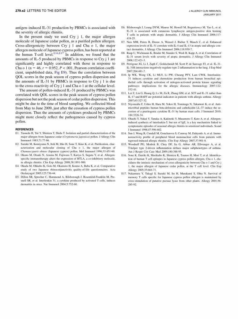
antigen–induced IL-31 production by PBMCs is associated withthe severity of allergic rhinitis.In the present study we used Cry j 1, the major allergen
molecule of Japanese cedar pollen, as a purified pollen allergen.Cross-allergenicity between Cry j 1 and Cha o 1, the majorallergenmolecule of Japanese cypress pollen, has been reported atthe human T-cell level.E16,E17 In addition, we found that theamounts of IL-5 produced by PBMCs in response to Cry j 1 aresignificantly and highly correlated with those in response toCha o 1 (n5 46, r5 0.952, P < .001, Pearson correlation coeffi-cient, unpublished data, Fig E9). Thus the correlation betweenQOL scores in the peak season of cypress pollen dispersion andthe amounts of IL-31 by PBMCs in response to Cry j 1 is dueto the cross-reactivity of Cry j 1 and Cha o 1 at the cellular level.The amount of pollen-induced IL-31 produced by PBMCs was
correlated with QOL scores in the peak season of cypress pollendispersion but not the peak season of cedar pollen dispersion. Thismight be due to the time of blood sampling. We collected bloodfrom May to June 2009, just after the cessation of cypress pollendispersion. Thus the amounts of cytokines produced by PBMCsmight more closely reflect the pathogenesis caused by cypresspollen.
REFERENCES
E1. Yasueda H, Yui Y, Shimizu T, Shida T. Isolation and partial characterization of the
major allergen from Japanese cedar (Cryptomeria japonica) pollen. J Allergy Clin
Immunol 1983;71:77-86.
E2. Suzuki M, Komiyama N, Itoh M, Itho H, Sone T, Kino K, et al. Purification, char-
acterization and molecular cloning of Cha o 1, the major allergen of
Chamaecyparis obtuse (Japanese cypress) pollen. Mol Immunol 1996;33:451-60.
E3. Okano M, Otsuki N, Azuma M, Fujiwara T, Kariya S, Sugata Y, et al. Allergen-
specific immunotherapy alters the expression of BTLA, a co-inhibitory molecule,
in allergic rhinitis. Clin Exp Allergy 2008;38:1891-900.
E4. Okuda M, Ohkubo K, Goto M, Okamoto H, Konno A, Baba K, et al. Comparative
study of two Japanese rhinoconjunctivitis quality-of-life questionnaires. Acta
Otolaryngol 2005;125:736-44.
E5. Dillon SR, Sprecher C, Hammond A, Bilsborough J, Rosenfeld-Franklin M, Pre-
snell SR, et al. Interleukin 31, a cytokine produced by activated T cells, induces
dermatitis in mice. Nat Immunol 2004;5:752-60.
E6. Bilsborough J, Leung DYM, Maurer M, Howell M, Boguniewcz M, Yao L, et al.
IL-31 is associated with cutaneous lymphocyte antigen-positive skin homing
T cells in patients with atopic dermatitis. J Allergy Clin Immunol 2006;117:
418-25.
E7. Neis MM, Peters B, Dreuw A, Wenzel J, Bieber T, Mauch C, et al. Enhanced
expression levels of IL-31 correlate with IL-4 and IL-13 in atopic and allergic con-
tact dermatitis. J Allergy Clin Immunol 2006;118:930-7.
E8. Raap U, Wichmann K, Bruder M, Stander S, Wedi B, Kapp A, et al. Correlation of
IL-31 serum levels with severity of atopic dermatitis. J Allergy Clin Immunol
2008;122:421-3.
E9. Perrigoue JG, Li J, Zaph C, Goldschmidt M, Scott P, de Sauvage FJ, et al. IL-31-
IL-31R interactions negatively regulate type 2 inflammation in the lung. J Exp Med
2007;204:481-7.
E10. Ip WK, Wong CK, Li MLY, Li PW, Cheung PFY, Lam CWK. Interleukin-
31 induces cytokine and chemokine production from human bronchial epi-
thelial cells through activation of mitogen-activated protein kinase signaling
pathways: implications for the allergic diseases. Immunology 2007;122:
532-41.
E11. Lei Z, Liu G, Huang Q, Lv M, Zu R, Zhang GM, et al. SCF and IL-31 rather than
IL-17 and BAFF are potential indicators in patients with allergic asthma. Allergy
2007;63:327-32.
E12. Niyonsaba F, Ushio H, Hara M, Yokoi H, Tominaga N, Takamori K, et al. Anti-
microbial peptides human beta-defensins and cathelicidin LL-37 induce the se-
cretion of a pruritogenic cytokine IL-31 by human mast cells. J Immunol 2010;
184:3526-34.
E13. Ohashi Y, Nakai Y, Tanaka A, Kakinoki Y, Masamoto T, Kato A, et al. Allergen-
induced synthesis of interleukin-5, but not of IgE, is a key mechanism linked to
symptomatic episodes of seasonal allergic rhinitis in sensitized individuals. Scand
J Immunol 1998;47:596-602.
E14. Sun J, Wong B, Cundall M, Goncharova S, Conway M, Dalrymle A, et al. Immu-
noreactivity profile of peripheral blood mononuclear cells from patients with
ragweed-induced allergic rhinitis. Clin Exp Allergy 2007;37:901-8.
E15. Woodruff PG, Modrek B, Choy DF, Jia G, Abbas AR, Ellwanger A, et al.
T-helper type 2-driven inflammation defines major subphenotypes of asthma.
Am J Respir Crit Care Med 2009;180:388-95.
E16. Sone K, Dairiki K, Morikubo K, Shimizu K, Tsunoo H, Mori T, et al. Identifica-
tion of human T cell epitopes in Japanese cypress pollen allergen, Cha o 1, elu-
cidates the intrinsic mechanism of cross-allergenicity between Cha o 1 and Cry j
1, the major allergen of Japanese cedar pollen, at the T cell level. Clin Exp
Allergy 2005;35:664-71.
E17. Nakamura Y, Takagi S, Suzuki M, Ito H, Murakami S, Ohta N. Survival of
memory T cells specific for Japanese cypress pollen allergen is maintained by
cross-stimulation of putative pectase lyase from other plants. Allergy 2001;56:
285-92.
J ALLERGY CLIN IMMUNOL
JANUARY 2011
279.e2 LETTERS TO THE EDITOR

FIG E1. Relationship between amounts of pollen antigen–induced IL-31 and TH1/TH2 cytokines produced by
PBMCs from patients with JCCP. Correlations between the amounts of IL-31 in response to Cry j 1 (A-C), ce-
dar crude antigen (D-F), and cypress crude antigen (G-I) and the amounts of IL-5 (Fig E1, A, D, and G), IL-13
(Fig E1, B, E, and H), and IFN-g (Fig E1, C, F, and I) in response to the respective antigens were determined by
using the Spearman correlation coefficient by rank. Ag, Antigen.
J ALLERGY CLIN IMMUNOL
VOLUME 127, NUMBER 1
LETTERS TO THE EDITOR 279.e3

FIG E2. Comparison of the amounts of IL-5 (A, D, and G), IL-13 (B, E, and H), and IFN-g (C, F, and I) in
response to Cry j 1 (Fig E2, A-C), cedar crude antigen (Fig E2, D-F), and cypress crude antigen (Fig E2,
G-I) between patients with positive and negative production of IL-31 in response to the respective antigens.
The horizontal line indicates the median. P values were determined by using the Mann-Whitney U test. Ag,
Antigen.
J ALLERGY CLIN IMMUNOL
JANUARY 2011
279.e4 LETTERS TO THE EDITOR

FIG E3. Comparison of naso-ocular symptoms (A-C) and QOL (D-F) during the peak season of cedar pollen
dispersion between patients with positive and negative production of IL-31 in response to Cry j 1 (Fig E3, A
and D), cedar crude antigen (Fig E3, B and E), and cypress crude antigen (Fig E3, C and F). The horizontal line
indicates the median. P values were determined by using the Mann-Whitney U test. Ag, Antigen.
J ALLERGY CLIN IMMUNOL
VOLUME 127, NUMBER 1
LETTERS TO THE EDITOR 279.e5
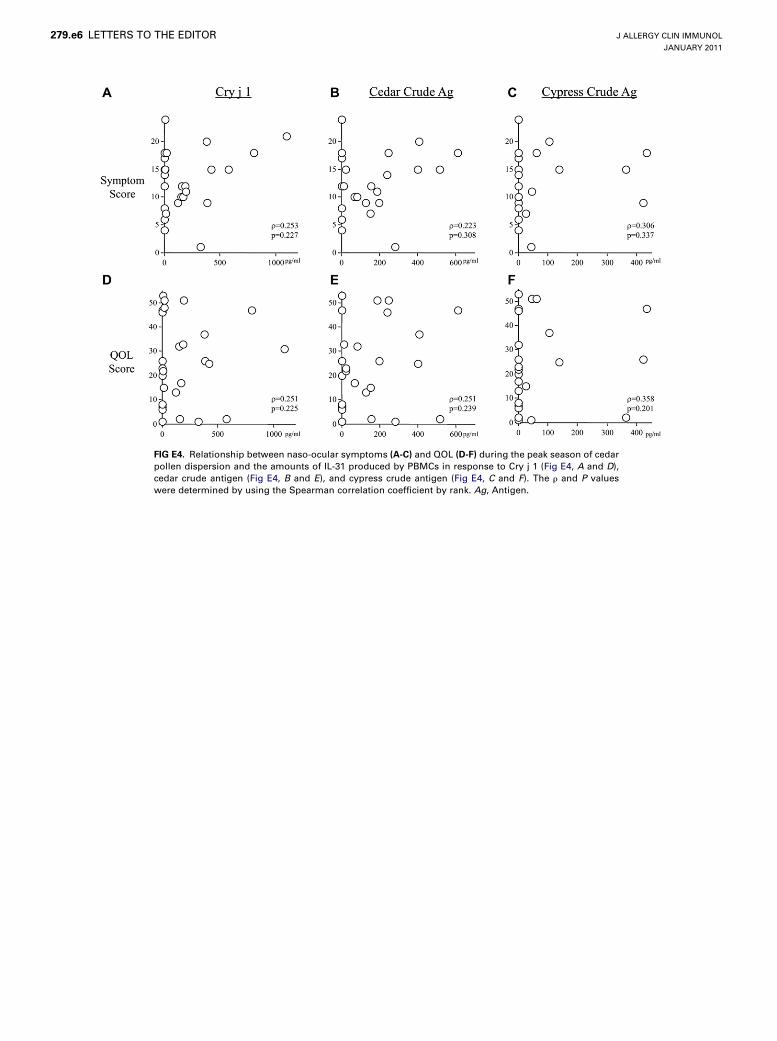
FIG E4. Relationship between naso-ocular symptoms (A-C) and QOL (D-F) during the peak season of cedar
pollen dispersion and the amounts of IL-31 produced by PBMCs in response to Cry j 1 (Fig E4, A and D),
cedar crude antigen (Fig E4, B and E), and cypress crude antigen (Fig E4, C and F). The r and P values
were determined by using the Spearman correlation coefficient by rank. Ag, Antigen.
J ALLERGY CLIN IMMUNOL
JANUARY 2011
279.e6 LETTERS TO THE EDITOR

FIG E5. Relationship between naso-ocular symptoms (A-C) and QOL (D-F) during the peak season of
cypress pollen dispersion and the amounts of IL-31 produced by PBMCs in response to Cry j 1 (Fig E5, A
and D), cedar crude antigen (Fig E5, B and E), and cypress crude antigen (Fig E5, C and F). The r and P values
were determined by using the Spearman correlation coefficient by rank. Ag, Antigen.
J ALLERGY CLIN IMMUNOL
VOLUME 127, NUMBER 1
LETTERS TO THE EDITOR 279.e7

FIG E6. Relationship between QOL during the peak season of cypress pollen dispersion and the amounts of
IL-5 (A-C) and IL-13 (D-F) produced by PBMCs in response to Cry j 1 (Fig E6, A and D), cedar crude antigen
(Fig E6, B and E), and cypress crude antigen (Fig E6, C and F). The r and P values were determined by using
the Spearman correlation coefficient by rank.
J ALLERGY CLIN IMMUNOL
JANUARY 2011
279.e8 LETTERS TO THE EDITOR
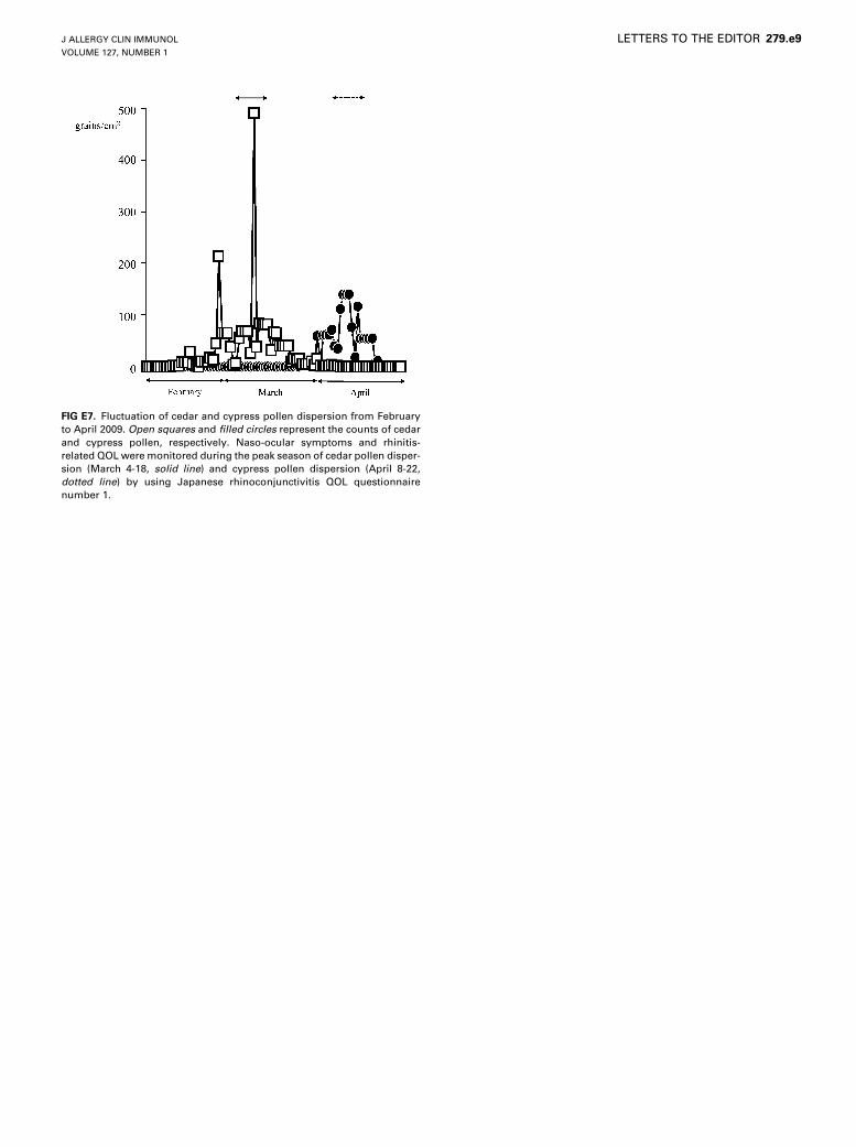
FIG E7. Fluctuation of cedar and cypress pollen dispersion from February
to April 2009. Open squares and filled circles represent the counts of cedar
and cypress pollen, respectively. Naso-ocular symptoms and rhinitis-
related QOL were monitored during the peak season of cedar pollen disper-
sion (March 4-18, solid line) and cypress pollen dispersion (April 8-22,
dotted line) by using Japanese rhinoconjunctivitis QOL questionnaire
number 1.
J ALLERGY CLIN IMMUNOL
VOLUME 127, NUMBER 1
LETTERS TO THE EDITOR 279.e9

FIG E8. Production of TH1 and TH2 cytokines of PBMCs against stimulation with pollen antigens. PBMCs
from healthy control subjects (Control), patients with JCCP without specific immunotherapy with standard-
ized extract of Japanese cedar pollen (Non-SIT), and patients with JCCP with specific immunotherapy (SIT)
were stimulated with Cry j 1 (A-C), cedar crude antigen (D-F), and cypress crude antigen (G-I) for 72 hours,
and then the concentration of IL-5 (Fig E8, A, D, andG), IL-13 (Fig E8, B, E, and H), and IFN-g (Fig E8, C, F, and
I) in the supernatants was measured bymeans of ELISA. The rectangle includes the range from the 25th and
75th percentiles, the horizontal line indicates the median, and the vertical line indicates the range from the
10th to the 90th percentiles. P values were determined by using the Mann-Whitney U test. Ag, Antigen.
J ALLERGY CLIN IMMUNOL
JANUARY 2011
279.e10 LETTERS TO THE EDITOR

FIG E9. Relationship between Cry j 1– and Cha o 1–induced IL-5 production
by PBMCs from patients with JCCP (n 5 46).
J ALLERGY CLIN IMMUNOL
VOLUME 127, NUMBER 1
LETTERS TO THE EDITOR 279.e11