Embed Size (px)
Citation preview

CHARACTERIZATION OF NITROGEN FIXATION (nif) GENES
FROM Paenibacillus polymyxa
YAM HOK CHAI
UNIVERSITI SAINS MALAYSIA
2007
CHARACTERIZATION OF NITROGEN FIXATION (nif) GENES
FROM Paenibacillus polymyxa
by
YAM HOK CHAI
Thesis submitted in fulfilment of the requirements
for the degree of
Master of Science
March 2007
ACKNOWLEDGEMENTS
Eventually, a book which is less than one inch was culminated. Each
page took an average of 10 days. Usually, a thesis does not reflect the real
effort that has been invested. Throughout this period, sadness is normally
caused by experimental failure; happiness is not only derived from successful
experiment but also something that can only be experienced when living in a
harmonious community.
The best way to describe the individual connection within this
community is ‘symbiotic relationship’. Basically, neither one of us is living
alone physically and mentally. The secret is in sharing. When happiness is
shared, it becomes greater. In contrast, when sadness is shared, it becomes
less burden one.
Professor Nazalan Najimudin who is an expert in molecular genetics
was so brave and dared to recruit someone like me (who didn’t even know
how to spell ‘science’ correctly) to be involved in a scientific research. It has
been an arduous task for him to raise me. Finally, I hope he had turned a
naughty boy into a disciplined researcher. My gratitude to him can not be
described by mere words.
My heartfelt thanks are also dedicated to Associate Professor Mohd
Razip Samian for providing laboratory support. Coincidentally, three of his
‘daughters’ in the lab became very important women in my life.
I am grateful to all the individuals who brought me happiness and made
my life complete. They include Aini, Ai Tee, Apai, Boon Choon, Boon Poh,
Chee Yong, Emmanuel, Eugene, Hanim, Ivan, Jeremy, Kem, Le Yau, Qis,

Shamanee, Shima, Tham, Yifen and Lab 409 members. I can’t imagine how
miserable my life would be without their companionship.
Neo Pei Chin, whatever you have said to me, I had it engraved in my
heart!
iv
Contents
Page
Acknowledgement ii
Contents iv
List of Tables vii
List of Figures viii
Symbols and abbreviations x
Abstrak xii
Abstract xiii
Chapter 1 Introduction 1
Chapter 2 Literature review 3
2.1 Nitrogen: Essential element for biomolecules 3
2.2 Biological nitrogen fixation 3
2.3 Diazotrophic microorganisms 4
2.4 Paenibacillus polymyxa 5
2.5 Key enzymes involved in biological nitrogen fixation
2.5.1 MoFe protein
2.5.2 Fe-protein
2.5.3 Stoichiometry
2.5.4 Alternative nitrogenase
6
6
7
7
8
2.6 Genes involved in biological nitrogen fixation 11
2.7 Regulation of nitrogenase expression
2.7.1 Oxygen control
2.7.2 Nitrogen control
2.7.3 nif promoter is regulated by DNA supercoiling
11
11
14
16
2.8 Promoter motive of nif genes 18
2.9 Gram-positive, free living diazotrophs
2.9.1 Clostridium pasteurianum
2.9.2 Paenibacillus massiliensis T7
2.9.3 Paenibacillus durus
19
20
21
22
2.10 Transcriptional Start Site (TSS) determination methods 22
2.11 Research objectives 24
v
Chapter 3 Material and methods 25
3.1 Bacterial strains, vectors and growth medium 25
3.2 General molecular biological methods 25
3.3 Isolation of nif genes 25
3.4 DNA isolation
3.4.1 Paenibacillus polymyxa genomic DNA isolation
3.4.2 Lambda DNA isolation
3.4.3 Plasmid DNA isolation
3.4.4 Gel DNA purification
28
28
29
29
29
3.5 PCR amplification of partial nifH and nifD fragments
3.5.1 Primers
3.5.2 PCR condition and ingredient
29
29
31
3.6 P. polymyxa genomic library construction
3.6.1 Partial digestion of genomic DNA
3.6.2 Ligation of inserts to vector LambdaGEM®-11 arms
3.6.3 Packaging of recombinant lambda DNA
3.6.4 Preparation of E. coli host cells for phage particle
infection
3.6.5 Plaque blotting
31
31
32
32
32
32
3.7 Southern hybridization analysis
3.7.1 Probe labelling
3.7.2 Southern hybridization analysis using fully digested
genomic DNA
3.7.3 Plaque hybridization: Primary and secondary
screening
3.7.4 Digested lambda DNA hybridization
33
33
33
35
35
3.8 E. coli DNA transformation 36
3.9 DNA sequencing 36
3.10 Total RNA isolation 37
3.11 cRACE: Circular or Concatemeric First-strand cDNA
mediated Rapid Amplification of cDNA Ends
37
3.12 cmRACE : Circularized mRNA Rapid Amplification of cDNA
Ends
41
vi
3.13 Phylogenetic analysis 43
Chapter 4 Results 46
4.1 Summary of overall result 46
4.2 PCR amplification of nifH fragment 46
4.3 Fully digested genomic DNA Southern
hybridization analysis
46
4.4 Genomic library 49
4.5 Genomic library screening 49
4.6 PCR amplication of nifD fragment 49
4.7 Southern analysis of digested λC2 lambda DNA 49
4.8 Subcloning and sequencing of positively hybridizing
fragments
54
4.9 NifH Phylogenetic analysis 62
4.10 cRACE (Circular or Concatemeric First-strand cDNA-
mediated Rapid Amplification of cDNA Ends)
62
4.11 cmRACE (Circularized mRNA Rapid amplification of cDNA
ends)
71
Chapter 5 Discussion 76
Chapter 6 Conclusion 83
Chapter 7 References 85
Chapter 8 Appendices 95

vii
List of Tables Page
Table 2.1 Nitrogen fixation (nif) genes products and functions 12
Table 3.1 Bacteria strains and vectors used in this study 26
Table 3.2 Primers used in amplifying partial nifD and nifH
fragments 30
Table 3.3a Primers employed in cRACE 40
Table 3.3b Primers employed in cmRACE 40
Table 3.4 Bacteria strains used in NifH phylogenetic analysis 44
Table 4.1 BlastX Result summary of NifBHDK proteins of P.
polymyxa 63
Table 4.2 Nucleotide sequence of the junction of the gene
specific primer and 3’ cDNA sequences 68
viii
List of Figures
Page
Figure 2.1 Schematic representation of the nitrogenase Fe
protein cycle
8
Figure 2.2 Model for oxygen control in K. pneumoniae 15
Figure 2.3 The nitrogen regulation Ntr system of enteric bacteria 17
Figure 3.1 Flow chart of nif genes isolation methods pathway 27
Figure 3.2 Flow chart showing the method pathway of the
cRACE applied in this research
38
Figure 3.3 Flow chart showing the method pathway of the
cRACE applied in this research.
42
Figure 4.1 PCR amplification of partial nifH gene 47
Figure 4.2a Fully digested Paenibacillus polymyxa genomic DNA 48
Figure 4.2b Autoradiograph of Southern hybridization analysis
employing nifH PCR fragment as the probe
48
Figure 4.3a Autoradiograph of primary screening of lambda
clones
50
Figure 4.3b Autoradiograph of secondary screening of positive
lambda clones
50
Figure 4.4 PCR amplification of a partial nifD fragment 51
Figure 4.5a DNA lambda clone λC2 digested with EcoRI 52
Figure 4.5b Autoradiograph of Southern hybridization analysis
using nifH fragment as a probe
52
Figure 4.6a DNA lambda clone λC2 digested with several
restriction enzymes
53
Figure 4.6b Autoradiograph of Southern hybridization analysis
using nifD fragment as the probe
53
Figure 4.7 Nucleotide sequence and deduced amino acid
sequence of complete nifBHD and partial nifK genes
55
Figure 4.8 Dendogram of NifH polypeptide phylogenetic tree 64
Figure 4.9 cRACE revealed two PCR products 65

ix
Figure 4.10 Flow chart showing the summary of the cRACE PCR
amplification process
66
Figure 4.11 Inferred transcriptional start sites and regulatory
region of nifB
69
Figure 4.12 Inferred transcriptional start sites and regulatory
region of nifH
70
Figure 4.13 Approximately 850 bp was obtained by nested PCR
in cmRACE approach
72
Figure 4.14 Outline of the cmRACE (Circularized mRNA Rapid
Amplification of cDNA Ends) approach to obtain the
850 bp fragment
73
Figure 4.15 Complete nifK ORF nucleotide and deduced amino
acid sequence
74
Figure 5.1a Presumptive promoter consensus element of nifB 82
Figure 5.2b Presumptive promoter consensus element of nifH 82
Figure 6.1 Physical maps of nifBHDK and its presumptive
regulatory regions
84

x
Symbols and Abbreviations
% percentage
ºC degree Celsius
σ sigma factor
λ lambda clone
μg micro gram
μl micro litre
μM micro molar
aa amino acid
acc. no. accession number
atm standard atmospheric pressure
ADP adenosine diphosphate
ATP adenosine triphosphate
bp base pair
DEPC Diethyl pyrocarbonate
DIG digoxigenin (non-radioactive DNA labelling)
DNA deoxyribonucleic acid
dNTP deoxynucleotide triphosphates
DTT dithiothereitol
et al. et alii (and others)
EDTA ethylenediaminetetraacetic
FAD flavin adenine dinucleotide
g gram
IPTG isopropyl β-thiogalactopyranoside
kb kilo base pair
kDa kilo Dalton
M molar
min minute
mM millimolar
OD optical density
ORF open reading frame
xi
RACE rapid amplification of cDNA ends
PCR polymerase chain reaction
RBS ribosome binding site
RNA ribonucleic acid
RT reverse transcription
rpm revolution per minute
sec second
SDS sodium dodecyl sulphate
SSC standard saline buffer
TAE Tris acetic acid EDTA
U unit
w/v weight per volume
X-gal 5-bromo-4-chloro-3-indolyl-β-galactoside

xii
PENCIRIAN GEN PENGIKAT NITROGEN (nif) DARIPADA Paenibacillus polymyxa
Abstrak
Paenibacillus polymyxa adalah sejenis bakteria Gram positif yang berupaya
menurunkan dinitrogen (N2) kepada ammonia. Satu fragmen nifH separa
telah diamplifikasi dengan menggunakan sepasang primer degenerat.
Fragmen tersebut digunakan sebagai prob untuk penyaringan perpustakaan
genomi lambda dan juga analisis penghibridan Southern terhadap genom P.
polymyxa. Berasakan keputusan analisis Southern, P. polymyxa dipercayai
mempunyai salinan tunggal homolog nifH. Daripada penyaringan
perpustakaan, lima klon lambda positif telah berjaya dipencilkan. Satu
fragmen nifD separa telah diamplifikasi daripada klon λC2 dengan
menggunakan sepasang primer degenerat untuk nifD. Prob nifH dan nifD
digunakan untuk penghibridan terhadap DNA lambda λC2 yang telah
dicernakan. Satu fragmen EcoRI 4 kb dan fragmen NdeI 5.5 kb telah
berhibridan secara positif masing-masing kepada prob nifH dan nifD. Kedua
fragmen telah disubklon dan dijujuk. Jujukan yang lengkap untuk rangka
bacaan terbuka nifBHD dan separa untuk nifK telah diperolehi. Tapak
permulaan transkripsi bagi nifB dan nifK telah ditentukan melalui kaedah
cRACE dan cmRACE. Klon PCR daripada cmRACE mempunyai jujukan 3’
nifK. Maka jujukan ORF nifBHDK yang sepenuhnya telah berjaya diperolehi.
Berasaskan tapak permulaan transkripsi, jujukan promoter bagi nifB dan nifH
didapati mempunyai jujukan konsensus yang jelas tetapi berlainan daripada
jujukan promoter nif yang tradisional. Ini mencadangkan yang ekspresi gen nif
dalam P. polymyxa adalah berkemungkinan menerusi sistem pengawalaturan
yang unik.

xiii
CHARACTERIZATION OF NITROGEN FIXATION GENES (nif)
FROM Paenibacillus polymyxa
Abstract
Paenibacilus polymyxa is a Gram positive bacterium capable of
converting dinitrogen (N2) to ammonia. A partial nifH fragment was amplified
by using a pair of nifH degenerate primers. The fragment was used as a
probe for screening a genomic library as well as a genomic Southern
hybridization analysis. Based on the Southern analysis results, P. polymyxa
was believed to possess a single copy of nifH homologue. From the library
screening, five positive lambda clones were successfully isolated. A partial
nifD fragment was amplified from lambda clone λC2 by using a pair of
degenerate primers for nifD. The nifH and nifD probes were then used to
hybridize digested lambda λC2 DNA. A 4 kb EcoRI and a 5.5 kb NdeI
fragment hybridized positively to the nifH and nifD probes, respectively. These
fragments were then subcloned and sequenced. An intact sequence of
nifBHD and a partial sequence of nifK open reading frames (ORFs) were
obtained. The transcriptional start sites of nifB and nifH were determined by
cRACE and cmRACE methods. The cmRACE PCR clones also carried the 3’
end sequence of nifK. Therefore, intact ORFs of nifBHDK were successfully
obtained. Based on their transcriptional start sites, the promoter regions of
nifB and nifH revealed consensus sequences that were distinguishable from
the traditional nif promoter. This suggested that the expression of nif genes in
P. polymyxa is possibly controlled by a unique regulation system.

1
Chapter 1 Introduction
The rapid growth of human population and agricultural activity are
closely related. The soil nitrogenous source is usually a limiting factor in any
increase in agricultural activity. Great endeavor is undertaken in order to solve
the nitrogen crisis before it becomes a significant problem.
The largest nitrogen reservoir is atmospheric dinitrogen (N2). Hence,
the seemingly economical way is to develop an industrial scale process to
convert N2 into a form that can be assimilated by plants. The Haber-Bosch
process finally realized the conversion of N2 to ammonia in 1908 under
conditions of high temperature (450ºC) and pressure (200 atm) in the
presence of an iron-based catalyst (Haber, 1922).
Prior to this, biological nitrogen fixation was the exclusive way to
provide nitrogen source into the biosphere (relatively small amount is
produced by terrestrial source, lightning and volcanic activity). Today, both
biological and chemical processes generate even amount of fixed nitrogen to
fulfill the demand of agricultural activity of the world population. However,
speedy increase in usage of chemically fixed nitrogen causes a profound
environmental problem (Smil, 2001). Thus, researchers of biological nitrogen
fixation hope to moderate the usage of chemical fertilizer as well as gain
priceless scientific knowledge.
Biological nitrogen fixation is a trademark of prokaryotes. More than
100 species were reported to have nitrogen fixation ability. It is found in most
bacterial phylogenetic groups and methanogens in Archaea (Raymond et al.,
2004). Conversion of N2 to ammonia is catalyzed by a metalloenzyme called

2
nitrogenase. The enzyme contains two components that are named according
to their metal constituents: Fe-protein and MoFe-protein (Dixon & Kahn, 2004).
The most well studied nitrogen fixers are Klebsiella pneumoniae and
Azotobacter vinelandii and both are Gram negative bacteria. Research
activities exploiting Gram positive diazotrophs are still few (Klipp et al., 2004).
Gram positive diazotrophs have been shown to have distinctly different nif
genes organization and regulation patterns (Wang et al. 1988, Harriott et al.,
1995). For instance, NifA protein in K. pneumoniae is a well known positive
regulator that mediates the expression of nitrogenase in respond to various
environmental signals such as oxygen and nitrogen. In contrast, nifA gene
does not even exist in the genome of the Gram positive diazotroph Frankia
alni (Harriott et al., 1995). The intact genome sequence of Frankia sp. CcI3
(Acc. no. NC_007777) also does not possess a nifA-like gene.
Therefore, we have undertaken investigation on several Gram positive
diazotrophs such as Paenibacillus durus, P. macerans and P. polymyxa. In
this research, P. polymyxa is used to uncover the genetics of nitrogen fixation
in a Gram positive bacterium. Using gene isolation methodology and
cmRACE approach, the complete DNA sequence of nifBHDK was obtained.
The transcriptional start sites of nifB and nifH were determined using
both cRACE and cmRACE approaches. The findings of this research are in
line with the notion that Gram positive diazotrophs apparently have a different
mode of gene regulation. This speculation is based on dissimilar promoter
motif, gene organization and transcriptional pattern.
3
Chapter 2 Literature Review
2.1 Nitrogen: Essential element for biomolecules
Nitrogen is an essential part of most biological compounds such as
amino and nucleic acids and this makes it vital to all life. It is the next most
abundant element in the living cell after carbon. For example, a typical
bacterial cell possesses 12% nitrogen in terms of dry mass. In nature,
nitrogen exists in both organic and inorganic forms. However, the bulk of
available nitrogen is in the inorganic form, either as ammonia (NH3), nitrate
(NO3-) or N2. Ammonia and nitrate can be used as nitrogen sources while N2
(nitrogen gas) requires a reduction process prior to assimilation by plants and
other living organisms. Unfortunately, nitrogen, the fifth most abundant
element in the universe and makes up about 78% of the earth's atmosphere is
inert. Therefore, biological nitrogen fixation is a crucially important process to
convert nitrogen gas into ammonia using an environmentally friendly and
natural mechanism (Arp, 2000; Finan et al., 2002).
2.2 Biological nitrogen fixation
Biological nitrogen fixation, the conversion of atmospheric nitrogen into
ammonia by symbiotic, associative and free-living bacteria, is responsible for
supplying more than 60% of the world’s annual new ammonia source
(Schlesinger, 1991). The amount of biologically fixed nitrogen produced is in
excess of 2 x 10¹³ g/year (Falkowski, 1997). The availability of fixed nitrogen
is normally the limiting factor for crop productivity. Thus there is a tremendous
demand on global agriculture to provide food security which correlates

4
dramatically to the increase in world’s population in the twenty-first century.
The increased utilization of chemical fertilizers, which constitutes the largest
human interference in the nitrogen cycle, has prompted concerns regarding
profound pollution impacts such as increased emissions of nitrogen oxides,
soil acidification and water eutrophication. Fortunately, the fixed nitrogen
provided by biological nitrogen fixation is less prone to leaching and
volatilization as it is utilized in situ. Therefore this biological process
contributes an important input into agriculture in a sustainable manner (Dixon
& Kahn, 2004; Gallon & Chaplin, 1987).
2.3 Diazotrophic microorganisms
Biological nitrogen fixation is exclusively an ability of prokaryotes.
These organisms are called diazotrophs and are widely distributed among the
prokarya and archaea with more than 100 species reported (Raymond et al.,
2004). The ability to fix nitrogen is found in most bacterial phylogenetic groups,
including green sulphur bacteria, Firmibacteria, actinomycetes, cyanobacteria
and all subdivisions of the Proteobacteria. In Archaea, nitrogen fixation is
mainly restricted to the methanogens. The ability to fix nitrogen is compatible
with a wide range of physiological state such as aerobic (for example,
Azotobacter), facultatively anaerobic (for example, Klebsiella) or anaerobic
(for example, Clostridium) heterotrophs; anoxygenic (for example,
Rhodobacter) or oxygenic (for example, Anabaena) phototrophs; and
chemolithotrophs (for example, Leptospirillum ferrooxidans). Diazotrophs are
also found in a wide variety of habitats including soil and water. They are also
found in various states of associations; symbioses with grasses, termite guts,
5
woody plants, and root–nodule formation in legumes (Raymond et al., 2004;
Madigan, 2000).
2.4 Paenibacillus polymyxa
Paenibacillus polymyxa was firstly isolated by Prazmowski (Buchanan
dan Gibbons, 1974). Paenibacillus durus (formerly P. azotofixans), P.
polymyxa and P. macerans are nitrogen fixers which form a monophyletic
cluster in the genus Paenibacillus. These are formerly labeled as Bacillus until
they were reclassified based on rDNA sequence analysis (Ash et al., 1993).
P. polymyxa is a bacterial species that is often found in soil and in the
rhizosphere and rhizoplane of grasses such as wheat, maize, sorghum and
sugarcane (Line and Loutit, 1971; Holl et al., 1988; Mavingui et al., 1992; von
der Weid et al., 2000). Many P. polymyxa strains are free-living nitrogen fixers
(Grau and Wilson 1962; Seldin et al., 1983) and there was evidence on the
secretion of plant growth-enhancing substances by root-associated P.
polymyxa (Holl et al., 1988). Besides their ecological importance as plant-
growth-promoting rhizobacteria, P. polymyxa strains are also important for the
pharmaceutical and food industries (Debabov, 1982; Priest, 1993). P.
polymyxa is known to produce two types of peptide antibiotics (Beatty &
Jensen, 2002). One group is active against bacteria and includes the
polymyxin-colistin-circulin family, polypeptins, jolipeptin, gavaserin, and
saltavalin, which contain a 2,4-diaminobutyric acid. The other is active against
fungi and actinomycetes and includes gatavalin and fusaricidins. This species
also synthesizes plant hormones auxin (Lebuhn et al., 1997) and cytokinin
(Timmusk et al., 1999).

6
2.5 Key enzymes involved in biological nitrogen fixation
The conversion of dinitrogen(N2) to ammonia (NH3) is catalysed by the
nitrogenase enzyme, a complex of metalloproteins with conserved structural
and mechanistic features. Nitrogenase is a combination of two soluble
proteins, known as MoFe protein and Fe protein, according to their metal
property. Fe protein functions as an ATP-dependent electron donor to the
larger component, the MoFe protein, which contains the enzyme catalytic site
(Rees et al., 2005).
2.5.1 MoFe protein
MoFe protein has a size of 200 to 250-kDa (also called dinitrogenase)
and is composed of four subunits (α2β2 tetramer). Each subunit has a size of
approximately 60-kDa; the α subunit is encoded by the nifD gene while the β
subunit is encoded by the nifK. The MoFe protein contains two types of metal-
sulfur clusters: the P-cluster (8Fe-7S cluster) and the FeMo cofactor (Mo-7Fe-
9S cluster). The latter is usually designated as FeMo-co and is believed to
contain the substrate binding site and reduction process (Rees et al., 2005).
The P-cluster is coordinated by six Cys ligands, bridges the α and β subunits
and intermediates the electron transport pathway between Fe-protein and
FeMo-co. FeMo-co cluster is connected to the protein by only two ligands
(Cys and His) located within the α subunit (Kim and Rees, 1994; Eady, 1995;
Igarasshi and Seefeldt, 2003).

7
2.5.2 Fe-protein
Fe-protein (also called dinitrogenase reductase) is a dimer composed
of two copies of a single α subunit and is encoded by nifH. It has a size of 55
to 65-kDa. The Fe-protein contains a metal cluster ([4Fe–4S] cluster) that
covalently connects the two α subunits. The [4Fe–4S] cluster is the redox-
active site involving in electron transfer to FeMo-protein which cycles between
reduced state and oxidized state (Eady, 1995; Igarashi and Seefeldt, 2003).
2.5.3 Stoichiometry
The overall stoichiometry of dinitrogen reduction under normal
condition is as follows:
N2 + 8 e– + 8 H+ + 16 MgATP→ 2 NH3 + H2 + 16 MgADP + 16 Pi
The enzyme mechanism starts with a reduction of the Fe protein by electron
donors such as ferredoxin and flavodoxin (Dixon and Kahn, 2004, Igarashi
and Seefeldt, 2003). This is followed by a transfer of a single electron from the
Fe protein to the MoFe protein (which is dependent on MgATP hydrolysis).
Finally, an internal electron transfer takes place in the MoFe protein by the P
cluster to the FeMo-co cluster at the substrate-binding site. Each electron-
transfer step requires a continuous cycling step of association and
dissociation of the Fe and MoFe proteins as shown in Figure 2.1. Nitrogenase
is a relatively slow enzyme with a turnover time of about 5s–1, and thus the
dissociation of the complex is the rate-limiting step. Complex formation has a
crucial role in the enzyme mechanism as it is required for the coupling of ATP
hydrolysis to electron transfer (Thornely & Lowe, 1985).

8
Figure 2.1 : Schematic representation of the nitrogenase Fe protein cycle. The
Fe protein dimer is shown in light blue with the cube representing
the [4Fe–4S] cluster coloured green to indicate the reduced form
and red to represent the oxidized form. The α and β subunits of
the MoFe protein are depicted as orange and purple, respectively,
the yellow squares represent the P cluster and the white diamond
represent the FeMo cofactor (adapted from Dixon & Kahn, 2004).
ATP
ATP
ADP
ADP
ATP
ATP
ADP
ADP
ATP
ADP
2
2 e-
Disassociation
Complex formation
MoFe protein
Fe protein (reduced)
Fe protein (oxidized)
Nucleotide exchange
Reduction
2Pi
Electron transfer ATP hydrolysis Phosphate release

9
There is a conformational change in the Fe protein upon ATP turnover
coupled to repositioning of the [4Fe–4S] cluster. This brings the cluster in
closer proximity to the MoFe protein, thereby facilitating inter-protein electron
transfer from the Fe protein to the MoFe protein (Schindelin et al., 1997).
2.5.4 Alternative nitrogenase
Some microorganisms contain alternative nitrogenase whereby Mo is
replaced by either Fe or V in condition lacking Mo source. Molybdenum-
lacking nitrogenase was firstly reported in Azotobacter vinelandii (Bishop et al.,
1980). These so-called alternative nitrogenases are found only in limited
diazotrophs and their presence is secondary to the MoFe protein. An
alternative nitrogenase from Azotobacter chroococcum was first purified and
characterized by Robson et al. (1986), who demonstrated that the enzyme
contained vanadium rather than molybdenum. The vanadium nitrogenase is
encoded by the vnf genes (Robson et al., 1989). After the description of the
vanadium nitrogenase, Chisnell et al. (1988) isolated a second alternative
nitrogenase apparently lacking any metal other than iron in its dinitrogenase
protein. The gene encoding this second alternative nitrogenase was
designated as anf genes (Joerger et al., 1989). Vanadium containing
nitrogenase is expressed preferentially if Fe and V are both present in Mo
lacking situation and also if the microorganism has all three types of
nitrogenases. The MoFe-co containing nitrogenase has highest affinity and
efficiency in nitrogen fixation compared to the other two nitrogenases in the
order of Mo>V > Fe (Joerger and Bishop, 1988).
10
Obviously, the structural genes of nif, vnf, and anf nitrogenases show
significant sequence similarity. The general properties of the component
proteins are also quite similar. All nitrogenase systems contain two
components; a dinitrogenase protein and a dinitrogenase reductase protein.
The usual nif-encoded dinitrogenase protein is α2β2 tetramer of the nifDK
gene products whereas the alternative dinitrogenases encoded by the vnfDK
and anfDK genes consist a third subunit known as VnfG and AnfG
respectively (Robson et al., 1989; Premakumar et al., 1989). In contrast to the
reduction of acetylene to ethylene by the nif-encoded nitrogenase, anf-
encoded nitrogenase and vnf-encoded nitrogenase are able to reduce
acetylene to a mixture of ethylene and ethane (Dilworth et al., 1988; Scott et
al., 1990).
The fourth family of nitrogenases is represented by enzyme from the
carboxydotrophic bacterium Streptomyces thermoautotrophicus (Ribbe et al.,
1997). This nitrogenase contains two component proteins. One component, a
CO-dehydrogenase, oxidizes CO to CO2 and reduces O2 to the superoxide
anion radical (O2−). The second component is a manganese-dependent
oxidoreductase that oxidizes O2−, providing electrons to the N2 and reducing
the MoFeS active site (Ribbe et al., 1997). Among the most striking properties
of the S. thermoautotrophicus nitrogenase system are the dependence on O2
and O2−, the complete insensitivity to neither O2 nor H2O2, the inability to
reduce acetylene or ethylene, and a low MgATP requirement. In addition, the
subunit structure of the S. thermoautotrophicus nitrogenase components and
the polypeptides involved distinctly dissimilar from the other three nitrogenase
families (Ribbe et al., 1997).
11
2.6 Genes involved in biological nitrogen fixation
Klebsiella pneumoniae and Azotobacter vinelandii are the best-studied
diazotrophs and most of the biological nitrogen fixation related genes were
discovered in these bacteria. About 20 genes were found to be responsible in
the complex nitrogen fixation mechanism for both nitrogenase protein
synthesis and genetic regulation of the process (Jacobson et al., 1989; Arnold
et al., 1988). Knowledge obtained from this two species became essential
reference for the studies of other diazotroph. Table 2.1 shows a summary of
nif genes and functions and their corresponding proteins.
2.7 Regulation of nitrogenase expression
2.7.1 Oxygen control
Nitrogenase is irreversibly inactivated by oxygen and hence
environmental oxygen tension is one of the major regulatory factors. In the
case of strict anaerobes, oxygen regulation has less importance since the
anaerobes can only survive in oxygen free environment. For the facultative
aerobes, oxygen regulation becomes very significant in order to avoid
nitrogenase denaturation by oxygen (Merrick, 1992).
In the case of the strict aerobe Azotobacter, a very high respiratory
activity rapidly reduces the oxygen, thus decreasing the internal oxygen
concentration and thereby protecting the nitrogenase (Philips &Johnson,
1961). Most aerobic diazotroph produce gummy colonies on agar medium
and this may also play a role in protecting nitrogenase from oxygen (Hill et al.,
1972).

12
Gene(nif) Function
Q Incorporation of molybdenum into nitrogenase (Imperial et al.,
1984)
B FeMo-cofactor synthesis (Curatti et al., 2006)
A Positive regulation (Lee et al., 1993; Eydmann et al., 1995;
Schmitz et al., 2002)
L Negative regulation (Lee et al., 1993; Eydmann et al., 1995;
Schmitz et al., 2002)
F Electron carrier : flavodoxin (Arnold et al., 1988; Taylor et al.,
1990)
M Nitrogenase reductase processing (Roberts et al., 1978; Arnold
et al., 1988)
Z Maturation and activation of FeMo-protein (Paul and Merrick,
1989)
W Maturation and activation; oxygen protection of FeMo-protein
(Paul and Merrick, 1989; Lee et al., 2000)
V FeMo-cofactor synthesis: homocitrate-synthase (Zheng et al.,
1997; Allen et al., 1994)
S Homodimeric cysteine desulfurase, S activation in metallocluster
synthesis (Zheng et al.,1993; Zheng and Dean, 1994)
U FeMo-protein processing (Harris et al., 1990; Fu et al., 1994)
X FeMo cofactor synthesis. Negative regulation (Lee et al.,2000)
E Synthesis and insertion of FeMo-co into dinitrogenase protein
(Orme-Johnson, 1985; Arnold et al., 1988)
Table 2.1 Nitrogen fixation (nif) genes products and functions.

13
Y Processing of MoFe-protein (Homer et al., 1993)
T Nitrogenase maturation (Simon et al., 1996)
K FeMo-protein β subunit (Arnold et al,. 1988; Kim and Rees, 1994)
D FeMo-protein α subunit (Arnold et al., 1988; Kim and Rees, 1994)
H Fe-protein subunit (Arnold et al,. 1988; Kim and Rees, 1994)
J Electron transfer: pyruvate-flavodoxin oxidoreductase (Schmitz et
al., 2001)
Table 2.1 (continued)
14
In heterocystous cyanobacteria, the enzyme nitrogenase seems to only
exist in the heterocysts-thick-walled cells that lack oxygen due to
photosynthetic process. Some heterocyst defective mutants are unable to fix
nitrogen. Therefore, heterocysts play an essential role in nitrogen fixation
(Fleming & Haselkorn, 1973).
In Klebsiella pneumoniae and Azotobacter vinelandii the nitrogen
regulatory proteins NifL and NifA tightly control the synthesis of nitrogen
fixation genes in response to oxygen (and nitrogen). The transcriptional
activator NifA is required for transcription of other nitrogen fixation (nif) genes.
In this regulation, the negative regulator NifL inhibits NifA activity.
Immunological studies, chromatography and complex formation analyses
using the yeast two-hybrid system demonstrates that NifA interacts directly
with NifL by protein-protein interaction (Henderson et al., 1989; Money et al.,
1999 and 2001; Lei et al., 1999). This indicates that the oxygen molecule
finally results in a complex formation between NifL and NifA which inhibits
NifA activity and thus prevents the transcription of other nif genes (Figure 2.2).
The inhibitor NifL is a flavoprotein which regulates NifA activity depending on
the reduction status of its N-terminally bound FAD-cofactor. It allows NifA to
function only under anaerobic conditions. Thus, the redox-sensitive FAD-
cofactor appears to be involved in oxygen signal-transduction (Hill et al., 1996;
Klopprogge et al., 2002; Grabbe & Schmitz, 2003).
2.7.2 Nitrogen control
Nitrogen itself is a major regulatory factor in free living diazotrophs.
These microorganisms can only fix nitrogen when a nitrogenous source is

15
Figure 2.2: Model for oxygen control in K. pneumoniae (Adapted
from Dixon and Kahn, 2004). In low oxygen condition, NifL
dissociates from NifA, thus allowing the latter to drive the
expression of nif genes.
o NifA
FAD
FADH2 NifA
H D K
nif genes transcription
-O2
+O2
NifL
NifL +
Positive regulation
16
limited. A general nitrogen regulation (ntr) system controls the expression of
many genes including the nif system in the diazotroph. Four key proteins are
involved in nitrogen control system; a uridylyltransferase(UTase) encoded by
glnD, a small tetrameric effector protein (PII) encoded by glnB, NtrB encoded
by ntrB and a transcriptional activator NtrC encoded by ntrC (Merrick, 1995).
UTase participates as the primary sensor of the cellular nitrogen status.
When cells are lacking nitrogen, Utase mediates the uridylation of PII by
transferring a uridylyl group onto a tyrosine residue on each of the four PII
subunits (Figure 2.3). The uridylylated PII (PII-UMP) indirectly causes the
phosphorylation of NtrC. It actually mediates autophosphorylation of NtrB
followed by the transfer of the phosphate to NtrC. Under nitrogen rich
condition, UTase acts as a uridylyl-removing enzyme, converting PII-UMP to
its original form, and NtrB now promotes the dephosphorilation of NtrC. In the
second level of regulation, phosphorylated NtrC activates the expression of
nifA and hence control the nitrogenase expression response to the low
nitrogen level in the cell (Merrick 1994; Merrick 2004).
2.7.3 nif promoter is regulated by DNA supercoiling
In prokaryotes, negatively supercoiled genomic DNA promotes DNA
recombination, replication and transcription (Snoep et al., 2002). In addition,
DNA supercoiling plays an important role in the cellular perception of
environmental signals. Changes in oxygen content directly affect DNA
superhelical density via the action of gyrase which increases negative
supercoiling. In terms of oxygen content, anaerobic conditions activate
bacterial gyrase, whereas aerobic conditions activate topoisomerase I. Indeed,

17
Figure 2.3: The nitrogen regulation Ntr system of enteric bacteria. The activity
of the response regulator NtrC is regulated in response to the
intracellular nitrogen status. UTase (glnD product) catalyses the
uridylylation and deuridylylation of PII (glnB product). PII in turn
regulates the activity of the sensor histidine kinase NtrB which
catalyses the phosphorylation and dephosphorylation of NtrC
(Adapted from Merrick, 2004).
NtrC-P NtrC NtrB + PII
PII(UMP)3
3UTP
3PPi
High N
Low N
3UMP
NtrC NtrC-P
UR/Utase glnD
(glnB)
18
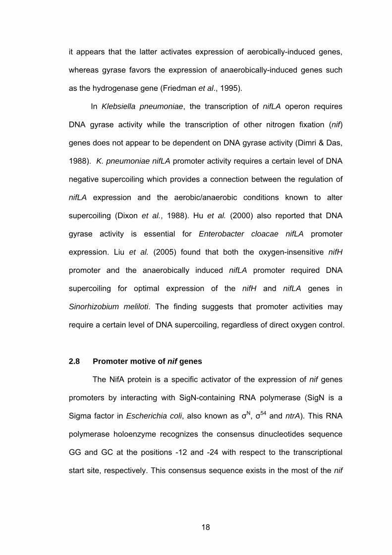
it appears that the latter activates expression of aerobically-induced genes,
whereas gyrase favors the expression of anaerobically-induced genes such
as the hydrogenase gene (Friedman et al., 1995).
In Klebsiella pneumoniae, the transcription of nifLA operon requires
DNA gyrase activity while the transcription of other nitrogen fixation (nif)
genes does not appear to be dependent on DNA gyrase activity (Dimri & Das,
1988). K. pneumoniae nifLA promoter activity requires a certain level of DNA
negative supercoiling which provides a connection between the regulation of
nifLA expression and the aerobic/anaerobic conditions known to alter
supercoiling (Dixon et al., 1988). Hu et al. (2000) also reported that DNA
gyrase activity is essential for Enterobacter cloacae nifLA promoter
expression. Liu et al. (2005) found that both the oxygen-insensitive nifH
promoter and the anaerobically induced nifLA promoter required DNA
supercoiling for optimal expression of the nifH and nifLA genes in
Sinorhizobium meliloti. The finding suggests that promoter activities may
require a certain level of DNA supercoiling, regardless of direct oxygen control.
2.8 Promoter motive of nif genes
The NifA protein is a specific activator of the expression of nif genes
promoters by interacting with SigN-containing RNA polymerase (SigN is a
Sigma factor in Escherichia coli, also known as σN, σ54 and ntrA). This RNA
polymerase holoenzyme recognizes the consensus dinucleotides sequence
GG and GC at the positions -12 and -24 with respect to the transcriptional
start site, respectively. This consensus sequence exists in the most of the nif

19
promoters in K. pneumoniea including nifH, nifE, nifU, nifM, nifF, nifL and nifB
(Barrios et al., 1999; Jack et al., 1999).
Purified SigN can bind to certain promoters in the absence of core RNA
polymerase, indicating that the DNA binding determinants of SigN can
function in the isolated protein (Buck and Cannon, 1992). This is in contrast to
the major sigma factor of enteric bacteria SigA (also known as σ70), in which
the DNA binding determinant is masked until the protein binds to the core
RNA polymerase (Dombroski et al., 1993).
Based on promoter study of K. pneumoniae, NifA (a member of
enhancer-binding proteins) binds to a specific upstream activating sequence,
UAS (also known as enhancer sequence), and interacts with the SigN-
containing RNA polymerase bound to the -12,-24 portion. Mutational analysis
of the nifH UAS has supported the suggestion that the TGT-N1O-ACA motif
(where N is any nucleotide), which characterizes the UAS of nif promoters, is
a NifA binding site (Buck et al., 1987; Morett et al., 1988). This UAS sequence
is normally found at the 100 to 200bp region upstream of nif genes. NifA
protein participates in the activation of transcription by binding to the UAS and
contacting the downstream SigN-containing RNA polymerase complex by
forming a loop forming in the DNA between the UAS and the -12,-24 (GG-N10-
GC) conserved sequence promoter element (Buck et al., 1987; Morett & Buck,
1989; Jack et al., 1999).
2.9 Gram-positive, free living diazotrophs
Most of nitrogen fixation related research focussed on Gram-negative
bacteria such as K. pneumoniae while Gram-positive diazotrophs received
20
very little attention. Interestingly, Gram-positive diazotrophs show exclusively
contrasting features either in their nif genes promoter motive or genes
arrangement compared to Gram-negative diazotroph (Wang et al., 1988;
Harriott et al., 1995). Thus, research on Gram-positive diazotrophs is much
needed, especially to look at new features of nif genes regulation. Below are
some examples of research of Gram-positive diazotrophs.
2.9.1 Clostridium pasteurianum
Clostridium pasteurianum is a Gram-positive anaerobic bacterium with
a low G+C content of 26 to 28% (Cummins & Johnson, 1971) which
distinguishes it from the rest of well-studied nitrogen-fixing microorganisms.
The primary structure of C. pasteurianum nitrogenase components is
significantly less related to that of nitrogenases from other microorganisms
(Chen et al., 1973). C. pasteurianum nitrogenase has high activity, but its
components are distinctly ineffective in forming active heterologous
complexes (Emerich & Burris, 1978, Smith et al., 1976) and are less sensitive
to H2 as an inhibitor (Guth & Burris, 1983).
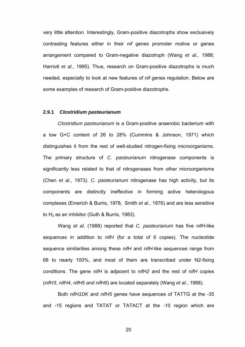
Wang et al. (1988) reported that C. pasteurianum has five nifH-like
sequences in addition to nifH (for a total of 6 copies). The nucleotide
sequence similarities among these nifH and nifH-like sequences range from
68 to nearly 100%, and most of them are transcribed under N2-fixing
conditions. The gene nifH is adjacent to nifH2 and the rest of nifH copies
(nifH3, nifH4, nifH5 and nifH6) are located separately (Wang et al., 1988).
Both nifH1DK and nifH5 genes have sequences of TATTG at the -35
and -15 regions and TATAT or TATACT at the -10 region which are
21
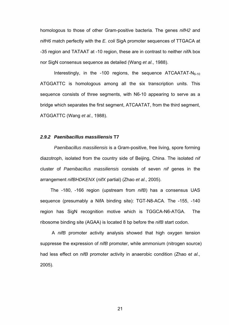
homologous to those of other Gram-positive bacteria. The genes nifH2 and
nifH6 match perfectly with the E. coli SigA promoter sequences of TTGACA at
-35 region and TATAAT at -10 region, these are in contrast to neither nifA box
nor SigN consensus sequence as detailed (Wang et al., 1988).
Interestingly, in the -100 regions, the sequence ATCAATAT-N6-10
ATGGATTC is homologous among all the six transcription units. This
sequence consists of three segments, with N6-10 appearing to serve as a
bridge which separates the first segment, ATCAATAT, from the third segment,
ATGGATTC (Wang et al., 1988).
2.9.2 Paenibacillus massiliensis T7
Paenibacillus massiliensis is a Gram-positive, free living, spore forming
diazotroph, isolated from the country side of Beijing, China. The isolated nif
cluster of Paenibacillus massiliensis consists of seven nif genes in the
arrangement nifBHDKENX (nifX partial) (Zhao et al., 2005).
The -180, -166 region (upstream from nifB) has a consensus UAS
sequence (presumably a NifA binding site): TGT-N8-ACA. The -155, -140
region has SigN recognition motive which is TGGCA-N6-ATGA. The
ribosome binding site (AGAA) is located 8 bp before the nifB start codon.
A nifB promoter activity analysis showed that high oxygen tension
suppresse the expression of nifB promoter, while ammonium (nitrogen source)
had less effect on nifB promoter activity in anaerobic condition (Zhao et al.,
2005).

22
2.9.3 Paenibacillus durus
Paenibacillus durus (formerly P. azotofixans) is a Gram-positive,
facultatively anaerobic diazotroph, classified into a broad cluster of nitrogen
fixers in rRNA group 3. This cluster also includes Paenibacillus macerans and
Paenibacillus polymyxa (Ash et al., 1991). It fixes atmospheric nitrogen with
high efficiency and this ability is not affected by the presence of nitrate which
is in contrast to the majority nitrogen fixers (Rosado et al., 1998).
Choo et al. (2003) reported the presence of three nifH homolog in the P.
durus and these are designated as nifH1, nifH2 and nifH3. Adjacent to nifH1
are nifB1, nifD1 and nifK1 in the following arrangement nifB1H1D1K1. It was
firstly found that the gene nifB is located upstream to nifH. NifH1 and NifH2
protein comparison yielded 97% identity. In contrast, comparing either nifH1
or nifH2 to nifH3 yielded a relatively low 43% identity (Choo et al., 2003).
An analysis of NifH phylogeny demonstrated clustering of P. durus
NifH1 and NifH2 within the Cyanobacteriaceae grouping while NifH3 clustered
with the NifH proteins of members of the Archaea domain. Interestingly, none
of the NifH proteins from P. durus clustered with the NifH of other gram-
positive diazotroph such as Frankia sp. (a high-G+C firmicute) and
Clostridium pasteurianum (a low-G+C firmicute). Unusual placement of NifH3
among the highly divergent members of the Archaea suggests the occurrence
of horizontal gene transmission (Choo et al., 2003).
2.10 Transcriptional Start Site (TSS) determination methods
Determination of the first nucleotide (5'-end) of mRNA is a crucial step
to identify and analyse a gene’s promoter. Several methods have been
23
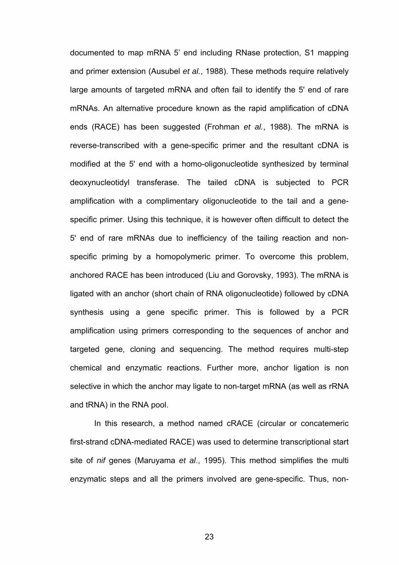
documented to map mRNA 5’ end including RNase protection, S1 mapping
and primer extension (Ausubel et al., 1988). These methods require relatively
large amounts of targeted mRNA and often fail to identify the 5' end of rare
mRNAs. An alternative procedure known as the rapid amplification of cDNA
ends (RACE) has been suggested (Frohman et al., 1988). The mRNA is
reverse-transcribed with a gene-specific primer and the resultant cDNA is
modified at the 5' end with a homo-oligonucleotide synthesized by terminal
deoxynucleotidyl transferase. The tailed cDNA is subjected to PCR
amplification with a complimentary oligonucleotide to the tail and a gene-
specific primer. Using this technique, it is however often difficult to detect the
5' end of rare mRNAs due to inefficiency of the tailing reaction and non-
specific priming by a homopolymeric primer. To overcome this problem,
anchored RACE has been introduced (Liu and Gorovsky, 1993). The mRNA is
ligated with an anchor (short chain of RNA oligonucleotide) followed by cDNA
synthesis using a gene specific primer. This is followed by a PCR
amplification using primers corresponding to the sequences of anchor and
targeted gene, cloning and sequencing. The method requires multi-step
chemical and enzymatic reactions. Further more, anchor ligation is non
selective in which the anchor may ligate to non-target mRNA (as well as rRNA
and tRNA) in the RNA pool.
In this research, a method named cRACE (circular or concatemeric
first-strand cDNA-mediated RACE) was used to determine transcriptional start
site of nif genes (Maruyama et al., 1995). This method simplifies the multi
enzymatic steps and all the primers involved are gene-specific. Thus, non-
24
specific PCR products should be less likely to be amplified. The complete
protocol is detailed in Chapter 3.
RT-PCR analysis of circularized mRNA is the only option to
concurrently determine the 5’ as well as 3’ mRNA sequences. This approach
requires target mRNA to be religated by using RNA ligase, followed by cDNA
synthesis and PCR amplification targeting the joined regions. The reliability of
this method was firstly ensured by Mandl et al. (1991) on identification 5’ and
3’-terminal regions of linear RNA genome from several tick-borne flaviviruses.
This method was also included in this research to refine the results obtained
from cRACE. An exhaustive protocol is detailed in Chapter 3.
2.11 Research objectives
The objectives of this research are summarized as below:
a. To obtain complete sequence of the nifH gene of P. polymyxa as well
as nif-related genes adjoining to nifH.
b. To analysis phylogenetic of nifH isolated from P. polymyxa.
c. To determine the transcriptional start sites of nif genes.
d. To postulate probable promoter and regulatory regions.