Embed Size (px)
Citation preview

Characterization of mice lacking candidate N-acyl ethanolamine
biosynthetic enzymes provides evidence for multiple pathways that
contribute to endocannabinoid production in vivowzGabriel M. Simon and Benjamin F. Cravatt*
Received 7th January 2010, Accepted 19th March 2010
First published as an Advance Article on the web 14th April 2010
DOI: 10.1039/c000237b
The biosynthesis of the endocannabinoid anandamide (AEA) and related N-acyl ethanolamine
(NAE) lipids is complex and appears to involve multiple pathways, including: (1) direct release
of NAEs from N-acyl phosphatidyl ethanolamine (NAPE) precursors by the phosphodiesterase
NAPE-PLD, and (2) double O-deacylation of NAPEs followed by phosphodiester bond
hydrolysis of the resulting glycero-phospho (GP)-NAEs. We recently identified GDE1 as a
GP-NAE phosphodiesterase that may be involved in the second pathway. Here, we report the
generation and characterization of GDE1(�/�) mice, which are viable and overtly normal in their
cage behavior. Brain homogenates from GDE1(�/�) mice exhibit a near-complete loss of
detectable GP-NAE phosphodiesterase activity; however, bulk brain levels of AEA and other
NAEs were unaltered in these animals. To address the possibility of compensatory pathways,
we generated GDE1(�/�)/NAPE-PLD(�/�) mice. Conversion of NAPE to NAE was virtually
undetectable in brain homogenates from these animals as measured under standard assay
conditions, but again, bulk changes in brain NAEs were not observed. Interestingly, significant
reductions in the accumulation of brain NAEs, including anandamide, were detected in
GDE1(�/�)/NAPE-PLD(�/�) mice treated with a fatty acid amide hydrolase (FAAH)
inhibitor that blocks NAE degradation. Finally, we determined that primary neurons from
GDE1(�/�)/NAPE-PLD(�/�) mice can convert NAPEs to NAEs by a pathway that is not
preserved following cell homogenization. In summary, combined inactivation of GDE1 and
NAPE-PLD results in partial disruption of NAE biosynthesis, while also pointing to the existence
of an additional enzymatic pathway(s) that converts NAPEs to NAEs. Characterization of this
pathway should provide clarity on the multifaceted nature of NAE biosynthesis.
Introduction
The endocannabinoid system plays a key role in both central
and peripheral tissues, where it participates in diverse physio-
logical processes including nociception, inflammation, appe-
tite, and metabolism.1–4 This system is comprised primarily of
the cannabinoid G-protein coupled receptors CB1 and
CB2, their two endogenous arachidonate-containing lipids
(‘‘endocannabinoids’’) anandamide (N-arachidonoyl ethanol-
amine, AEA) and 2-arachidonoyl glycerol (2-AG), and the
enzymatic pathways responsible for endocannabinoid bio-
synthesis and degradation.5,6
Classical neurotransmitters, such as amino acids, mono-
amines, and neuropeptides, are water-soluble and are stored in
vesicles prior to release into the synaptic cleft upon neuronal
depolarization. In contrast, lipid messengers such as the
endocannabinoids are considered to be too hydrophobic for
storage in vesicles and are therefore believed to be synthesized
‘‘on demand’’ from phospholipid precursors.7 The signaling
potential of endocannabinoids is consequently under strict
regulation by the enzymes and proteins that catalyze their
biosynthesis, transport, and degradation.5,8–10 The identifica-
tion and characterization of these enzymes is crucial to obtaining
a complete understanding of endocannabinoid signaling and
its role in mammalian physiology, as well as to ascertain the
potentially distinct functions played by AEA and 2-AG.
AEA is a member of a large class of amidated lipids termed
the N-acyl ethanolamines (NAEs)11 and is the principal
member of this family with CB1 agonist activity.12,13 Early
studies on changes to the lipidome resulting from ischemic
shock in dog heart and brain tissue revealed rapid simul-
taneous accumulation of NAEs and an unusual phospholipid
species,N-acyl phosphatidyl ethanolamines (NAPEs), suggesting
a precursor–product relationship between these two classes of
lipids.14,15 Several alternative hypotheses concerning the bio-
synthesis of NAEs have been proposed including direct con-
jugation of ethanolamine with fatty acids or acyl CoAs;16,17
however, multiple lines of evidence point to NAPEs as the
endogenous precursors to NAEs.18 In the early 1980s, several
The Department of Chemical Physiology and The Skaggs Institute forChemical Biology, The Scripps Research Institute, 10550 N. TorreyPines Road, La Jolla, California 92037, USA.E-mail: [email protected] This article is part of a Molecular BioSystems themed issue onChemical Genomics.z Electronic supplementary information (ESI) available: Three supple-mental figures are provided that describe additional in vitro and in situexperiments investigating NAE levels and NAPE metabolism. SeeDOI: 10.1039/c000237b
This journal is �c The Royal Society of Chemistry 2010 Mol. BioSyst., 2010, 6, 1411–1418 | 1411
PAPER www.rsc.org/molecularbiosystems | Molecular BioSystems

pathways were proposed by which NAEs could be released
from NAPEs, including direct hydrolysis by a D-type phospho-
lipase, as well as single- or double-O-deacylation followed by
phosphodiesterase activity on the resulting lysophosphatidyl-
or glycero-phospho-N-acyl ethanolamines (GP-NAEs).19
Direct release from NAPEs was found to be activated by
millimolar concentrations of calcium in vitro, and this D-type
phospholipase activity could be detected in multiple rodent
tissues, from where it was ultimately purified and molecularly
characterized.20–22 This enzyme, dubbed NAPE-PLD, was
found to be exquisitely selective for NAPEs in vitro and highly
expressed in tissues that utilize endocannabinoid signaling,
such as nervous tissue and testis.
Consistent with a role for NAPE-PLD in NAE biosynthesis,
mice lacking this enzyme [NAPE-PLD(�/�) mice] display
profound reductions in very long-chain saturated and mono-
unsaturated NAEs in the central nervous system.23 Interestingly,
however, poly-unsaturated NAEs, including AEA, were
unaltered in the nervous system of NAPE-PLD(�/�) mice.
This finding engendered the hypothesis that multiple enzy-
matic pathways are involved in the conversion of NAPEs to
NAEs in vivo, with NAPE-PLD controlling the biosynthesis of
long-chain saturated NAEs, and additional enzymes partici-
pating in the generation of shorter chain and poly-unsaturated
NAEs (including AEA). Attempts to detect an additional
D-type phospholipase activity in NAPE-PLD(�/�) tissues
were unsuccessful, but robust O-deacylation of NAPE was
observed, suggesting that lysoNAPEs or GP-NAEs might
serve as direct precursors to NAEs.24 In support of this
deacylation hypothesis, it was observed that inhibitors of
A/B-type phospholipases blocked the release of NAE
from NAPE and lysoNAPE in brain homogenates from
NAPE-PLD(�/�) mice indicating that total O-deacylation
must occur prior to phosphodiester bond cleavage under these
in vitro conditions.
Recent work from our lab has identified candidate enzymes
that can perform each step in this proposed deacylation–
phosphodiesterase pathway for NAE biosynthesis. The previously
uncharacterized a-b hydrolase enzyme ABH4 acts as a B-type
phospholipase selective for (lyso)NAPE and produces GP-NAE
from NAPE in vitro. The resulting GP-NAE is a substrate for
the metal-dependent enzyme GDE125 (glycerophosphodiester
phosphodiesterase 1, also known as MIR16), which was
previously characterized in vitro as a glycerophosphodiesterase
that acts on multiple glycerophosphodiester substrates.26,27 To
test the contribution of this pathway to NAE biosynthesis
in vivo, we herein report the generation and characterization of
mice bearing a targeted disruption of the GDE1 gene.
Results and discussion
Generation of GDE1(�/�) mice
Construction of mice bearing a disruption in the GDE1 gene
was achieved using a standard targeting strategy directly in
embryonic stem (ES) cells from the C57BL/6 background. A
targeting construct was designed to disrupt exon two of the
GDE1 gene locus, which contains residues that are required
for catalytic activity (Fig. 1A). A homologously recombined
ES-cell clone was identified by Southern blotting (Fig. 1B)
and used to generate chimeric mice on an albino C57BL/6
background. These chimeric mice gave germline transfer of
the mutated locus (Fig. 1C), and the resulting pups served
as founders for the GDE1(�/�) strain (Fig. 1D). Western
blotting of tissue extracts from GDE1(�/�) brains confirmed
loss of the GDE1 protein (Fig. 1E).
Characterization of GP-NAE and NAE metabolism in
GDE1(�/�) mice
GDE1(�/�) mice were born at the expected Mendelian
frequency, were viable and healthy, and showed no overt
differences in their cage behavior compared to wild-type litter-
mates. Initial biochemical characterization of the GDE1(�/�)mice focused on measurement of GP-NAE phosphodiesterase
activity in brain homogenates (Fig. 2A). These experiments
revealed that GDE1 is responsible for nearly all (>95%) of
the detectable GP-NAE phosphodiesterase activity in brain
tissue. Additionally, we found that conversion of lysoNAPE to
NAE was also impaired in tissue extracts (Fig. 2B), confirming
that conversion of lysoNAPE to NAE proceeds through a
GP-NAE intermediate in vitro. Previously, we described an experi-
ment in which brain homogenates accumulated ‘‘endogenous’’
GP-NAEs in an MAFP-sensitive, EDTA-induced manner,
and we hypothesized that GDE1, a metal-dependent enzyme,
was the EDTA-sensitive phosphodiesterase responsible for
this accumulation.25 To test this hypothesis, a similar experi-
ment was performed in which brains from GDE1(+/�) and(�/�) mice were incubated with or without EDTA and the
accumulation of GP-NAE was measured by LC-MS/MS
(Fig. 2C). Consistent with previous studies, GP-NAEs accumu-
lated in brain tissue from GDE1(+/�) mice in an EDTA-
dependent manner. Interestingly, GP-NAE levels in GDE1(�/�)brains were basally high after 4 hour incubation and equivalent
in magnitude to those observed in EDTA-treated GDE1(+/�)brains. No additional EDTA-induced accumulation of
GP-NAEs was observed in GDE1(�/�) brains. These results
confirm that GDE1 is the principal EDTA-sensitive GP-NAE
phosphodiesterase in brain homogenates.
To determine if the deacylation–phosphodiesterase pathway
is operant in vivo, we undertook targeted measurements of
endogenous metabolites via LC-MS/MS. First, we measured
GP-NAEs in the brains of GDE1(+/�) and (�/�) animals but,
surprisingly, observed no significant differences between these
genotypes (Fig. 3). Basal NAE levels were also unchanged in
GDE1(�/�) brains (Fig. 4).
Characterization of GP-NAE and NAE metabolism
in GDE1(�/�), NAPE-PLD(�/�), and GDE1(�/�)/NAPE-PLD(�/�) mice
As multiple enzymes are known to utilize NAPE as a substrate
(Fig. 5A), we next crossed GDE1 and NAPE-PLD lines to
generate GDE1(�/�)/NAPE-PLD(�/�) double knockout
mice to measure the combined effects of ablation of these
enzymes on NAE metabolism. Brain tissue from wild-type,
double-knockout, and each single-knockout was assayed for
the ability to release 14C-NAE from 14C-NAPE. As has been
previously reported,23 the choice of assay conditions affected
1412 | Mol. BioSyst., 2010, 6, 1411–1418 This journal is �c The Royal Society of Chemistry 2010

the relative contributions of these two pathways, with the
presence or absence of calcium proving particularly dramatic.
Furthermore, we found that selection of reaction buffer and
inclusion of detergents also influenced the relative contribu-
tions made by each pathway (Fig. S1 and S2, ESIw). Depending
on the assay conditions employed, absence of either NAPE-PLD
or GDE1 resulted in major reductions in NAPE-to-NAE
conversion, but, under all conditions tested, the double-knockout
showed near-total loss of NAPE-to-NAE conversion in brain
homogenates (Fig. 5B). Similar results were obtained in testis
homogenates (not shown).
Despite the near-complete loss of NAPE-to-NAE conver-
sion in brain homogenates from GDE1(�/�)/NAPE-PLD(�/�)double knockout mice, no significant differences in bulk brain
levels of NAEs were observed between NAPE-PLD(�/�) andthe GDE1(�/�)/NAPE-PLD(�/�) mice (Fig. 4). We next
asked whether the rate of NAE production might be impaired
by GDE1 and/or NAPE-PLD disruption by treating mice with
PF-3845, a recently described inhibitor of the NAE-degrading
enzyme fatty acid amide hydrolase (FAAH).28 FAAH inhibition
has previously been shown to produce a steady and dramatic
rise in NAE levels that plateau after about 2–3 hours.29,30 We
therefore measured brain NAE levels in various mouse models
at 3 hours following treatment with PF-3845. Interestingly, at
this time point, GDE1(�/�)/NAPE-PLD(�/�) mice displayed
significant reductions in several NAEs including AEA (Fig. 6).
The magnitude of these reductions was rather modest
(B25–40%), but clearly suggests that GDE1 and NAPE-PLD,
together, contribute to NAE biosynthesis. Similar effects on
NAE accumulation were observed in testis from these mice
(Fig. S3, ESIw). Furthermore, the fact that these reductions
were only observed following disruption of both GDE1 and
NAPE-PLD indicates that these pathways have the potential
to crosstalk with one another to regulate NAE production.
Evidence for additional NAPE-to-NAE enzymatic pathways in
neurons
Taken together, our results with GDE1(�/�) and NAPE-
PLD(�/�) mice are somewhat perplexing. Mice lacking both
GDE1 and NAPE-PLD display wild-type levels of NAEs and
only moderately compromised rates of NAE production,
Fig. 1 Generation of GDE1(�/�) mice. (A) The genomic structure of the GDE1 gene locus on chromosome 7 is shown, along with the targeting
construct used to delete exon 2 (which contains several residues essential for catalysis, Glu97, Asp99 and His112; asterisk) and the final recombined
locus. Only relevant restriction sites are designated. The locations of DNA probes used for Southern blotting 50 and 30 to the targeting con-
struct are also shown. (B) Southern blot of EcoRV-digested ES cell DNA showing the heterozygous (targeted) clone #301. (C) Southern blot of
BspHI/XhoI-digested tail-DNA from heterozygous and wild-type mice demonstrating germ-line transmission. (D) PCR strategy in which the
wild-type locus is identified by a 238 bp band and the targeted locus is identified by a 417 bp band. (E) Western blot of the particulate fraction from
brain tissue confirming loss of GDE1 protein. GDE1 is a PNGaseF-sensitive glycosylated protein.
This journal is �c The Royal Society of Chemistry 2010 Mol. BioSyst., 2010, 6, 1411–1418 | 1413

despite a total loss of NAE release from NAPE in brain
homogenates. Two possible explanations for this situation
present themselves: (1) NAEs do not, in fact, come from
NAPEs in vivo, or (2) NAEs are released from NAPEs
in vivo by an enzyme or enzymes whose activities are not
maintained in vitro. To explore the latter possibility, we sought
to develop a live-cell system in which the conversion of NAPE
to NAE could be monitored in situ. To achieve this goal, we
cultured primary neurons from neonatal wild-type, GDE1(�/�),NAPE-PLD(�/�), or GDE1(�/�)/NAPE-PLD(�/�) pups
and grew them for 6 days in vitro. Neurons were then treated
with the FAAH inhibitor PF-3845 (to prevent turnover of
NAE) and 14C-NAPE was added to the media. In this way, we
observed clear time-dependent release of NAE from NAPE in
live neurons (Fig. 7A). Previous studies have monitored the
formation of NAE in neurons by employing either radio-
labeled ethanolamine or arachidonate,7,31 which do not provide
information on the direct precursor of NAE biosynthesis.
Under our experimental conditions, the presence of a FAAH
inhibitor prevented hydrolysis of the amide bond linking14C-fatty acid to phosphatidylethanolamine thereby ensuring
that all NAE formed must be released from NAPE. Once this
experimental paradigm was established, we tested the ability of
neurons from single- and double knockout animals to release
NAE from NAPE. To our surprise, we found that all geno-
types including GDE1(�/�)/NAPE-PLD(�/�) released NAE
from NAPE at equivalent levels (Fig. 7B and C). This result
implies the existence of an additional enzyme(s) distinct from
NAPE-PLD and GDE1 that can release NAEs from NAPEs.
To test whether this in situ-active enzymatic activity requires
O-deacylation or whether it operates directly on NAPE, we
employed a non-hydrolyzable analogue of NAPE that
contains ether-linkages in the sn-1 and sn-2 positions.24 This
ether–NAPE analogue did not serve as an effective substrate
for NAE formation (Fig. 7D), suggesting that the in situ
(GDE1- and NAPE-PLD-independent) pathway for NAE
generation requires O-deacylation prior to phosphodiester
bond cleavage.
Conclusion
In summary, brain tissue from mice lacking GDE1 and
NAPE-PLD shows a near-complete loss in NAPE conversion
to NAE. Despite this result, however, bulk brain levels of
NAEs were unaltered in mice lacking GDE1 and NAPE-PLD.
A defect in the rate of NAE production was observed in
Fig. 2 GP-NAE phosphodiesterase activity is lost in GDE1(�/�) tissue. (A) Near-complete loss of conversion of 14C-GP-NAE to 14C-NAE is
evident from a representative thin layer radiochromatogram (left) and quantified data from replicate experiments (right). (B) Loss of conversion of14C-lysoNAPE to 14C-NAE is evident from a representative thin layer radiochromatogram (left) and quantified data from replicate experiments
(right). Note also the accumulation of the GP-NAE intermediate in the assay with GDE1(�/�) tissue. All assays were performed in PBS buffer
with tissue homogenates from brains of GDE1(+/+) and (�/�) mice. (C) Ex vivo assay demonstrating that EDTA induces GP-NAE
accumulation in brain homogenates due to disruption of GDE1 activity. Brain tissue from GDE1(+/�) and (�/�) mice was homogenized in
the presence or absence of 10 mM EDTA (which inhibits GDE1) and incubated at room temperature for 4 hours. The addition of EDTA leads
to a rapid accumulation of GP-NAEs in wild-type brain, as previously reported. GP-NAE accumulation was observed in brain tissue from
GDE1(�/�) mice with or without EDTA, demonstrating that GDE1 is the EDTA-sensitive GP-NAE phosphodiesterase in wild-type tissue. n= 4
mice per group. Results are presented as means� standard error. Asterisks designate po 0.05, po 0.01, po 0.005 (Student’s t-test) for *, **, and
***, respectively. Asterisks represent difference from wild-type in panels (A) and (B), and difference from EDTA-treated GDE1(+/�) tissue in
panel (C).
Fig. 3 GP-NAE levels are unchanged in GDE1(�/�) brains. Endo-genous GP-NAEs were measured in brain extracts from GDE1(+/�)and (�/�) mice via LC-MS/MS. No significant differences were
observed. n = 8 mice per group. Results are presented as means �standard error.
1414 | Mol. BioSyst., 2010, 6, 1411–1418 This journal is �c The Royal Society of Chemistry 2010
GDE1(�/�)/NAPE-PLD(�/�) mice, suggesting that both
GDE1 and NAPE-PLD make partial contributions to the
biosynthesis of anandamide and other NAEs in vivo. Con-
sidering further that all of our measurements were made
with bulk brain tissue, it is also possible that GDE1 and/or
NAPE-PLD play even more substantial roles in AEA/NAE
biosynthesis in specific neuronal circuits or brain regions.
When NAPE metabolism was studied in live neurons
(‘‘in situ’’), we discovered that neurons lacking both GDE1
and NAPE-PLD are capable of converting NAPEs to NAEs.
This result supports the existence of additional NAPE-
catabolizing enzymes that are operant in situ, and likely in vivo,
but artificially inactivated in brain homogenates. When con-
sidering the history of other phosphodiesterase enzymes, one
finds that this situation is not exceptional. The characteriza-
tion of the prototypical mammalian phospholipase D (PLD1)
involved years of frustration during which activity in intact cell
systems was observed, but lost upon cell-lysis. In fact, several
reports similar to this one can be found in the literature in
which PLD activity was measured in intact cell prepara-
tions, but could not be reconstituted in vitro.24,32,33 The
breakthrough discovery came when it was found that PLD1
activity was dramatically enhanced in vitro by performing
substrate assays under precise conditions: addition of the
non-hydrolyzable GTP analogue GTPgS, addition of the
ADP-ribosylation factor (ARF) protein, as well as substrate
presentation in mixed liposomes that include phospha-
tidylinositol-4,5-bisphosphate (PIP2).34 Indeed, omission of
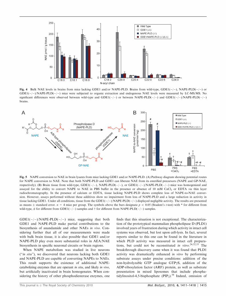
Fig. 4 Bulk NAE levels in brains from mice lacking GDE1 and/or NAPE-PLD. Brains from wild-type, GDE1(�/�), NAPE-PLD(�/�) orGDE1(�/�)/NAPE-PLD(�/�) mice were subjected to organic extraction and endogenous NAE levels were measured by LC-MS/MS. No
significant differences were observed between wild-type and GDE1(�/�) or between NAPE-PLD(�/�) and GDE1(�/�)/NAPE-PLD(�/�)brains.
Fig. 5 NAPE conversion to NAE in brain lysates from mice lacking GDE1 and/or NAPE-PLD. (A) Pathway diagram showing potential routes
for NAPE conversion to NAE. Note that both NAPE-PLD and GDE1 can liberate NAE from its esterified precursors (NAPE and GP-NAE,
respectively). (B) Brain tissue from wild-type, GDE1(�/�), NAPE-PLD(�/�), or GDE1(�/�)/NAPE-PLD(�/�) mice was homogenized and
assayed for the ability to convert NAPE to NAE in PBS buffer in the presence or absence of 10 mM CaCl2 or EDTA via thin layer
radiochromatography. In the presence of calcium or EDTA, tissue lacking NAPE-PLD shows complete loss of NAPE-to-NAE conver-
sion. However, assays performed without these additives show no impairment from loss of NAPE-PLD and a large reduction in activity in
tissue lacking GDE1. Under all conditions, tissue from the GDE1(�/�)/NAPE-PLD(�/�) displayed negligible activity. The results are presented
as means � standard error. n = 4 mice per group. The symbols above the bars designate p o 0.05 (Student’s t-test) with * for different from
wild-type, # for different from GDE1(�/�) samples and w for different from NAPE-PLD(�/�) samples.
This journal is �c The Royal Society of Chemistry 2010 Mol. BioSyst., 2010, 6, 1411–1418 | 1415

any of these factors resulted in almost complete loss of
activity. That enzyme, PLD1, is selective for phosphatidyl-
choline (PC), although to our knowledge it has not been tested
with N-acylated substrates. Additionally, there are another six
PLD isoforms in mammalian genomes, five of which are
virtually unstudied due to inability to show activity in vitro.
Furthermore, there are several GDE1 homologues that, to our
knowledge, have not yet been shown to display activity
in vitro. Finally, other pathways have been proposed for
NAPE catabolism involving C-type phospholipase (PLC)
activity (resulting in phospho-NAE, which is a substrate for
one or more phosphatases),35,36 although direct measurement
of NAPE-PLC activity has not yet been described. This does
not necessarily argue against a PLC pathway, since mammalian
C-type phospholipases have also been shown to have complex
requirements for co-factors and protein activators for reconsti-
tution of activity in vitro.37 Looking forward, the ability of
various co-factors to stimulate endogenous NAPE catabolic
pathway(s) should be explored to more clearly define their
composition (e.g., do they involve A/B-, C-, or D-type phospho-
lipases, or some combination thereof?) and possible role in
AEA biosynthesis in vivo. Finally, considering the recent
discovery of several classes of endogenous N-acyl amino acids
in brain tissue,38 it is worth investigating whether alternative
Fig. 6 Impaired NAE accumulation is observed following FAAH inhibition in brains from mice lacking GDE1 and NAPE-PLD. Mice were
treated with the selective FAAH inhibitor PF-3845 (10 mg kg�1, i.p.) and, after 3 hours, sacrificed and brain NAE levels measured. Significant
reductions were observed for several NAEs, including the endocannabinoid AEA (C20:4), in brains from GDE1(�/�)/NAPE-PLD(�/�) mice.
The results are presented as means � standard error. n = 4 mice per group. The symbols above the bars designate p o 0.05, p o 0.01, p o 0.005
(Student’s t-test) with *, **, *** for different from wild-type, #, ##, ### for different from GDE1(�/�) samples and w, ww, www for different from
NAPE-PLD(�/�) samples, respectively.
Fig. 7 In situNAE release fromNAPE in primary neuron cultures frommice lacking GDE1 andNAPE-PLD. (A) Time-dependence of NAE release
in the presence or absence of the FAAH inhibitor PF-3845. NAE release in the absence of neurons was negligible on this timescale (not shown).
(B) Assays conducted in primary neurons for 6 hours in the presence of PF-3845. Comparison of wild-type neurons to neurons from GDE1(�/�) orNAPE-PLD(�/�) mice reveals no impairment in the ability to release NAE from NAPE in situ. (C) Comparison of NAPE-PLD(�/�) neurons toGDE1(�/�)/NAPE-PLD(�/�) neurons reveals wild-type levels of NAE release in GDE1(�/�)/NAPE-PLD(�/�) neurons. (D) NAE release
from 14C-NAPE or an ether-linked (non-hydrolyzable) NAPE analogue in neurons from GDE1(�/�)/NAPE-PLD(�/�) mice reveals the
necessity of de-acylation prior to NAE release. These data represent single experiments performed in triplicate, representative of multiple experiments.
*, p o 0.05.
1416 | Mol. BioSyst., 2010, 6, 1411–1418 This journal is �c The Royal Society of Chemistry 2010

pathways for AEA/NAE production may exist that do not
involve NAPEs as obligate precursors.
Experimental
Generation of GDE1(�/�) mice
All animal experiments were performed in compliance with
protocols approved by The Scripps Research Institute’s Institu-
tional Animal Care and Use Committee. The GDE1 locus was
obtained as part of a BAC clone from the RPCI-23 library of
genomic DNA from a female C57BL/6J mouse. The targeting
construct was generated using PCR-amplified 50 and 30 homo-
logous recombination fragments surrounding exon 2 of theGDE1
gene, which were subcloned into NotI/EcoRI and XhoI/HindIII
sites in the pKO-NTKV vector. Homologous recombinant Bruce-
4 stem cell clones (C57BL/6 strain of origin) were identified by
Southern blot analysis, and one such clone was used to generate
chimeric mice on an albino C57BL/6 background, several of
which yielded germline transmission of the mutated gene.
In vitro enzyme assays
All assays were performed in tissue homogenates. Mice were
anesthetized with isoflurane and killed by decapitation. Tissues
were immediately removed and snap-frozen in liquid N2. Frozen
tissue was homogenized in phosphate buffered saline (PBS,
except where noted) using a Dounce-homogenizer and centri-
fuged at 1000� g to remove tissue debris. The supernatant from
this centrifugation step was adjusted to 2 mg mL�1 with PBS
and stored in single-use aliquots at �80 1C. Except where noted,assays were performed in glass vials in PBS at 37 1C for 30 min
in 100 mL total volume. Final protein concentration for enzyme
assays was 1 mg mL�1 for NAPE and lysoNAPE substrates,
and 0.1 mg mL�1 for GP-NAE substrates. Substrates (1,2-
dioleoyl-sn-glycero-3-phospho(N-[10-14C]-palmitoyl)ethanolamine,
1-oleoyl-2-hydroxy-sn-glycero-3-phospho(N-[10-14C]-palmitoyl)-
ethanolamine, and 1,2-dihydroxy-sn-glycero-3-phospho(N-[10-14C]-
palmitoyl)ethanolamine) were prepared as previously described.24,25
Substrates were stored as 2 mM stock in EtOH at �80 1C
and 5 mL was added to each enzyme assay (for 100 mM final
concentration). After 30 min, 1.5 mL of 2 : 1 CHCl3 : MeOH
was added to quench the reactions and an additional 0.4 mL
of 1% formic acid was added. Vials were mixed and
briefly centrifuged to separate phases and the organic
(bottom) phase was removed to fresh vials and evaporated
to dryness under N2. Lipids thus extracted were resuspended
in 20 mL 2 : 1 CHCl3 : MeOH and spotted on thin layer silica
plates and developed in 40 : 10 : 1 CHCl3 : MeOH : 28%
NH4OH. Distribution of radioactivity on the plates was quantified
by a phosphorimaging device (Packard) and products were identi-
fied by comparison to synthetic standards. Heat-denatured or
no-protein controls were included with all assays and these values
were subtracted as background. As noted in the text, some assays
included one of the following additives: 10 mM EDTA, 10 mM
CaCl2 or 0.1% Triton X-100.
Primary neuron culture and in situ assays
Culturing of primary neurons was performed essentially as
previously described.39 Briefly, P0 pups were taken from their
mothers and kept at B37 1C prior to dissection. Pups were
tailed and PCR-genotyped within 2 h of separation from their
mothers (Fig. 1D). Pups with the desired genotypes were
sacrificed and forebrains were dissected and dissociated by
incubation for 7 min at 37 1C in digestion solution containing
6 mg mL�1 trypsin, 0.5 mg mL�1 DNAse, 137 mM NaCl,
5 mM KCl, 7 mM Na2HPO4, and 25 mM HEPES, pH 7.2.
The dissociated cells were washed once with Hank’s balanced
salt solution (HBS) containing 20% fetal bovine serum (FBS)
and once in serum-free HBS, and further dissociated by gentle
trituration in HBS containing 12 mM MgSO4 and 0.5 mg mL�1
DNAse. The cell suspension was centrifuged for 3 min
at 1000 � g and plated in Matrigel-coated 6-well dishes
in MEM supplemented with glutamine, insulin, transferrin,
glucose, and 10% FBS. Cells from the forebrain of a single
pup were used to plate 6 wells in a 6-well dish. After B12 h,
the media was removed and replaced with MEM supple-
mented with glucose, transferrin, B27, glutamine, and 5%
FBS (‘‘growth media’’). After 3 days of culture (when glial
cells reached B40–50%), 50% of the culture-medium was
replaced with fresh growth medium containing 10 mM Ara-C
(for 5 mM final concentration) to prevent glial proliferation.
The cultures were maintained in medium containing 5 mMAra-C. After 6 days in vitro, 2 mL PF-3845 in DMSO or
DMSO was added to a final concentration of 10 mM and cells
were returned to the incubator. After 1 h, media was removed,
and replaced with 1 mL of fresh growth media containing
10 mMPF-3845 as well as 14C-labeled substrate at 20 mM (final
concentration), and neurons were returned to the incubator
for various time points, typically 6 h. After 6 h, each well was
scraped and neurons and media were transferred to a glass vial
containing 6 mL of 2 : 1 CHCl3 : MeOH and 1 mL of 1%
formic acid for 8 mL total. Vials were vortexed to mix,
centrifuged to separate phases, and the organic (bottom) phase
was removed to a fresh glass vial and evaporated to dryness
under a stream of N2. Dried reactions were then analyzed by
thin layer radiochromatography as described above.
Brain-lipid extraction and tandem MS analysis of NAE and
GP-NAE
Endogenous lipid measurements were performed essentially as
previously described. Briefly, male mice between 2 and 6
months of age were anesthetized with isoflurane and killed
by decapitation. Brains were rapidly removed, sectioned into
left and right hemispheres, and immediately frozen in liquid
N2. Frozen brains were placed in a Dounce homogenizer with
8 mL of 2 : 1 : 1 CHCl3 : MeOH : 50 mM Tris, pH 8.0, and
200 pmol of 1,2-dihydroxy-sn-glycero-3-phospho(N-penta-
decenoyl)ethanolamine (C15:1 GP-NAE) or 2 pmol of arachi-
donoyl-(d4)-ethanolamine for GP-NAE or NAEmeasurements,
respectively. Lipids were extracted via homogenization with
8–10 strokes of the Dounce homogenizer at which point
homogenates were transferred to 8 mL glass vials and centri-
fuged for 10 min at 1400 � g. The organic (bottom) phase was
transferred to a clean vial and the remaining aqueous
phase was re-extracted with an additional 4 mL of CHCl3.
After centrifugation, the organic phases were pooled and
evaporated to dryness under a stream of N2. Lipid extracts
This journal is �c The Royal Society of Chemistry 2010 Mol. BioSyst., 2010, 6, 1411–1418 | 1417

from each half-brain were then resuspended in 120 mL 2 : 1
CHCl3 : MeOH and transferred to LC-MS vials for immediate
analysis or stored at �80 1C. 15 mL of resuspended lipid was
injected into an Agilent 6410 triple quadrupole mass spectro-
meter with an Agilent 1200 autosampler. Samples were
chromatographed on a Gemini 5 mm C18 column (50 � 4.6 mm,
Phenomenex, Torrance, CA) in the negative-ion mode for
GP-NAE measurements and the positive-ion mode for NAE
measurements at a flow-rate of 0.4 mL min�1. GP-NAEs and
NAEs were monitored by multiple reaction monitoring
(MRM). For GP-NAEs the transition from [M � H]� to
[M � H � 74]� with collision energy 35 V was monitored
and compared to the internal C15:1 GP-NAE standard and
measurements of endogenous GP-NAEs were corrected for
extraction efficiency as previously described.25 For NAEs, the
MRM transition from [M + H]+ to m/z 62+ (ethanolamine
fragment) with collision energy 11 V was used, except for the
d4-AEA standard which monitored the transition from
[M + H]+ to m/z 66+ (d4 ethanolamine fragment). NAEs
were quantified by comparison to the d4-AEA standard. The
dwell-time was set to 100 ms for all lipids. For ex vivo analysis
of GP-NAEs, half-brains were homogenized in a Dounce
homogenizer in 2 mL of 50 mM Tris pH 8.0 with or without
10 mM EDTA. Samples were incubated at room temperature
for 4 h with gentle rotation prior to extraction and GP-NAE
quantification as described above.
References
1 P. Pacher, S. Batkai and G. Kunos, Pharmacol. Rev., 2006, 58,389–462.
2 A. C. Howlett, C. S. Breivogel, S. R. Childers, S. A. Deadwyler,R. E. Hampson and L. J. Porrino, Neuropharmacology, 2004, 47,345–358.
3 L. Walter and N. Stella, Br. J. Pharmacol., 2004, 141, 775–785.4 C. J. Fowler, S. Holt, O. Nilsson, K. O. Jonsson, G. Tiger andS. O. Jacobsson, Pharmacol., Biochem. Behav., 2005, 81, 248–262.
5 K. Ahn, M. K. McKinney and B. F. Cravatt, Chem. Rev., 2008,108, 1687–1707.
6 V. Di Marzo, Rev. Physiol., Biochem., Pharmacol., 2008, 160, 1–24.7 H. Cadas, S. Gaillet, M. Beltramo, L. Venance and D. Piomelli,J. Neurosci., 1996, 16, 3934–3942.
8 C. J. Fowler, Fundam. Clin. Pharmacol., 2006, 20, 549–562.9 S. T. Glaser, M. Kaczocha and D. G. Deutsch, Life Sci., 2005, 77,1584–1604.
10 D. G. Deutsch, N. Ueda and S. Yamamoto, Prostaglandins,Leukotrienes Essent. Fatty Acids, 2002, 66, 201–210.
11 H. H. Schmid, Chem. Phys. Lipids, 2000, 108, 71–87.12 W. A. Devane, L. Hanus, A. Breuer, R. G. Pertwee,
L. A. Stevenson, G. Griffin, D. Gibson, A. Mandelbaum,A. Etinger and R. Mechoulam, Science, 1992, 258, 1946–1949.
13 C. C. Felder, E. M. Briley, J. Axelrod, J. T. Simpson, K. Mackieand W. A. Devane, Proc. Natl. Acad. Sci. U. S. A., 1993, 90,7656–7660.
14 D. E. Epps, P. C. Schmid, V. Natarajan and H. H. O. Schmid,Biochem. Biophys. Res. Commun., 1979, 90, 628–633.
15 V. Natarajan, P. C. Schmid, P. V. Reddy, M. L. Zuzarte-Augustinand H. H. O. Schmid, J. Neurochem., 1983, 41, 1303–1312.
16 M. Colodzin, N. R. Bachur, H. Weissbach and S. Udenfriend,Biochem. Biophys. Res. Commun., 1963, 10, 165–170.
17 D. G. Deutsch and S. A. Chin, Biochem. Pharmacol., 1993, 46,791–796.
18 T. Sugiura, S. Kondo, A. Sukagawa, T. Tonegawa, S. Nakane,A. Yamshita and K. Waku, Eur. J. Biochem., 1996, 240,53–62.
19 V. Natarajan, P. C. Schmid, P. V. Reddy and H. H. Schmid,J. Neurochem., 1984, 42, 1613–1619.
20 Y. Okamoto, J. Morishita, K. Tsuboi, T. Tonai and N. Ueda,J. Biol. Chem., 2003, 279, 5298–5305.
21 N. Ueda, Q. Liu and K. Yamanaka, Biochim. Biophys. Acta, Mol.Cell Biol. Lipids, 2001, 1532, 121–127.
22 J. Wang, Y. Okamoto, J. Morishita, K. Tsuboi, A. Miyatake andN. Ueda, J. Biol. Chem., 2006, 281, 12325–12335.
23 D. Leung, A. Saghatelian, G. M. Simon and B. F. Cravatt,Biochemistry, 2006, 45, 4720–4726.
24 G. M. Simon and B. F. Cravatt, J. Biol. Chem., 2006, 281,26465–26472.
25 G. M. Simon and B. F. Cravatt, J. Biol. Chem., 2008, 283,9341–9349.
26 B. Zheng, D. Chen and M. G. Farquhar, Proc. Natl. Acad. Sci.U. S. A., 2000, 97, 3999–4004.
27 B. Zheng, C. P. Berrie, D. Corda and M. G. Farquhar, Proc. Natl.Acad. Sci. U. S. A., 2003, 100, 1745–1750.
28 D. S. Johnson, K. Ahn, S. Kesten, S. E. Lazerwith, Y. Song,M. Morris, L. Fay, T. Gregory, C. Stiff, J. B. Dunbar, Jr.,M. Liimatta, D. Beidler, S. Smith, T. K. Nomanbhoy andB. F. Cravatt, Bioorg. Med. Chem. Lett., 2009, 19, 2865–2869.
29 K. Ahn, D. S. Johnson, M. Mileni, D. Beidler, J. Z. Long,M. K. McKinney, E. Weerapana, N. Sadagopan, M. Liimatta,S. E. Smith, S. Lazerwith, C. Stiff, S. Kamtekar, K. Bhattacharya,Y. Zhang, S. Swaney, K. Van Becelaere, R. C. Stevens andB. F. Cravatt, Chem. Biol., 2009, 16, 411–420.
30 D. Fegley, S. Gaetani, A. Duranti, A. Tontini, M. Mor,G. Tarzia and D. Piomelli, J. Pharmacol. Exp. Ther., 2004, 313,352–358.
31 V. Di Marzo, A. Fontana, H. Cadas, S. Schinelli, G. Cimino,J.-C. Schwartz and D. Piomelli, Nature, 1994, 372, 686–691.
32 S. Cockcroft, Biochim. Biophys. Acta, 1992, 1113, 135–160.33 T. W. Martin and K. Michaelis, J. Biol. Chem., 1989, 264,
8847–8856.34 H. A. Brown, S. Gutowski, C. R. Moomaw, C. Slaughter and
P. C. Sternwels, Cell (Cambridge, Mass.), 1993, 75, 1137–1144.35 J. Liu, L. Wang, J. Harvey-White, B. X. Huang, H.-Y. Kim,
S. Luquet, R. D. Palmiter, G. Krystal, R. Rai, A. Mahadevan,R. K. Razdan and G. Kunos, Neuropharmacology, 2008, 54,1–7.
36 J. Liu, L. Wang, J. Harvey-White, D. Osei-Hyiaman, R. Razdan,Q. Gong, A. C. Chan, Z. Zhou, B. X. Huang, H. Y. Kim andG. Kunos, Proc. Natl. Acad. Sci. U. S. A., 2006, 103, 13345–13350.
37 A. V. Smrcka, J. R. Hepler, K. O. Brown and P. C. Sternweis,Science, 1991, 251, 804–807.
38 B. Tan, D. K. O’Dell, Y. W. Yu, M. F. Monn, H. V. Hughes,S. Burstein and J. M. Walker, J. Lipid Res., 2009,M900198–JLR900200.
39 A. Maximov, Z. P. Pang, D. G. R. Tervo and T. C. Sudhof,J. Neurosci. Methods, 2007, 161, 75–87.
1418 | Mol. BioSyst., 2010, 6, 1411–1418 This journal is �c The Royal Society of Chemistry 2010