Embed Size (px)
Citation preview

29TH DAAAM INTERNATIONAL SYMPOSIUM ON INTELLIGENT MANUFACTURING AND AUTOMATION
DOI: 10.2507/29th.daaam.proceedings.136
CHARACTERIZATION OF MATERIALS USED IN 3D-PRINTING
TECHNOLOGY WITH DIFFERENT ANALYSIS TECHNIQUES
Mª Angeles Castro-Sastre, Ana Isabel Fernández-Abia,
Pablo Rodríguez-González, Susana Martínez-Pellitero & Joaquín Barreiro
This Publication has to be referred as: Castro-Sastre, A[ngeles]; Fernández-Abia, A[na] I[sabel]; Rodriguez-Gonzalez,
P[ablo]; Martínez-Pellitero, S[usana] & Barreiro, J[oaquin] (2018). Characterization of Materials Used in 3D-Printing
Technology with Different Analysis Techniques, Proceedings of the 29th DAAAM International Symposium, pp.0947-
0954, B. Katalinic (Ed.), Published by DAAAM International, ISBN 978-3-902734-20-4, ISSN 1726-9679, Vienna,
Austria
DOI: 10.2507/29th.daaam.proceedings.136
Abstract
3D inkjet printing is mainly a powder-based method where layers of solid particles are bounded together by means of a
printed liquid material to generate a 3D model. In this process, it is necessary to control different parameters that can
significantly affect the quality of 3D printed parts, such as: chemical composition, structural elucidation, particles
morphology, among others. For this reason, the purpose of this research was to develop a study of the chemical and
physical properties of the two involved materials: the powder, hemihydrate calcium sulphate 80-90% of purity, and the
binder, 2-pyrrolidone 1% of purity. Both materials were studied with different techniques to characterize different
parameters. Raman and FT-IR spectrometry and X-ray diffractometer (XRD) were used to study structural elucidation
and presence to others phases of this sulphate powder; for knowing the kind of impurities and morphological aspect; we
used scanning electron microscope (SEM), equipped with an energy-dispersive X-ray detecting system (ED-XRS), and
inductive coupling plasma (ICP) with optical emission spectrophotometer (OES). The results showed the real composition
of solid powder and liquid binder. The images of secondary electrons obtained showed the presence of alpha and beta
phase of called hemihydrated sulphate. The presence of other phases of sulphate, such as dihydrate and anhydrous, was
also detected. This information is important to know the properties of the manufactured part.
Keywords: 3D inkjet printing; material characterization; additive manufacturing; powder bed printing; calcium sulphate
1. Introduction
Additive Manufacturing (AM) has emerged as a technology for manufacturing objects with complex geometries from
three-dimensional (3D) model data. New AM methods, materials and applications are being developed continuously. A
review of materials, methods, applications and challenges can be found in [1]. One of the most important AM methods
for ceramic components is three-dimensional printing (3DP). This method is a powder-based process where fine powder
particles are bound together with a liquid binder to create a 3D part. The powder particles are spread and packed on a
platform and a liquid binder is deposited onto the powder layer, using a printer head, to join the particles.
- 0947 -

29TH DAAAM INTERNATIONAL SYMPOSIUM ON INTELLIGENT MANUFACTURING AND AUTOMATION
The process is repeated by spreading a new layer of powder on top of the previous layer that results in the creation of
a 3D structure [2] [3]. 3DP technology can use a wide variety of materials (powder and binder) for different applications.
In order to build ceramic components, the most used powders are aluminium oxide (Al2O3), zirconium oxide (ZrO2),
silicium oxide (SiO2), calcium hemihydrate (CaSO4 ½ H2O) and calcium hydroxyapatite (Ca5(PO4)3H)). On the other
hand, different types of binder have also been used: 2-Pyrrolidone (C4H7NO), -n-butyl cyanoacrylate (NBCA)
(C8H11NO2), Polyvinyl Alcohol (PVA) ((C2H4O)x) and 10 wt% phosphoric acid (H3PO4) among others [4].
Calcium hemihydrate, also called Plaster of Paris (Bassanite), was the first material used for 3D Printing technology.
This material has been widely used in many areas such as precision instrument moulds, ceramics, industrial arts and
architecture, due to its workability [5]. However, applications of this material using 3DP technology are focused in
medical and dental applications. There are numerous works that study the optimization of the process for bone scaffolds
fabrication [6] [7] and to make dental models [8]. The advantage of using Calcium hemihydrate for medical applications
is because it is harmless to humans and has the ability to directly print porous scaffolds with designed shape, controlled
chemistry and interconnected porosity [9]. For these applications and for any other, it is very important to know the
chemical and physical properties of the material. The material for 3DP technology is supplied as powder, and its
characteristics such as size, shape and distribution of particle, chemical composition, physical properties and
microstructure will significantly influence the resulting structure and, therefore, affect the properties of the printed part.
All these characteristics are related to the method of obtaining the material.
Calcium hemihydrate is produced commercially by partial dehydration of gypsum (CaSO4.2H2O). This process of
dehydration consists in removing ¾ of the combined water from the gypsum, using sometimes small amounts of chemical
additives. Depending on the production method that is used, wet (e.g. autoclaving) or dry (e.g. calcining), the hemihydrate
is into one of two phases, or , respectively. Several authors have different opinions about the physical differences
about the two phases of the hemihydrate [10]. On the other hand, use of chemical additives affects the chemical and
physical properties of calcium hemihydrate (morphology and growth of crystals). These properties may change depending
on the quantity, form and stage of the process in which the additives are added. Also, the nature of the additives (organic
or inorganic) modifies the calcium hemihydrate properties. Therefore, we can conclude that the calcium hemihydrate
morphology depends on the formation conditions and the content of chemical additives.
To characterize the material, the most used techniques, selected by their usefulness, are generally SEM-EDX, Raman
Spectroscopy, X-Ray Diffraction (XDR) or Dumas Analysis [11] [12] [13]. The present work studies the real chemical
composition and physical properties (microstructure and morphology) of raw materials, using the techniques advised on
the bibliography and others complementary techniques (ICP-OES; FTIR), in order to know the properties of raw
materials, with the aim of finding different applications for this material using 3DP technology.
2. Experimental Procedure
In this study a commercial plaster based powder CaSO4. 1/2H2O (VisiJet PXL Core) with a purity of 80-90% with
an appropriate water based binder solution of 2-Pyrrolidone C4H7NO (purity of 0-1%) (VisiJet PXL Colors Clear) were
used as raw materials. Both materials are used in a 3DP machine Project 660Pro (3DSystem, USA) and have the properties
showed in Table 1 according to the manufacturer’s safety data sheet.
Binder Plaster based powder
pH (20ºC) 9,8 -
Boiling/Melting point (ºC) 100 1450
Density (g/cm3) 1 2,6 – 2,7
Table 1. Binder and powder properties according to the manufacturer’s safety data sheet
The morphological shape of the powder particles and their chemical composition were assessed using electron
microscope analysis. A JEOL scanning electron microscope (Model JSM-6100) equipped with energy dispersive
spectroscopy (EDS) (LINK) operated under recommended conditions (20kV acceleration voltage and 5nA probe current).
Carbon tape was used to fix the specimens on the sample holder to discharge the negative charge in the microscope.
Subsequently, backscattered electron images were obtained and the spectra of energy dispersive X-ray microanalysis were
obtained.
In order to know the presence of impurities on the raw materials, we used inductive coupling plasma (ICP) with optical
emission spectrophotometer (OES) (ICP-OES). The model was Perkin Elmer Optima 2000DV. To analyze the powder,
0.5 g of the gypsum sample was resuspended in 10 ml of 63% nitric acid. Appropriate dilutions were made for the analysis,
adding 5ppm of Sc as indicated by internal standard, to perform the calibration line. The binder did not need to be diluted
because its composition is mainly water.
- 0948 -
29TH DAAAM INTERNATIONAL SYMPOSIUM ON INTELLIGENT MANUFACTURING AND AUTOMATION
The structure analysis was assessed by a PhilipsPW1830 (high voltage generator) powder X-ray diffractometer (XRD)
(Philips, Almelo, The Netherlands) with a Philips PW1710/00 (diffractometer controller), working in Bragg–Brentano
diffraction geometry. The current and voltage used were 30 mA and 40 kV, respectively. Powder samples were prepared
in the form of thin layer on a zero background Si (911) substrate using Cu (Kα) as incident radiation.
The scattered intensities were recorded in the 2θ span of 15–80°. The powder was also analysed using Raman
spectroscopy with a portable BWTEK (i-Raman) spectrometer fitted with a CCD refrigerated detector. Raman
spectrometry measurements were performed at room temperature using the 785 nm line of an argon–ion laser as the
excitation source, model Clean Laze (>300mW). The power level was set nominally at 100%, but it had to be reduced on
several occasions due to saturation of the detector. The experimental conditions were 10 s accumulation time, 1 min
acquisition time and spectra were scanned from 200 to 2500 cm-1. With this technique we cannot study the presence of
water molecules, since it only allows a working range up to 3000 cm-1 and water bands appear far away this value. Taking
into account that calcium sulphate hemihydrate has different functional groups that absorb infrared light at different
wavelengths to those of water, we used Fourier transform infrared (FTIR) spectroscopy (IFS66v/S, Bruker, Germany).
The spectra were collected in transmittance mode in the 4000– 450 cm−1 range.
Moreover the powder was analysed with the Dumas method to know the presence of carbon. This method consists in
a complete combustion in an atmosphere with oxygen with reduction of the not interesting elements in a reactor. Helium
is used as carrier gas at the end of the process. After passing through a water trap, N2 and CO2 are obtained and introduced
in a chromatographic oven with TCD, which generates the corresponding chromatograms. The equipment used was
EuroVector Elemental Analyser, EURO model EA 3000 / J1F. The sample was finely ground. Therefore, no previous
pre-processing was necessary. It was weighed, in triplicate, in tin capsules. The working parameters were the following:
carrier flow (ml/min) 100 +/- 5; carrier (KPa) 130; purge (ml/min) 130-120; oxygen (ml) 20; sampling delay (s) 8; run
time (s) 140; front furnace temp (ºC) 980; GC Oven temp (ºC) 100. Four patterns were used with known concentrations
of C.
3. Results and Discussion
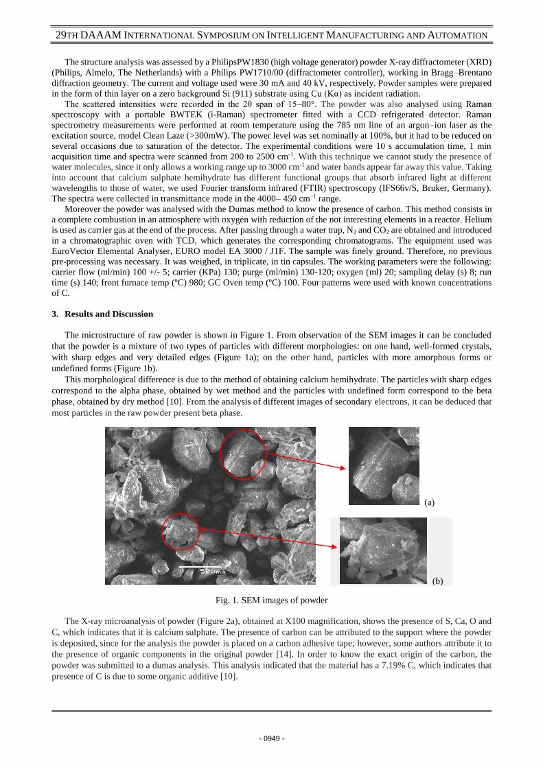
The microstructure of raw powder is shown in Figure 1. From observation of the SEM images it can be concluded
that the powder is a mixture of two types of particles with different morphologies: on one hand, well-formed crystals,
with sharp edges and very detailed edges (Figure 1a); on the other hand, particles with more amorphous forms or
undefined forms (Figure 1b).
This morphological difference is due to the method of obtaining calcium hemihydrate. The particles with sharp edges
correspond to the alpha phase, obtained by wet method and the particles with undefined form correspond to the beta
phase, obtained by dry method [10]. From the analysis of different images of secondary electrons, it can be deduced that
most particles in the raw powder present beta phase.
(a)
(b)
Fig. 1. SEM images of powder
The X-ray microanalysis of powder (Figure 2a), obtained at X100 magnification, shows the presence of S, Ca, O and
C, which indicates that it is calcium sulphate. The presence of carbon can be attributed to the support where the powder
is deposited, since for the analysis the powder is placed on a carbon adhesive tape; however, some authors attribute it to
the presence of organic components in the original powder [14]. In order to know the exact origin of the carbon, the
powder was submitted to a dumas analysis. This analysis indicated that the material has a 7.19% C, which indicates that
presence of C is due to some organic additive [10].
- 0949 -

29TH DAAAM INTERNATIONAL SYMPOSIUM ON INTELLIGENT MANUFACTURING AND AUTOMATION
Fig. 2. SEM microanalysis of powder
Results obtained in additional areas on the particles also confirmed the presence of other elements (Figure 2b and 2c).
The spectra obtained showed the presence of Si, K, Al, Mg and Na, which are on traces respect to S, O and Ca. Intensity
of peaks relative to these elements are very low, as well as the weight percentage.
Therefore, these additional elements are considered impurities (Table 2). Except for Si, which is attributed to the
original mineral from which the gypsum is obtained, the rest of elements are supplementary additives included to improve
workability of the gypsum. This chemical composition of material is important because affects the properties of the 3DP
printed product, such as porosity, hardness and mechanical strength. Moreover, impurities like K, Na and Mg are known
to play a key role in the formation of gypsum and the dehydration process.
Element O S Ca C Na Al Mg Si K
%Weight 40,31 13,79 17,89 28,01 0,28 0,29 0,95 1,40 0,67
Table 2. Chemical composition of powder
Due to the importance of impurities content in the properties in the final product, a more detailed analysis was carried
out. The analysis performed was ICP-OES, since it allows knowing exactly the impurities composition in the raw material.
The result indicated that the predominant elements are Na and K, which are added to calcium sulphate to facilitate the
setting process. There is also a high content of Si, which is present in the material used to obtain the plaster. Mg and Al
are also present as trace elements.
Analite Mg (285,213) Na (589,592) K (766,490) Al (396,153) Si (251,611)
CaSO4•1/2H2O
(mg/Kg) <4 508,26 679,66 <4 253,75
Table 3. ICP-OES powder analysis
Once the elemental composition of the powder is known, the next step consisted in analyse the phases in the
CaSO4.nH2O system, as well as a structural study of it. For this purpose a diffraction analysis was carried out. Figure 3
displays the XRD spectra of the powder. Diffraction lines with higher intensity were found in the follow diffraction
angles: 2θ = 14.71°, 25.65°, 29.72° and 31.8º, corresponding to the respective planes (200), (220), (400) and (204). The
peaks with lower intensities were observed at 2θ = 31.79°, 42.24°, 49.18°, 52.92°, 54.09° and 55.11°, associated with the
planes (422), (424), (207), (604) and (620), respectively. All these peaks are attributed to bassanite (calcium sulphate
hemihydrate) [16].
Some peaks of minimum intensities, such as 2θ = 25.33°, 39.66° and 47.58° are attributed to the anhydrite. These
materials are a subproduct that comes from the gypsum calcination process. According to the XDR analysis, gypsum is
also present, as the peak at 11.54º indicates. Also, the powder contains quartz, as indicated by the peak at 27,64º. In the
previous XRay analysis it was detected Si which is an element contained in quartz. Quartz and gypsum are the species
present in the mineral utilised for obtaining bassanite.
According to the pdf ICDD card 00-033-0310, 01-081-1848 consulted in the diffraction equipment database we can
determine that the powder contains a higher bassanite quantity (60,9 % and 35,1 %) compared with the lesser amounts of
the other sulphate phases (anhydrite and gypsum, 3,2 % and 0,7 % respectively) and quartz (0,2%). These quantitative
analysis of present phases was confirmed using (1) proposed by Panttanayak [15].
% 𝒑𝒉𝒂𝒔𝒆 =𝑰𝒏𝒕𝒆𝒏𝒔𝒊𝒕𝒚 𝒐𝒇 𝒎𝒂𝒋𝒐𝒓 𝒑𝒆𝒂𝒌 𝒐𝒇 𝒕𝒉𝒆 𝒑𝒉𝒂𝒔𝒆 𝒕𝒐 𝒃𝒆 𝒅𝒆𝒕𝒆𝒓𝒎𝒊𝒏𝒆𝒅
∑ 𝑰𝒏𝒕𝒆𝒏𝒔𝒊𝒕𝒚 𝒐𝒇 𝒎𝒂𝒋𝒐𝒓 𝒑𝒆𝒂𝒌𝒔 𝒐𝒇 𝒂𝒍𝒍 𝒕𝒉𝒆 𝒑𝒉𝒂𝒔𝒆𝒔 (1)
- 0950 -

29TH DAAAM INTERNATIONAL SYMPOSIUM ON INTELLIGENT MANUFACTURING AND AUTOMATION
Fig. 3. XRD powder pattern. (A: Anhydrite; B: Bassanite; G: Gypsum)
According to pdf ICCD letters, bassanite has two different structures: orthorhombic and hexagonal. These two
structures are indicated by the intensity relation of the 14,73º and 29,72º peaks. In Figure 3 it can be observed that I29,72 >
I14,73, which indicates that most of the structure is orthorhombic. The presence of peaks with high intensities at 29.29º and
31.82º suggests the presence of hexagonal bassanite. Orthorhombic structure has been attributed to phase of the
bassanite [16]. These results are according to SEM images (Figure 1). This information is important for the porosity study
of the 3D printed part using this material [17].
In order to confirm the material characterization, the powder was analysed using Raman and FT-IR. In Raman
technique it is known that the system of calcium sulphate (CaSO4.nH2O) has a main Raman band (Table 4). In the Raman
spectra (Figure 4), the area of interest was between 1000 - 1300 cm-1, where the most intense peaks appear relative to the
different phases of CaSO4.nH2O. Both spectrum carried out on the material indicate the presence of three phases: gypsum,
anhydrite and basanite at these absorption peaks 1009,16 cm-1;1017,90 cm-1 and 1015,62 cm-1 respectively, confirming
that bassanite is the main phase in the powder.
Gypsum Bassanite Anhydrite III Anhydrite II Anhydrite I
Main Raman Band 1008cm-1 1015cm-1 1025cm-1 1017cm-1 1017cm-1
Table 4. Summary of the different Raman band of the hydration−dehydration system of CaSO4•nH2O
Fig. 4. Raman spectra of the powder
- 0951 -

29TH DAAAM INTERNATIONAL SYMPOSIUM ON INTELLIGENT MANUFACTURING AND AUTOMATION
Unfortunately, the Raman equipment used did not allow us to study the area from 2900cm-1, which would let to know
evolution of water content in the material when it is heated. To study the evolution of water, we performed the analysis
FT-IR. Calcium sulphate hemihydrate only contains one type of water molecules, and its characteristic mode of vibration
in IR corresponds to a peak at 1617cm-1, usually accompanied by two less intense and wider peaks at 3600cm-1 and
3550cm-1. All these modes of vibration correspond to the water contained in the structure of calcium sulphate [18]. In the
FT-IR spectra showed in Figure 5, it is clear the presence of bands positioned at 3549 cm-1 and 3607 cm-1, attributed to
the water content of bassanite [16]. Groups of SO42- were also identified (593,96; 660,48 and 1005 to 1132cm-1).
Fig. 5. FT-IR spectra of the powder
The binder used in the 3D printing process to join the powder particles was also analysed. According the safety date-
sheet provided by 3D System, its chemical composition is 2-pyrrolidone with a purity of 1%. It is necessary to know the
exact binder composition for studying the agglomeration process with the raw powder and also to be able to perform
adequate thermal post-treatments. For this analysis two techniques were used: ICP-OES and FT-IR. The FT-IR spectra
of the binder and pure water appear in Figure 6. The absorption peaks in the region from 1500cm-1 to 4000cm-1 are
characteristic of pure water. Figure 6 shows the coincidence of water and binder absorption bands in this region. We can
conclude that pure water is the majority component on the binder. At lower absorption values (1042,33 cm-1 and
1112,72cm-1) appear peaks which correspond to the 2-pyrrolidone [19].
Fig. 6. FT-IR spectra of the binder
Additionally to this study, it was decided to analyse the presence of impurities on the binder. Similarly to the previous
study carried out on the powder we used ICP-OES technique. The elements found and their quantity are listed in Table
5. It can be observed the presence of Na, K and Mg. All of them are used to improve the setting process, as aforementioned.
Analite Mg (285,213) Na (589,592) K (766,490) Al (396,153)
Binder (mg/l) 0,13 73,20 2,24 <0,10
Table 5. Chemical composition impurities in the binder
- 0952 -

29TH DAAAM INTERNATIONAL SYMPOSIUM ON INTELLIGENT MANUFACTURING AND AUTOMATION
4. Conclusions
Three-dimensional printing process (3DP) highly depends on the used material. Hence, to carry out a in depth study
of both powder and binder materials used in 3DP is necessary. Knowledge of composition and properties of materials can
improve significantly the 3DP process, allowing extending the use of this technology to new applications. Properties of
materials, such as existence of different phases, impurities content, size or shape of crystals affect the printing process
and the final part properties. These properties affected are mechanical strength, porosity, surface roughness and post-
processing behaviour. In order to obtain parts which satisfy the requirements for different applications, to know
composition and properties of materials used for printing the part is necessary. Therefore, the aim of this study was to
characterize one of the most used materials in 3DP technology, hemihydrate calcium sulphate, and the binder, 2-
pyrrolidone 1% of purity. Based on the results discussed in this study, the following remarks can be drawn:
The results of different microscopy and spectroscopy techniques show that the powder exhibits all the species of the
system CaSO4•n H2O. These species are gypsum, hemihydrate and anhydrite, being hemihydrate the main specie.
Moreover, it is confirmed that this hemihydrate is a mix between two phases (alpha and beta), being beta the main phase.
The alpha and beta phases generate different porous structure, which affect the mechanical properties of the printed parts.
The beta phase generates micro cracks and interconnected porous and the alpha phase generates a more homogeneous
porosity.
The results obtained with ICP-OES technique showed the exact composition of the materials used in three-dimensional
printing (3DP). Both, powder and binder, have impurities such as Na, K and Mg, which are added to improve the setting
process. Moreover, these impurities can improve the mechanical (resistance to compression) and morphological
properties (porosity).
The above information plays a major role in determining the final properties of the 3D printed parts. For example, for
applications in which mechanical strength is necessary, a material with more alpha phase content and presence of
impurities is required. On the other hand, for applications that require higher porosity in the part, a material with high
content of beta phase should be chosen; it is due to alpha phase have crystals with flat faces, so that when joining they
imbricate each other giving the whole a great continuity, thus observing a lower porosity than in betha plaster.
Characterization of material is the first step necessary for further development of 3DP specific implementations. Once
the composition and properties of the raw materials are known, other studies can be addressed for improving the printing
process parameters, the properties of final part or the post-treatment techniques.
5. Acknowledgments
Authors thank to Ministry of Science, Innovation and Universities of Spain for the support through the research project
with reference DPI2017-89840-R and to the Junta de Castilla y León through the research project with reference FEDER
P17-LE027P17.
6. References
[1] Ngo, T., Kashani, A., Imbalzano, G., Nguyen, K. T. Q. & Hui, D. (2018). Additive manufacturing (3D printing): A
review of materials, methods, applications and challenges. Composites Part B Engineering, Vol. 143, 2018, pp. 172-
196, DOI: 10.1016/j.compositesb.2018.02.012.
[2] Peltola, S. M., Melchels, F. P. W., Grijpma, D. W. & Kellomäki, M. (2008). A review of rapid prototyping
techniques for tissue engineering purposes. Annals of Medicine. Vol. 40, Nº 4, 2008, pp. 268-280,
DOI:10.1080/07853890701881788.
[3] Leukers, B., Gülkan, H., Irsen, S.H. et al. (2005). Hydroxyapatite scaffolds for bone tissue engineering made by 3D
printing. Journal of Materials Science: Materials in Medicine. Vol. 16, Nº 12, 2005, pp. 1121-1124,
DOI:10.1007/s10856-005-4716-5. [4] Hwa, L. Ch., Rajoo, S., Noor, A. M., Ahmad, N. & Uday, M.B. (2017). Recent advances in 3D printing of porous
ceramics: A review. Current Opinion in Solid State and Materials Science. Vol. 21, 2017, pp. 323-347, DOI:10.1016/j.cossms.2017.08.002
[5] Lewry, A.J. & Williamson, J. (1994). The setting of gypsum plaster. Part II. The development of microstructure and
strength. Journal of Materials Science. Vol. 29, Nº 21, 1994, pp. 5524-5528, DOI:10.1007/BF00349943
[6] Farzadi, A., Waran, V., Solati-Hashjin, M., Abdul Rahman, Z. A., Asadi, M. & Abu Osman, N. A. (2015). Effect of
layer printing delay on mechanical properties and dimensional accuracy of 3D printed porous prototypes in bone
tissue engineering. Ceramics International, Vol. 41, 2015, pp. 8320-8330, DOI: 10.1016/j.ceramint.2015.03.004. [7] Zhou, Z., Mitchell, Ch. A., Buchanan, F. J. & Dunne, N. J. (2013). Effects of heat treatment on the mechanical and
degradation properties of 3D-Printed calcium-sulphate-based scaffolds. ISRN Biomaterials, Hindawi Publishing Corporation, Vol. 2013, 10 pages, article ID 750720, DOI: 10.5402/2013/750720
[8] Ledingham, A., English, J., Akyalcin, S., Cozad, B, Ontiveros, J. & Kasper, F. (2016). Accuracy and mechanical properties of orthodontic models printed 3-dimensionally from calcium sulfate before and after various postprinting treatments. American Journal of Orthodontics and Dentofacial Orthopedics, Vol. 150, Nº 6, 2016, pp. 1056-1062, DOI: 10.1016/j.ajodo.2016.04.027
- 0953 -
29TH DAAAM INTERNATIONAL SYMPOSIUM ON INTELLIGENT MANUFACTURING AND AUTOMATION
[9] Bose, S., Vahabzadeh, S. & Bandyopadhyay, A. (2013). Bone tissue engineering using 3D printing. Materials Today, Vol. 16, Nº 12, 2013, pp. 496-504, DOI: 10.1016/j.mattod.2013.11.017
[10] Singh, N.B.& Middendorf, B. (2007). Calcium sulphate hemihydrate hydration leading to gypsum crystallization. Progress in Crystal Growth and Characterization of Materials. Vol. 53, 2007, pp. 57-77, DOI:10.1016/j.pcrysgrow.2007.01.002.
[11] Zetkova, I; Kucerova, L; Zetek, M; Cesanek, J; Hanzl, P & Dana, M. (2017). Evaluation of Metal Powder for
Additive Manufacturing of Margaring Steel, Proceedings of the 28th DAAAM International Symposium, pp.0410-
0416, B. Katalinic (Ed.), Published by DAAAM International, ISBN 978-3-902734-11-2, ISSN 1726-9679,
Vienna, Austria. DOI: 10.2507/28th.daaam.proceedings.057
[12] Rubesova, K; Jenicek, S; Kana, J & Zetkova, I. (2016). Microstructure of MS1 Maraging Steel in 3D-Printed
Products After Semi-Solid Processing, Proceedings of the 27th DAAAM International Symposium, pp.0467-0472,
B. Katalinic (Ed.), Published by DAAAM International, ISBN 978-3-902734-08-2, ISSN 1726-9679, Vienna,
Austria. DOI: 10.2507/27th.daaam.proceedings.070
[13] Jenicek, S; Bublikova, D; Jirkova, H & Kana, J (2017). Stability of Retained Austenite in High-Strength
Martensitic Steels during Cold Deformation, Proceedings of the 28th DAAAM International Symposium, pp.0289-
0294, B. Katalinic (Ed.), Published by DAAAM International, ISBN 978- 3-902734-11-2, ISSN 1726-9679,
Vienna, Austria. DOI: 10.2507/28th.daaam.proceedings.039
[14] Asadi-Eydivand,M., Solati-Hashjin, M., Farzad, A. & Abu Osman, N. A. (2016). Effect of technical parameters on
porous structure and strength of 3D printed calcium sulfate prototypes. Robotics and Computer-Integrated
Manufacturing. Vol. 37, 2016, pp.57-67, DOI: 10.1016/j.rcim.2015.06.005 [15] Pattanayak, d. K., Dash, R., Prasad, R.C., Rao, B.T. & Rama Mohan, T.R. (2007). Synthesis and sintered properties
evaluation of calcium phosphate ceramics. Materials Science and Engineering: C, Vol. 27, No. 4, 2007, pp. 684–
690, DOI: 10.1016/j.msec.2006.06.021
[16] Barbosa, A. A., Ferraz, A. V. & Santos, G. A. Chemical, mechanical and morphological characterization of gypsum
obtained at Araripe, PE, Brazil. Cermica, Vol. 60, 2014, pp. 501-508, ISSN 0366-6913, DOI: 10.1590/S0366-
69132014000400007
[17] Song, K. M., Mitchell, J. & Gladden, L.F. (2009). Observing Microstructural Evolution During Plaster Hydration.
Diffusion-Fundamentals , Vol. 10, 2009, pp. 22.1 - 22.3,
[18] Hamad, S. EI.D. (1981). A study of solid phases in the system CaSO4 – H2O: II. Infrared spectra of the hemihydrates
and soluble anhydrides, Trans. J. Br. Ceram. Soc, Vol. 80, 1981, pp. 51-55.
[19] http://webbook.nist.gov/cgi/cbook.cgi?ID=C616455&Mask=80#IR-Spec. (2017) by the U.S. Secretary of
Commerce on behalf of the United States of America. Data compiled by: NIST Mass Spec Data Center, S.E. Stein,
director.
- 0954 -