Embed Size (px)
Citation preview

Characterization of a Novel ArsR-Like Regulator Encodedby Rv2034 in Mycobacterium tuberculosisChun-hui Gao, Min Yang, Zheng-Guo He*
National Key Laboratory of Agricultural Microbiology, Center for Proteomics Research, College of Life Science and Technology, Huazhong Agricultural University, Wuhan,
China
Abstract
The genome of Mycobacterium tuberculosis, the causative agent of tuberculosis, encodes a large number of putativetranscriptional regulators. However, the identity and target genes of only a few of them have been clearly identified to date.In a recent study, the ArsR family regulator Rv2034 was characterized as a novel positive regulator of phoP. In the currentstudy, we characterized the auto-repressive capabilities of Rv2034 and identified several residues in the protein critical for itsDNA binding activities. We also provide evidence that Rv2034 forms dimers in vitro. Furthermore, by using DNaseIfootprinting assays, a palindromic sequence was identified as its binding site. Notably, we found that the dosR promoterregion contains the binding motif for Rv2034, and that Rv2034 positively regulates the expression of the dosR gene. Thepotential roles of Rv2034 in the regulation of lipid metabolism and hypoxic adaptation are discussed.
Citation: Gao C-h, Yang M, He Z-G (2012) Characterization of a Novel ArsR-Like Regulator Encoded by Rv2034 in Mycobacterium tuberculosis. PLoS ONE 7(4):e36255. doi:10.1371/journal.pone.0036255
Editor: Riccardo Manganelli, University of Padova, Medical School, Italy
Received December 23, 2011; Accepted April 3, 2012; Published April 27, 2012
Copyright: � 2012 Gao et al. This is an open-access article distributed under the terms of the Creative Commons Attribution License, which permits unrestricteduse, distribution, and reproduction in any medium, provided the original author and source are credited.
Funding: This work is supported by the National Natural Science Foundation of China (30930003 and 31025002), the Fundamental Research Funds for theCentral Universities (2011PY140) and Hubei Chutian Scholar Program (to Z.-G.H.). The funders had no role in study design, data collection and analysis, decision topublish, or preparation of the manuscript.
Competing Interests: The authors have declared that no competing interests exist.
* E-mail: [email protected]
Introduction
Mycobacterium tuberculosis, the causative agent of tuberculosis, is a
leading cause of death worldwide [1]. One-third of the global
population is latently infected with this pathogen and millions die
each year due to active tuberculosis [2]. The success of M.
tuberculosis bacilli is linked to its ability to adapt to hypoxia and
persist within humans for long periods without causing any overt
disease symptoms [3–6]. Uncovering the mechanisms that regulate
its adaptability and persistence is therefore of great clinical and
public health significance.
Multiple studies in vitro and in vivo have indicated that DosR is
the key regulator that mediates hypoxic responses in M. tuberculosis
[3,6,7]. However, a recent study claimed that only the initial
hypoxic response genes are regulated by dosR [8] while the
regulatory mechanism of enduring hypoxic response genes
remains unclear.
The ArsR family regulator is widely expressed in many bacterial
or archaeal species and is highly abundant in some of them [9].
For example, Corynebacterium gutamicum and Streptomyces coelicolor
have 12 and 23 copies of ArsR homology, respectively [9]. M.
tuberculosis has 12 ArsR homologies [10]. Generally, the ArsR
transcriptional regulators in many bacterial species or archeae act
as metal sensors that repress gene expression during peacetime,
and release from the promoters to de-repress gene expression
when metal ions become abundant [11–15]. However, some of the
ArsR-type regulators have also been found to be involved in
bacterial pathogenesis. In Vibrio cholerae, the regulator HlyU
regulates expression of the virulence determinant HlyA [16]. In
Vibrio vulnificus, the HlyU protein upregulates the expression of the
essential virulence factor rtxA1 [17,18]. Besides, the SloR protein
from Streptococcus mutans [19] and the PagR protein from Bacillus
anthracis [20] have both been shown to be essential for bacterial
pathogenesis. These findings suggest that ArsR regulators may also
act as regulators of bacterial pathogenesis.
To our knowledge, four of the twelve ArsR family regulators in
M. tuberculosis have been characterized as metal sensors: KmtR
(encoded by Rv0827c) and NmtR (encode by Rv3744) sense
nickel-cobalt [21,22], CmtR (encoded by Rv1994c) senses
cadmium and lead [12,23], and SmtB (encoded by Rv2358)
senses zinc [21,24]. Among them, CmtR and SmtB are capable of
self-regulation [12,23,24]. In our previous study, we characterized
the ArsR family regulator Rv2034 as a novel positive regulator of
phoP and groEL2 [10]. However, the potential of Rv2034 for self-
regulation was unclear.
In the present study, we have characterized the ability of
Rv2034 for auto-regulation in M. tuberculosis. The binding site of
Rv2034 to its promoter DNA was mapped out and some residues
critical in Rv2034 for DNA binding activity were also identified.
Furthermore, a novel gene target of Rv2034, the dosR gene, was
identified. Based on our analysis of its target genes, we conclude
that Rv2034 is a transcriptional regulator involved in the
regulation of lipid metabolism and hypoxic response in M.
tuberculosis.
Results
Rv2034 specifically binds to its own promoter DNAsequence
We used a bacterial one-hybrid system [25], which detects
protein-DNA interactions based on transcriptional activation of
PLoS ONE | www.plosone.org 1 April 2012 | Volume 7 | Issue 4 | e36255
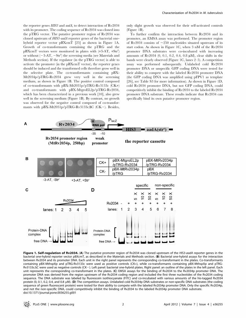
the reporter genes HIS3 and aadA, to detect interaction of Rv2034
with its promoter. The coding sequence of Rv2034 was cloned into
the pTRG vector. The putative promoter region of Rv2034 was
cloned upstream of HIS3-aadA reporter genes of the bacterial one-
hybrid reporter vector pBXcmT [25] as shown in Figure 1A.
Growth of co-transformants containing the pTRG and the
pBXcmT vectors were monitored in plates with (+3-AT, +Strr)
or without (23-AT, 2Strr) the screening media (see Materials and
Methods section). If the regulator (in the pTRG vector) is able to
activate the promoter (in the pBXcmT vector), the reporter genes
should be induced and the transformed cells therefore grow well in
the selective plate. The co-transformants containing pBX-
Mt2034p/pTRG-Rv2034 grew very well in the screening
medium, as shown in Figure 1B. The positive control composed
of co-transformants with pBX-Mt2031p/pTRG-Rv3133c (CK+)
and co-transformants with pBX-MtgroEL2p/pTRG-Rv2034,
which has been characterized in a previous work [10], also grew
well in the screening medium (Figure 1B). By contrast, no growth
was observed for the negative control composed of co-transfor-
mants with pBX-Mt2031p/pTRG-Rv3133cDC (CK2). Besides,
only slight growth was observed for their self-activated controls
(Figure 1B).
To further confirm the interaction between Rv2034 and its
promoter, an EMSA assay was performed. The promoter region
of Rv2034 consists of ,250 nucleotides situated upstream of its
start codon. As shown in Figure 1C, when 3 nM of the Rv2034
promoter DNA substrates were co-incubated with increasing
amounts of Rv2034 (0, 0.1, 0.2, 0.4, 0.8 mM), clear shifts in the
bands were clearly observed (Figure 1C, lanes 2–5). A competition
assay was performed subsequently. Unlabeled cold Rv2034
promoter DNA or unspecific GFP coding DNA were tested for
their ability to compete with the labeled Rv2034 promoter DNA
(the GFP coding DNA was amplified using pFPV1 as template
[26], see Table S3 for more information). As shown in Figure 1D,
cold Rv2034 promoter DNA, but not GFP coding DNA, could
competitively inhibit the binding of Rv2034 to the labeled Rv2034
promoter DNA substrate. These results indicate that Rv2034 can
specifically bind its own putative promoter region.
Figure 1. Self-regulation of Rv2034. (A) The putative promoter region of Rv2034 was cloned upstream of the HIS3-aadA reporter genes in thebacterial one-hybrid reporter vector pBXcmT, as described in the Materials and Methods section. (B) Bacterial one-hybrid assays for the interactionbetween Rv2034 and its promoter DNA. Each unit in the right panel represents the corresponding co-transformant in the plates. Co-transformantscontaining pBX-MthspXp and pTRG-Rv3133c were used as positive controls (CK+), while co-transformants containing pBX-MthspXp and pTRG-Rv3133cDC were used as negative controls (CK2). Left panel: bacterial one-hybrid plates. Right panel: an outline of the plates in the left panel. Eachunit represents the corresponding co-transformant in the plates. (C) EMSA assays for the binding of Rv2034 to the Rv2034p promoter DNA. Thepromoter DNA was derived from the region upstream of the Rv2034 coding region and included the first three nucleotides of the Rv2034 codingsequence. The DNA substrate was labeled by fluorescein isothiocyanate (FITC) and co-incubated with various amounts of the his-tagged Rv2034protein (0, 0.1, 0.2, 0.4, and 0.8 mM). (D) The competition assays. Unlabeled cold Rv2034p DNA substrates or non-specific DNA substrates (the codingsequence of green fluorescent protein) were tested for their ability to compete with the labeled Rv2034p promoter DNA. Only the specific Rv2034p,and not the non-specific DNA, could competitively inhibit the binding of Rv2034 to the labeled Rv2034p promoter DNA substrate.doi:10.1371/journal.pone.0036255.g001
Characterization of Rv2034 in M. tuberculosis
PLoS ONE | www.plosone.org 2 April 2012 | Volume 7 | Issue 4 | e36255
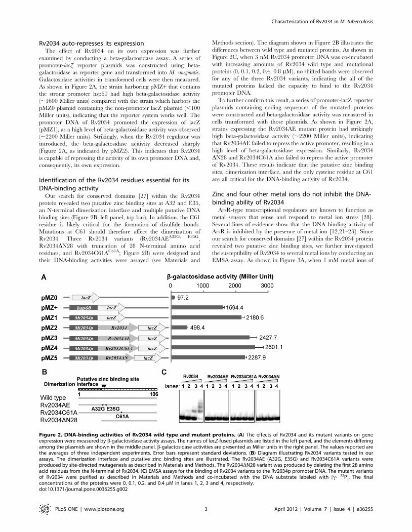
Rv2034 auto-represses its expressionThe effect of Rv2034 on its own expression was further
examined by conducting a beta-galactosidase assay. A series of
promoter-lacZ reporter plasmids was constructed using beta-
galactosidase as reporter gene and transformed into M. smegmatis.
Galactosidase activities in transformed cells were then measured.
As shown in Figure 2A, the strain harboring pMZ+ that contains
the strong promoter hsp60 had high beta-galactosidase activity
(,1600 Miller units) compared with the strain which harbors the
pMZ0 plasmid containing the non-promoter lacZ plasmid (,100
Miller units), indicating that the reporter system works well. The
promoter DNA of Rv2034 promoted the expression of lacZ
(pMZ1), as a high level of beta-galactosidase activity was observed
(,2200 Miller units). Strikingly, when the Rv2034 regulator was
introduced, the beta-galactosidase acitivity decreased sharply
(Figure 2A, as indicated by pMZ2). This indicates that Rv2034
is capable of repressing the activity of its own promoter DNA and,
consequently, its own expression.
Identification of the Rv2034 residues essential for itsDNA-binding activity
Our search for conserved domains [27] within the Rv2034
protein revealed two putative zinc binding sites at A32 and E35,
an N-terminal dimerization interface and multiple putative DNA
binding sites (Figure 2B, left panel, top bar). In addition, the C61
residue is likely critical for the formation of disulfide bonds.
Mutations at C61 should therefore affect the dimerization of
Rv2034. Three Rv2034 variants (Rv2034AEA32G, E35G,
Rv2034DN28 with truncation of 28 N-terminal amino acid
residues, and Rv2034C61AC61A; Figure 2B) were designed and
their DNA-binding activities were assayed (see Materials and
Methods section). The diagram shown in Figure 2B illustrates the
differences between wild type and mutated proteins. As shown in
Figure 2C, when 3 nM Rv2034 promoter DNA was co-incubated
with increasing amounts of Rv2034 wild type and mutational
proteins (0, 0.1, 0.2, 0.4, 0.8 mM), no shifted bands were observed
for any of the three Rv2034 variants, indicating the all of the
mutated proteins lacked the capacity to bind to the Rv2034
promoter DNA.
To further confirm this result, a series of promoter-lacZ reporter
plasmids containing coding sequences of the mutated proteins
were constructed and beta-galactosidase activity was measured in
cells transformed with those plasmids. As shown in Figure 2A,
strains expressing the Rv2034AE mutant protein had strikingly
high beta-galactosidase activity (,2200 Miller units), indicating
that Rv2034AE failed to repress the active promoter, resulting in a
high level of beta-galactosidase expression. Similarly, Rv2034
DN28 and Rv2034C61A also failed to repress the active promoter
of Rv2034. These results indicate that the putative zinc binding
sites, dimerization interface, and the only cysteine residue at C61
are all critical for the DNA-binding activity of Rv2034.
Zinc and four other metal ions do not inhibit the DNA-binding ability of Rv2034
ArsR-type transcriptional regulators are known to function as
metal sensors that sense and respond to metal ion stress [28].
Several lines of evidence show that the DNA binding activity of
ArsR is inhibited by the presence of metal ion [12,21–23]. Since
our search for conserved domains [27] within the Rv2034 protein
revealed two putative zinc binding sites, we further investigated
the susceptibility of Rv2034 to several metal ions by conducting an
EMSA assay. As shown in Figure 3A, when 1 mM metal ions of
Figure 2. DNA-binding activities of Rv2034 wild type and mutant proteins. (A) The effects of Rv2034 and its mutant variants on geneexpression were measured by b-galactosidase activity assays. The names of lacZ-fused plasmids are listed in the left panel, and the elements differingamong the plasmids are shown in the middle panel. b-galactosidase activities are presented as Miller units in the right panel. The values reported arethe averages of three independent experiments. Error bars represent standard deviations. (B) Diagram illustrating Rv2034 variants tested in ourassays. The dimerization interface and putative zinc binding sites are illustrated. The Rv2034AE (A32G, E35G) and Rv2034C61A variants wereproduced by site-directed mutagenesis as described in Materials and Methods. The Rv2034DN28 variant was produced by deleting the first 28 aminoacid residues from the N-terminal of Rv2034. (C) EMSA assays for the binding of Rv2034 variants to the Rv2034p promoter DNA. The mutant variantsof Rv2034 were purified as described in Materials and Methods and co-incubated with the DNA substrate labeled with [c- 32P]. The finalconcentrations of the proteins were 0, 0.1, 0.2, and 0.4 mM in lanes 1, 2, 3 and 4, respectively.doi:10.1371/journal.pone.0036255.g002
Characterization of Rv2034 in M. tuberculosis
PLoS ONE | www.plosone.org 3 April 2012 | Volume 7 | Issue 4 | e36255
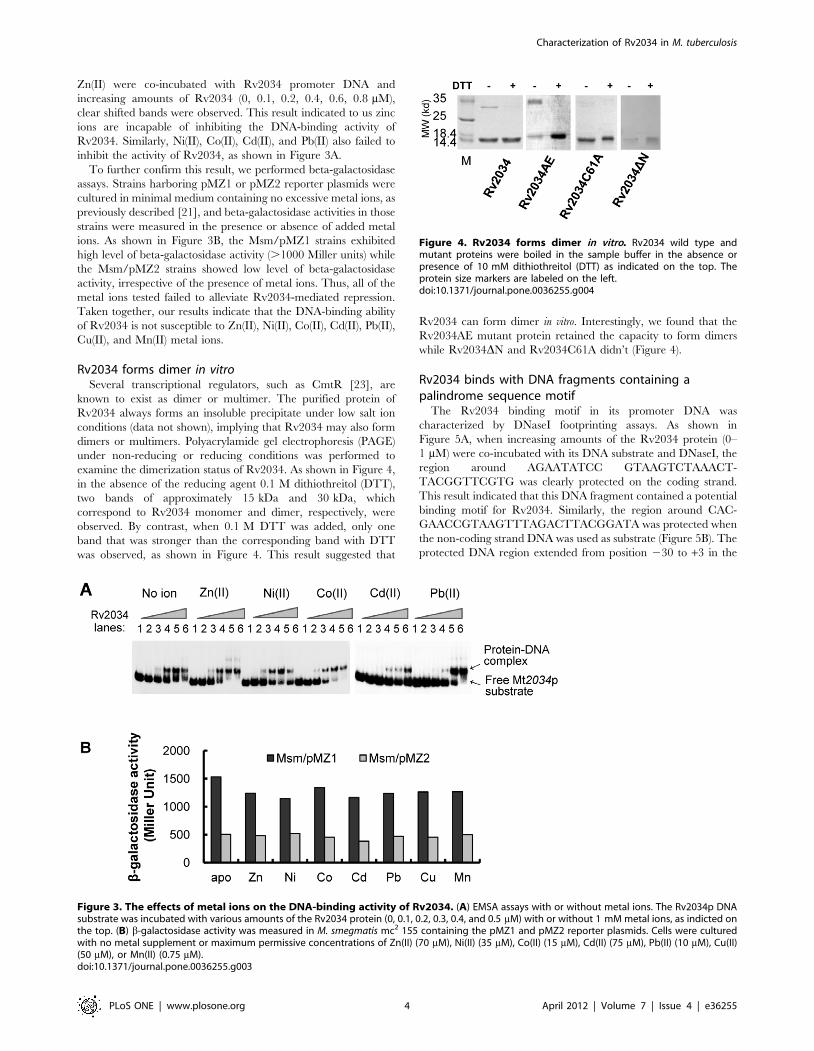
Zn(II) were co-incubated with Rv2034 promoter DNA and
increasing amounts of Rv2034 (0, 0.1, 0.2, 0.4, 0.6, 0.8 mM),
clear shifted bands were observed. This result indicated to us zinc
ions are incapable of inhibiting the DNA-binding activity of
Rv2034. Similarly, Ni(II), Co(II), Cd(II), and Pb(II) also failed to
inhibit the activity of Rv2034, as shown in Figure 3A.
To further confirm this result, we performed beta-galactosidase
assays. Strains harboring pMZ1 or pMZ2 reporter plasmids were
cultured in minimal medium containing no excessive metal ions, as
previously described [21], and beta-galactosidase activities in those
strains were measured in the presence or absence of added metal
ions. As shown in Figure 3B, the Msm/pMZ1 strains exhibited
high level of beta-galactosidase activity (.1000 Miller units) while
the Msm/pMZ2 strains showed low level of beta-galactosidase
activity, irrespective of the presence of metal ions. Thus, all of the
metal ions tested failed to alleviate Rv2034-mediated repression.
Taken together, our results indicate that the DNA-binding ability
of Rv2034 is not susceptible to Zn(II), Ni(II), Co(II), Cd(II), Pb(II),
Cu(II), and Mn(II) metal ions.
Rv2034 forms dimer in vitroSeveral transcriptional regulators, such as CmtR [23], are
known to exist as dimer or multimer. The purified protein of
Rv2034 always forms an insoluble precipitate under low salt ion
conditions (data not shown), implying that Rv2034 may also form
dimers or multimers. Polyacrylamide gel electrophoresis (PAGE)
under non-reducing or reducing conditions was performed to
examine the dimerization status of Rv2034. As shown in Figure 4,
in the absence of the reducing agent 0.1 M dithiothreitol (DTT),
two bands of approximately 15 kDa and 30 kDa, which
correspond to Rv2034 monomer and dimer, respectively, were
observed. By contrast, when 0.1 M DTT was added, only one
band that was stronger than the corresponding band with DTT
was observed, as shown in Figure 4. This result suggested that
Rv2034 can form dimer in vitro. Interestingly, we found that the
Rv2034AE mutant protein retained the capacity to form dimers
while Rv2034DN and Rv2034C61A didn’t (Figure 4).
Rv2034 binds with DNA fragments containing apalindrome sequence motif
The Rv2034 binding motif in its promoter DNA was
characterized by DNaseI footprinting assays. As shown in
Figure 5A, when increasing amounts of the Rv2034 protein (0–
1 mM) were co-incubated with its DNA substrate and DNaseI, the
region around AGAATATCC GTAAGTCTAAACT-
TACGGTTCGTG was clearly protected on the coding strand.
This result indicated that this DNA fragment contained a potential
binding motif for Rv2034. Similarly, the region around CAC-
GAACCGTAAGTTTAGACTTACGGATA was protected when
the non-coding strand DNA was used as substrate (Figure 5B). The
protected DNA region extended from position 230 to +3 in the
Figure 3. The effects of metal ions on the DNA-binding activity of Rv2034. (A) EMSA assays with or without metal ions. The Rv2034p DNAsubstrate was incubated with various amounts of the Rv2034 protein (0, 0.1, 0.2, 0.3, 0.4, and 0.5 mM) with or without 1 mM metal ions, as indicted onthe top. (B) b-galactosidase activity was measured in M. smegmatis mc2 155 containing the pMZ1 and pMZ2 reporter plasmids. Cells were culturedwith no metal supplement or maximum permissive concentrations of Zn(II) (70 mM), Ni(II) (35 mM), Co(II) (15 mM), Cd(II) (75 mM), Pb(II) (10 mM), Cu(II)(50 mM), or Mn(II) (0.75 mM).doi:10.1371/journal.pone.0036255.g003
Figure 4. Rv2034 forms dimer in vitro. Rv2034 wild type andmutant proteins were boiled in the sample buffer in the absence orpresence of 10 mM dithiothreitol (DTT) as indicated on the top. Theprotein size markers are labeled on the left.doi:10.1371/journal.pone.0036255.g004
Characterization of Rv2034 in M. tuberculosis
PLoS ONE | www.plosone.org 4 April 2012 | Volume 7 | Issue 4 | e36255
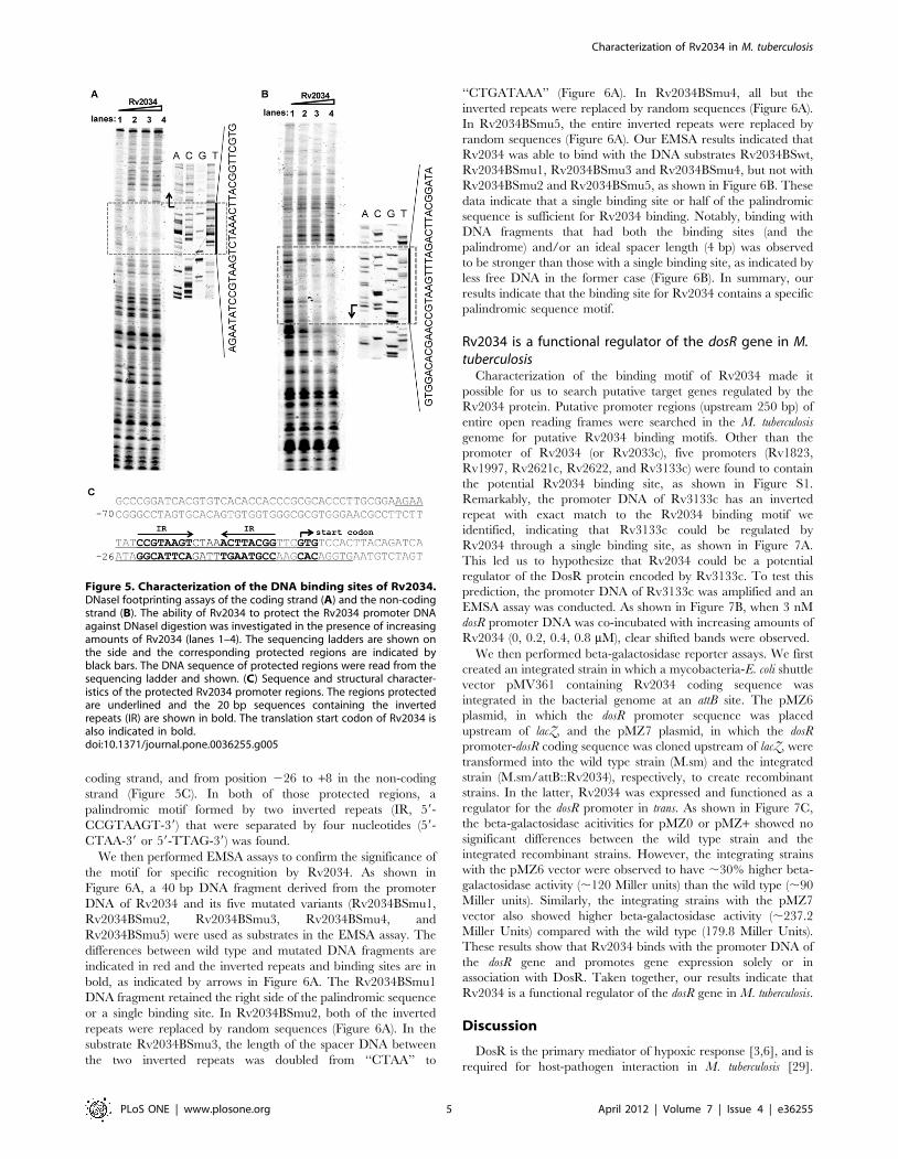
coding strand, and from position 226 to +8 in the non-coding
strand (Figure 5C). In both of those protected regions, a
palindromic motif formed by two inverted repeats (IR, 59-
CCGTAAGT-39) that were separated by four nucleotides (59-
CTAA-39 or 59-TTAG-39) was found.
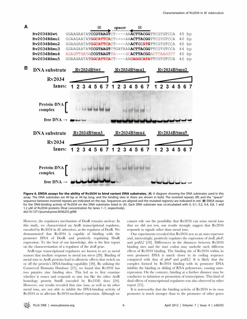
We then performed EMSA assays to confirm the significance of
the motif for specific recognition by Rv2034. As shown in
Figure 6A, a 40 bp DNA fragment derived from the promoter
DNA of Rv2034 and its five mutated variants (Rv2034BSmu1,
Rv2034BSmu2, Rv2034BSmu3, Rv2034BSmu4, and
Rv2034BSmu5) were used as substrates in the EMSA assay. The
differences between wild type and mutated DNA fragments are
indicated in red and the inverted repeats and binding sites are in
bold, as indicated by arrows in Figure 6A. The Rv2034BSmu1
DNA fragment retained the right side of the palindromic sequence
or a single binding site. In Rv2034BSmu2, both of the inverted
repeats were replaced by random sequences (Figure 6A). In the
substrate Rv2034BSmu3, the length of the spacer DNA between
the two inverted repeats was doubled from ‘‘CTAA’’ to
‘‘CTGATAAA’’ (Figure 6A). In Rv2034BSmu4, all but the
inverted repeats were replaced by random sequences (Figure 6A).
In Rv2034BSmu5, the entire inverted repeats were replaced by
random sequences (Figure 6A). Our EMSA results indicated that
Rv2034 was able to bind with the DNA substrates Rv2034BSwt,
Rv2034BSmu1, Rv2034BSmu3 and Rv2034BSmu4, but not with
Rv2034BSmu2 and Rv2034BSmu5, as shown in Figure 6B. These
data indicate that a single binding site or half of the palindromic
sequence is sufficient for Rv2034 binding. Notably, binding with
DNA fragments that had both the binding sites (and the
palindrome) and/or an ideal spacer length (4 bp) was observed
to be stronger than those with a single binding site, as indicated by
less free DNA in the former case (Figure 6B). In summary, our
results indicate that the binding site for Rv2034 contains a specific
palindromic sequence motif.
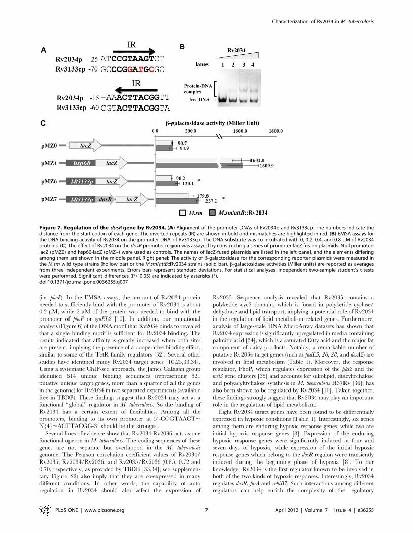
Rv2034 is a functional regulator of the dosR gene in M.tuberculosis
Characterization of the binding motif of Rv2034 made it
possible for us to search putative target genes regulated by the
Rv2034 protein. Putative promoter regions (upstream 250 bp) of
entire open reading frames were searched in the M. tuberculosis
genome for putative Rv2034 binding motifs. Other than the
promoter of Rv2034 (or Rv2033c), five promoters (Rv1823,
Rv1997, Rv2621c, Rv2622, and Rv3133c) were found to contain
the potential Rv2034 binding site, as shown in Figure S1.
Remarkably, the promoter DNA of Rv3133c has an inverted
repeat with exact match to the Rv2034 binding motif we
identified, indicating that Rv3133c could be regulated by
Rv2034 through a single binding site, as shown in Figure 7A.
This led us to hypothesize that Rv2034 could be a potential
regulator of the DosR protein encoded by Rv3133c. To test this
prediction, the promoter DNA of Rv3133c was amplified and an
EMSA assay was conducted. As shown in Figure 7B, when 3 nM
dosR promoter DNA was co-incubated with increasing amounts of
Rv2034 (0, 0.2, 0.4, 0.8 mM), clear shifted bands were observed.
We then performed beta-galactosidase reporter assays. We first
created an integrated strain in which a mycobacteria-E. coli shuttle
vector pMV361 containing Rv2034 coding sequence was
integrated in the bacterial genome at an attB site. The pMZ6
plasmid, in which the dosR promoter sequence was placed
upstream of lacZ, and the pMZ7 plasmid, in which the dosR
promoter-dosR coding sequence was cloned upstream of lacZ, were
transformed into the wild type strain (M.sm) and the integrated
strain (M.sm/attB::Rv2034), respectively, to create recombinant
strains. In the latter, Rv2034 was expressed and functioned as a
regulator for the dosR promoter in trans. As shown in Figure 7C,
the beta-galactosidase acitivities for pMZ0 or pMZ+ showed no
significant differences between the wild type strain and the
integrated recombinant strains. However, the integrating strains
with the pMZ6 vector were observed to have ,30% higher beta-
galactosidase activity (,120 Miller units) than the wild type (,90
Miller units). Similarly, the integrating strains with the pMZ7
vector also showed higher beta-galactosidase activity (,237.2
Miller Units) compared with the wild type (179.8 Miller Units).
These results show that Rv2034 binds with the promoter DNA of
the dosR gene and promotes gene expression solely or in
association with DosR. Taken together, our results indicate that
Rv2034 is a functional regulator of the dosR gene in M. tuberculosis.
Discussion
DosR is the primary mediator of hypoxic response [3,6], and is
required for host-pathogen interaction in M. tuberculosis [29].
Figure 5. Characterization of the DNA binding sites of Rv2034.DNaseI footprinting assays of the coding strand (A) and the non-codingstrand (B). The ability of Rv2034 to protect the Rv2034 promoter DNAagainst DNaseI digestion was investigated in the presence of increasingamounts of Rv2034 (lanes 1–4). The sequencing ladders are shown onthe side and the corresponding protected regions are indicated byblack bars. The DNA sequence of protected regions were read from thesequencing ladder and shown. (C) Sequence and structural character-istics of the protected Rv2034 promoter regions. The regions protectedare underlined and the 20 bp sequences containing the invertedrepeats (IR) are shown in bold. The translation start codon of Rv2034 isalso indicated in bold.doi:10.1371/journal.pone.0036255.g005
Characterization of Rv2034 in M. tuberculosis
PLoS ONE | www.plosone.org 5 April 2012 | Volume 7 | Issue 4 | e36255
However, the regulatory mechanism of DosR remains unclear. In
this study, we characterized an ArsR transcriptional regulator,
encoded by Rv2034 in M. tuberculosis, as the regulator of DosR. We
demonstrated that Rv2034 is capable of binding with the
promoter DNA of DosR and positively regulating DosR
expression. To the best of our knowledge, this is the first report
on the characterization of a regulator of the dosR gene.
ArsR-type transcriptional regulators are known to act as metal
sensors that mediate response to metal ion stress [28]. Binding of
metal ions to ArsR proteins lead to allosteric effects that switch on
or off the protein’s DNA-binding capability [30]. By utilizing the
Conserved Domains Database [27], we found that Rv2034 has
two putative zinc binding sites. This led us to first examine
whether it senses and responds to zinc ion like the other ArsR
homology protein SmtB (encoded by Rv2358) does [24].
However, our results revealed that zinc ions, as well as six other
metal ions, are not able to inhibit the DNA-binding activity of
Rv2034 or to alleviate Rv2034-mediated repression. Although we
cannot rule out the possibility that Rv2034 can sense metal ions
that we did not test, our results strongly suggest that Rv2034
responds to signals other than metal ions.
Our experiments revealed that Rv2034 acts as an auto repressor
and, interestingly, positively regulates the expression of dosR, phoP,
and groEL2 [10]. Differences in the distances between Rv2034
binding sites and the start codon may underlie such different
effects of Rv2034 binding. The binding site of Rv2034 within its
own promoter DNA is much closer to its coding sequence
compared with that of phoP and groEL2. It is likely that the
complex formed by Rv2034 binding with its promoter DNA
inhibits the binding or sliding of RNA polymerase, causing auto-
repression. On the contrary, binding at a further distance may be
conducive to initiation or promotion of transcription. This kind of
dual effects of transcriptional regulators was also observed in other
report [31].
It is noteworthy that the binding activity of Rv2034 to its own
promoter is much stronger than to the promoter of other genes
Figure 6. EMSA assays for the ability of Rv2034 to bind variant DNA substrates. (A) A diagram showing the DNA substrates used in thisassay. The DNA substrates are 40 bp or 44 bp long, and the binding sites in them are shown in bold. The inverted repeats (IR) and the ‘‘spacer’’sequence between inverted repeats are indicated on the top. Sequences are aligned and the mutated region(s) are indicated in red. (B) EMSA assaysfor the DNA-binding activity of Rv2034 on the DNA substrates listed in (A). Each DNA substrate was co-incubated with 0, 0.1, 0.2, 0.4, 0.8, 1 and1.2 mM of Rv2034 proteins (final concentration for lanes 1–7, respectively).doi:10.1371/journal.pone.0036255.g006
Characterization of Rv2034 in M. tuberculosis
PLoS ONE | www.plosone.org 6 April 2012 | Volume 7 | Issue 4 | e36255
(i.e. phoP). In the EMSA assays, the amount of Rv2034 protein
needed to sufficiently bind with the promoter of Rv2034 is about
0.2 mM, while 2 mM of the protein was needed to bind with the
promoter of phoP or groEL2 [10]. In addition, our mutational
analysis (Figure 6) of the DNA motif that Rv2034 binds to revealed
that a single binding motif is sufficient for Rv2034 binding. The
results indicated that affinity is greatly increased when both sites
are present, implying the presence of a cooperative binding effect,
similar to some of the TetR family regulators [32]. Several other
studies have identified many Rv2034 target genes [10,25,33,34].
Using a systematic ChIP-seq approach, the James Galagan group
identified 614 unique binding sequences (representing 821
putative unique target genes, more than a quarter of all the genes
in the genome) for Rv2034 in two separated experiments (available
free in TBDB). These findings suggest that Rv2034 may act as a
functional ‘‘global’’ regulator in M. tuberculosis. So the binding of
Rv2034 has a certain extent of flexibilities. Among all the
promoters, binding to its own promoter at 59-CCGTAAGT,N{4},ACTTACGG-39 should be the strongest.
Several lines of evidence show that Rv2034-Rv2036 acts as one
functional operon in M. tuberculosis. The coding sequences of these
genes are not separate but overlapped in the M. tuberculosis
genome. The Pearson correlation coefficient values of Rv2034/
Rv2035, Rv2034/Rv2036, and Rv2035/Rv2036 (0.85, 0.72 and
0.70, respectively, as provided by TBDB [33,34]; see supplemen-
tary Figure S2) also imply that they are co-expressed in many
different conditions. In other words, the capability of auto
regulation in Rv2034 should also affect the expression of
Rv2035. Sequence analysis revealed that Rv2035 contains a
polyketide_cyc2 domain, which is found in polyketide cyclase/
dehydrase and lipid transport, implying a potential role of Rv2034
in the regulation of lipid metabolism related genes. Furthermore,
analysis of large-scale DNA MicroArray datasets has shown that
Rv2034 expression is significantly upregulated in media containing
palmitic acid [34], which is a saturated fatty acid and the major fat
component of dairy products. Notably, a remarkable number of
putative Rv2034 target genes (such as fadE5, 26, 28, and desA2) are
involved in lipid metabolism (Table 1). Moreover, the response
regulator, PhoP, which regulates expression of the pks2 and the
msl3 gene clusters [35] and accounts for sulfolipid, diacyltrehalose
and polyacyltrehalose synthesis in M. tuberculosis H37Rv [36], has
also been shown to be regulated by Rv2034 [10]. Taken together,
these findings strongly suggest that Rv2034 may play an important
role in the regulation of lipid metabolism.
Eight Rv2034 target genes have been found to be differentially
expressed in hypoxic conditions (Table 1). Interestingly, six genes
among them are enduring hypoxic response genes, while two are
initial hypoxic response genes [8]. Expression of the enduring
hypoxic response genes were significantly induced at four and
seven days of hypoxia, while expression of the initial hypoxic
response genes which belong to the dosR regulon were transiently
induced during the beginning phase of hypoxia [8]. To our
knowledge, Rv2034 is the first regulator known to be involved in
both of the two kinds of hypoxic responses. Interestingly, Rv2034
regulates dosR, furA and whiB7. Such interactions among different
regulators can help enrich the complexity of the regulatory
Figure 7. Regulation of the dosR gene by Rv2034. (A) Alignment of the promoter DNAs of Rv2034p and Rv3133cp. The numbers indicate thedistance from the start codon of each gene. The inverted repeats (IR) are shown in bold and mismatches are highlighted in red. (B) EMSA assays forthe DNA-binding activity of Rv2034 on the promoter DNA of Rv3133cp. The DNA substrate was co-incubated with 0, 0.2, 0.4, and 0.8 mM of Rv2034proteins. (C) The effect of Rv2034 on the dosR promoter region was assayed by constructing a series of promoter-lacZ fusion plasmids. Null promoter-lacZ (pMZ0) and hsp60-lacZ (pMZ+) were used as controls. The names of lacZ-fused plasmids are listed in the left panel, and the elements differingamong them are shown in the middle panel. Right panel: The activity of b-galactosidase for the corresponding reporter plasmids were measured inthe M.sm wild type strains (hollow bar) or the M.sm/attB::Rv2034 strains (solid bar). b-galactosidase activities (Miller units) are reported as averagesfrom three independent experiments. Errors bars represent standard deviations. For statistical analyses, independent two-sample student’s t-testswere performed. Significant differences (P,0.05) are indicated by asterisks (*).doi:10.1371/journal.pone.0036255.g007
Characterization of Rv2034 in M. tuberculosis
PLoS ONE | www.plosone.org 7 April 2012 | Volume 7 | Issue 4 | e36255
network and benefit the adaptation of the cell to hypoxic
conditions. In addition, it is possible that Rv2034 is involved in
the transition of hypoxic responses. Besides, in the ArsR knock-out
strain of M. smegmatis, in which the coding region of Rv2034
homologous gene (encoded by Ms6762) was replaced by the
hygromycin gene, is more resistant to hydrogen peroxide (H2O2),
as compared with the wild type and complementary strains (see
supplementary figure S3). This implies that the homologous gene
of Rv2034 play a role in the adaptation of oxidative stress in M.
smegmatis and shed a light on the possible role of Rv2034 in M.
tuberculosis H37Rv. A comprehensive understanding of the function
of Rv2034 should be uncovered along with more advances in
system biology.
In summary, we characterized the auto repression of the ArsR
transcriptional regulator Rv2034 and identified a palindromic
sequence as its binding site. Several residues in Rv2034 critical to
its DNA binding activities were successfully characterized.
Notably, we found that Rv2034 regulates the expression of the
dosR gene. Our findings establish a direct link of the ArsR
regulator to both lipid metabolism and hypoxic adaptation in M.
tuberculosis.
Materials and Methods
Plasmids, strains, enzymes and chemicalsThe Escherichia coli BL21 cells and the pET28a vector were
purchased from Novagen. All the enzymes including restriction
enzymes, ligase, and DNA polymerase were purchased from
TaKaRa Biotech. Deoxynucleoside triphosphates (dNTPs) and all
antibiotics were purchased from TaKaRa Biotech as well. PCR
primers were synthesized by Invitrogen (See Table S1). All the
derived plasmids were listed in Table S2. Ni-NTA (Ni2+-
nitrilotriacetate) agarose was obtained from Qiagen.
Cloning, expression, and purification of mycobacterialproteins
Protein expression and purification were performed as previ-
ously described [10]. Rv2034AEA32G, E35G and Rv2034C61AC61A
mutated variants were generated by site-directed mutagenesis
using the overlap extension polymerase chain reaction technique
(overlapping-PCR). Primers used in the overlapping-PCR are
listed in supplementary Table S1. The Rv2034DN mutated
variant was generated by using a truncated forward primer
Rv2034DNf-EcoRI and the primer Mt2034pr-XbaI for PCR (see
Table S1).
Electrophoretic mobility shift assayLarge DNA segments with more than 70 base pairs were
acquired using polymerase chain reaction (PCR), and short DNA
segments were synthesized and directly annealed in vitro. The
primers used for PCR are listed in Supplemental Table S1. All the
DNA substrates used in this assay are listed in Table S3. DNA
fragments used in this assay were labeled at the 59-end with [c-32P]
ATP or with fluorescein isothiocyanate (FITC). Mixtures contain-
ing the labeled DNA (,10 nM) and increasing concentrations of
the protein (as indicated in the legend of the corresponding figure)
were incubated in EMSA buffer (100 mM Tris-HCl, pH 8.0,
100 mM NaCl, 1 mM DTT and 10% glycerol) for 30 min at
room temperature, then subjected to 5% native polyacrylamide gel
electrophoresis (PAGE). Electrophoresis was performed at 150 V
at room temperature for about 1.5 hours in 0.56 Tris-borate-
EDTA buffer (TBE). Radioactive gels were exposed to a storage-
phosphor screen (GE healthcare) overnight at room temperature.
Images of gels were acquired using a Typhoon Scanner (GE
healthcare).
Bacterial one-hybrid assayBacterial one-hybrid assays were performed as previously
described [25]. In the present study, the promoter region of
Rv2034 and other reference DNA fragments were amplified using
corresponding primer pairs (see Table S1) and cloned upstream of
the reporter genes (HIS3 and aadA) in the pBXcmT plasmid [25].
In this way, a series of reporter plasmids (see Table S2) were
created. The reporter plasmids were co-transformed with the
pTRG-Rv2034 plasmid (see Table S2) into Escherichia coli XR
competent cells. Co-transformants were grown on selective
medium containing 20 mM 3-amino-1, 2, 4-triazole (3-AT),
16 mg/mL streptomycin, 15 mg/mL tetracycline, 34 mg/mL
chloramphenicol, and 50 mg/mL kanamycin. Plates were incu-
bated at 30 degree centigrade for 3–4 days. Co-transformants
containing the pBX-MthspXp and pTRG-Rv3133c plasmids [25]
served as the positive control while co-transformants containing
the pBX-MthspXp and pTRG-Rv3133cDC plasmids [25] served
as the negative control. Co-transformants containing the empty
vectors pBX and/or pTRG were used as self-activation controls.
DnaseI footprinting assayThe substrate DNAs used in this assay were about 220 base
pairs long, including 140 base pairs upstream from the start codon
and the first 82 base pairs of the Rv2034 coding region. Both the
coding and non-coding strands were amplified by PCR using their
specific primers labeled with Fluorescein Isothiocyanate (FITC)
(see Table S3). PCR products were purified with BioFlux PCR
DNA Purification kit (BioFlux) and subjected to the same binding
reaction as in EMSA. DNaseI footprinting was performed as
previously described [37]. The ladders were produced using the
Sanger dideoxy method.
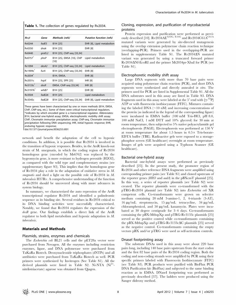
Table 1. The collection of genes regulated by Rv2034.
Rv# Gene Methods (refs) Putative function (refs)
Rv0244 fadE5 B1H [25] EHR [8], Lipid metabolism
Rv0350 dnak B1H [25] EHR [8]
Rv0440 groEL2 B1H [25], ChIP-seq [33,34]
Rv0757* phoP B1H [25], EMSA [10], ChIP[10]
Lipid metabolism
Rv1094 desA2 B1H [25], ChIP-seq [33,34] Lipid metabolism
Rv1909c* furA B1H [25], ChIP-seq [33,34] EHR [8]
Rv2034* B1H, EMSA, EHR [8]
Rv2031c hspX B1H [25], SPR [25] IHR [8]
Rv3133c* dosR EMSA, ChIP-seq [33,34] IHR [8]
Rv3197A* whiB7 B1H [25] EHR [8]
Rv3504 fadE26 B1H [25] Lipid metabolism
Rv3545c fadE28 B1H [25], ChIP-seq [33,34] EHR [8], Lipid metabolism
These genes have been characterized by one or more methods (B1H, EMSA,ChIP, ChIP-seq, etc.). Some of them were critical transcriptional regulators.*indicates by which encoded gene is a transcriptional regulator. Abbreviates:B1H, bacterial one-hybrid assay; EMSA, electrophoretic mobility shift assay;ChIP, Chromatin immunize precipitation assay; ChIP-seq, Chromatin immunizeprecipitation following DNA sequencing; IHR, initial hypoxic response; EHR,enduring hypoxic response;doi:10.1371/journal.pone.0036255.t001
Characterization of Rv2034 in M. tuberculosis
PLoS ONE | www.plosone.org 8 April 2012 | Volume 7 | Issue 4 | e36255
b-galactosidase activity assayb-galactosidase activity assays were performed as previously
described [37]. In the present study, the experiments were
performed by creating operon-lacZ fusions based on the
expression vector pMV261 [38]. To obtain the plasmid pMZ1
to pMZ9, a series of digestion and ligation reactions were carried
out. The promoter sequences, the corresponding regulatory
protein coding sequences and the reporter gene lacZ were cloned
into the pMV261 backbone step by step (See Table S2). Taking
the construction of the pMZ3 plasmid as an example, the putative
promoter region of Rv2034 Mt2034p was first cloned into the
pMV261 backbone using XbaI/EcoRI. Then, the coding sequence
of Rv2034AE was cloned using EcoR/HindIII. Finally, the reporter
gene lacZ was cloned using HindIII/NheI. To construct the pMZ+plasmid, which was used as a positive control in our assays, the
reporter gene lacZ was inserted into pMV261 downstream of the
enhanced promoter of hsp60 using HindIII/NheI. To construct the
negative control plasmid pMZ-, the promoter region of pMZ+ was
removed by XbaI/EcoRI double-digest and blunt-end ligation. The
reporter plasmids were transformed into the M. smegmatis str. mc2
155 host strain and the corresponding recombinant reporter
strains were obtained. All strains were grown at 37uC to an OD600
of ,1.5. Cell suspensions (1 ml) were then collected and washed
with cold PBS buffer (137 mM NaCl, 2.7 mM KCl, 8 mM
Na2HPO4, 1.46 mM KH2PO4, pH 7.4) once or twice. For cell
permeabilization, 600 ml of the Z buffer (60 mM Na2HPO4,
40 mM NaH2PO4, 10 mM KCl, 1 mM MgSO4 and 59 mM b-
mercaptoethanol, pH 7.5) was dispensed into the microcentrifuge
tube to resuspend the cells. Three hundred microliters of
resuspended cells was saved to determine the A600 value. After
adding 50 ml of 0.1% SDS (sodium dodecyl sulfonate) and 100 ml
of chloroform, the mixture was vortexed and incubated at 28uCfor 5 min. A total of 200 ml of ONPG (o-nitrophenyl-D-
galactoside, 4 mg/ml) was then added to a final concentration of
0.66 mg/ml. Each tube was incubated at room temperature for 5–
10 min. Five hundred microliters of 1 M Na2CO3 was added to
end this reaction. Each tube was centrifuged at 1200 rpm for
5 min and the supernatant was used to determine the A420 and
A550 values. b-galactosidase activities were calculated as described
previously [39]. This system is similar to the pJEM system [40,41]
and works well in terms of repeatability, distinction and
reproducibility as according to the practical operations [10,37].
Supporting Information
Figure S1 Search for putative target genes regulated bythe Rv2034 protein using the binding motif of Rv2034.Putative promoter regions (upstream 250 bp) of entire open
reading frames in the M. tuberculosis genome were searched based
on the Rv2034 binding motif (or the palindrome sequence) that we
identified. Other than the promoter of Rv2034 (or Rv2033c), we
found that five promoters (Rv1823, Rv1997, Rv2621c, Rv2622,
and Rv3133c) contained the potential Rv2034 binding site. The
promoter regions were aligned and mismatches are shown on the
right.
(TIF)
Figure S2 Pearson correlation coefficient values ofRv2034/Rv2035, Rv2034/Rv2036, and Rv2035/Rv2036pairs. Correlation coefficient values were obtained from TBDB.
Co-expressed genes are likely to have higher coefficients than ones
that are not co-expressed (from 0 to 1).
(TIF)
Figure S3 Construction of an ArsR-knockout strain andthe growth determination in the presence and absence ofhydrogen peroxide (H2O2). (A) Construction of the Ms6762
knockout strain of M. smegmatis and Southern blot assays. Ms6762
is the homologous gene of Rv2034 in M. smegmatis. Left panel:
Using a recombination-based strategy, the coding sequence of
Ms6762 was replaced by a hygromycin resistance gene (indicated
as hyg in the schematic representation). The restriction sites for
NarI are indicated with arrows. The DNA probes used in
Southern blotting are indicated with black bars. Right panel:
Southern blot assays. The DNA fragment corresponding to the
region upstream of Ms6762 in M. smegmatis was obtained by PCR
and labeled with digoxigenin dUTP (Boehringer Mannheim, Inc.,
Germany). The probe was used to detect the change in size of the
NarI-digested genomic fragment of M. smegmatis wild type (WT,
,600 bp) and knock-out (KO, ,900 bp) strains. The knock-out,
the wild type and the complement strains were cultured in the
presence of 0 mM (B), 15 mM (C) and 20 mM (D) of hydrogen
peroxide, as indicated on the top. The complement strain was
generated by transforming an inducible expression vector of
pMind-Ms6762 into the verified Ms6762 knock-out strain. To
induce the expression of Ms6762 in the complement strains,
,25 ng/ml of the inducer (tetracycline) was added in the
corresponding culture. The experiments were performed thrice
with similar results.
(TIF)
Table S1 Primers used in this study.(DOC)
Table S2 Plasmids used in this study.(DOC)
Table S3 DNA substrates used in this study.(DOC)
Acknowledgments
The ChIP-Seq data used was freely downloaded from TBDB (www.tbdb.
org).
Author Contributions
Conceived and designed the experiments: CHG ZGH. Performed the
experiments: CHG MY. Analyzed the data: CHG. Contributed reagents/
materials/analysis tools: ZGH. Wrote the paper: CHG ZGH.
References
1. World Health Organization (2010) Global tuberculosis control WHO report
2010. Geneva: World Health Organization.
2. Ginsberg AM, Spigelman M (2007) Challenges in tuberculosis drug research and
development. Nat Med 13: 290–294. doi:10.1038/nm0307–290.
3. Rustad TR, Sherrid AM, Minch KJ, Sherman DR (2009) Hypoxia: a window
into Mycobacterium tuberculosis latency. Cell Microbiol 11: 1151–1159.
doi:10.1111/j.1462-5822.2009.01325.x.
4. Kendall SL, Rison SCG, Movahedzadeh F, Frita R, Stoker NG (2004) What do
microarrays really tell us about M. tuberculosis? Trends Microbiol 12: 537–544.
doi:10.1016/j.tim.2004.10.005.
5. Vergne I, Chua J, Singh SB, Deretic V (2004) Cell biology of mycobacterium
tuberculosis phagosome. Annu Rev Cell Dev Biol 20: 367–394. doi:10.1146/
annurev.cellbio.20.010403.114015.
6. Park H-D, Guinn KM, Harrell MI, Liao R, Voskuil MI, et al. (2003) Rv3133c/
dosR is a transcription factor that mediates the hypoxic response of
Mycobacterium tuberculosis. Mol Microbiol 48: 833–843.
7. Chauhan S, Sharma D, Singh A, Surolia A, Tyagi JS (2011) Comprehensive
insights into Mycobacterium tuberculosis DevR (DosR) regulon activation
switch. Nucleic Acids Res 39: 7400–7414. doi:10.1093/nar/gkr375.
Characterization of Rv2034 in M. tuberculosis
PLoS ONE | www.plosone.org 9 April 2012 | Volume 7 | Issue 4 | e36255
8. Rustad TR, Harrell MI, Liao R, Sherman DR (2008) The enduring hypoxic
response of Mycobacterium tuberculosis. PLoS ONE 3: e1502. doi:10.1371/journal.pone.0001502.
9. Finn RD, Tate J, Mistry J, Coggill PC, Sammut SJ, et al. (2008) The Pfam
protein families database. Nucl Acids Res 36: D281–288. doi:10.1093/nar/gkm960.
10. Gao C-H, Yang M, He Z-G (2011) An ArsR-like transcriptional factorrecognizes a conserved sequence motif and positively regulates the expression of
phoP in mycobacteria. Biochem Biophys Res Commun 411: 726–731.
doi:10.1016/j.bbrc.2011.07.014.11. Barbosa RL, Benedetti CE (2007) BigR, a Transcriptional Repressor from Plant-
Associated Bacteria, Regulates an Operon Implicated in Biofilm Growth.J Bacteriol 189: 6185–6194. doi:10.1128/JB.00331-07.
12. Chauhan S, Kumar A, Singhal A, Tyagi JS, Prasad HK (2009) CmtR, acadmium-sensing ArsR-SmtB repressor, cooperatively interacts with multiple
operator sites to autorepress its transcription in Mycobacterium tuberculosis.
FEBS Journal 276: 3428–3439. doi:10.1111/j.1742-4658.2009.07066.x.13. Liu W, Vierke G, Wenke A-K, Thomm M, Ladenstein R (2007) Crystal
structure of the archaeal heat shock regulator from Pyrococcus furiosus: amolecular chimera representing eukaryal and bacterial features. J Mol Biol 369:
474–488. doi:10.1016/j.jmb.2007.03.044.
14. Murphy JN, Saltikov CW (2009) The ArsR Repressor Mediates Arsenite-Dependent Regulation of Arsenate Respiration and Detoxification Operons of
Shewanella sp. Strain ANA-3. J Bacteriol 191: 6722–6731. doi:10.1128/JB.00801-09.
15. Wu J, Rosen BP (1991) The ArsR protein is a trans-acting regulatory protein.Mol Microbiol 5: 1331–1336.
16. Williams SG, Attridge SR, Manning PA (1993) The transcriptional activator
HlyU of Vibrio cholerae: nucleotide sequence and role in virulence geneexpression. Mol Microbiol 9: 751–760.
17. Liu M, Alice AF, Naka H, Crosa JH (2007) The HlyU protein is a positiveregulator of rtxA1, a gene responsible for cytotoxicity and virulence in the
human pathogen Vibrio vulnificus. Infect Immun 75: 3282–3289. doi:10.1128/
IAI.00045-07.18. Lee JH, Kim MW, Kim BS, Kim SM, Lee BC, et al. (2007) Identification and
characterization of the Vibrio vulnificus rtxA essential for cytotoxicity in vitroand virulence in mice. J Microbiol 45: 146–152.
19. O’Rourke KP, Shaw JD, Pesesky MW, Cook BT, Roberts SM, et al. (2010)Genome-wide characterization of the SloR metalloregulome in Streptococcus
mutans. J Bacteriol 192: 1433–1443. doi:10.1128/JB.01161-09.
20. Zhao H, Volkov A, Veldore VH, Hoch JA, Varughese KI (2010) Crystalstructure of the transcriptional repressor PagR of Bacillus anthracis. Microbi-
ology (Reading, Engl.) 156: 385–391. doi:10.1099/mic.0.033548-0.21. Campbell DR, Chapman KE, Waldron KJ, Tottey S, Kendall S, et al. (2007)
Mycobacterial cells have dual nickel-cobalt sensors: sequence relationships and
metal sites of metal-responsive repressors are not congruent. J Biol Chem 282:32298–32310. doi:10.1074/jbc.M703451200.
22. Cavet JS, Meng W, Pennella MA, Appelhoff RJ, Giedroc DP, et al. (2002) Anickel-cobalt-sensing ArsR-SmtB family repressor. Contributions of cytosol and
effector binding sites to metal selectivity. J Biol Chem 277: 38441–38448.doi:10.1074/jbc.M207677200.
23. Cavet JS, Graham AI, Meng W, Robinson NJ (2003) A cadmium-lead-sensing
ArsR-SmtB repressor with novel sensory sites. Complementary metal discrim-ination by NmtR AND CmtR in a common cytosol. J Biol Chem 278:
44560–44566. doi:10.1074/jbc.M307877200.24. Canneva F, Branzoni M, Riccardi G, Provvedi R, Milano A (2005) Rv2358 and
FurB: two transcriptional regulators from Mycobacterium tuberculosis which
respond to zinc. J Bacteriol 187: 5837–5840. doi:10.1128/JB.187.16.5837-
5840.2005.25. Guo M, Feng H, Zhang J, Wang W, Wang Y, et al. (2009) Dissecting
transcription regulatory pathways through a new bacterial one-hybrid reporter
system. Genome Res 19: 1301–1308. doi:10.1101/gr.086595.108.26. Valdivia RH, Hromockyj AE, Monack D, Ramakrishnan L, Falkow S (1996)
Applications for green fluorescent protein (GFP) in the study of host-pathogeninteractions. Gene 173: 47–52.
27. Marchler-Bauer A, Lu S, Anderson JB, Chitsaz F, Derbyshire MK, et al. (2011)
CDD: a Conserved Domain Database for the functional annotation of proteins.Nucleic Acids Res 39: D225–229. doi:10.1093/nar/gkq1189.
28. Summers AO (2009) Damage control: regulating defenses against toxic metalsand metalloids. Curr Opin Microbiol 12: 138–144. doi:10.1016/
j.mib.2009.02.003.29. Shiloh MU, Manzanillo P, Cox JS (2008) Mycobacterium tuberculosis senses
host-derived carbon monoxide during macrophage infection. Cell Host Microbe
3: 323–330. doi:10.1016/j.chom.2008.03.007.30. Arunkumar AI, Campanello GC, Giedroc DP (2009) Solution structure of a
paradigm ArsR family zinc sensor in the DNA-bound state. Proc Natl AcadSci U S A 106: 18177–18182. doi:10.1073/pnas.0905558106.
31. Molina-Henares AJ, Krell T, Eugenia Guazzaroni M, Segura A, Ramos JL
(2006) Members of the IclR family of bacterial transcriptional regulatorsfunction as activators and/or repressors. FEMS Microbiol Rev 30: 157–186.
doi:10.1111/j.1574-6976.2005.00008.x.32. Ramos JL, Martınez-Bueno M, Molina-Henares AJ, Teran W, Watanabe K, et
al. (2005) The TetR family of transcriptional repressors. Microbiol Mol Biol Rev69: 326–356. doi:10.1128/MMBR.69.2.326-356.2005.
33. Galagan JE, Sisk P, Stolte C, Weiner B, Koehrsen M, et al. (2010) TB database
2010: overview and update. Tuberculosis (Edinb) 90: 225–235. doi:10.1016/j.tube.2010.03.010.
34. Reddy TBK, Riley R, Wymore F, Montgomery P, DeCaprio D, et al. (2009) TBdatabase: an integrated platform for tuberculosis research. Nucleic Acids Res 37:
D499–508. doi:10.1093/nar/gkn652.
35. Gonzalo-Asensio J, Soto CY, Arbues A, Sancho J, del Carmen Menendez M, etal. (2008) The Mycobacterium tuberculosis phoPR operon is positively
autoregulated in the virulent strain H37Rv. J Bacteriol 190: 7068–7078.doi:10.1128/JB.00712-08.
36. Chesne-Seck M-L, Barilone N, Boudou F, Gonzalo Asensio J, Kolattukudy PE,et al. (2008) A point mutation in the two-component regulator PhoP-PhoR
accounts for the absence of polyketide-derived acyltrehaloses but not that of
phthiocerol dimycocerosates in Mycobacterium tuberculosis H37Ra. J Bacteriol190: 1329–1334. doi:10.1128/JB.01465-07.
37. Yang M, Gao C, Cui T, An J, He Z-G (2012) A TetR-like regulator broadlyaffects the expressions of diverse genes in Mycobacterium smegmatis. Nucleic
Acids Res 40: 1009–1020. doi:10.1093/nar/gkr830.
38. Stover CK, de la Cruz VF, Fuerst TR, Burlein JE, Benson LA, et al. (1991) Newuse of BCG for recombinant vaccines. Nature 351: 456–460. doi:10.1038/
351456a0.39. Miller JH (1972) Experiments in Molecular Genetics. Cold Spring Harbor
Laboratory Pr. pp 352–355.40. Timm J, Lim EM, Gicquel B (1994) Escherichia coli-mycobacteria shuttle
vectors for operon and gene fusions to lacZ: the pJEM series. J Bacteriol 176:
6749–6753.41. Korch SB, Contreras H, Clark-Curtiss JE (2009) Three Mycobacterium
tuberculosis Rel toxin-antitoxin modules inhibit mycobacterial growth and areexpressed in infected human macrophages. J Bacteriol 191: 1618–1630.
doi:10.1128/JB.01318-08.
Characterization of Rv2034 in M. tuberculosis
PLoS ONE | www.plosone.org 10 April 2012 | Volume 7 | Issue 4 | e36255