Embed Size (px)
Citation preview

FULL PAPER Anatomy
Characterization and Distributien of an Arginine Vasotocin Receptor in Mouse
Masaru USUI’), Hitoshi AOSHIMA2), Yoshimi YAMAMOTO’), Claudius LUZIGA’) and Koichi MAMBAi)“
iJDeparrment of Veterinar y Sciences, Faculty ofAgriculntre, Yamaguchi University, Yamaguchi 753Bs15 and 2)Department ofPhysics,
Biology and lnformatics, Faculty ofScience, Yamagttchi University, Yamaguchi 753-8512, Japan
(Received 21 November 2005/Accepted 1 March 2006)
ABsTRAcT. A cDNA, which has a high homology with teleost Platichthys.flesus [ArgS] vasotocin (AVT) receptor (GenBank: AKO339S7),
vvas found in mouse genome database. Analyses of the deduced am ino acid sequence revealed that a cDNA has several features of AVT
receptor. We tentatively named it as a mouse vasotocin receptor (MVTR). A two-electrodes voltage clamp technique was applied to
characterize the MVTR expressed in Xenopus laevis oocytes. AVT induced Ca2’一dependent Cl一 currents in Xenopus oocytes inj ected
with MVTR cRNA. On the other hand, [Argg] vasopressin, oxytocin and isotocin did not induce such currents. RT-PCR shovved that
MVTR mRNA was specifically expressed in the brain. ln situ hybridization analysis demonstrated significant expression of MVTR
mRNA in suprachiasmatic nucleus, arcuate nucleus and medial habenular nucleus of mouse brain. These results suggest that MVTR
may mediate a variety of physiological functions in mouse.
KEY woRDs:[Arg8]vasotocin, G protein-coupled receptor,’〃s伽hybridization, voltage clamp.
J. Vet. Med. Sci. 68(7): 655-661,2006
[Arg8] vasotocin (AVT) is one of neurohypophyseal hor-
mones [40]. Neurohypophyseal hormones are composed of
nine amino acid stmctures with a disulphide bridge linking
two cystines at position 1 and 6 to give a ring structure.
AVT possess a hybrid structure, having the C-terminal
sequence of [Arg8] vasopressin (AVP) and the N-terminal
ring of oxytocin (OT).
The role of AVT and its receptor have been well charac-
terized in lower vertebrates including lungfish, amphibians,
reptlles and birds [12]. AVT is synthesized in the cell bod-
ies of magnocellular neurons of the hypothalamic pre-optic
nucleus as a larger precursor molecule, called pro-vasotocin,
and is stored in and released from the pituitary [28]. ln the
central nervous system, AVT works as a neuromodulator to
control reproductive behavior [34, 38]. ln the periphery,
AVT plays a role in regulating osmotic and electrolyte bal-
ance and blood pressure [13].
A fevv studies about AVT and its receptor have been
reported in mammals. Some reports showed the presence of
AVT in the pineal gland of sheep [24], bovine [2] and rat
[3 1, 33]. ln addition, in vitro experiments also showed that
rat pineal releases AVT when it is stimulated by acetylcho-
line [37]. lt has been observed that AVT has severai func-
tions in mammals [15]. Most effects ofAVT on mammalian
brain have been explained by cross-reaction with AVP or
OT receptors. However, the effects on sleep-wake cycles
such as the cat REM sleep and LH releasing are selective to
AVT. ln mature cats, inj ection’of AVT into third ventricle
decreases the amount of REM sleep, whereas inj ection of
AVT antiserum increases REM sleep. Peripheral adminis-
tration of AVT in newboM rats increases the quiet state
sleep and decreases the active state sleep [17]. Electrophys-
iological studies on neurons in the rat brain have also shown
*CoRREspoND酬cE To:MAMBA, K., Department of Veterinary Sci-
ences, Faculty of Agriculture, Yarnaguchi University, Yamagu-
chi 753-8515, Japan.
that the effects of AVT are more potent tiaan either those of
AVP and OT [18, 26]. From all these data, it has been spec-
ulated that there exist AVT-specific receptor in mammals.
Recent reports took advantage of the FANTOM2 (Func-
tional Annotation Meeting of Mouse cDNA 2) project,
which aimed to collect full-length cDNAs inclusively from
mouse tissues, and found 410 candidates for G protein-cou-
pled receptor (GPCR) cDNAs [20]. Forty-eight genes of
them were new in mouse and their characters are still not
known. ln this experiment, we searched for cDNAs encod-
ing AVT receptor in mouse genome database using several
sequence analyses. One ofthe cDNAs was predicted to be a
cDNA encoding AVT receptor in mouse. To investigate
whether it is a fimctional AVT receptor, we expressed it in
Xenopus oocytes and measured the response against AVT.
Distribution of its mRNA was examined by RT-PCR and in
situ hybridization.
MATERIALS AND METHODS
」)reparation{ゾ01)ル1:cDNAs having high sequence
homology with Ptatichthys flesus AVT receptor (GenBank:
AF i 84966) [41] were searched in mouse genome database
using FASTA program (http://fasta.genomeJpD. Several
cDNAs were selected as candidates for mouse AVT recep-
tor cDNA. For such cDNAs, hydrophobicity analysis ofthe
deduced amino acid sequences was done using TMHMM
program (htip://www.cbs. dtu. dk7(service/TMHMMD. To
search for sequence motifs, the amino acid sequences were
scanned using PRO SITE program (htip://kr. expasy. org/
prosite). Homology analysis was catried out using Clustal
W prograni (h’ttp://atign.genome.yl ). From these analyses,
one of the cDNA (GenBank: AKO33957) was predicted to
be a cDNA encoding mouse AVT receptor. lt was tenta-
tively named as a motise vasotocin receptor (MVTR). Plas-
mids pFLC i containing the MVTR cDNA was purchased
656 M USUI ET AL.
from Dnaform (Tokyo, Japan).
Preparation of cRNA: Routine molecular cloning tech-
niques were used as described briefiy [35].
DNA fragment eontaining MVTR cDNA was isolated by
cleavage of plasmid pFLCI with SJfil (TaKaRa, Otsu,
Japan). After blunting with T4DNA polymerase (TaKaRa),
the DNA fragment was ligated into pBluescript SK (+)
(Stratagene, La Jollo, CA) which was cleaved with EcoR V
(TaKaRa). After cleaving the plasmid with BamHI, com-
plementary RNA of the MVTR was synthesized in vitro
using RiboMAX Large’Scale RNA Production Systems(Promega, Madison, WI). For a 5’ prime capping of the
cRNA, m’G5’ ppp5’G Cap Analog (Promega) was added to
the reaction mixture. All reactions were carried out accord-
ing to the manufacture’s instructions. After reaction cRNA
was purified by phenol-chloroform treatment and free
nucleotides were removed using a Micro Bio-Spin Columns
P-30 (Bio-Rad, Marnes-la-coquette. France). Then, the
cRNA was recovered by ethanol precipitation, and it was
dissolved in RNase-free water.
Oocyte expression: Oocytes were obtained from Xenopus
laevis left in ice for l hr and incubated for 1 hr at room tem-
perature with l mg/ml collagenase in Ba曲’s medium(5
mM Tris-HCI, pH 7.6, containing 88 mM NaCl, 1 mM KCI,
2.4 mM NaHCO3, O.33 mM Ca(NO3)2, O.41 mM CaC12,
0.82 mM MgSO4) containing 18 unitsiml penicillin and 18
”g/ml streptomycin. After follicular cell layers were
removed with forceps, MVTR cRNA was injected into the
oocyte at stage V or VI. Before recording, oocytes were
incubated at 190C for 2-5 days in Barth’s medium.
Currents were recorded with two-electrodes voltage
clamp technique using an amplifier (TEV-200A., Dagan
Co., Minneapolis, MN). Electrodes were filled with 3 M
KCI. During experiments, oocytes were perfused with a
constant stream of frog Ringer’s solution (Tris-HCI, pH 7.2,
contai血g 115 mM NaC1, l mM KCI,1.8 mM CaC12,5
mM) at 210C. The oocyte membrane was voltage-clamped
at 一40 mV. To detect physiological responses with peptide
ligands, peptides diluted with frog Ringer’s solution were
applied to the perfusion chaniber every 15 min to prevent
desensitization.
AVP was purchased from Calbiochem-Novabiochem AG
(Lauferfingen, Switzerland). AVT, OT and isotocin (IT)
were purchased from Peptide lnstitute (Osaka Japan).
As a positive control, plasmids containing cDNA encod-
ing the AVP receptor (V l aR) was gifts from Dr. Tanoue
(National Research lnstimte for Child Health and Develop-
ment). Control experiments were carried out by the proce-
dures described above.
RT-PCR: Total RNAs were extracted from mouse(C57BL/6 adult male) tissues using Rneasy Kit (QIAGEN,
Santa C larita CA). RT-PCR was performed with the gene
specific primers, forward: 5’一CCG GAT GAC TCC TAC
TGG ACC-3’ and reverse: 5’一CGG CTT GGA GAG AAT
CTG CAT-3’, which corresponds to position 604-624 and.
1084-1 104 of MVTR sequence, using RT-PCR high 一plus一
(TOYOBO, Osaka, Japan). RT-PCR reactions were’ per一
formed with one cycle of 60。C fbr 30 min,94。C for 2 min,
then 40 cycles of94。C fヒ)r l min,49。C fbr l.5 min, fbllowed
by one cycle of 49。C fbr 7 min. Control RT-PCR was per・
formed with primers specific for G3PDH, fbrward:5’一TCC
ACC ACC CTG TTG CTG TA・3’and reverse:5’一ACC
ACA GTC CAT GCC ATC AC・3’. RT。PCR reactions were
pe㎡formed with one cyde of 60。C fbr 30 min,94。C fbr 2
min, then 40 cycles of 94。C fbr l min,60。C fbr l 5 min, fbl-
lowed by one cycle of 60。C fbr 7 min.
To confirm the specificity ofthe RT-PCR reaction, nested
PCR was perfbrmed with the primers, fbrward:5’一CCG
GAT GAC TCC TAC TGG ACC-3’and reverse:5’一GGC
ATA GAA ACG CTC CTT GGT・3’, which corresponds to
position 604-624 and g l 3-9330f MVTR sequence, using
Taq DNA polymerase(TaKaRa). Nested PCR reactionswere performed with 30 cycles of 950C for 30 sec,45。C fbr
30sec 72。C fbr l min. ウ
In sitzl hybridization:DNA fragments containing nested
PCR product of 330bp was clolled into a pGEM T easy vec-
tor(Promega), and the sequence and insert direction were
confirmed by sequence analysis. Digoxygenin(DIG)一
labeled antisense and sense cRNA probes were synthesized
with above c董ones using a DIG RNA labeling kit(Roche
Diagnostics, Basel, SwitZerland). To prepare mouse tissue
sections, brains were dissected from adult male(C57BL/6),
fixed in 4%parafbrmaldehyde and embedded in paraffin.
AII steps prior to and during hybridization were conducted
under RNase-free conditions. Sections(6μm)were depar-
affinized with xylene and rehydrated through descending
ethanol concentrations(3 min each)and PB S. The sections
were treated with proteinase K,20μg/ml in PBS, pH 7.4 at
room temperature fbr 5 min. Then, they were immersed in
O.2%(w/v)glycine in PBS(5 min), fbllowed by hybridiza-
tion in a humidified chamber overnight at 50。C with prehy-
bridization solution containing l O%dextran sulfate and 300
pg/μ10f the DIG-UTP labeled antisense or sense RNA
probes. The sections were then washed廿lree times fbr lO
min each time with 2×SSC(1×SSC is O.15MNaCl,0.015
MNa citrate, pH 7.4)and O.5×SSC at 50。C. After wash-
ing, sections were prepared fbr immunodetection by i璋cu-
bating them in Buffer A(100 mM Tris-HC1, pH 7.5,
containing l 50 mM:NaCI)containing 3%normal goats
serum and I%bovine serum albumin fbr 30 min at room
tcmperature. The sections were then exposed to anti-DIG-
Alkaline phosphatase co nj ugate(1:500 dilution, Roche
Diagnostics)in the s.arne buffer for I.5 hr at room tempera-
tUre followed by extensive washing with Buffer A and then
with Buffer B(100 mM Tris・HCI, pH 9.5, containing 100
mM NaCl and 50 mM MgC12). The bound antibody was
detected by incubating the sections with 5-Bromo-4-Chloro-
3-Indolyl Phosphatc/Nitroblue Tetrazolium(BCIP∠NBT)
substrate(Roche Diagnost董cs), producing precipitate reac-
tion product. The reaction was stopped by washing sections
with distllled water followed by mounting ln entellan
(MERCK, Damlstadt, Germany). Sections were examined
with a Zeiss microscope(Axiophot 2).
Similar experiments were carried out us董ng different sets

MOUSE HAS AN ARGININE VASOTOCIN RECEPTOR 657
ofprobes. PCR was performed with the gene specific prim-
ers, forward: 5’一CAG TAG CTT GCA CTG AAA TTG-3’
and・reverse: 5 S-GGC CAT GAA GTC TCC TGT GAA-3’,
which corresponds to position 68一一88 and 347-367 of
MVTR sequence.
RESULTS
Seqttences analysis of MVTR: Nucleotide sequence and
deduced amino acid sequence of MVTR is shown in Fig. 1.
Hydrophobicity analysis of the deduced amino acid
sequence of MVTR revealed seven hydrophobic regions
characteristic for the seven transmembrane domains of
GPCR. Scanning ofthe PROSITE database revealed a num-
ber of motifs characteristic for GPCR in the MVTR
sequence, a GPCR signature at position 134-150, N-glyco-
sylation sites at position 47, 13一一16 and 250-253, phospho-
rylation sites for protein kinase C at positions 4749, 74“76,
352-354 and 357一一359. The N-glycosylation sites may have
a function in targeting the receptor protein to the plasma
membrane. The sites of phosphorylation have been mapped
mainly to the C-terminal tail and have been linked regula-
tory processes, such as desensitization and internalization
[1 1]. The D-R-Y sequence, positions at 145-147, is highly
conserved (Fig. 2). This amino acid triplet is present in the
cytoplasmic end of transmembrane III and is thought to be
important in G-protein coupling [10]. Two cysteine resi-
dues composing a disulphide bridge, which are important
for ligand binding, are conserved at position in the extracel-
Iular II and III (positions at 121 and 197). Other important
site for ligand binding analyzed of the white sucker AVT
receptor bY mutational analyses also conserved at Q in the
transmembrane III and F in the transmembrane IV [16].
MVTR also have these residues (positions at Q 128 and F
185). The sequence comparison of MVTR with other mem-
bers of AVT receptor was carried out using their deduced
・一№
王
四更5
多11
量δ三
宝竃圭
至子象
歪1圭
蓬葦含
口書i
皇ま琶
arnino acid sequences. MVTR has 68.30/o sequence similar-
ity (34.30/e identity) with P. ,tZesus AVT receptor, 65.20/o
similarity (33.30/o identity) with R. catesbeiana AVT recep-
tor (GenBank: AY277924), and 67.80/e similarity (32.80/o
identity) with G. gallus AVT receptor (GenBank:
AF 147743). These data show that MVTR has significant
sequence relationship with AVT receptors and retains com-
mon conserved sequence elements.
Erpressions in the oo(ツ彪:InoXenopus oocytes, the inter-
action of an agonist with its GPCR induces an increase in
the intracellular Ca2’ via phospholipase C (PLC) and leads
to the activation ofa Ca2’一dependent Cl一 channel, which can
be evaluated by direct observation of the resultant inward
Cl一 current. This system has been employed for functional
analyses of AVP/OT receptors [21, 30]. To define which
lignads activate the MVTR, we expressed its cRNA in
Xenopus oocytes. The physiological response of the recep-
tor was tested by application of various neurohypophyseal
hormones, which are structurally similar to AVT. A signif-
icant inward current was observed when injected oocytes
were exposed to 10 nM AVT. This physiological response
was dose-dependent over the range of 50 nM to 10 nM of
AVT (Fig. 3), retaining the same response until 200 nM. ln
order to ascertain whether this experiments was carried out
under optimal condition,’@AVP were administered to oocytes
injected with V l aR that is well known AVP receptor. ln
these oocytes, high responses were elicited with AVP (Data
not shown).
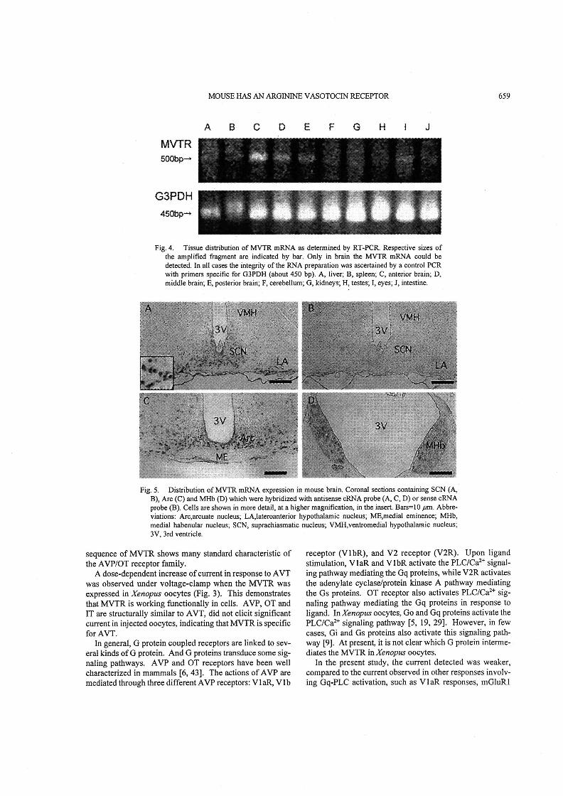
RT一一PCR analysis: To know tissue distribution of MVTR
mRNA, RT-PCR vvas carried out in several tissues of
mouse. lt revealed that MVTR mRNA was expressed in the
brain (Fig. 4). Brain was divided into three regions: ante-
rior, middie and posterior, followed by RT-PCR. There was
no significant difference in intensity oftranscript signals. ln
order to confirm specificity of RT-PCR reaction, nested RT-
PCR was performed, and similar results were obtained
G《A (τ( TτC ACT GAG G了G GGC TCA G6G AGG GくT CTG TGC CT5 (GT TCr G(τ G(二《 GGT GC瓦 GAG AC《 G了G AGA ⊂CT GAC ⊂Cτ GCC τGA GCC
・iG CS” GSIく幣C〔IC^鯉G Gε⊂^IC耳τ略T G£(竿((6G^凹G C呂6^li6(IA‘合τ丁9 ’gC CC・^9^‘鰻‘^孚丁墜A凹G A鰹(Ag GIY GSiA
き
rG G凹丁6EG GeG TSG GS Tg⊂TIC T今C ’g⊂TgC ’1’ ”RG^響替噺(i6 A{A A等C CIGτ評G‘⊂rG甲’%C鰭(Ag A王丁G‘G Gき幣;Tg’ GI’ GIG
口 1
9G ・EC ・9C^i6 ’εC▲霞^^巨^轡^霞^漿‘τ墓⊂^隅G^;C 1(上口鱒G A等^C凹G G£C^王(^S^Gsc Tgc T;c^¥G GE((正Gム王(ze(^王C ’TG “f^6SC
一 鱈
・王・^9・SG CR・1(^早G薯^Gec Ec^轟G張(CS’ Ge⊂(IIG GIT T2(^津G凹C(餅τ今C ffG %6 GY G’,C gG gG ’e’ GliC ’1τ ”¥C T今(Gl⊂C凹G
ぬ・9・ ・1・・gC ・1・‘合C^實^丁今C C肴T G蛋C^凹T T令C C盲‘A轟‘榮‘πT CI’ CaA GE^‘歪G楼G㍗‘ΣC轡G》C CτC▲王C騨AI^GXG ’SG^gC CIC ’gG ’FC
CIG TI・TξC AIT C5⊂AfG⊂王G AHA平響町A震G A睾A(r T葦‘幣T G鯉q‘喜‘鴇G TεC T鱈A‘王G TaG⊂壽‘殆τ殆CτξC了今(噺武等‘C轟G T⇔C
A轟・・宇・^1・〈1・・妄・V・(1・ ・IG ・今・1・・IT⊂霧(兄G G餅H‘A興7^IC ’e’ Gg CI’ GIG^王((ff 9^F T麟‘A轟‘攣^gC ”ly ”¥C Cfi’ G歪G
誓GG‘・A王了「g・Aftc TE‘ T9^殆マ‘1⊂轡rA T薯‘τ罐‘^1⊂T㈱⊂⊂欝聴G⊂1⊂A王⊂丁筆丁築G G艶攣A王‘喰G GΣ(AIC eeG ’eT AgC AI(GU⊂AIA A{⊂
・τ雫耳・・}C・EC・εC ’SG Agc cs^TeC gC Cl‘Ψ殆C^王^n^W ZeC 1⊂ZeC GIC CT「CS“ GSC▲iC eG GeG⊂鱈C T金T G£C T野G甜F^1(
“G幣C⊂IG(S・・X¶G繁・▲讐了G蛋C△r繁((SC 9⊂^王⊂了eC TEC A王(耳‘^1こA§⊂了gC A王(TE⊂TgC(SC ’E(ARG^IG CeC “li“ ’Y CS‘GBT TξC ARA
の罫^M’G A9 ’e⊂C霞^G含G A霞^Alic G含6^巨^CAC G2G^珊G^y CI(了ξ‘榮r C唇G G凹く^王C TN
■ ■
Fig. 1. Nuc!eotide sequence and deduced amino acid sequence of the MVTR (AKO33957). Only the open reading frame and a
part of the 5’ untranslated leader sequenge are shovvn. In-frame stop codon is indicated by asterisk. The .sevep Sransm-embrapg
domains are underlined and numbered. The potential phosphorylation sites for protein kinase C are indicated by 一. Potential
N-glycosylation sites are marked by IAr.

658 M. USUI ET AL.
》瓶りPf
抑「R層Rく
町盈一Gg
漁VTII脚Pf
》丁貧・欺.
VTR・.鋤
闘》1醸
VTR-Pfy猟勘Rζ
》撒礪
聯》丁翼一F,tt
》了又卵陵ζ
》撫殉鵬飛
臓一Pfv1R一腔ζ
糠一{幻
繍
藝lll三課馨i懸i難解購i離,1灘難羅
1叢難語難i購灘il…鍵…麟灘i灘….…灘難
リ ム や ヨ
鵜;難騨灘・…… 箋.….…1….鑛1.肇 購蒙
ム ヤ
lil灘門門難三門. li欝欝鰹繋纒騨
や の
継惑乱美簿醸:::::1::::::二1::1:
ζ武‘,▼‘.」ρ臨 .亀」.L,..亀塾 .}8.辱巳.r∂ρ ‘9.9脅.齢臨‘晶 ‘鯖}.鼻.脳」.口.一...r,卜亀...順...凸・・¶も
贋覇騰;
羅 ヨワ や
雛野晒t、鑛鞍
一 脚,一縛tζ¢s職 鶏姦 κ 《1 s 1隣
罐難嚢難灘
5…:CSf》(KMQRSQpss綴マ簸瓢蜘ε纏し~卿.EF峯......,..
” , , , 凸
。 t - t -
一 一 一
亀 亀 ㍉ 昌
一 1 一 一
十 ・ 旨
J t i
l b -
t t i
一 一 一
髄1eeS183
節5z臼@
三輪
2ag
Z{;5
Z97
2?S
zs・i
36S
騨フ
363
算裏
g8441937e
Fig. 2. Comparison of deduced amino acid sequence for the MVTR with sequences of AVT receptor of P. ,flesus (VTR-Pf,
AF 184966), the AVT receptor of Rana catesbeiana (VTR-Rc; AY277924) and the Gallus gallus AVT receptor (VTR-Gg;
AF 147743). Lines under the MVTR sequence indicate transmembrane regions. The important sequence in G-protein coupling
site is boxed. Two cystine residues in extraceliular II and III, which composed a disulfide bridge, are marked by A. The impor-
tant sequence in ligand binding sites are marked by “,
Av-r sonM
me
snA
30sec
,一
Fig. 3. Functional expression of the MVTR in A’enopus oocytes as recorded using voltage clamp techniques, Recorded
membrane cunent trace ofan oocyte expressing the MVTR protein was shown when 50 iiM AVT was applied (A). After a
washout, 100 nM AVT was applied (B). An inward current is shown as a downward curve and the upper bars show the
app1ication of AVT at the described concentration. AVP, OT and IT induced no response, whose concentrations were
equivalent to that of AVT. Application ofAVT to noninjected oocytes failed to elicit the cu町ent.
(Data not shown).
In sittt hybridization: ln situ hybridization using a cRNA
probe compiementary to MVTR mRNA (antisense probe)
was performcd on coronal sections through various regions
of mouse brain. MVTR mRNAs were expressed highly in
the neurons of suprachiasmatic nucleus (SCN)(Fig. 5A),
arcuate nucleus (Arc)(Fig. 5C) and medial habenular
nucleus (MHb)(Fig. 5D). High magnification micrograph
showed that cytoplasms were uniquely stained in a11 these
regions. Another locus did not show a significant signal.
Sense probe did not stain in these regions (Fig. 5B). ln situ
hybridization performed using a cRNA probe made from
different region ofthe cDNA produced similar results (Data
not shown).
DISCUSSION
From data accumulated in the GenBank database, we
attempted to find a DNA encoding MVTR. P. flesus AVT
receptor had already been well characterized using receptor
expression experiments in Xenopus oocytes. Therefore, we
searched for candidates for cDNA encoding MVTR by com-
paring with the cDNA ofP. .flesus AVT receptor. We found
that deduced amino acid sequence of MVTR, one of the can-
didates, has significant homology with that ofP. .flesus AVT
receptor and other AVT receptors. Moreover amino acid
MOUSE HAS AN ARGININE VASOTOCIN RECEPTOR 659
MVTR50bop一
G3PDH
450bp嚇
A B c D E F G H 嚢 J
Fig. 4. Tissue distribution of MVTR mRNA as determined by RT-PCR. Respective sizes of
the amplified fragment are indicated by bar. Only in brain the MVTR mRNA could be
detected. ln all cases the integrity of the RNA preparation was ascertained by a control PCR
with primers speciftc for G3PDH (about 450 bp). A, liver; B, spleen; C, anterior brain; D,
middle brain; E, posterier brain; F, cerebellum; G, kidneys; H, testes; 1, eyes; J, intestine.
’∫:
?蜉]
灘
Fig. 5. Distribution of MVTR mRNA expression in mouse bTain. Coronal sections containing SCN (A,
B), Arc (C) and MHb (D) which were hybridized with antisense cRNA probe (A, C, D) or sense cRNA
probe(B). Cells are shown in more detai1, at a higher magnification, in the insert. Bars=・1 0 ptm. Abbre-
viations: Arc,arcuate nucleus; LA,lateroanterior hypothalarnic nucieus; ME,medial eminence; MHb,
media且habenu!ar nucleus;SCN, suprachiasmatic nucleus;VMH,ventromedial hypothalamic nucleus;
3V. 3rd ventricle. ’
sequence of MVTR shows many standard characteristic of
the AVPIOT receptor family.
A dose-dependent incrcase ofcurrent in response to AVT
was observed under voltage-clamp when the MVTR vvas
expressed in Xenopus oocytes (Fig. 3). This demonstrates
that MVTR is working fUnctionally in cells. AVP, OT and
IT are structurally similar to AVT, did not elicit significant
current in injected oocytes, indicating that MVTR is specific
for AVT.
ln general, G protein coupled receptors are linked to sev-
eral kinds of G protein. And G proteins transduce some sig-
naling pathways. AVP aiid OT receptors have been well
characterized in mammals [6, 43]. The actions of AVP are
皿ediated through three different AVP receptors:VlaR, V l b
receptor (VlbR), and V2 receptor (V2R). Upon ligand
stimulation, V l aR and V lbR activate the PLC/Ca2’ signal-
ing pathway mediating the Gq proteins, while V2R activates
the adenylate cyclase/protein kinase A pathway mediating
the Gs proteins. OT receptor also activates PLC/Ca2’ sig-
naling pathway mediating the Gq proteins in response to
ligand. ln Xenopus oocytes, Go and Gq proteins activate the
PLCICa2’ signaling pathway [5, 19, 29]. However, in few
cases, Gi and Gs proteins also activate this signaling path-
way [9]. At preseng it is not clear which G protein interme-
diates the MVTR in Xenopus oocytes.
In the present study, the current detected was weaker,
compared to the current observed in other responses involv-
ing Gq-PLC activation, such as V l aR responses, mGluRl
660 M. USUI ET AL.
metabotropic glutamate. receptor responses [25] and Ml-
type muscarinic acetylcholine receptor responses [22]. Sim-
ilar weak responses were observed in receptor responses
mediating other PLCICa2’ signaling pathway. For exaniple,
6 opioid receptor activates PLC activation via Gi proteins in
Xenopus oocytes [27]. But the intrinsic activity to activate
PLC is less potent, compared to Gq proteins. ln addition, Gi
proteins stimulate the PLC/Ca2’ signaling pathway less
effective than Go proteins [7].
The distribution of the MVTR mRNA in mouse brain
indicates new and as yet undetermined roles for AVT in
brain. ln situ hybridization analyses demonstrated the sig-
nificant expression of MVTR mRNA in SCN, Arc and
MHb. SCN is well known to regulate circadian rhythm and
reproduction in mammals [1, 3]. The excitatory effect of
AVP and its potential contribution to the circadian cycle of
electrical activity in the SCN of the rat was investigated
using extracellular recording from hypothalamic slices of
rats (26]. The maj ority of neurons tested for their responses
to AVP and AVT displayed coincident, dose-dependent
excitation by both peptides, although the relative efficacy
varied between neurons, with some showing a highly prefer-
ential excitation by AVT. These results show that AVT
works specifically in SCN and does not confiict wnh our
results that show the expression of MVTR mRNA in this
reglon.
Arc, known to play a role in energy homeostasis and
reptoduction [8, 23], is labeled by a[3H]d(CH2)s[Tyr(Me)]VP, AVP antagonist, in rat brain
[39], but it is not labeled by either [3H]AVP or [3H]OT.
These results suggest that AVT specific receptor or novel
AVP receptor subtype would be present in this region.
Habenular complex (Hb) is well known to take part in a
variety of biological functions such as, pain processing,
reproductive behavior, reward, food and water intake, stress
response, sleep wake cycles and learning [36]. AVT pro-
duces its specific effects when injected into the third ventri-
cle of the brain. The electrolytic destruction of the Hb
completely suppressed the AVT effects and neither AVP
nor OT was able to mimick these effects [32]. ln addition,
Hb specifically bound synthetic AVT [14]. These results
predict the action of AVT on the Hb, which in turn consist
with our results ofMVTR mRNA expression in this region.
MVTR distribution is partially similar to V l aR [4]. But
in this study, MVTR did not respond to AVP, indicating that
MVTR is different from V l aR.
Recently a neuropeptide S receptor (NPSR) has been
reported [42]. lts gene sequence is consistent with that of
MVTR. NPSR mRNA was widely distributed in rat brain,
the strongest expression signals were found in several dis-
crete nuclei or regions, such as the anterior olfactory
nucleus, endopiriform nucleus, amygdala, precommissural
nucleus, paraventricular thalamic nucleus, and subiculum.
High levels of expression were found in multiple nuclei of
hypothalamus. Their results using rat coincided with our
results that were obtained using mouse. However, there
were some differences between them. ln mouse, MVTR
mRNA was distributed gspecially in SCN, Arc and MHb.
In conclusion, mouse has an AVT reactive receptor. lt is
responsive to AVT but also neuropeptide S and it may’be
playing roles related to sleep, reproduction and a variety of
physiological functions.
ACKNOWLEDGMENT. We thank Dr. Tanoue (NationalResearch lnstitute for Child Health and Development) for
providing plasmid containing cDNA encoding V l aR.
REFERENCES
1.
2.
3.
4.
5.
6.
7.
8.
9.
10.
IL
12.
13.
14.
互5.
Ande, MC and Silver, R.2005. Orchestrating time:arrange-
ments of the brain circadian clock.7》’endsハXeurosc’.28:145-
151.
Badiu, C,, Coculescu, M, and Moller, M.1999. Arginlne vaso.
tocin mRNA revealed by in situ hybridization ln bovine pineal
gland cells. Cε〃Tissue Rθ∫.295:225一一229.
Barbacka-Surowiak G Surowiak J. and Stoklosowa S.2GO3. り つ ひ コThe involvement of suprachiasmatic nuclei in the regulation of
estrous cycles敢l rodents、 Reprod. B’01.3:99-129.
Barberis, C. and Tribollet, E.1996. Vasopressin and oxytocin
receptors in the central nervous system. C,’it. Rev.ハleto‘obiol.
10:119-154.
Berridge, MJ.1993. Inositol trisphosphate and calcium signal-
ing. Natitre(乙ond.ノ361:315-325.
Birnbaumer, M.2000. Vasopressin receptors. Trencts Endo-
crino1.ルfetab.11:406-410.
Blumenau, C., Berger, E, Fauteck, J.D., Madeja, M., Witt-
kowski, W., Speckmann, EJ. and Musshoff, U.2001. Expres-
sion and functional characterization of the mtl melatonin
receptor from rat brain in Xenopus oocytes:evidence for cou・
pling to the phosphoinositol pathway. J.」P加θo〃~es.30:139-
146.
Cone, RD., Cowley, MA., Butler, A.A., Fan, W., Marks, D.L.
and Low MJ.2001.The arcuate nucleus as a conduit for ウ
diverse signals relevant to energy homeostasis. Int.ノ:Obes.
Relat. Metab. Disord.(SupρL/5:S63-67.
de la Pena, P., del Camino, D,,Pardo, L.A., Dominguez, P. and
Barros, F.1995. Gs couples thyrotropin-releasing hormone
receptors expressed in Xenopus oocytes to phospholipase C. J.
Bゴ01.Chem。270:3554-3559.
Dixon, RA., Siga1,1.S.,Cande[ore, M.R。, Register, RB., Scat-
tergood, W., Rands, E. and Strader, C.D.1987. Structural fea-
tures required fbr董igand binding to the beta-adrenergic
receptor, E:?レBO J.6:3269-3275.
Ferguson, S.S.1998, Using green fluorescent protein to under-
stand the mechanisms of G-protein-coupled receptor regula-
tion. Braz.」.ルfed. Biol. Res,31:1471-1477.
Figueroa, J., Morley, SD., Heierhorst, J., Krentler, C., Lederis,
K.and Rlchter, D.1989. Two isotocin genes are present in the
white sucker Catostomus commersoni both lacking introns in
their protein coding regions. EルfBO J.8:2873-2877.
Gilchriest, BJ., Tipping, DR., Levy, A. and Baker, B.1.1998.
Diurnal changes in the expression of genes encoding for argin-
ine vasotocin and pituitary pro-opiomelanocortin in the rain-
bow trout(Oncorhynchus mykiss):correlation with changes in
plasma hormones. J.ハ「eitroendocrinoL 10:937-943.
Goldstein, R.1989. HabenuIar nuclei specifically bind syn-
thet三。 arginine vasotocin.・Endocrinologア27二237」239.
Goldstein, R.1992. Arginine-vasotocin(AVT>一a pineal hor」
16.
17.
18.
19.
20.
21.
22.
23.
24.
25.
26.
27.
28.
29.
MQUSE HAS AN ARGININE VASOTOCIN RECEPTOR 661
mone in marnmals. Rom. 」. EndocrinoL 30: 21-44.
Hausmann, H., Richters, A., Kreieiikamp, H.J., Meyerhof, W,,
Mattes, H.; Lederis, K., Zwiers, H. and Richter, D. 1996. Muta-
tional analysis and molecular modeling ofthe nonapeptide hor-
mone binding domains of the [Argg] vasotocin receptor. Proc.
1>btl. Aced.5ヒガ.こノニS.A.93:6907」6912.
Hilakivi, 1., Taira, T. and Hiiakivi, L.A. 1988. Effect of periph-
eral administration of arginine vasotocin on neonatal sleep in
rats. Peptides 9: 487491 .
Ingram, C.D. and Tolchard, S. 1994. [Arg8] vasotocin excites
neurones in the dorsal vagal complex in yitro: evidence for an
action through novel class(es) of CNS receptors. 」. Neuroendo-
crinoL 6: 415422.
Kasahara, J. and Sugiyameq H. 1994. lnositol phospholipid
metabolism in Xenopus oocytes mediated by endogenous G(o)
and Gi proteins. FEBS Lett. 355: 41-44.
Kawasawa, Y., McKenzie, L.M,, H川, D.P., Bono, H., Yanag-
isawa, M.; RIKEN GER Group; GSR Members. 2003. G pro-
tein-co叩led receptor genes in the FANTOM2 database。
Genome Res. 13: 1466-1477.
Kimura, T., Tanizavveg O., Mori, K, Brownstein, M.J. and
Okayama, H. 1992. Structure and expression of a human oxy-
tocin receptor. Natui「e(Lond.ノ356:526-529.
Kubo, T., Fukuda, K, Mikami, A., Maeda, A., Takahashi, H.,
Mishina, M., Hageg T., Hageg K., lchiyama A., Kangawa, K. et
al, 1986. Cloning, sequencing and expression of complemen-
tary DNA encoding the muscarinic acetylcholine receptor.
Nature (Lond.) 323: 411-416.
Leedom, L., Lewis, C., Garcia-Segura, L.M. and Naftolin, F.
1994. Regulation of arcuate nucleus synaptology by estrogen.
Ann.ハ「en・}”ork Acad. Sci.743:61-71,
Liu, B. and Burbach, J.P. 1987. Characterization ofvasopressin
and oxytocin immunoreactivity in the sheep and rat pineal
gland: absence of vasotocin and detection of a vasopressin-like
peptide. Peptides 8: 7-11.
Masu, M., Tanabe, Y., Tsuchida K., Shigemoto, R. and
Nakanishi, S. 1991. Sequence and expression of a metabotro-
pic glutamate receptor. Natttre (Lond.) 349: 760-765.
Mihai, R., Cocul escu, M., Wakerley, J,B. and Ingram, C.D.
1994. The effects of [Argg] vasopressin and [Argg] vasotocin
on the firing rate of suprachiasmatic neurons in vitro. Neuro-
science 62: 783-792.
Miyarnae, T., Fukushimeq N., Misu, Y. and Ueda, H. 1993.
Delta opioid receptor mediates phospholipase C activation via
Gi in Xenopus oocytes. EEBS Lett. 333: 31 1-3 14.
Moons, L., Cambre, M., Batten, T.F. and Vandesande, F. 1989.
Autoradiographic localization ofbinding sites for vasotocin in
the brain and pituitary of the sea bass (Dicentrarchus labrax).
ハlei‘rosci. Z,ett.100:11・一16。
Moriarty, T.M., Padrell, E., Carty, D.J., Omri, G., Landau,
30.
3L
32.
33.
34.
3S.
36.
37.
38.
39.
40.
4L
42.
43.
EM. and lyengar, R.1990. Go protein as signal transducer in
the pertussis tOxin-sensitive phosphat皇dylinositol pathway.
ハratt〃re(Lond/343:79-82.
Nathanson, M H., Moyer, M.S。, Burgstahler, AD.,0℃arroll,
AM., Brownstein, M.J, and Lolait, S.J.1992, Mechanisms of
subcellu1ar cytosolic Ca2+signahng evoked by stimulation of
the vasopressin V l a receptor.」.」B’o’.(フhe〃1.267:23282-
23289.
Pavel, S,1978、 Arginine vasotocin as a pineal hormone.ノ:
ハlei‘raL乃「ansm。(SitppL/13:135-155.
PaveしS. and Eisner, C 1984. A GABAergic habenulo-raphe
pathway mediates both serotoninergic and hypnogenic eff6cts
of vasotocin in cats. Brain Res. BttlL l3:623一一627.
Prechel, M.M,Audhya, T.K. and Simmons, W.H.1989. In伽・
ence of age on August levels of pineal immunoreactive argin.
ine vasotocin in rats. J. Pineal 1~es.6:1-7.
Rose J.D. and Moore F.L。2002. Behavioral neuroendocrinol一、 ウ ウ
ogy of vasotocin and vasopressin and the sensorimotor pro-
cessing卦ypothesis. Front」ハleuroendoci’inoL 23;317-341・
Sambrook, J. and Russel, DW.2001. A Laboratory Manual,
3rd ed.,.Cold Spring Harbor Laboratory Press, New York,
Sandyk, R.1991,Relevance of the habenular complex to neu-
ropsychiatry:a review and hypothesis. Int.ノ:ハXe urosc’.61:
189-219
Sartin, J.L., Brout, B℃. and Orts, R,J.1979. Neurotransm itter
rggulation of arginine vasotocin release from rat pineal g!ands.
in vitro. Acta. EndocrinoL(Coρenh/91:571-576.
Thompson, R.R. and Moore, F.L 2000. Vasotocin stimulates
appetitive responses to the visual and pheromona夏stimuli used
by male roughskin newts during courtsh{p, H()rm. Behav.38:
75-85.
van Leeuwen, F.W., van der Beek, E.M,van Heerikhuize, JJ.,
Wolters, P., van der Meulen, G. and wan, Y.P.1987, Quantita-
tive light microscopic autoradiographic localization of binding
sites labelled with[3H】vasopressin antagonistd(CH2)5Tyr(Me)VP in the rat brain, pituitary and kidney. Neu-
rosci.」Cett.80:121-126.
Warne, J.2002. The role of arginine vasotocin in teleost fish
osmoregulation. S〃mp. Soc.瓦Ψ. B’oL 54:83-95.
Warne, J.M.2001。Cloning and characterization of an arginine
vasotocin receptor from the euryhaline flounder Platichthys
flesus. Gen. Co㎎P.万ndocrinol.122:312-319.
Xu, Y.L., Reinscheid, RK., Huitron-Resendiz, S., Ciark, S.D.,
Wang, Z., Lin, S.H.,B則cher, F.A.,Zeng, J.,Ly, NK., Henrik-
sen, SJ., de Lecea, L, and CiveUi,0.2004, Neuropeptide S:a
neuropeptide promoting arousal and anx{olytic・like effects.
ハXe;tron 43:487L497.
Zingg, H.H. and Laporte, S.A.2003, The oxytocm receptor。
Trends・EndocrinoL Metab.14:222-227.