Embed Size (px)
Citation preview
Characterization and Biocompatibility Studies of Novel HumicAcids Based Films as Membrane Material for an Implantable
Glucose Sensor
Izabela Galeska,† Tammy Hickey,‡ Francis Moussy,‡ Donald Kreutzer,‡ andFotios Papadimitrakopoulos*,†
Nanomaterials Optoelectronic Laboratory, Department of Chemistry, Polymer Program, Institute ofMaterials Science, University of Connecticut, Storrs, Connecticut 06269; and Center for Biomaterials &
Surgical Research Center, University of Connecticut Health Center, Farmington, CT 06030-1615.
Received June 29, 2001
Multilayered films of humic acids (HAs) (naturally occurring biopolymers) were investigated as a potentialsemipermeable membrane for implantable glucose sensors. These films were grown using a layer-by-layerself-assembly process of HAs and oppositely charged ferric ions. The growth of these assemblies exhibitedstrong dependence on the pH and ionic strength of HAs solutions, which correlated with the degree ofionization of the carboxyl groups and neutralization-induced surface spreading. Quartz crystal microbalance(QCM) and ellipsometric studies have shown repeatable, stepwise increase in mass (as high as 5.63µg/cm2) and in film thickness (ca. 24.3 nm per layer) for these assemblies. The permeability of glucose throughthese membranes can be regulated by varying the number of self-assembled HAs/Fe3+ layers. Moreover, a200 nm thick HAs/Fe3+ film (in its hydrated state) had a shear modulus of about 80 MPa, implying stabilityupon implantation. These films were determined to be biocompatible since in vivo studies indicated onlymild tissue reaction along with some neovascularization.
Introduction
In vivo membrane degradation, fouling, inflammation, andfibrosis (i.e., fibrous encapsulation) have so far preventedthe widespread use of implantable biosensors.1,2 Nafion hasbeen successfully used in nonbiological sensors either as asemipermeable or protective membrane.3-6 However, inimplantable devices Nafion has been associated with (1)calcification and (2) fibrosis of the surrounding tissue, leadingto loss of vascularization and restricted analyte flow, andtogether resulting in premature sensor failure.7 Hence thereis an urgent need for inert, bioacceptable materials capableof withstanding the strong oxidative environment in vivo.Humic acids (HAs) are biopolymers found in soil, sediments,water, and some plants such as tobacco.8-11 Although theirorigin is ambiguous, it is speculated that they may haveevolved from the auto-oxidation and condensation of ligninand polyphenols during the humification process12 or areproduced during in vivo biosynthesis in plants.9 Humic acidsare deemed as the final product of the biodegradative andoxidative route, with a mean residence time up to 1200 yearsin soil,13 and probably will not undergo any further break-down unless they are exposed to agents they have notpreviously encountered.14 HAs are very heterogeneous innature and contain species with molecular weight rangingfrom a few thousand daltons to hundreds of kilodaltons. Theirstructures,15,16 although still under investigation, are domi-
nated by aromatic moieties containing carboxylic, carbonyl,phenol, catechol, and quinone, along with a few aminegroups.17
The physiological and toxicological effects of humic acidson humans and animals have been explored to some degree.HAs are known to exist in the gastrointestinal tract of humansand animals,18 have been found in the blood19 and also areknown to be metabolized in the liver.20 Oral doses of HAsreduce heavy metal adsorption in animals19 and also decreasepesticide toxicity.21 Antiviral, antiinflammatory and profi-brinolytic activity of HAs have been previously reported.19
HAs also have the ability to interact with metals22 and absorbto a variety of surfaces such as minerals,23-27 cellulose,28
and chitin and chitosan29 as well as to bacteria.25 Althoughingestion of large quantities of HAs via drinking water havebeen reported to be one of the causative factors of Kahin-Beck disease30 (an endemic osteoarthritic disorder) and ofBlack Foot disease31 (a peripheral vascular disease), thephysiological effects associated with these diseases requirethe HAs to be available in solution. On the basis of theabove-mentioned attributes we have investigated multilayeredfilms of HAs and ferric ions, as a possible membrane materialfor an implantable glucose sensor. The present paperexamines the effect of experimental conditions on the HAs/Fe3+ electrostatic layer-by-layer assembly, glucose perme-ability and biocompatibility of the resulting films.
The self-assembly involving oppositely charged moieties32
has the potential to address membrane mineralization, oftenexperienced in anionic biomaterials.7,33-34 Along the linesof what was demonstrated in our previous work for Nafion/
* To whom correspondence should be addressed: Telephone: (860)-486-3447. Fax: (860)-486-4745. E-mail: [email protected].
† University of Connecticut.‡ University of Connecticut Health Center.
1249Biomacromolecules 2001,2, 1249-1255
10.1021/bm010112y CCC: $20.00 © 2001 American Chemical SocietyPublished on Web 10/12/2001

Fe3+ system,35 the carboxylic groups in HAs will be saturatedwith iron(III) during the self-assembly process, thus poten-tially preventing calcification and fouling on the sensor’ssurface.36 Furthermore, the rich chemical environment in theHAs structure and the water-based self-assembly process maybe easily employed for the attachment or coupling biologi-cally active molecules such as growth factors, antifoulingagents and drug delivery systems, which would furtherminimize the adverse tissue reactions to the implanted sensor.
Experimental Section
A. Electrostatic Layer-by-Layer Film Growth andCharacterization. Materials and Reagents.Humic acid,lot no. 11909LR, was purchased from Aldrich and was usedwithout any further purification. The HAs sample used inour studies was characterized by elemental and trace metalanalysis, as well as molecular weight and functional groupsdetermination. In particular elemental analysis (as suppliedby Aldrich Chemical Co., Inc. and verified within 1% byGalbrith Laboratory) for C, N, and H indicated (%): C, 43.7;O, 41.8; H, 4.02; N, 0.94; Na, 6.2; Ca, 0.9; S, 0.7; Fe, 0.8;Al, 0.6; Mg, 0.2. Trace metal analysis (in ppm) (as suppliedby Aldrich Chemical Co.) detected: K, 535; Si, 260; Mn,175; Ti, 164; Sr, 139; Ba, 90; V, 10; Zr, 9; Cu, 6; Li, 4; Cr,2. Gel permeation chromatography (GPC) inN-methylpyrolidone/water (9:10) mixture indicated a weight-averagemolecular weight of 169 kDa with polydispersity indexMw/Mn ) 2.58. Volumetric titration (from pH 2.5 to 12) showeda cumulative acidic functionality content on the order of 820mequiv/100 g of HAs. The intermediate deflection points inthe titration curve ((I) pH 3.2; (II) pH 5.8 and 6.8; (III) pH10.3) indicated presence of different types of acidic groups((I) aromatic carboxylic groups ortho to phenolic substitu-tions; (II) weak aromatic and aliphatic carboxylic acids; (III)phenolic and catecholic-OH).
A 1 mg/mL HAs solution in deionized water was usedfor all experiments. pH of the HAs solutions used for theassembly was adjusted with diluted hydrochloric acid (J. T.Baker) and the ionic strength of these solutions weremodified by the addition of ACS Certified potassium chloride(Fischer). A 5 mg/mL solution of iron(III) chloride hexahy-drate (FeCl3‚6H2O) (Aldrich) was used for the self-assemblywith HAs. Millipore quality deionized water with resistivity>18 MΩ was utilized in all experiments.
The self-assembled films were deposited either on siliconwafers or quartz crystal resonators. Silicon wafers werecleaned in piranha solution (H2SO4/H2O2 (7:3)), rinsed withdeionized water and methanol, kept in deionized waterovernight, and used for the self-assembly without furthersurface modification. For all QCM experiments, 9 MHz ATcut quartz crystal resonators with an electrode surface areaof 0.32 cm2 (USI, Japan) were used. The resonators withgold electrodes were used for the shear modulus experimentsand those with silver electrodes for all other experiments.Prior to film deposition, the resonators were immersed forca. 20 s. in ethanol/water/potassium hydroxide mixture (59/40/1), rinsed in deionized water and used without furthersurface modification.
Techniques and Instrumentation.An HMS series pro-grammable slide stainer from Carl Zeiss, Inc., was used forthe layer-by-layer assembly of humic acids with Fe3+. Thesample holder in the HMS Series Slide Stainer was coveredto reduce solvent evaporation thereby improving film quality.
A Hewlett-Packard Network analyzer (HP-1005A, 10 kHz- 300 MHz) was utilized to detect changes in both thefundamental frequency and in the higher harmonics afterevery dip cycle during the self-assembly. The Sauerbreyequation37 was used to quantify the change in frequency withthe apparent mass deposited on the resonator (eq 1), where
fo is the resonant frequency of the quartz resonator,∆m isthe mass change,A is the active surface area,µQ is the shearmodulus of quartz, andFQ is the density of quartz.
The higher frequency harmonics measured for an emptyresonator and after every dip cycle during the HA/Fe3+
assembly were utilized for the shear modulus calculation.The shear compliance (J′) can be determined by plotting theequivalent mass (meq) (eq 2), derived from different harmon-ics, vs the square of the frequencyω2 ) (2πf)2.38 The trueequivalent mass, deposited on the resonator, can be obtainedfrom the intercept of this plot and the shear compliance andshear modulus can be further calculated from the slope,utilizing eq 3.38 The equivalent mass obtained by thisprocedure gives much better representation of the true massdeposited on the resonator by taking into account theviscoelastic losses of the self-assembled film.
whereZq ) 8.8× 10⁶ kg m-2 s-1 is the piezoelectric stiffenedacoustic impedance,f0 is the fundamental frequency of anempty quartz,δf/f ) (fin - fn)/fn; fin is the resonant frequencyof the “ith” layer for the “nth” harmonic andfn is the resonantfrequency of the empty resonator for the “nth” harmonic.J′is the shear compliance, andd is the thickness of the film,determined by ellipsometry.
The thickness of self-assembled films was independentlydetermined by ellipsometry. Ellipsometric data was acquiredusing a variable-angle WVASE32 spectroscopic ellipsometerfrom J. A. Woolman Co., Inc. Films were scanned in therange 250-1000 nm at the incident angles of 65, 70, and75°. Data were collected every 10 nm at 10 revolutions permeasurement. A standard Cauchy model was used to fit forthickness and optical constants in the entire optical range.
A dialysis chamber was used to determine permeabilityof 1 µm glass fiber membranes (Gelman Sciences) coatedwith self-assembled HA/Fe3+ layers. The interior of thedialysis chamber was filled with 1 mL of 1 M glucose in0.9% NaCl solution. Subsequently, the concentration ofglucose that diffused out from the dialysis chamber to thedialysis buffer (50 mL of 0.9% NaCl solution, no glucose)was determined by a Beckman glucose analyzer. The detailedprocedure has been described elsewhere.39 The apparentdiffusion coefficients (DApp) of glucose through the self-
∆f ) -2fo2∆m/A (µQFQ)0.5 (1)
meq ) -(Zq/2f0)(δfn/fn) (2)
slope) J′(meq)2d/3 (3)
1250 Biomacromolecules, Vol. 2, No. 4, 2001 Galeska et al.
assembled membranes were estimated based on the modeldescribed previously.35 The following assumptions weretaken into account: (i) the glass fiber membrane wasconsidered extremely porous and did not contribute to theresistance; hence the flux was assumed to be through theself-assembled layer; (ii) the flow rate did not effect the fluxof glucose through the membrane; (iii) rapid diffusion wasassumed through the boundary layers.
Fourier transform infrared (FTIR) spectroscopy data wasobtained from a Nicolet Magna 560 using TeGeSe detectorwith 4 cm-1 resolution. HAs/Fe3+ films were assembled ondouble polished Si wafer and held at an angle of 45° withrespect to the incident IR beam. This increases the samplingpath length and eliminates the interference fringes from thedouble polished silicon wafer. A minimum of 128 scans wassignal averaged and the background, consisting of double-polished Si wafers and air was carefully subtracted from thesample’s spectra.
The stability of the HAs/Fe3+ films used for the in vivoexperiments was evaluated by depositing them on glass slidesand monitoring change in UV absorption as a function ofimmersion time in 100 mL of PBS buffer (phosphate buffersaline, Sigma Diagnostics) in a thermostated bath at 37°C.A Perkin-Elmer Lambda 6 UV/vis spectrophotometer wasused for these experiments.
B. In Vivo Biocompatibility Studies. Humic acidssolution at pH 5, with and without 0.01 M KCl, was used tocoat medical grade silastic tubing, which is a well-knownbiocompatible material.40 Nine layers of HA/Fe3+ were self-assembled. The 1 cm long silastic tubing samples weresterilized in ethylene oxide (3M steri-gas at 55°C for 2.5 hand 12 h of aeration cycle) before implantation. Three groupsof three Sprague-Dawley male rats were used for this study.Both types of humic acids coatings on silastic tubing,prepared with and without salt, as well plain medical-gradesilastic tubing as a control, were implanted into each rat.The first group of rats was euthanized after 1 day, the secondafter 1 week, and the last after 1 month. Samples wereinserted subcutaneously using sterile 18 gauge iv catheters.The samples were slid into the rats through the lumen ofthe catheter. The backs of the rats had been shaved the daybefore, to minimize the inflammatory response due to theirritation of the shaving procedure. During implantation andshaving procedures the rats were anesthetized with isofluo-rane.
After the rats were euthanized with carbon dioxide, anapproximately 1 cm by 2 cm rectangle of tissue containingthe implanted tubing was removed. It was fixed in formalinand later cut into several sections across its length, so thatthe tubing was cut in cross section. These tissue sampleswere prepared for the histology by embedding them inparaffin wax and staining by hematoxylin and eosin (H&E).Hematoxylin is known to dye the nucleus dark purple-blue,42
while the eosin stains other cell structures pink, especiallythe cytoplasm.42 Gomori’s one-step trichrome was also usedto stain fibrous tissue (i.e., collagen fibers).42 These stainingprocedures help in elucidating the inflammatory responseinduced by the foreign implant.
Results and Discussion
A. Characterization. The technique of electrostatic layer-by-layer self-assembly32 was used to deposit films of HAswith oppositely charged ferric ions. The HAs/Fe3+ self-assembly dip cycle consists of consecutive dipping in HAsand ferric chloride solution followed in each case by washingin deionized water. The wash step between the dipping stepshelps to remove the loosely bounded moieties on the surface.The acidic functional groups in humic acids, primarilycarboxylic (pKa 4-5)43 and cateholic (pKa 8-11)43 provideactive sites for the positively charged ferric ions, which resultin electrostatic and complexation driven interactions.44,45Theexcess charge of the trivalent iron ions reverses the chargeon the adsorbed HAs layer rendering it positive and thusallowing the next layer of HAs to assemble. Thus, multi-layered HAs/Fe3+ films can be deposited both as a functionof the pH and ionic strength of the humic acids solution. Adetailed experimental description and discussion on the self-assembly process is provided in a previous study on Nafion/Fe3+ assemblies.35
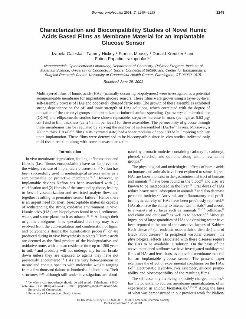
Figure 1 shows the ellipsometrically determined thicknessof HAs/Fe3+ films vs the number of HAs/Fe3+ layers (i.e.dip cycles) as a function of the pH and ionic strength ofhumic acids solutions. The ellipsometric studies indicate astepwise increase in film thickness. However, in someinstances, particularly at a higher pH (which yields lesserionization of the Si/SiOx surface), steady state is reached onlyafter 3-5 dip cycles (see curve C in Figure 1). The growthcharacteristics of these assemblies exhibit strong dependenceon the pH and ionic strength of the HAs solution, whichcan be correlated to the HAs conformation in solution andalso that attained during surface deposition, as describedbelow. The net charge on HAs is governed by the degree ofionization of its carboxylic and phenolic groups, which inturn is a function of solution pH.46 At a high pH these weakacid functionalities in HAs are ionized and attain a negativecharge resulting in both inter- and intramolecular electrostaticrepulsion. Therefore, in solutions of high pH, HAs areexpected to attain a more expanded conformation. pH playsa very important role on the nature of the substrate surfacethat HAs have to assemble on due to iron(III) precipitationin its hydroxide form (Ksp ∼ 6 × 10-39).47 Above pH 4.3,the formation of iron hydroxides transforms the iron domi-
Figure 1. Ellipsometrically determined thickness vs dip cycle as afunction of pH and ionic strength of HAs solutions for HAs/Fe3+
assemblies: (A) pH 5, 0.01 M KCl; (B) pH 5, no salt; (C) pH 9.5,0.01 M KCl; (D) pH 9.5, no salt.
Novel Humic Acids Based Films Biomacromolecules, Vol. 2, No. 4, 2001 1251

nated surface into a strong base that induces greater surfacespreading of HAs due to acid-base interactions and affectsthe number of available complexation sites.35 Thus, the filmgrowth is considerably slower, (i.e., less than 2 nm per dipcycle at pH 9.5) as compared to that at pH 5, where thegrowth rate is on the order of 19.4 nm per dip cycle. Similaracid-base neutralization-induced surface spreading leadingto a lower growth rate was also observed in our previouslyreported Nafion/Fe3+ assemblies.35 Thus, neutralization-induced surface spreading of HAs on the iron(III) surface isbelieved to be one of the governing factors in theseassemblies. Another important factor could be the conforma-tion dependence of HAs on pH. At a lower pH, the degreeof ionization on HAs is lesser, which causes them to attaina tighter, globular conformation and simultaneously ag-gregate in solution.48 Upon adsorption on the surface, thehydrophobic interactions that are responsible for the globu-larization of the HAs, compete with neutralization-inducedsurface spreading leading to an enhanced growth rate i.e.,19.4 nm per dip cycle. The notable acceleration in filmgrowth is observed upon increasing the ionic strength of HAssolution by adding potassium chloride (i.e. 24.3 nm per HA/Fe3+ layer at pH 5 and 9.2 nm at pH 9.5). This faster film
growth is attributed to salt induced charge screening49 thatleads to more compact HAs conformation capable of furtherresisting neutralization-induced surface spreading.35
Along with ellipsometry, QCM was utilized to obtainbetter understanding of the growth of HA/Fe3+ assemblies.QCM is an analytical method for thein situ probing ofinterfacial processes and has attracted considerable interestin the scientific community, with applications in variousareas.38,50-52 For the HA/Fe3+ assembly, change in theresonant frequency (f0) of the QCM resonator was monitoredafter each dip cycle, where the decrease inf0 signifies theapparent mass adsorption. Because of the well-defined quartzresonant frequency, high precision measurements allow thedetection of minute amounts of deposited material. Themeasured frequency changes can be correlated with mass
Figure 2. Frequency change as determined by QCM (left axes) uponself-assembly on the resonator and the calculated apparent mass(right axes) vs dip cycle as a function of the pH of HAs solutions forHAs/Fe3+ assemblies: (A) pH 3; (B) pH 5; (C) pH 7; (D) pH 9.5.
Figure 3. Frequency change as determined by QCM in (left axes)upon self-assembly on the resonator and the calculated apparentmass (right axes) vs dip cycle as a function of pH and ionic strengthof HAs solutions for HAs/Fe3+ assemblies: (A) pH 5, 0.01 M KCl;(B) pH 5, no salt; (C) pH 9.5, 0.01 M KCl; (D) pH 9.5, no salt.
Table 1. Summary of Experimentally Determined HAs/Fe3+ FilmThickness and Apparent Mass Deposited per Dip Cycle on QCMResonators as a Function of pH and Ionic Strength of HAsSolution
deposited mass
pH KCl [M]layer thickness
per dip cycle, nm µga µg/cm2
9 1.3 0.34 1.069 0.01 9.2 0.83 2.595 19.4 1.34 4.205 0.01 24.3 1.80 5.633 22.1 2.04 6.36
a Mass deposited on QCM resonator in one dip cycle.
Figure 4. Changes in shear modulus obtained from QCM (right axis(b)) and equivalent mass (left axis (0)) of a HAs (pH 5.0, no salt)/Fe3+ film as a function of self-assembled layers.
Figure 5. Effect of film thickness on glucose permeability throughthe self-assembled HAs films (pH 5/Fe3+) supported on 1 µm glassfiber membranes. (-) Empty membrane (curve A); (9) 1 layer (curveB); (b) 2 layers; (1) 4 layers; (+) 6 layers; (/) 8 layers; (|) 20 layers(curve C).
1252 Biomacromolecules, Vol. 2, No. 4, 2001 Galeska et al.

deposition occurring at the QCM surface via the Sauerbreyequation (eq 1).37 However, we presently use the termapparent to signify the fact that mass values represent truemass only if the films are rigid and not viscoelastic.53
The deposition of the self-assembled HA/Fe3+ film on theQCM resonator as a function of pH and ionic strength ofthe HAs solution are depicted in Figures 2 and 3, respec-tively. The left ordinate axis shows change in frequency,whereas the right axis corresponds to deposited apparentmass of the self-assembled film. The similarity in film growthon the silicon substrates, as determined by ellipsometry(Figure 1) and by QCM (Figures 2 and 3), suggests that HAs/
Fe3+ self-assembly is substrate independent. The averageapparent mass deposited per dip cycle varies from 1.06µg/cm2 for pH 9.5 to 6.36µg/cm2 for pH 3 in the presence ofsalt (Table 1).
The viscoelastic properties of layer-by-layer assembledHAs/Fe3+ films are of profound importance. The viscoelasticprocesses occurring during film deposition were characterizedusing QCM impedance spectroscopy to obtain shear modulusof these assemblies. This provides insight into film stabilityand resilience, which are important factors in the design ofan implantable sensor. The equivalent mass and the shearmodulus were calculated as described in the ExperimentalSection and are shown in Figure 4 on the left and righty-axis,respectively. The shear modulus increased linearly with thenumber of layers deposited. This is presently attributed toimproved HAs interlayer penetration that along with ad-ditionally formed Fe3+ interactions yields stiffer assemblies(ca. 80 MPa for a 200 nm thick film in its hydrated form)(Figure 4). This value is ca. 2-3 orders of magnitude largerthan that of most soft tissues.54-56 Subsequent drying of thefilm for 12 h in a vacuum oven resulted in an increase inthe shear modulus to ca. 120 MPa. Upon implantation theseassemblies will soften since the in vivo environment willapproaches their glass transition temperature (ca. 42°C).57
The control of glucose diffusion through the sensor’smembrane is one of the most important characteristics orthe biosensor membrane. Figure 5 illustrates glucose perme-
Figure 6. UV/vis spectra of 10 layers HAs/Fe3+ film immersed inPBS buffer (pH 7.4) for one to 4 weeks at 37 °C.
Figure 7. Histology slides: (i) silastic tubing alone, implanted for (a) 1 day, (d) 1 week, and (g) 1 month; (ii) HAs (pH 5, 0.01 M KCl)/Fe3+-coated silastic tubing, implanted for (b) 1 day, (e) 1 week, and (h) 1 month and (iii) HAs (pH 5, no salt)/Fe3+-coated silastic tubing, implantedfor (c) 1 day, (f) 1 week, and (i) 1 month.
Novel Humic Acids Based Films Biomacromolecules, Vol. 2, No. 4, 2001 1253

ability through HAs/Fe3+ films (pH 5, in the absence of salt,supported on 1µm glass fiber membranes) as a function ofself-assembled HAs/Fe3+ layers. A comparison of curves A(porous supporting membrane) and B (membrane after theassembly of one HAs/Fe3+ layer) implies that the perme-ability of glucose through the membrane is dramaticallyaltered after the very first dip cycle. Additional filmdeposition gradually decreases glucose permeability as thefilm grows on the glass fibers filling the interstitial pores ofthe supporting membrane. This trend suggests that glucosepermeability can be selectively tuned as a function of pHand ionic strength of the HAs solution.
The apparent diffusion coefficient, derived based on ourpreviously reported model35 for a ca. 400 nm film (20 dipcycles) on the glass fiber membrane (curve C), was on theorder of 5× 10⁻⁸ cm2/s. This is about 2 orders of magnitudelarger than for the Nafion/Fe3+ system,35 suggesting arelatively porous structure for the hydrated HA/Fe3+ mem-branes.
The long-term stability of HA/Fe3+ films is of interest fortheir in vivo applications. Our in vitro studies (Figure 6)indicate a relatively high stability for these assemblies witha partial leaching of the HAs/Fe3+ membrane (ca. 15% lossof UV-active species i.e., HAs and Fe3+). The in vitro studieswere performed by immersing the substrates in PBS buffer(pH 7.4) for a period of 4 weeks. However, extensive invitro and in vivo experiments are necessary to confirm ourinitial observation and fully understand this process.
B. In Vivo Evaluation in Rats. Since HAs present in soilhave been shown to interact with ethylene oxide duringsterilization (primarily due to hydroxyalkylation of labilehydrogen atoms)41 the extent of chemical modification ofHAs/Fe3+ membrane has been evaluated by FTIR. Unlikethe HAs from the soil samples, in the self-assembled filmsmost of the functional groups except for those on the surfaceare satiated with Fe3+ and thus not available for esterification.This hypothesis was corroborated by FTIR results, wherean increase in intensity of aliphatic band (2850-2950 cm-1)and the appearance of a very weak ester band (1725 cm-1)was observed upon sterilization. Significantly there was nodetectable change in the optical thickness of these films aftersterilization as measured by ellipsometry, suggesting theformation of a monolayer of ethylene oxide and not itspolymerization. This is further corroborated by the lack ofthe typical polyether bands in the FTIR spectra. However,there was a slight decrease in hydrophilicity of theseassemblies after sterilization (contact angle change from 49to 67°) presumably due to some esterification of thefunctional groups on the film surface.
Figure 7 shows some examples of the hematoxylin andeosin (H&E) stained tissue for each type of implantedsample: silastic tubing (a, d, and g), HAs/Fe3+-coated silastictubing in the presence of salt (b, e, and h) and HAs/Fe3+-coated silastic tubing without salt (c, f, and i). The implants,removed during histology processing, were located on theleft of each picture. The arc bordering the clear area wasthe implant/tissue interface. At day one (a-c), monocytesand some polymorphonuclear leukocytes (PMN’s), which areassociated with the acute inflammatory response are clearly
seen. The nucleus of these cells is stained dark purple byhematoxylin. After 1 week (d-f), the acute inflammatoryresponse has dissipated leaving only residual PMN’s andmonocytes and the chronic and/or fibrotic responses havebegun. The band of darker pink material at the tissue/implantinterface is a fibrous encapsulation of the implants. This isseen more clearly in the samples implanted for 1 month(g-i).
The three types of samples used in this study [(i) silastic,(ii) silastic + HAs (pH ) 5)/Fe3+/no salt and (iii) silastic+HAs (pH ) 5)/Fe3+/salt] have induced the same types oftissue reaction following subcutaneous implantation in rats.After 1 day of implantation (Figure 7, parts a-c), all sampleshave shown a moderate to fairly intense acute inflammation.This reaction appears to have been mostly caused by thetissue damage created during implantation since there wasno significant difference between the silastic tubing (control)and the HAs-coated samples. The tissue reaction wascharacterized by many monocytes and a few PMNs. Oneweek after implantation (Figure 7, parts d-f), the tissuereaction in each sample was characterized by very littleresidual acute inflammation, a low-grade fibroblast encap-sulation and a few focal points of blood vessels. Four weeksafter implantation (Figure 7, parts g-i), the tissue reactionshowed almost no inflammation, light to moderate fibrosiswith some neovascularization and no giant cells. Since therewas no difference in the intensity and nature of the tissuereaction observed among these samples and silastic tubingcontrols, the HAs/Fe3+ assemblies appear to be well-toleratedby the tissue.
Conclusions
Novel humic acids (HAs) based films grown via layer-by-layer self-assembly are presently reported as a plausiblemembrane material for implantable glucose sensors. TheHAs/Fe3+ film growth shows strong dependence on the pHand ionic strength of the HAs solution. The underlyingreasons of this behavior appears to be related to pH-dependent conformational rearrangement of HAs moleculesin solution as well as neutralization-induced spreading onthe surface. Film growth can be significantly altered by theincorporation of salt, where the charge-screening effectcontributes to thicker film deposition, irrespective of pH.With a shear modulus in the range of 80 MPa, HAs-basedassemblies are soft and pliable, qualities that should poten-tially minimize tissue damage. The glucose permeabilitythrough these films can be tuned by varying the number ofself-assembled layers. These films can be considered bio-compatible since in vivo studies in rats indicate mild tissuereaction along with some neovascularization around the HAs/Fe3+-coated implants. Moreover, the absence of significantfibrosis around the implant is a considerable advantage overthe Nafion based membranes for biosensors that rely onglucose diffusion.58-60
Acknowledgment. The authors wish to thank our col-laborators: Drs. D. J. Burgess, S. Huang, J. T. Koberstein,T. K. Kim, and D. Chattopadhyay for stimulating discussions.
1254 Biomacromolecules, Vol. 2, No. 4, 2001 Galeska et al.
This work was supported by the NIH-R01RR14171, NSF-Career DMR-970220, and ONR N00014-00-1-0333 grants.
References and Notes
(1) Gerritsen, M.; Jansen, J. A.; Kros, A.; Nolte, R. J.; Lutterman, J. A.J. InVest. Surg.1998, 11, 163-74.
(2) Fraser, D. M.Biosensors in the body. continuous inViVo monitoring;Wiley: New York, 1998.
(3) Hodgson, A. W. E.; Jacquinot, P.; Hauser, P. C.Anal. Chem.1999,71, 2831-2837.
(4) Yasuzawa, M.; Matsuki, T.; Mitsui, H.; Kunugi, A.; Nakaya, T.Chem.Sens.1998, 14, 137-140.
(5) Ishiji, T.; Kudo, K.; Kaneko, M.Sensors Actuators B1994, 22, 205-210.
(6) Barton, S. A. C.; Murach, B. L.; Fuller, T. F.; West, A. C.J.Electrochem. Soc.1998, 145, 3783-3788.
(7) Mercado, R. C.; Moussy, F.Biosens. Bioelect.1998, 13, 133-145.(8) Davies, G.; Fataftah, A.; Radwan, A.; Raffauf, R. F.; Ghabbour, E.
A.; Jansen, S. A.Sci. Tot. EnViron. 1997, 201, 79-87.(9) Ghabbour, E. A.; Khairy, A. H.; Cheney, D. P.; Gross, V.; Davis,
G.; Gilbert, T. R.; Zhang, X. J.J. Appl. Phycol.1994, 6, 459.(10) Radwan, A.; Willey, R. J.; Davies, G.; Fataftah, A.; Ghabbour, E.
A.; Jansen, S. A.J. Appl. Phycol.1997, 8, 545-551.(11) Radwan, A.; Davies, G.; Fataftah, A.; Ghabbour, E. A.; Jansen, S.
A.; Willey, R. J. J. Appl. Phycol.1997, 8, 553-562.(12) Steelink, C. J.J. Chem. Educ.1963, 40, 379-384.(13) Stevenson, F. J. InHumus chemistry. Genesis, composition, reactions;
Stevenson, F. J., Ed.; Wiley & Sons: New York, 1994; pp 1-23.(14) Jones, M. N.; Bryan, N. D.AdV. Colloid Interface Sci.1998, 78,
1-48.(15) Stevenson, F. J. InHumus Chemistry: Genesis composition, reac-
tions; Stevenson, F. J., Ed.; Wiley: New York, 1994; pp 258-263.(16) Paciolla, M. D.; Davis, G.; Jansen, S. A.EnViron. Sci. Technol.1999,
33, 1814-1818.(17) Stevenson, F. J. InHumus chemistry. Genesis, composition, reactions;
Stevenson, F. J., Ed.; Wiley: New York, 1994; pp 221-243.(18) Visser, S. A.Acta Biol. Med. Germ.1973, 31, 569-81.(19) Klocking, R. InHumic Substances in the Global EnVironment and
Implications on Human Health. Proceedings of the 6th InternationalMeeting of the International Humic Substances Society, Monopoli(Bari), Italy; 20-25 September, 1992; Senesi, N., Miano, T. M., Eds.;Elsevier: Amsterdam, Netherlands, 1994; pp 1245-1257.
(20) Sato, T.; Ose, Y.; Nagase, H.Mutat. Res.1986, 162, 173-8.(21) Lin, J. K.; Lee, S. F.Mutat. Res.1992, 229, 217-24.(22) Livens, F. R.EnViron. Poll. 1991, 70, 183-208.(23) Vermeer, A. W. P.; Koopal, L. K.Langmuir1998, 14, 4210-4216.(24) Vermeer, A. W. P.; Riemsdijk, W. H.; Koopal, L. K.Langmuir1998,
14, 2810-2819.(25) Fein, J. B.; Boily, J.-F.; Guclu, K.; Kaulbach, E.Chem. Geol.1999,
162, 33-45.(26) Schulthess, C. P.; Huang, C. P.Soil Sci. Soc. Am. J.1991, 55.(27) Schlautman, M. A.; Morgan, J. J.Geochim. Cosmochim. Acta1994,
58, 4293-4303.(28) Jones, K. L.; O’Melia, C. R.J. Membr. Sci.2000, 165, 31-46.(29) Ngah, W. S. W.; Musa, A.J. Appl. Polym. Sci.1998, 69, 2305-
2310.(30) Yang, H.-L.; Chiu, H.-C.; Lu, F.-J.Am. J. Hemat.1996, 51, 200-
206.
(31) Liang, H.-J.; Tsai, C.-L.; Chen, P.-Q.; Lu, F.-J.Life Sci.1999, 65,1163-1173.
(32) Decher, G.Science1997, 277, 1232-1237.(33) Phillips, R. E.; Frautschi, J.Polym. Mater. Sci. Eng.1990, 62, 787-
96.(34) Thoma, R. J.; Tan, F. R.; Phillips, R. E.J. Biomater. Appl.1988, 3,
180-206.(35) Galeska, I.; Chattopadhyay, D.; Moussy, F.; Papadimitrakopoulos,
F. Biomacromolecules2000, 1, 202-207.(36) Galeska, I.; Moussy, F.; Kreutzer, D.; Papadimitrakopoulos, F. Work
in progress.(37) Sauerbrey, G. Z.Z. Phys.1957, 155, 206.(38) Domack, A.; Johannsmann, D.J. Appl. Phys.1996, 80, 2599-2604.(39) Valdes, T. I.; Moussy, F.Biosens. Bioelect.1999, 14, 579-585.(40) Implantation biology: The host response and biomedical deVices;
Greco, R. S., Ed.; CRC Press: Boca Raton, FL, 1994.(41) Negre, M.; Genari, M.; Crecchio, C.; Ruggiero, P.Soil Sci.1995,
159, 199-206.(42) Ross, M. H.; Romrell, L. J.; Kayes, G. I.Histology: A Text and
Atlas; Williams & Wilkins: Baltimore, MD, 1995.(43) March, J. InSeries in AdVanced Chemistry, 2nd ed.; McGraw-Hill:
New York, 1977.(44) Schnitzer, M.; Skinner, S. I. M.Soil Sci.1965, 99, 278-284.(45) Stevenson, F. J. InHumus chemistry. Genesis, composition, reactions;
Stevenson, F. J., Ed.; Wiley: New York, 1994; pp 348-354.(46) Swift, R. S. InHumic Substances II. In Search of Structure; Hayes,
M. H. B., MacCarthy, P., Malcolm, R. L., Swift, R. S., Eds.; Wiley:New York, 1989; pp 449-467.
(47) Ebbing, D. D. General Chemistry, 5th ed.; Houghton MifflinCompany: Boston, MA, 1996.
(48) Wershaw, R. L. InHumic Substances II. In Search of Structure;Hayes, M. H. B., MacCarthy, P., Malcolm, R. L., Swift, R. S., Eds.;Wiley: New York, 1989; pp 545-561.
(49) Stevenson, F. J. InHumus chemistry. Genesis, composition, reactions;Stevenson, F. J., Ed.; Wiley: New York, 1994; pp 285-308.
(50) Lvov, Y.; Rusling, J.; Thomsen, D. L.; Papadimitrakopoulos, F.;Kawakami, T.; Kunitake, T.Chem. Commun.1998, 11, 1229-1230.
(51) Johannsmann, D.Macromol. Chem. Phys.1999, 200, 501-516.(52) Krotzer, A.; Nordin, S. A.; Kasemo, B.J. Colloid Interface Sci.1995,
176, 479-484.(53) Buttry, D. A. InElectrochemical interfaces: modern techniques for
in-situ interface characterization; Abruna, H. D., Ed.; VCH Publish-ers: New York, 1991.
(54) Mak, A. F. T.; Zhang, M. InHandbook of Biomaterials Properties;Black, J.; Hastings, G., Eds.; Chapman & Hall: London, 1998.
(55) Bader, D. L.; Bowker, P.Biomaterials1983, 4, 305-8.(56) Yamada, H.Strength of Biological Tissues; Williams & Wilkins:
Baltimore, MD, 1970.(57) LeBoeuf, E. J.; Weber, W. J., Jr.EnViron. Sci. Technol.1997, 31,
1697-1702.(58) Sharkawy, A. A.; Klitzman, B.; Truskey, G. A.; Reichert, W. M.J.
Biomed. Mater. Res.1998, 40, 598-605.(59) Sharkawy, A. A.; Klitzman, B.; Truskey, G. A.; Reichert, W. M.J.
Biomed. Mater. Res.1997, 37, 401-412.(60) Sharkawy, A. A.; Klitzman, B.; Truskey, G. A.; Reichert, W. M.J.
Biomed. Mater. Res.1998, 40, 586-597.
BM010112Y
Novel Humic Acids Based Films Biomacromolecules, Vol. 2, No. 4, 2001 1255