Embed Size (px)
Citation preview
RESEARCH Open Access
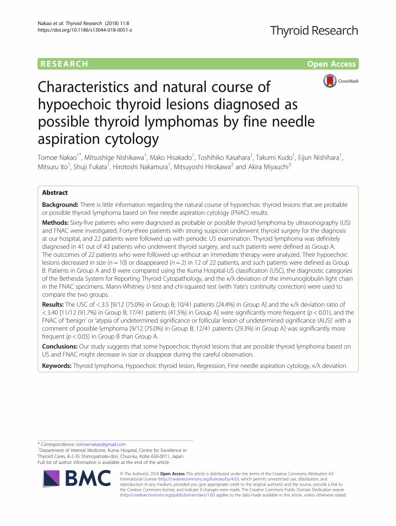
Characteristics and natural course ofhypoechoic thyroid lesions diagnosed aspossible thyroid lymphomas by fine needleaspiration cytologyTomoe Nakao1*, Mitsushige Nishikawa1, Mako Hisakado1, Toshihiko Kasahara1, Takumi Kudo1, Eijun Nishihara1,Mitsuru Ito1, Shuji Fukata1, Hirotoshi Nakamura1, Mitsuyoshi Hirokawa2 and Akira Miyauchi3
Abstract
Background: There is little information regarding the natural course of hypoechoic thyroid lesions that are probableor possible thyroid lymphoma based on fine needle aspiration cytology (FNAC) results.
Methods: Sixty-five patients who were diagnosed as probable or possible thyroid lymphoma by ultrasonography (US)and FNAC were investigated. Forty-three patients with strong suspicion underwent thyroid surgery for the diagnosisat our hospital, and 22 patients were followed up with periodic US examination. Thyroid lymphoma was definitelydiagnosed in 41 out of 43 patients who underwent thyroid surgery, and such patients were defined as Group A.The outcomes of 22 patients who were followed up without an immediate therapy were analyzed. Their hypoechoiclesions decreased in size (n = 10) or disappeared (n = 2) in 12 of 22 patients, and such patients were defined as GroupB. Patients in Group A and B were compared using the Kuma Hospital-US classification (USC), the diagnostic categoriesof the Bethesda System for Reporting Thyroid Cytopathology, and the κ/λ deviation of the immunoglobulin light chainin the FNAC specimens. Mann-Whitney U-test and chi-squared test (with Yate’s continuity correction) were used tocompare the two groups.
Results: The USC of < 3.5 [9/12 (75.0%) in Group B; 10/41 patients (24.4%) in Group A] and the κ/λ deviation ratio of< 3.40 [11/12 (91.7%) in Group B; 17/41 patients (41.5%) in Group A] were significantly more frequent (p < 0.01), and theFNAC of ‘benign’ or ‘atypia of undetermined significance or follicular lesion of undetermined significance (AUS)’ with acomment of possible lymphoma [9/12 (75.0%) in Group B; 12/41 patients (29.3%) in Group A] was significantly morefrequent (p < 0.05) in Group B than Group A.
Conclusions: Our study suggests that some hypoechoic thyroid lesions that are possible thyroid lymphoma based onUS and FNAC might decrease in size or disappear during the careful observation.
Keywords: Thyroid lymphoma, Hypoechoic thyroid lesion, Regression, Fine needle aspiration cytology, κ/λ deviation
* Correspondence: [email protected] of Internal Medicine, Kuma Hospital, Centre for Excellence inThyroid Cares, 8-2-35 Shimoyamate-dori, Chuo-ku, Kobe 650-0011, JapanFull list of author information is available at the end of the article
© The Author(s). 2018 Open Access This article is distributed under the terms of the Creative Commons Attribution 4.0International License (http://creativecommons.org/licenses/by/4.0/), which permits unrestricted use, distribution, andreproduction in any medium, provided you give appropriate credit to the original author(s) and the source, provide a link tothe Creative Commons license, and indicate if changes were made. The Creative Commons Public Domain Dedication waiver(http://creativecommons.org/publicdomain/zero/1.0/) applies to the data made available in this article, unless otherwise stated.
Nakao et al. Thyroid Research (2018) 11:8 https://doi.org/10.1186/s13044-018-0051-z

BackgroundPrimary thyroid lymphoma is a rare cause of malignancy,accounting for 2–5% of all thyroid malignancies [1] and< 2% of extranodal lymphomas [2, 3]. A rapidly enlargingmass was the most common clinical manifestations inearlier series, but recently small lesions are found at theearly stage [4]. It is a potentially lethal disease, but oftenresponds well to appropriate treatments [5]. Thyroidlymphoma is not always diagnosed easily, especially inits early phase, because most of the cases are associatedwith Hashimoto’s thyroiditis.Ultrasonography (US) of the thyroid is initially used for
the diagnosis of thyroid lymphoma. On US, lymphoma isshown as hypoechoic lesions, but subacute thyroiditis,focal chronic thyroiditis, some thyroid cancers, and meta-static thyroid cancer is also shown as hypoechoic lesionsmimicking thyroid lymphoma. Based on US findings ofinternal echo levels, borders, and posterior echoes, thyroidlymphoma can be classified as the nodular, diffuse, ormixed type [6]. Although the nodular type often resemblesfollicular tumor or adenomatous nodule and the mixedtype often resembles adenomatous goiter on US, theselesions can be often distinguished by an enhancement ofposterior echoes. The diffuse type shows homogeneousand hypoechoic internal echoes, but these findings arealso typical for severe chronic thyroiditis [6, 7].Fine needle aspiration cytology (FNAC) is the next
diagnostic strategy for thyroid lymphoma, but is challen-ging, particularly due to its histological similarities withmucosa-associated lymphoid tissue (MALT) lymphomaand chronic thyroiditis [8, 9]. The flow cytometry withCD45 gating on the FNAC specimen can be used toanalyze the proportions of lymphocytic cells with κ andλ immunoglobulin light chains. The κ/λ deviation ratioof the light chain assessment is an important criterionfor discriminating between polyclonal reactive processessuch as chronic thyroiditis and monoclonal lymphomas.Strong deviation in the κ/λ ratio is regarded as suggest-ive monoclonal growth of lymphocytes, thus indicatingthyroid lymphoma [10].Even with these modalities, a definite diagnosis is not
established easily. A surgical interventions is often neededfor the histopathological diagnosis, but it might beunnecessary for the benign lesions. This is especially truefor the small or moderate-size lesions. Needle biopsy isusually useful for the diagnosis of diffuse large B-cell typelymphoma, but is often insufficient in the case of MALTlymphoma.We have observed some hypoechoic lesions that were
diagnosed as possible thyroid lymphoma based on theFNAC decrease in size or disappear during their clinicalcourses without definitive treatment. However, there islittle information regarding the natural course of suchhypoechoic lesions that were possible thyroid lymphoma
based on FNAC findings. We conducted the presentstudy to (1) clarify the natural course of hypoechoicthyroid lesions that were possible thyroid lymphoma,and (2) identify clinical features that might be used todiscriminate benign non-progressive lesions from thosewith progressive character.
MethodsBetween April 2012 and July 2016, 136 patients weresuspected of having thyroid lymphoma on US examin-ation at Kuma Hospital, where approximately 15,000new patients with thyroid diseases are evaluated annu-ally. US examinations were routinely performed by ourwell-experienced operators. US was performed using anAPLIO 500 TUS-A500 system (Toshiba MedicalSystems Co., Ltd., Otawara, Japan) with a PLT-805AT(Toshiba) or PLT-1005BT probe (Toshiba). Patients withsystemic lymphoma or previously diagnosed lymphomawere excluded. Of these 136 patients, 122 were diag-nosed as having probable or possible thyroid lymphomaby FNAC (Fig. 1). The remaining 14 patients were ex-cluded from the study, because they had other diagnosesbased on FNAC findings (eight specimens were normalor benign, one anaplastic carcinoma of the thyroid, onesubacute thyroiditis or thyroid papillary carcinoma, andone metastatic renal cell carcinoma; the remaining threespecimens were inadequate for diagnosis). Among the122 patients, 57 were excluded from the study, because56 were referred to other hospitals for the definitivediagnosis and chemo radiotherapy, and one patientdropped out from the follow-up study. Thus, theremaining 65 patients were investigated in this study.Forty-three patients strongly suspected of having thyroidlymphoma underwent thyroid surgery at our hospital forthe definite diagnosis and 22 patients with comments ofpossible lymphoma on cytology were followed up withperiodic US examination for 1–41 (median 11.5)months. Whether surgery or follow-up examination wasdetermined by each doctor in charge based on clinicaland radiological findings, including the rapidity of theenlargement of the thyroid mass, and the US and FNACfindings. Of 43 patients who underwent thyroid surgeryat our hospital, 41 (95.3%) were diagnosed as definitethyroid lymphoma, and the other two (4.7%) werediagnosed as chronic thyroiditis (Fig. 1). Among the 41patients who were diagnosed as definite thyroid lymph-oma, 34 were diagnosed as MALT lymphoma and sevenwere diagnosed as diffuse large B-cell lymphoma.Whether the lesions of all 41 patients were limited tothe thyroid was not known because all were referred toother hospitals to undergo further diagnostic examina-tions, including the determination of the disease stage,and treatment.
Nakao et al. Thyroid Research (2018) 11:8 Page 2 of 8

Hypoechoic lesions of the periodically followed-up 22patients were analyzed. The US examination was repeated1–3 month-interval at first and then 6 month-intervalwhen no deterioration was observed. None of 22 patientswere treated with steroids or immunosuppressive drugs
during the follow-up examination. Other medications orsupplementations, or dietary habit including iodine intakewere not known in detail. When the maximum diameterof a hypoechoic lesion increased by ≥3 mm, we definedthe case as an “increase” of the hypoechoic lesion. When
Suspicious of thyroid lymphoma by US (n=136)
14 patients excluded by FNAC*
Probable or possible thyroid lymphoma by FNAC (n=122)
Follow-up examination(n=22)
Thyroid surgery(n=43)
Referred to other hospitals (n=56)
Dropped out (n=1)
Thyroid lymphoma (n=41)
Group AChronic thyroiditis (n=2)
Decrease (n=10)Disappear (n=2)
Group B
No change (n=1) Increase (n=3) Drop out (n=6)
*described in text. Group A: patients with definite thyroid lymphoma diagnosed pathologically. Group B: patients with hypoechoiclesions which decreased in diameter or disappeared.
Subjects of the present study (n=65)
Fig. 1 Clinical flow and outcomes of patients with possible thyroid lymphoma based on US and FNAC
A1
A2
B1
B2
C1
C2Fig. 2 Echograms of three representative patients whose hypoechoic lesions decreased or disappeared on careful follow-up examination. a A 74-year-old woman with a nodular hypoechoic lesion at presentation (A1) that had markedly decreased in size one month later (A2) (case No. 9 in Table 2).b A 69-year-old woman with a severely hypoechoic lesion involving the both thyroid lobes (B1) that had almost disappeared 10 months later (B2),although the irregularity remained possibly due to underlying chronic thyroiditis (case No. 4 in Table 2). c A 53-year-old woman with a diffuse severelyhypoechoic lesion involving the whole thyroid (C1) that had markedly decreased in size 27 months later (C2) (case No. 1 in Table 2)
Nakao et al. Thyroid Research (2018) 11:8 Page 3 of 8

the maximum diameter of a hypoechoic lesion reduced by≥3 mm, we defined the case as a “decrease.” When thehypoechoic lesions were not detected clearly at follow-upexamination, it was defined as “disappeared.” We estab-lished this parameter because, in our previous study, plusor minus 2 mm was recognized as an observation vari-ation [11]. In the present study, in some cases in whichthe entire thyroid volume was observed to have decreased,the maximum diameter of the hypoechoic lesions wasmeasured and analyzed.Representative US images showing “decrease” and “dis-
appearance” are shown in Fig. 2. Our hospital uses its ownultrasound classification system for the diagnosis of thy-roid nodules, which consists of five US classes (USC)based on the characteristics of thyroid nodules, such as aregular or irregular shape, solid or cystic content, thepresence or absence of microcalcifications, extraglandalinvasion, and other factors. The classification consists offive classes from 1 to 5. Intermediate classes from class 2to class 4 (designated as classes 2.5 and 3.5) are also used(Table 1) [12]. Some cases of lymphoma with diffuse ormixed lesion are hardly classified according to USC,because this classification is mainly scored to thyroid nod-ules. However, in the present study, the US classificationwas applied to the hypoechoic lesions. The present studywas approved by the Institutional Review Board of KumaHospital and the Ethics Committee in Kuma Hospital.The hypoechoic lesions in 22 patients who were followed
up with periodic US examination decreased in size (n = 10),disappeared (n = 2), showed no change (n = 1) or increasedin size (n = 3). The remaining six patients dropped out fromthe follow-up examination. Of the three patients whosehypoechoic lesions increased in size, one patient wasreferred to another hospital and diagnosed as diffuse largeB-cell lymphoma; the other two patients were carefullyfollowed up without further progression (Fig. 1).Forty-one patients with histopathologically diagnosed
thyroid lymphoma were defined as Group A, and 12patients whose hypoechoic lesions decreased in diameteror disappeared as defined Group B (Fig. 1). Their clinicalfeatures, the diameter of hypoechoic lesions, US featuresat the first presentation, and the diagnostic categories ofthe Bethesda System for Reporting Thyroid Cytopathol-ogy (BethSys categories) were compared between Group
A and B. Mann-Whitney U-test and chi-squared test(with Yate’s continuity correction) were used to comparethe two groups.
ResultsTable 2 summarizes the clinical features of 16 patients whounderwent follow-up US examinations of hypoechoiclesions that were diagnosed as possible thyroid lymphoma.Thyroid function tests at presentation showed hypothyroid(n = 1), subclinical hypothyroid (n = 4) and euthyroid (n =11). Some patients were taking levothyroxine (LT4) beforeand during the follow-up examination. However, whetherLT4 supplementation was involved in the decrease ordisappearance of the hypoechoic lesions is unclear, becauseof the small number of the cases. All Group B patients werethyroglobulin antibody (TgAb)- and/or thyroid peroxidaseantibody (TPOAb)-positive. As a result of FNAC, ‘atypia ofundetermined significance or follicular lesion of undeter-mined significance (AUS)’ was diagnosed in eight, ‘suspi-cious for ‘malignancy’ in two, ‘malignancy’ in one, and‘benign’ in one patient. The pathological report of case 2 inTable 2 said that it was probably benign; however, lymph-oma could not be completely excluded. On flow cytometryof the fine needle aspirations, the κ/λ ratio varied from 0.31to 4.61. When κ/λ ratio was lower than 1.00, we convertedκ/λ ratio to λ/κ ratio (deviation ratio) to discriminate be-tween the polyclonal reactive process and the monoclonalreactive process that is characteristic to lymphomas.There were no significant differences in age, sex, thyroid
function, TgAb, TPOAb, echo pattern of hypoechoiclesions, USC, BethSys, the diameter of hypoechoic lesions,κ/λ deviation ratio, LT4 supplementation or TSH betweenthe patients whose hypoechoic lesions decreased in size ordisappeared and patients whose lesions were unchangedor increased in size.There were no significant differences in patient age,
sex, or the diameter of the hypoechoic lesions betweenGroups A and B (Mann-Whitney U-test) (Table 3). TheUSC and the κ/λ deviation ratio were significantly lowerin Group B than Group A (p < 0.001 and p < 0.02,respectively). The USC of < 3.5 [9/12 (75.0%) in GroupB; 10/41 patients (24.4%) in Group A] was significantlymore frequent (p < 0.01), and the FNAC finding of ‘be-nign’ or ‘AUS’ [9/12 (75.0%) in Group B; 12/41 patients
Table 1 Ultrasonographic classification for thyroid nodule at Kuma Hospital
Class Description
1 Round or anechoic lesion.
2 Regular-shaped nodule with cystic change. The echo level of solid lesion is similar to that of normal thyroid.
3 Solid and regular-shaped nodule. Internal echo is homogeneous, or may have strong echoes internally or at the capsule.
4 Solid and irregular-shaped nodule. Internal echo is usually low and may have fine strong echogenic spots.
5 Solid and irregular-shaped nodule with extrathyroid extension.
Intermediate classes from class 2 to class 5 (designated as classes 2.5,3.5 and 4.5) are also used
Nakao et al. Thyroid Research (2018) 11:8 Page 4 of 8

Table
2Clinicalfeatures
of16
patientswho
unde
rwen
tfollow-upUSexam
inations
ofhypo
echo
iclesion
sthat
werediagno
sedas
possiblethyroidlymph
oma
Case
Age
Sex
Thyroid
functio
naTgAb
(IU/m
L)TPOAb
(IU/m
L)Echo
pattern
ofhypo
echo
iclesion
s
US
class
BethSysb
Durationof
follow-up
(mon
ths)
Before
(mm)
Afte
r(m
m)
κ/λ
deviation
ratio
c
LT4supp
lemen
tatio
n(μg/day)
TSHdu
ring
follow-up
Outcome
153
Fsubclinical
hypo
thyroid
(with
LT4
50μg
/day)
4000≦
535
diffu
se3
AUSd
2761
471.21
75no
rmal
decrease
262
Fhypo
thyroid
4000≦
600≦
diffu
se3.5
benign
e30
120
801.50
112.5
supp
ressed
decrease
366
Feuthyroid
2889
nodu
lar
3.5
AUS
1225
114.61
50no
rmal
decrease
469
Feuthyroid
120.7
600≦
nodu
lar
3AUS
1037
disapp
earance
3.14
–no
rmal
disapp
earance
570
Meuthyroid
54118
nodu
lar
3AUS
3715
121.84
–no
rmal
decrease
671
Msubclinical
hypo
thyroid
1171
–no
dular
3malignancy
637
233.20
50no
rmal
decrease
772
Feuthyroid
906
–no
dular
2.5
Suspicious
formalignancy
327
142.90
50no
rmal
decrease
874
Feuthyroid
512.9
151
nodu
lar
3AUS
4112
disapp
earance
2.75
–no
rmal
disapp
earance
974
Feuthyroid
688.8
16no
dular
3Suspicious
formalignancy
1105
251.72
–no
rmal
decrease
1074
Feuthyroid
121.8
–no
dular
3AUS
716
92.06
75supp
ressed
decrease
1177
Fsubclinical
hypo
thyroid
1402
–diffu
se3
AUS
2546
401.53
50no
rmal
decrease
1283
Feuthyroid
89.9
–no
dular
3.5
AUS
623
193.21
–no
rmal
decrease
1366
Feuthyroid
235.5
–no
dular
4AUS
2222
220.86
75supp
ressed
nochange
1446
Feuthyroid
609.6
–no
dular
3Suspicious
formalignancy
722
30–
–no
rmal
increase
1587
Meuthyroid
750.4
≦16
nodu
lar
3Suspicious
formalignancy
619
234.09
–no
rmal
increase
1694
Fsubclinical
hypo
thyroid
586.3
26.5
diffu
se3
Suspicious
formalignancy
1165
160
1.81
62.5
norm
alincrease
Theno
rmal
rang
eof
TgAbis≤39
.9U/m
L;TP
OAb:
≤27
.9U/m
L.–:TP
OAbno
tdo
neor
nolevo
thyroxine(LT4)supp
lemen
tatio
nInterm
ediate
USclassesfrom
class2to
class4(designa
tedas
classes2.5an
d3.5)
arealso
used
a Thy
roid
functio
nat
thefirst
exam
inationan
ddiag
nosisas
possible
thyroidlymph
omaby
FNAC
bTh
eBe
thesda
System
forRe
portingTh
yroidCytop
atho
logy
c The
κ/λde
viationratio
was
calculated
byλ/κratio
whe
ntheκ/λratio
was
<1.00
dAtypiaof
unde
term
ined
sign
ificanceor
follicularlesion
ofun
determ
ined
sign
ificance
e Lym
phom
acann
otbe
denied
Nakao et al. Thyroid Research (2018) 11:8 Page 5 of 8

(29.3%) in Group A] was significantly more frequent (p< 0.05) in Group B than Group A (Tables 4 and 5).Lastly, the κ/λ deviation ratio of the immunoglobulin(Ig) light chain in the FNAC specimens of < 3.40 [11/12(91.7%) in Group B; 17/41 patients (41.5%) in Group A]was significantly more frequent in Group B than GroupA (p < 0.01) (Table 6).
DiscussionThe characteristics and natural course of hypoechoiclesions that were diagnosed as possible thyroid lymph-oma based on the FNAC findings were investigated.Careful US examinations were repeatedly performed in22 of 65 patients, and hypoechoic lesions decreased indiameter in 10 patients (10 of the 22; 45.5% or 10 of 65patients; 15.4%), and disappeared in two patients (two of22; 9.1% or two of 65 patients; 3.1%).The USC and the κ/λ deviation ratio were significantly
lower in Group B than Group A, and the FNAC findingof ‘benign’ or ‘AUS’ was significantly more frequent inGroup B than Group A. Although thyroid lymphomatypically presents with a rapidly enlarging neck massleading to compressive symptoms [4], some hypoechoiclesions diagnosed as possible thyroid lymphoma mightregress in their natural courses in the present study.Several studies have demonstrated that Helicobacter
pylori infection is associated with low-grade gastric MALTlymphoma, and that the eradication of Helicobacter pylorican cause histological regression of the lymphoma [13, 14].Uohashi et al. reported that a hypoechoic lesion in the rightlobe disappeared spontaneously after being diagnosedpathologically as non-Hodgkin’s lymphoma following acontralateral lobe resection, and suggested spontaneousregression of acute inflammation [15]. Okamoto et al. re-ported a patient whose primary thyroid lymphoma of
cytotoxic T-cell origin regressed spontaneously [16]. Al-though its cause remains unknown, it may be attributableto an association of acute inflammation [17, 18], andimmune mechanisms such as those involving T-helper cellsor natural killer cells of the peripheral blood [19].However, to our best knowledge, there has been no
report that the natural course of hypoechoic lesions sus-pected of being possible thyroid lymphoma was studiedextensively. With the combination of US, FNAC, andCD gating analysis for κ/λ deviation, candidate for thesurgery could be accurately selected in 41 out of 43patients (95%) as demonstrated in Group A. However,the management of not strongly suggestive of lymphomamay be controversial. In the present study, the hypoe-choic lesion decreased in size in 10, and disappeared intwo out of 22 patients, suggesting the careful observa-tion for such lesions.The limitation of the present study concerns the retro-
spective analysis that may eliminate the ability to obtain thestrong conclusions. Selection of open surgery or careful ob-servation depends on various factors including the resultsof FNAC and patients’ previous courses. Therefore, theproper diagnostic rate of Group A was higher than in thatof Group B, and the selection bias might cause the differ-ence of the course of the two groups. The nature of thehypoechoic lesions that regressed during the follow-upexamination is unclear in the present study, because openbiopsies were not performed in these patients. Some couldbe actually low-grade thyroid lymphoma, but others couldbe focal or regional lymphocytic thyroiditis, atypicalsubacute thyroiditis, or other etiology-unknown lesion(s).Further prospective studies including histopathology andfactor(s) involved in the regression or progression of the
Table 3 Comparison of clinical features of Group A and B
Group A (n = 41) Group B (n = 12) p-value
Age (year) 67 (48–91) 72 (53–83) 0.35
Sex (Male, number) 11 2 0.48
Diameter of hypoechoic lesions (mm) 35 (16–75) 38 (12–120) 0.51
Group A is the patients with thyroid lymphoma diagnosed histopathologically following thyroid surgery. Group B is the patients whose hypoechoic lesionsdecreased or disappeared. Data are median and (ranges). The p-values were calculated by Mann-Whitney U-test
Table 4 Comparisons of US classification between the Group A(n = 41) and Group B (n = 12)
US class Group A Group B
2.5 1 1
3 9 8
3.5 9 3
4 21 0
P < 0.01Classes 2.5 and 3.5 designate intermediate classes between 2 &3 and 3 &4, respectively
Table 5 Comparisons of the diagnostic categories of theBethesda System for Reporting Thyroid Cytopathology betweenthe Group A (n = 41) and Group B (n = 12)
Diagnostic categories Group A Group B
Benign 1 1
AUS 11 8
Suspicious for malignancy 22 2
Malignant 7 1
P < 0.02US: ultrasound. AUS: atypia of undetermined significance or follicular lesion ofundetermined significanceBenign: benign, but with the comment that lymphoma cannot be denied
Nakao et al. Thyroid Research (2018) 11:8 Page 6 of 8

lesions are needed to investigate the true nature of suchlesions, and to clarify the best method of managing suchpatients.Hypoechoic lesions suspected of having possible lymph-
oma decreased in size or disappeared in 12 out of 22 pa-tients (55%), and such a regression should be emphasizedin the present study. Because most of these lesions cannotbe diagnosed definitely by core needle biopsy, open sur-gery might be considered. But unnecessary surgeries forbenign lesions might be avoided.
ConclusionOur study suggests that some hypoechoic thyroid lesionsthat are possible thyroid lymphoma based on US andFNAC might decrease in size or disappear during thecareful observation. A careful observation before surgeryis suggested for those lesions such as USC < 3.5, a κ/λdeviation ratio < 3.4, and the FNAC classification of‘benign’ or ‘atypia of determined significance or follicularlesions of undetermined significance’. However, an openbiopsy or thyroid surgery should be considered in caseof the increasing lesion in diameter on follow-up USexamination.
AbbreviationsAUS: Atypia of undetermined significance or follicular lesion ofundetermined significance; BethSys categories: Bethesda System forReporting Thyroid Cytopathology; FNAC: Fine needle aspiration cytology;Ig: Immunoglobulin; MALT: Mucosa-associated lymphoid tissue;TgAb: Thyroglobulin antibody; TPOAb: Thyroid peroxidase antibody;US: Ultrasonography; USC: US classification
Availability of data and materialsData sharing not applicable to this article as no datasets were generated oranalyzed during the current study.
Authors’ contributionsTN and MN were involved in study design, acquisition of data, analysis andinterpretation of data and drafting and revising the manuscript. TN, MN, MH,TK, TK, EN, MI, SF, HN, MH and AM were involved in data collection andmanuscript drafting. All authors read and approved the final manuscript.
Ethics approval and consent to participateThis study was approved by the Ethics Committee in Kuma Hospital. Allprocedures performed in studies involving human participants were inaccordance with the ethical standards of the institutional and with the1964 Declaration of Helsinki and its later amendments or comparableethical standards.
Consent for publicationThe patient has provided their consent for the contents of this report to bepublished.
Competing interestsThe authors declare that they have no competing interests.
Publisher’s NoteSpringer Nature remains neutral with regard to jurisdictional claims inpublished maps and institutional affiliations.
Author details1Department of Internal Medicine, Kuma Hospital, Centre for Excellence inThyroid Cares, 8-2-35 Shimoyamate-dori, Chuo-ku, Kobe 650-0011, Japan.2Department of Diagnostic Pathology and Cytology, Kuma Hospital, Centrefor Excellence in Thyroid Cares, 8-2-35 Shimoyamate-dori, Chuo-ku, Kobe650-0011, Japan. 3Department of Surgery, Kuma Hospital, Centre forExcellence in Thyroid Care, 8-2-35 Shimoyamate-dori, Chuo-ku, Kobe650-0011, Japan.
Received: 17 February 2018 Accepted: 22 May 2018
References1. Klopper JP, Kane MA, Haugen BR. Anaplastic thyroid cancer and
miscellaneous tumors of the thyroid. In: Braverman LE, Cooper DS, editors.Werner & Ingbar’s The Thyroid. 10th ed; 2013. p. 765–74.
2. Freeman C, Berg JW, Occurrence CSJ. Prognosis of extranodal lymphomas.Cancer. 1972;29:252–60.
3. Thieblemont C, Mayer A, Dumontet C, Barbier Y, Callet-Bauchu E, et al.Primary thyroid lymphoma is a heterogeneous disease. J Clin EndocrinolMetab. 2002;87:105–11.
4. Matsuzuka F, Miyauchi A, Katayama S, Narabayashi I, Ikeda H, et al. Clinicalaspects of primary thyroid lymphoma: diagnosis and treatment based onour experience of 119 cases. Thyroid. 1993;3:93–9.
5. Watanabe N, Noh JY, Narimatsu H, Takeuchi K, Yamaguchi T, et al.Clinicopathological features of 171 cases of primary thyroid lymphoma: along-term study involving 24553 patients with Hashimoto’s disease. Brit JHematol. 2011;153:236–43.
6. Ota H, Ito Y, Matsuzuka F, Kuma S, Fukata S, et al. Usefulness of ultrasonographyfor diagnosis of malignant lymphoma of the thyroid. Thyroid. 2006;16:983–7.
7. Mizokami T, Hamada K, Maruta T, Higashi K, Yamashita H, et al.Development of primary thyroid lymphoma during an ultrasonographicfollow-up of Hashimoto's thyroiditis: a report of 9 cases. Intern Med.2016;55:943–8.
8. Sangalli G, Serio G, Zampatti C, Lomuscio G, Colombo L. Fine needleaspiration cytology of primary lymphoma of the thyroid: a report of 17cases. Cytopathology. 2001;12:257–63.
9. Aleskow S, Wartofsky L. Primary thyroid lymphoma: a clinical review. J ClinEndocrnol Metab. 2013;98:3131–8.
10. Zeppa P, Cozzolino I, Peluso AL, Troncone G, Lucariello A, et al. Cytologic,flow cytometry, and molecular assessment of lymphoid infiltrate in fine-needle cytology samples of Hashimoto thyroiditis. Cancer Cytopathology.2009;117:174–84.
11. Ito Y, Miyauchi A, Inoue H, Fukushima M, Kihara M, et al. Anobservational trial for papillary thyroid microcarcinoma in Japanesepatients. World J Surg. 2010;34:28–35.
12. Ito Y, Amino N, Yokozawa T, Ota H, Ohshita M, et al. Ultrasonographic evaluationof thyroid nodules in 900 patients: comparison among ultrasonographic,cytological, and histological findings. Thyroid. 2007;17:1269–76.
13. Montalban C, Manzanal A, Boixeda D, Redondo C, Alvarez I.Helicobacter pylori eradication for the treatment of low-grade gastricMALT lymphoma: follow-up together with sequential molecular studies.Ann Oncol. 1997;2:37–9.
14. Grgov S, Katić V, Krstić M, Nagorni A, Radovanović-Dinić B. Treatment oflow-grade gastric MALT lymphoma using Helicobactert pylori eradicateon.Vojnosanit Pregl. 2015;72:431–6.
15. Uohashi A, Imoto S, Matsui T, Murayama T, Okimura Y, et al. Spontaneousregression of diffuse large-cell lymphoma associated with Hashimoto'sthyroiditis. Am J Hematol. 1996;53:201–11.
Table 6 Comparisons of Groups A and B regarding the κ/λdeviation ratio of Ig light chain
κ/λ deviation ratio Group A Group B Total
≥3.40 24 1 25
< 3.40 17 11 28
Total 41 12 53
The κ/λ deviation ratio of < 3.40 was significantly more frequent in Group Bcompared to Group A (p < 0.01, chi-square test (with Yate’s continuitycorrection)). When the κ/λ deviation ratio was lower than 1.00, we converted itto the λ/κ ratio (deviation ratio)
Nakao et al. Thyroid Research (2018) 11:8 Page 7 of 8

16. Okamoto A, Namura K, Uchiyama H, Kajita Y, Inaba T, et al. Cytotoxic T-cellnon-Hodgkin's lymphoma of the thyroid gland. Am J Hematol. 2005;80:77–8.
17. Drobyski WR, Qazi R. Spontaneous regression in non-Hodgkin's lymphoma.Clinical and pathologic considerations. Am J Hematol. 1989;31:138–41.
18. Seachrist L. Spontaneous cancer remissions spark questions. J Natl CancerInst. 1993;85:1892–5.
19. Papac RJ. Spontaneous regression of cancer: possible mechanisms. InVivo. 1998;12:571–8.
Nakao et al. Thyroid Research (2018) 11:8 Page 8 of 8